Immunologic Disorders Chapter 18. Type I Hypersensitivities: Immediate IgE-Mediated IgE causes...
21
Immunologic Disorders Chapter 18
-
Upload
clement-banks -
Category
Documents
-
view
216 -
download
0
Transcript of Immunologic Disorders Chapter 18. Type I Hypersensitivities: Immediate IgE-Mediated IgE causes...

Immunologic Disorders
Chapter 18

Type I Hypersensitivities:Immediate IgE-Mediated
IgE causes immediate (type I) hypersensitivities Characterized by reaction the sensitized individual
immediately Generally within minutes of exposure
Tendencies to have type I hypersensitivities is inherited Reactions occur in at least 20% to 30% of population
Type I reactions can be classified as local anaphylaxis or generalized anaphylaxis Anaphylaxis name given for IgE mediated allergic reaction

Sensitization occurs when antigen makes contact with some part of body and induces response
IgE antibodies bind to receptors on mast cells and basophils Antigen readily binds to cells
fixed with IgE antibodies Within seconds mast cells
degranulate releasing mediators that initiate immune reaction including, hives, hay fever and anaphylaxis
Type I Hypersensitivities:Immediate IgE-Mediated
Type I Hypersensitivities:Immediate IgE-Mediated
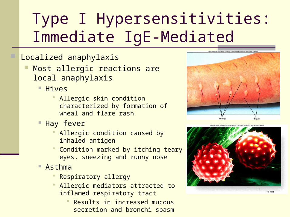
Localized anaphylaxis Most allergic reactions are local
anaphylaxis Hives
Allergic skin condition characterized by formation of wheal and flare rash
Hay fever Allergic condition caused by inhaled
antigen Condition marked by itching teary eyes,
sneezing and runny nose Asthma
Respiratory allergy Allergic mediators attracted to inflamed
respiratory tract Results in increased mucous
secretion and bronchi spasm

Generalized anaphylaxis Rare but more serious Antigen enters bloodstream and becomes
widespread Reactions affects almost entire body Can induce shock
Shock is state in which blood pressure too low to supply required blood flow
Massive release of mediators causes extensive blood vessel dilation and fluid loss
Causes fall in pressure leading to flow insufficiency
Type I Hypersensitivities:Immediate IgE-Mediated

Type I Hypersensitivities:Immediate IgE-Mediated
Immunotherapy General term for techniques
used to modify immune system for favorable effect
Procedure is to inject individual with extremely dilute suspension of allergen
Called desensitization or hyposensitization
Concentration of allergen gradually increased over time
Individual gradually becomes less sensitive
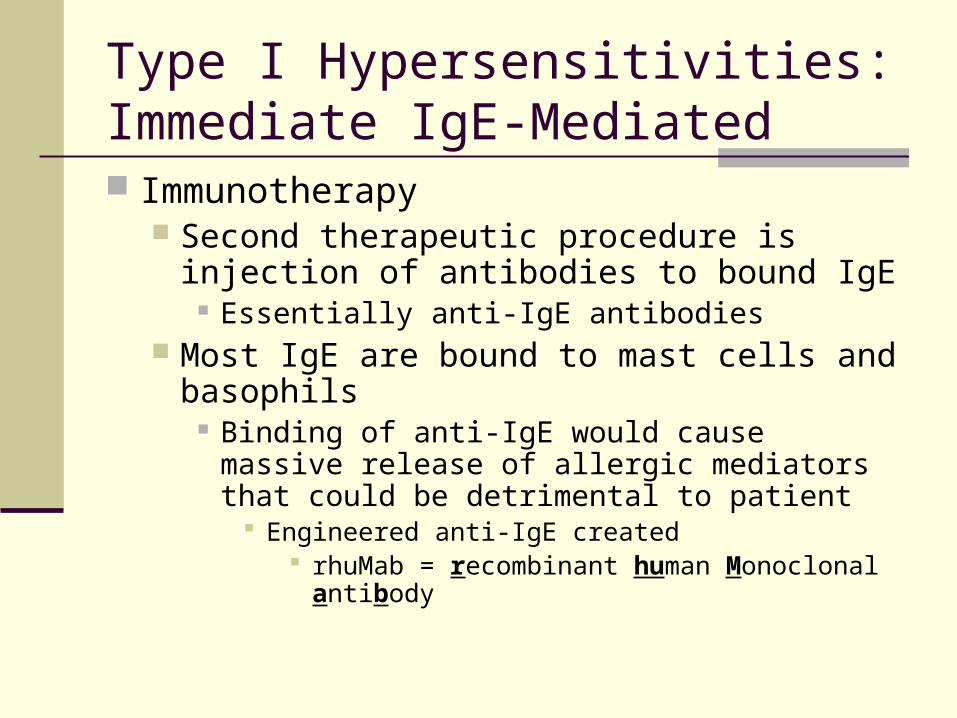
Immunotherapy Second therapeutic procedure is injection of
antibodies to bound IgE Essentially anti-IgE antibodies
Most IgE are bound to mast cells and basophils
Binding of anti-IgE would cause massive release of allergic mediators that could be detrimental to patient
Engineered anti-IgE created rhuMab = recombinant human Monoclonal antibody
Type I Hypersensitivities:Immediate IgE-Mediated

Type II Hypersensitivities:Cytotoxic
Complement-fixing antibodies react with cell surface antigens causing cell injury or death
Cells can be destroyed in type II reactions through complement fixation and antibody-dependent cellular cytotoxicity (ADCC)
Examples of Type II hypersensitivities are Transfusion reactions Hemolytic disease of the newborn
Type II Hypersensitivities:Cytotoxic
Transfusion reactions Normal red blood cells have different surface
antigens Antigens differ from person to person
People are designated type A, B, AB or O Transfused blood that is antigenically different
can be lysed by recipient immune cells Cross-matching blood is used to ensure
compatibility between donor and recipient IgM antibodies cause type II reactions Symptoms include low blood pressure, pain,
nausea and vomiting

Type II Hypersensitivities:Cytotoxic
Hemolytic disease of the newborn Basis of disease is incompatibility of
Rh factor between mother and child Rh factor RBC cell surface antigen
Rh positive = Rh antigen present Rh negative = Rh antigen missing
Anti-Rh antibodies form in Rh negative mother pregnant with Rh positive fetus
First Rh positive fetus unharmed Second Rh positive fetus provokes
strong secondary immune response IgG antibodies of secondary
response cross placenta causing extensive damage to fetal red blood cells

Type III Hypersensitivities:Immune Complex-Mediated
Immune complexes consist of antigen and antibody bound together
Usually adhere to Fc receptors on cells Complexes are destroyed
and removed Certain instances complexes
persist in circulation or at sites of formation Initiate blood clotting
mechanism Activate complement
contributing to inflammation
Complexes commonly deposited in skin, joints and kidney
Complexes also cause disseminated intravascular coagulation (DIC) Clots in small vessels
Leads to system failure

Type IV Hypersensitivities:Delayed Cell-Mediated Delayed hypersensitivities caused by cell-mediated
immunity Slowly developing response to antigen
Reactions peak in 2 to 3 days instead of minutes
T cells are responsible for reactions Reactions can occur nearly anywhere in the body
Delayed hypersensitivity reactions responsible for contact dermatitis, tissue damage, rejection of tissue grafts and some autoimmune disease

Type IV Hypersensitivities:Delayed Cell-Mediated
Tuberculin skin test Test involves introduction of
small quantities of protein antigens from tubercle bacillus into skin
In positive skin test injection site reddens and gradually thickens
Reaction reaches peak in 2 to 3 days
Reactions result from sensitized T cells, release of cytokines and influx of macrophages

Type IV Hypersensitivities:Delayed Cell-Mediated
Contact Hypersensitivities Mediated by the T cells
T cells release cytokines Cytokines initiate inflammation
that attracts macrophages Macrophages release
mediators to add to inflammation
Common examples of contact allergies include
Poison ivy and poison oak Nickel in metal jewelry Chromium salts in leather Latex products
Transplant Immunity
Major drawback to graft transplantation is possible immunological rejection Differences between donor and recipient tissues basis for
rejection Rejection is predominantly type IV reaction
Killing of graft cells occurs through complex combination of mechanisms Contact with sensitized cytotoxic T cells and natural killer cells
Combination of agents commonly used to prevent graft rejection Cyclosporin A Steroids Basiliximab
Monoclonal antibody preparation Blocks binding of immune mediators Blocks binding sites of T cells
Prevents formation of antibodies

Autoimmune Diseases
Body usually recognizes self antigens Destroys cells that would destroy self Malfunction in immune recognition basis
for autoimmunity Autoimmune diseases may result from
reaction to antigen that are similar to MHC self antigens
Autoimmunity may occur after tissue injury Self antigens released from injured organ
Autoantibodies form and interact with injured tissues

Spectrum of autoimmune diseases Reactions occur over spectrum
Organ-specific to widespread responses Organ-specific
Thyroid disease Only thyroid is affected
Widespread response Lupus
Autoantibodies made against nuclear constituents of all body cells
Rheumatoid arthritis Immune response made against collagen in connective tissue
Myasthenia gravis Autoantibody-mediated disease
Autoantibody to acetylcholine receptor proteins
Autoimmune Diseases

Treatment of autoimmune diseases Treatment aimed at:
Kill dividing cells Immunosuppressant
Controlling T cell signaling cyclosporin
Anti-inflammatory medications Cortico steroids
Replacement therapy insulin
Autoimmune Diseases

Immunodeficiency Disorders
Immunodeficiency disorders are marked by the body’s inability to make and sustain an adequate immune response
Two basic types of disorders Primary or congenital
Inborn as a result of genetic defect or developmental abnormality
Secondary or acquired Can be acquired as result of infection or other
stressor

Primary immunodeficiencies Generally rare Examples
Agammaglobulinemia Few or no antibodies produces Occurs in 1 in 50,00 people
Sever combined immunodeficiency disorder (SCID) Neither B nor T lymphocytes are functional Occurs in 1 in 500,000 live births
Selective IgA deficiency Little or no IgA produced Most common disorder
One in 333 to 700
Immunodeficiency Disorders

Secondary immunodeficiencies Result from environmental rather than genetic factors
Malignancies, advanced age certain infections, immunosuppressive drugs and malnutrition are just a few
Often results from depletion of certain cells of the immune system
Syphilis, leprosy and malaria effect T cell population and macrophage function
Malignancies of lymphoid system decrease antibody-mediated immunity
Most serious widespread immunodeficiency is AIDS Destroys helper T cells
Inhibits initiation of cellular and humoral immunity
Immunodeficiency Disorders


![Food Hypersensitivities and Atopic Dermatitis in Toddlers Dic 2011cancun[1]](https://static.fdocuments.in/doc/165x107/577ce4c31a28abf1038f1b4f/food-hypersensitivities-and-atopic-dermatitis-in-toddlers-dic-2011cancun1.jpg)