Immunologic and Structural Studies of Lupus/Sjogren's...
6
Immunologic and Structural Studies of the Lupus/Sjogren's Syndrome Autoantigen, La/SSB, with a Monoclonal Antibody John B. Harley, Myra 0. Rosario, Hajime Yamagata, Owen F. Fox, and Eugen Koren Departments of Medicine and Microbiology, University of Oklahoma Health Sciences Center, and the Arthritis and Immunology Program and Monoclonal Antibody Laboratory of the Oklahoma Medical Research Foundation, Oklahoma City, Oklahoma 73104 Abstract La/SSB is a small nuclear RNA protein against which precip- itating autoantibodies are made in many patients with systemic lupus erythematosus or Sjogren's syndrome. The recent puri- fication of La/SSB has made structural and immunologic studies possible. Consequently, a mouse hybridoma antibody (Lal) was raised, after immunization and fusion, that reacted with bovine La/SSB. Results of inhibition tests with tissue extracts and fluorescent antinuclear antibody tests demonstrated that Lal reacted with bovine extracts and cells, but not with those from human, mouse, or rabbit sources. Lal reacted in Western blot and in an adapted anti-La/SSB enzyme-linked immunosorbent assay with only the 41-k) bovine La/SSB peptide and not with the smaller 29-kD bovine La/SSB peptide. RNA gels showed that Lal bound the La/SSB particle that contained the predominant La/SSB RNA species near 90 nucleotides as well as the minor RNA species, both of which were bound by the human autoimmune anti-La/SSB serum. A solid-phase assay for human autoimmune anti-La/SSB antibody using Lal was more sensitive for the detection of human anti- La/SSB than was a comparable assay using purified La/SSB, and showed that anti-La/SSB is present in nearly all Ro/SSA precipitin-positive sera. Thus, this study demonstrates that monoclonal antibody can be raised against La/SSB; that the protein moiety of bovine La/SSB differs from human, mouse, and rabbit at an epitope on the 41-kD La/SSB peptide; that the RNA bound to the Lal-reactive particle was as heteroge- neous as that binding the anti-La/SSB autoimmune serum; and that anti-Ro/SSA and anti-La/SSB are closely associated. Introduction Autoantibodies reacting with an RNA protein particle now known as La/SSB are commonly found in systemic lupus erythematosus (SLE),' Sjogren's syndrome, and subacute cuta- neous lupus. The La/SSB particle is -60 kD and is probably composed of a single peptide and a heterogeneous RNA (1, Address reprint requests to Dr. Harley, Department of Medicine, University of Oklahoma, Health Sciences Center, P.O. Box 26901, Oklahoma City, OK 73104. Received for publication 4 October 1985. 1. Abbreviations used in this paper: ELISA, enzyme-linked immuno- sorbent assay; MDBK, Madin-Darby bovine kidney; SLE, systemic lupus erythematosus. 2). La/SSB RNA appears to be at least initially associated with all RNA polymerase III transcripts (3). Autoantigen structure and the molecular interactions of autoantibody with autoantigen are avenues of investigation that we hope will lead to better understanding of the immu- nopathogenic mechanisms of these diseases. As such, mono- clonal antibodies to autoimmune antigens have recently become an area of intense interest, especially with respect to SLE (4- 6). These reagents have the potential of leading to important insights concerning the structural properties of autoantibodies and autoantigens relevant to the disease process in man as well as in animal models. Monoclonal autoantibodies against DNA have been most intensively studied, though fusions done with the autoimmune mouse strains have also generated monoclonal autoantibodies against ribonucleoprotein and erythrocyte surface antigens (7, 8). Our approach has been to attempt to generate monoclonal heteroantibodies against La/ SSB. This study describes such a monoclonal antibody and its application to the analysis of La/SSB and anti-La/SSB. Methods Purification of La/SSB. An affinity chromatography method was used to purify La/SSB from calf thymus as described elsewhere (1). The final preparation usually contained the La/SSB RNA and both bovine La/SSB peptides (41 and 29 kD), though occasional preparations contained only the smaller peptide. These materials were used for immunization and analysis. Immunization. A BALB/c mouse was immunized with 50 ug bovine La/SSB in complete Freund's adjuvant by a subcutaneous route. The mouse was boosted with 10 ug La/SSB suspended in incomplete Freund's adjuvant into the peritoneal cavity. On each of the 3 d before fusion, the mouse was boosted (10 ug) by the peritoneal route, except for the last boost, which was done by the tail vein. Hybridomas. Spleen cells from the immunized BALB/c mouse were fused with the mouse myeloma cells P3X63-Ag86.5.3 and anti- La/SSB producing hybridomas were cloned and expanded by the procedure of Galfre and Milstein (9). Five producing wells with anti- La/SSB activity were found, two of which were successfully cloned. Mouse anti-La/SSB assay. The enzyme-linked immunosorbent assay (ELISA) technique that we adapted from Engvall and Perlmann (10) for human anti-La/SSB was modified to detect mouse anti-La/ SSB by using a goat anti-mouse IgG conjugate to alkaline phosphatase (Sigma Chemical Co., St. Louis, MO). Variations in this basic protocol were used in a number of experiments. Preparations containing only the smaller La/SSB peptide (29 kD) were also used to determine monoclonal antibody specificities. Preincubation of anti-La/SSB with La/SSB or crude concentrated extracts in phosphate-buffered saline of rabbit thymus acetone powder (Pel-Freeze Biologicals, Rogers, AR), human spleen, or calf thymus were carried out for 4 h at room temperature before being added to the La/SSB-coated plate. IgG purification and F(a)2 fragment preparation. IgG was prepared from ascites fluid by Affi-Gel Blue (Bio-Rad Laboratories, Richmond, CA) column chromatography (11). F(ab')2 fragments were generated by pepsin digestion and purified by gel filtration over Sephadex G100 Applications of an Anti-La/SSB Monoclonal Antibody 801 J. Clin. Invest. C The American Society for Clinical Investigation, Inc. 0021-9738/85/08/0801/06 $1.00 Volume 76, August 1985, 801-806
Transcript of Immunologic and Structural Studies of Lupus/Sjogren's...
Immunologic and Structural Studies of the Lupus/Sjogren's SyndromeAutoantigen, La/SSB, with a Monoclonal AntibodyJohn B. Harley, Myra 0. Rosario, Hajime Yamagata, Owen F. Fox, and Eugen KorenDepartments ofMedicine and Microbiology, University of Oklahoma Health Sciences Center, and the Arthritis and ImmunologyProgram and Monoclonal Antibody Laboratory of the Oklahoma Medical Research Foundation, Oklahoma City, Oklahoma 73104
Abstract
La/SSB is a small nuclear RNAprotein against which precip-itating autoantibodies are made in many patients with systemiclupus erythematosus or Sjogren's syndrome. The recent puri-fication of La/SSB has made structural and immunologicstudies possible. Consequently, a mouse hybridoma antibody(Lal) was raised, after immunization and fusion, that reactedwith bovine La/SSB. Results of inhibition tests with tissueextracts and fluorescent antinuclear antibody tests demonstratedthat Lal reacted with bovine extracts and cells, but not withthose from human, mouse, or rabbit sources. Lal reacted inWestern blot and in an adapted anti-La/SSB enzyme-linkedimmunosorbent assay with only the 41-k) bovine La/SSBpeptide and not with the smaller 29-kD bovine La/SSBpeptide. RNAgels showed that Lal bound the La/SSB particlethat contained the predominant La/SSB RNAspecies near 90nucleotides as well as the minor RNAspecies, both of whichwere bound by the human autoimmune anti-La/SSB serum. Asolid-phase assay for human autoimmune anti-La/SSB antibodyusing Lal was more sensitive for the detection of human anti-La/SSB than was a comparable assay using purified La/SSB,and showed that anti-La/SSB is present in nearly all Ro/SSAprecipitin-positive sera. Thus, this study demonstrates thatmonoclonal antibody can be raised against La/SSB; that theprotein moiety of bovine La/SSB differs from human, mouse,and rabbit at an epitope on the 41-kD La/SSB peptide; thatthe RNAbound to the Lal-reactive particle was as heteroge-neous as that binding the anti-La/SSB autoimmune serum;and that anti-Ro/SSA and anti-La/SSB are closely associated.
Introduction
Autoantibodies reacting with an RNA protein particle nowknown as La/SSB are commonly found in systemic lupuserythematosus (SLE),' Sjogren's syndrome, and subacute cuta-neous lupus. The La/SSB particle is -60 kD and is probablycomposed of a single peptide and a heterogeneous RNA (1,
Address reprint requests to Dr. Harley, Department of Medicine,University of Oklahoma, Health Sciences Center, P.O. Box 26901,Oklahoma City, OK73104.
Received for publication 4 October 1985.
1. Abbreviations used in this paper: ELISA, enzyme-linked immuno-sorbent assay; MDBK, Madin-Darby bovine kidney; SLE, systemiclupus erythematosus.
2). La/SSB RNAappears to be at least initially associated withall RNApolymerase III transcripts (3).
Autoantigen structure and the molecular interactions ofautoantibody with autoantigen are avenues of investigationthat we hope will lead to better understanding of the immu-nopathogenic mechanisms of these diseases. As such, mono-clonal antibodies to autoimmune antigens have recently becomean area of intense interest, especially with respect to SLE (4-6). These reagents have the potential of leading to importantinsights concerning the structural properties of autoantibodiesand autoantigens relevant to the disease process in man aswell as in animal models. Monoclonal autoantibodies againstDNAhave been most intensively studied, though fusions donewith the autoimmune mouse strains have also generatedmonoclonal autoantibodies against ribonucleoprotein anderythrocyte surface antigens (7, 8). Our approach has been toattempt to generate monoclonal heteroantibodies against La/SSB. This study describes such a monoclonal antibody and itsapplication to the analysis of La/SSB and anti-La/SSB.
Methods
Purification of La/SSB. An affinity chromatography method was usedto purify La/SSB from calf thymus as described elsewhere (1). Thefinal preparation usually contained the La/SSB RNAand both bovineLa/SSB peptides (41 and 29 kD), though occasional preparationscontained only the smaller peptide. These materials were used forimmunization and analysis.
Immunization. A BALB/c mouse was immunized with 50 ugbovine La/SSB in complete Freund's adjuvant by a subcutaneousroute. The mouse was boosted with 10 ug La/SSB suspended inincomplete Freund's adjuvant into the peritoneal cavity. On each ofthe 3 d before fusion, the mouse was boosted (10 ug) by the peritonealroute, except for the last boost, which was done by the tail vein.
Hybridomas. Spleen cells from the immunized BALB/c mousewere fused with the mouse myeloma cells P3X63-Ag86.5.3 and anti-La/SSB producing hybridomas were cloned and expanded by theprocedure of Galfre and Milstein (9). Five producing wells with anti-La/SSB activity were found, two of which were successfully cloned.
Mouse anti-La/SSB assay. The enzyme-linked immunosorbentassay (ELISA) technique that we adapted from Engvall and Perlmann(10) for human anti-La/SSB was modified to detect mouse anti-La/SSB by using a goat anti-mouse IgG conjugate to alkaline phosphatase(Sigma Chemical Co., St. Louis, MO).
Variations in this basic protocol were used in a number ofexperiments. Preparations containing only the smaller La/SSB peptide(29 kD) were also used to determine monoclonal antibody specificities.Preincubation of anti-La/SSB with La/SSB or crude concentratedextracts in phosphate-buffered saline of rabbit thymus acetone powder(Pel-Freeze Biologicals, Rogers, AR), human spleen, or calf thymuswere carried out for 4 h at room temperature before being added tothe La/SSB-coated plate.
IgG purification and F(a)2 fragment preparation. IgG was preparedfrom ascites fluid by Affi-Gel Blue (Bio-Rad Laboratories, Richmond,CA) column chromatography (11). F(ab')2 fragments were generatedby pepsin digestion and purified by gel filtration over Sephadex G100
Applications of an Anti-La/SSB Monoclonal Antibody 801
J. Clin. Invest.C The American Society for Clinical Investigation, Inc.0021-9738/85/08/0801/06 $1.00Volume 76, August 1985, 801-806
(Pharmacia Fine Chemicals, Piscataway, NJ) (12). No intact IgG wasdetected in this F(ab')2 preparation.
Western blot and RNA gels. The La/SSB antigenic peptides weredetected in Western blot by standard techniques (1). The RNAof theLal-binding particle was isolated and compared with the human anti-La/SSB binding by the following procedure. An extract of calf thymuswas prepared in the presence of ribonuclease inhibitor from humanplacenta (Sigma Chemical Co.) and concentrated over DE52. The 500mMNaCl eluate was first passed over on anti-Ro/SSA affinity column.This effluent was split into two equal parts. One was passed over aLal affinity column and the other was passed over an affinity columnconstructed from an anti-La/SSB that contained human serum. The 3M MgCl2 eluates were dialyzed and concentrated. The nucleic acidwas extracted and the RNAwas 3' end-labeled with 32P by previouslydescribed methods (13). The labeled RNAwas electrophoresed in 7 Murea-10% polyacrylamide gels and detected by autoradiography.
Fluorescent antinuclear antibody. Cell cultures from different specieswere used for immunofluorescence. A rabbit corneal epithelial line(SIRC), the National Cancer Tissue Culture (NCTC) clone of the 929mouse fibroblast line, and the Madin-Darby bovine kidney (MDBK)line (all from American Type Culture Collection, Rockville, MD;CCL60, CCL1, and CCL22, respectively) were grown on slides inRPMI 1640 with 50 lug/ml gentamycin, 5 mMglutamine, and 10%fetal calf serum. Slides of the human epidermoid carcinoma line, HEp-2, were purchased (Breit Laboratories, Inc., West Sacramento, CA).We used standard techniques for immunofluorescence on these sub-strates, employing goat anti-human fluoresceinated IgG (Breit Labo-ratories, Inc.) or goat anti-mouse fluoresceinated IgG (Sigma ChemicalCo.) as was appropriate for the source of antibody being studied.
Anti-La/SSB ELISA using Lal. The anti-La/SSB monoclonalantibody Lal was adapted to the ELISA technique. Plates were coatedwith an optimum of 10 ,sg/ml Lal. Both the IgG and F(ab')2 fragmentswere used, but the assay that used the F(ab')2 fragments resulted in alower background and was more sensitive. Then a bovine spleenextract that contained the La/SSB antigen, but which had previouslybeen depleted of Ro/SSA antigen by affinity chromatography, wasincubated on the plate at a saturating concentration. After washingwith phosphate-buffered saline (PBS)-Tween, the appropriate dilutionof human serum was incubated overnight at 4°C and then washedwith PBS-Tween. A conjugate of goat anti-human IgG to alkalinephosphatase (Sigma Chemical Co.) was incubated on the plate for 4-18 h at 4°C, followed by incubation with a substrate solution.
Results
Of the five uncloned hybridoma cell lines that made anti-La/SSB, three were available in adequate amounts for initialcharacterization of species specificity. The anti-bovine La/SSBactivity from all three was inhibited by preincubation withonly bovine La/SSB as either purified bovine La/SSB, calfthymus extract, or bovine spleen extract. Preincubation withpurified human La/SSB, human spleen extract, or rabbitthymus extract did not inhibit the binding of the hybridomaanti-La/SSB antibodies to bovine La/SSB.
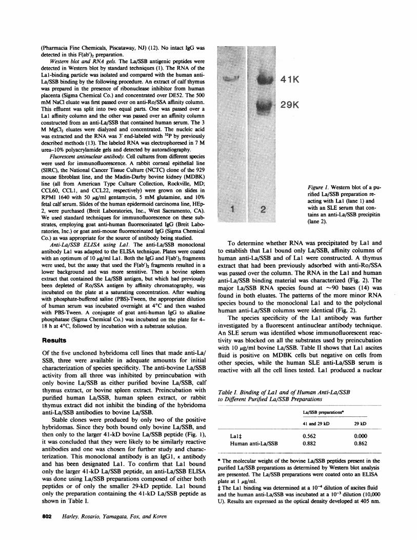
Stable clones were produced by only two of the positivehybridomas. Since they both bound only bovine La/SSB, andthen only to the larger 41-kD bovine La/SSB peptide (Fig. 1),it was concluded that they were likely to be similarly reactiveantibodies and one was chosen for further study and charac-terization. This monoclonal antibody is an IgGl, K antibodyand has been designated Lal. To confirm that Lal boundonly the larger 41-kD La/SSB peptide, an anti-La/SSB ELISAwas done using La/SSB preparations composed of either bothpeptides or of only the smaller 29-kD peptide. Lal boundonly the preparation containing the 41-kD La/SSB peptide asshown in Table I.
41
I F 29K
1 2
Figure 1. Western blot of a pu-rified La/SSB preparation re-acting with Lal (lane 1) andwith an SLE serum that con-tains an anti-La/SSB precipitin(lane 2).
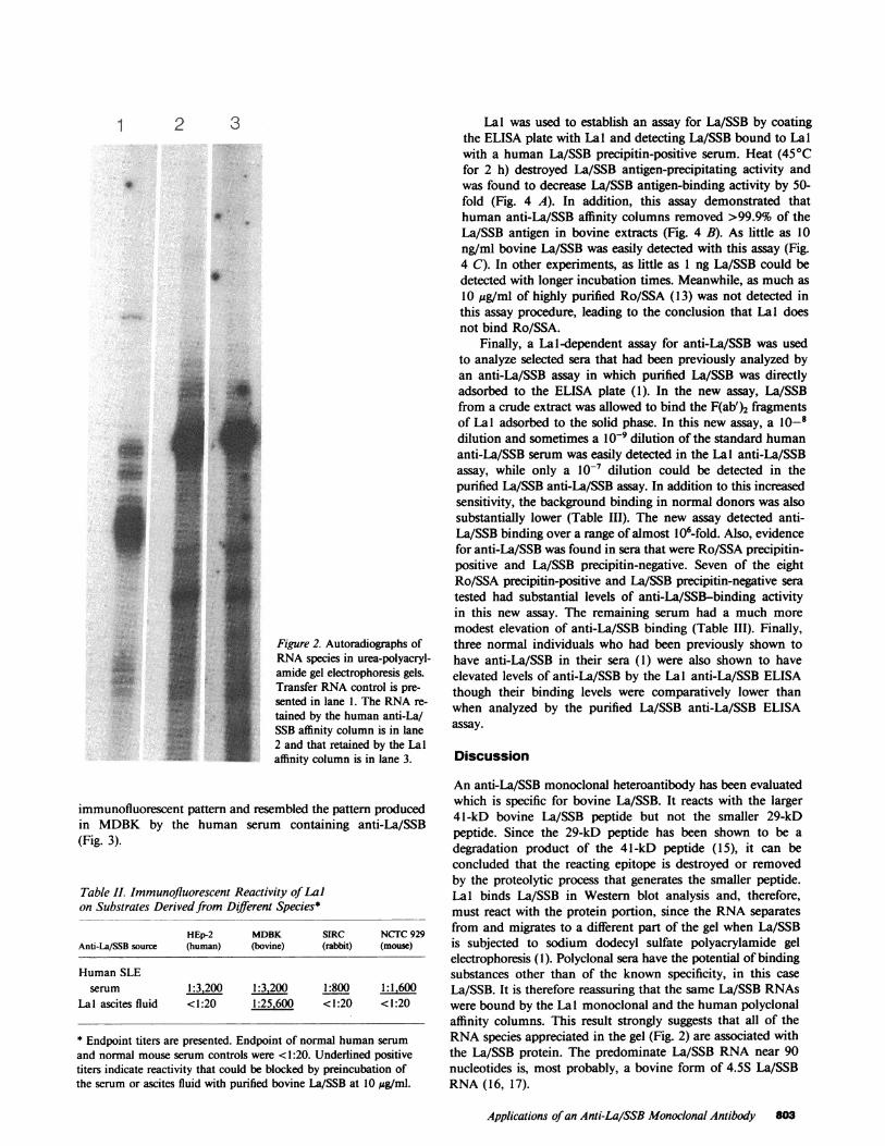
To determine whether RNAwas precipitated by Lal andto establish that Lal bound only La/SSB, affinity columns ofhuman anti-La/SSB and of Lal were constructed. A thymusextract that had been previously adsorbed with anti-Ro/SSAwas passed over the column. The RNAin the Lal and humananti-La/SSB binding material was characterized (Fig. 2). Themajor La/SSB RNA species found at -90 bases (14) wasfound in both eluates. The patterns of the more minor RNAspecies bound to the monoclonal Lal and to the polyclonalhuman anti-La/SSB columns were identical (Fig. 2).
The species specificity of the Lal antibody was furtherinvestigated by a fluorescent antinuclear antibody technique.An SLE serum was identified whose immunofluorescent reac-tivity was blocked on all the substrates used by preincubationwith 10 ,g/ml bovine La/SSB. Table II shows that Lal ascitesfluid is positive on MDBKcells but negative on cells fromother species, while the human SLE anti-La/SSB serum isreactive with all the cell lines tested. Lal produced a nuclear
Table I. Binding of Lal and of HumanAnti-La/SSBto Different Purified La/SSB Preparations
La/SSB preparations*
41 and 29 kD 29 kD
Lalt 0.562 0.000Human anti-La/SSB 0.882 0.862
* The molecular weight of the bovine La/SSB peptides present in thepurified La/SSB preparations as determined by Western blot analysisare presented. The La/SSB preparations were coated onto an ELISAplate at 1 jsg/ml.* The Lal binding was determined at a 10-4 dilution of ascites fluidand the human anti-La/SSB was incubated at a l0-3 dilution (10,000U). Results are expressed as the optical density developed at 405 nm.
802 Harley, Rosario, Yamagata, Fox, and Koren
0Ao IJ
1 2 3
S
0
Figure 2. Autoradiographs ofRNAspecies in urea-polyacryl-amide gel electrophoresis gels.Transfer RNAcontrol is pre-sented in lane 1. The RNAre-tained by the human anti-La/SSB affinity column is in lane2 and that retained by the Lalaffinity column is in lane 3.
immunofluorescent pattern and resembled the pattern producedin MDBKby the human serum containing anti-La/SSB(Fig. 3).
Table II. Immunofluorescent Reactivity of Lalon Substrates Derived from Different Species*
HEp-2 MDBK SIRC NCTC929Anti-La/SSB source (human) (bovine) (rabbit) (mouse)
Human SLEserum 1:3,200 1:3,200 1:800 1
Lal ascites fluid < 1:20 1:25,600 < 1:20 < 1:20
* Endpoint titers are presented. Endpoint of normal human serumand normal mouse serum controls were <1:20. Underlined positivetiters indicate reactivity that could be blocked by preincubation ofthe serum or ascites fluid with purified bovine La/SSB at 10 jg/ml.
Lal was used to establish an assay for La/SSB by coatingthe ELISA plate with Lal and detecting La/SSB bound to Lalwith a human La/SSB precipitin-positive serum. Heat (450Cfor 2 h) destroyed La/SSB antigen-precipitating activity andwas found to decrease La/SSB antigen-binding activity by 50-fold (Fig. 4 A). In addition, this assay demonstrated thathuman anti-La/SSB affinity columns removed >99.9% of theLa/SSB antigen in bovine extracts (Fig. 4 B). As little as 10ng/ml bovine La/SSB was easily detected with this assay (Fig.4 C). In other experiments, as little as 1 ng La/SSB could bedetected with longer incubation times. Meanwhile, as much as10 Ag/ml of highly purified Ro/SSA (13) was not detected inthis assay procedure, leading to the conclusion that Lal doesnot bind Ro/SSA.
Finally, a La1-dependent assay for anti-La/SSB was usedto analyze selected sera that had been previously analyzed byan anti-La/SSB assay in which purified La/SSB was directlyadsorbed to the ELISA plate (1). In the new assay, La/SSBfrom a crude extract was allowed to bind the F(ab')2 fragmentsof Lal adsorbed to the solid phase. In this new assay, a 10-_dilution and sometimes a IO-' dilution of the standard humananti-La/SSB serum was easily detected in the Lal anti-La/SSBassay, while only a lO-7 dilution could be detected in thepurified La/SSB anti-La/SSB assay. In addition to this increasedsensitivity, the background binding in normal donors was alsosubstantially lower (Table III). The new assay detected anti-La/SSB binding over a range of almost 106-fold. Also, evidencefor anti-La/SSB was found in sera that were Ro/SSA precipitin-positive and La/SSB precipitin-negative. Seven of the eightRo/SSA precipitin-positive and La/SSB precipitin-negative seratested had substantial levels of anti-La/SSB-binding activityin this new assay. The remaining serum had a much moremodest elevation of anti-La/SSB binding (Table III). Finally,three normal individuals who had been previously shown tohave anti-La/SSB in their sera (1) were also shown to haveelevated levels of anti-La/SSB by the Lal anti-La/SSB ELISAthough their binding levels were comparatively lower thanwhen analyzed by the purified La/SSB anti-La/SSB ELISAassay.
Discussion
An anti-La/SSB monoclonal heteroantibody has been evaluatedwhich is specific for bovine La/SSB. It reacts with the larger41-kD bovine La/SSB peptide but not the smaller 29-kDpeptide. Since the 29-kD peptide has been shown to be adegradation product of the 41-kD peptide (15), it can beconcluded that the reacting epitope is destroyed or removedby the proteolytic process that generates the smaller peptide.Lal binds La/SSB in Western blot analysis and, therefore,must react with the protein portion, since the RNAseparatesfrom and migrates to a different part of the gel when La/SSBis subjected to sodium dodecyl sulfate polyacrylamide gelelectrophoresis (1). Polyclonal sera have the potential of bindingsubstances other than of the known specificity, in this caseLa/SSB. It is therefore reassuring that the same La/SSB RNAswere bound by the La 1 monoclonal and the human polyclonalaffinity columns. This result strongly suggests that all of theRNAspecies appreciated in the gel (Fig. 2) are associated withthe La/SSB protein. The predominate La/SSB RNAnear 90nucleotides is, most probably, a bovine form of 4.5S La/SSBRNA(16, 17).
Applications of an Anti-La/SSB Monoclonal Antibody 803
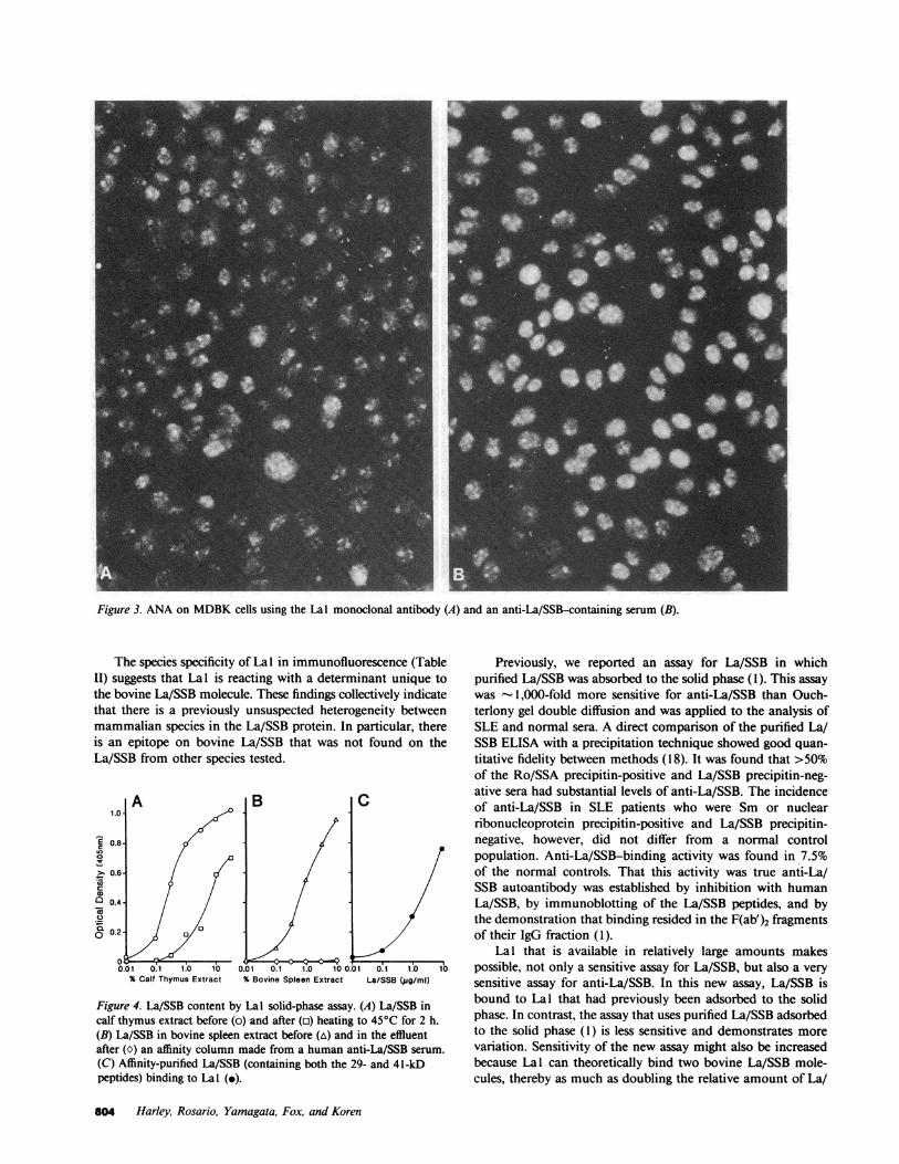
Figure 3. ANAon MDBKcells using the Lal monoclonal antibody (A) and an anti-La/SSB-containing serum (B).
The species specificity of Lal in immunofluorescence (TableII) suggests that Lal is reacting with a determinant unique tothe bovine La/SSB molecule. These findings collectively indicatethat there is a previously unsuspected heterogeneity betweenmammalian species in the La/SSB protein. In particular, thereis an epitope on bovine La/SSB that was not found on theLa/SSB from other species tested.
A B C1.0-
>, 0,,,6-,E0.8-
Q. 0.6-0
0.01 0.1 1.0 10 0.01 0.1 1.0 10 0.01 0.1 1.0 10% Calf Thymus Extract % Bovine Spleen Extract La/SSB (jg/ml)
Figure 4. La/SSB content by Lal solid-phase assay. (A) La/SSB incalf thymus extract before (o) and after (o) heating to 450C for 2 h.(B) La/SSB in bovine spleen extract before (A) and in the effluentafter (0) an affinity column made from a human anti-La/SSB serum.(C) Affinity-purified La/SSB (containing both the 29- and 41-kDpeptides) binding to Lal (-).
Previously, we reported an assay for La/SSB in whichpurified La/SSB was absorbed to the solid phase (1). This assaywas - 1,000-fold more sensitive for anti-La/SSB than Ouch-terlony gel double diffusion and was applied to the analysis ofSLE and normal sera. A direct comparison of the purified La/SSB ELISA with a precipitation technique showed good quan-titative fidelity between methods (18). It was found that >50%of the Ro/SSA precipitin-positive and La/SSB precipitin-neg-ative sera had substantial levels of anti-La/SSB. The incidenceof anti-La/SSB in SLE patients who were Sm or nuclearribonucleoprotein precipitin-positive and La/SSB precipitin-negative, however, did not differ from a normal controlpopulation. Anti-La/SSB-binding activity was found in 7.5%of the normal controls. That this activity was true anti-La/SSB autoantibody was established by inhibition with humanLa/SSB, by immunoblotting of the La/SSB peptides, and bythe demonstration that binding resided in the F(ab')2 fragmentsof their IgG fraction (1).
Lal that is available in relatively large amounts makespossible, not only a sensitive assay for La/SSB, but also a verysensitive assay for anti-La/SSB. In this new assay, La/SSB isbound to Lal that had previously been adsorbed to the solidphase. In contrast, the assay that uses purified La/SSB adsorbedto the solid phase (1) is less sensitive and demonstrates morevariation. Sensitivity of the new assay might also be increasedbecause Lal can theoretically bind two bovine La/SSB mole-cules, thereby as much as doubling the relative amount of La/
804 Harley, Rosario, Yamagata, Fox, and Koren
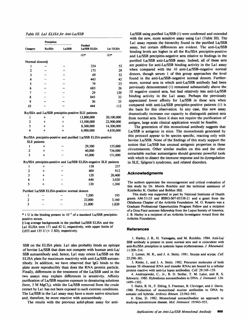
Table III. Lal ELISA for Anti-La/SSB
PrecipitinsPurified
Category Ro/SSA La/55B La/SSB ELISA Lal ELISA
(U)* (U)*
Normal donorsfI - _2 - _3 - _4 - _5 - _6 - _7 - _8 - _9 - _
10 - -
22417569
44379
68329
84530
444
Ro/SSA and La/SSB precipitin-positive SLE patientsI + + 13,800,0002 + + 13,500,0003 + + 8,300,0004 + + 6,900,000
20,100,00022,900,00014,300,0004,830,000
Ro/SSA precipitin-positive and purified La/SSB ELISA-positiveSLE patients
I +2 + _3 + _
29,50040,00045,000
155,000756,000151,000
Ro/SSA precipitin-positive and La/SSB ELISA-negative SLE patientsI + - 158 23'2 + - 409 91:3 + - 120 20,40(4 + - 640 3,02(5 + - 120 1,26C
Purified La/SSB ELISA-positive normal donorsI _ _2 - _3 - _
La/SSB using purified La/SSB (1) were confirmed and extendedwith the new, more sensitive assay using Lal (Table III). TheLal assay repeats the hierarchy found in the purified La/SSBassay, but certain differences are evident. The anti-La/SSBbinding levels are higher in all the Ro/SSA precipitin-positiveand -La/SSB precipitin-negative sera relative to findings in thepurified La/SSB anti-La/SSB assay. Indeed, all of these seraare positive for anti-La/SSB binding activity in the Lal assaywhen compared with the 10 anti-La/SSB-negative normaldonors, though serum 1 of this group approaches the levelfound in the anti-La/SSB-negative normal donors. Further-more, normal sera in which anti-La/SSB antibody had beenpreviously demonstrated (1) remained substantially above the10 negative control sera, but had relatively less anti-La/SSBbinding activity in the Lal assay. Perhaps the previouslyappreciated lower affinity for La/SSB in these sera whencompared with anti-La/SSB precipitin-positive patients (1) isthe basis for this observation. In any case, -this new assaydramatically increases our capacity to distinguish patient serafrom normal sera. Since it does not require the purification ofantigen, large scale clinical application would be feasible.
The generation of this monoclonal antibody suggests thatLa/SSB is antigenic in mice. The monoclonals generated bythis protocol appear to be species specific, reacting only withbovine La/SSB. None of the findings of this study support thenotion that La/SSB has unusual antigenic properties in thesecircumstances. Other similar studies on this and the otherextractable nuclear autoantigens should generate powerful toolswith which to dissect the immune response and its dysregulationin SLE, Sjogren's syndrome, and related disorders.
7
7,20022,00021,000
* I U is the binding present in 10-7 of a standard La/SSB precipitin-positive serum.t Log average backgrounds in the purified La/SSB ELISA and theLal ELISA were 171 and 42 U, respectively, with upper limits of2,053 and 135 U (+ 2 SD), respectively.
SSB on the ELISA plate. Lal also probably binds an epitopeof bovine La/SSB that does not compete with human anti-La/SSB autoantibody and, hence, Lal may orient La/SSB on theELISA plate for maximum reactivity with anti-La/SSB autoan-tibody. In addition, we have observed that IgG binds to theplate more reproducibly than does the RNAprotein particle.Finally, differences in the treatment of the La/SSB used in thetwo assays may explain differences in sensitivity. Affinitypurification of La/SSB requires exposure to denaturing solutions(here, 3 MMgCl2), while the La/SSB removed from the crudeextract by Lal has not been exposed to such extreme conditions.The La/SSB in the Lal assay may have a more native structureand, therefore, be more reactive with autoantibody.
The results with the previous solid-phase assay for anti-
Acknowledgments
The authors appreciate the encouragement and critical evaluation ofthis study by Dr. Morris Reichlin and the technical assistance ofKimberley K. Gaither and Bobbye Hill.
This study was supported in part by National Institutes of Healthgrants AM-31133 and BSRG-S07-05538-21 and a grant from theOklahoma Chapter of the Arthritis Foundation. M. 0. Rosario was aGraduate Professional Opportunities Program Fellow and a recipientof a Gina Finzi summer fellowship from the Lupus Society of America.J. B. Harley is a recipient of an Arthritis Investigator Award from theArthritis Foundation.
References
1. Harley, J. B., H. Yamagata, and M. Reichlin. 1984. Anti-La/SSB antibody is present in some normal sera and is coincident withanti-Ro/SSA precipitins in systemic lupus erythematosus. J Rheumatol.11:309-314.
2. Lerner, M. R., and J. A. Steitz. 1981. Snurps and scyrps. Cell.25:298-301.
3. Rinke, J., and J. A. Steitz. 1982. Precursor molecules of bothhuman 5S ribosomal RNAand transfer RNAsare bound by a cellularprotein reactive with anti-La lupus antibodies. Cell. 29:149-159.
4. Andrzejewski, C., Jr., B. D. Stollar, T. M. Lalor, and R. S.Schwartz. 1980. Hybridoma autoantibodies to DNA. J. Immunol. 124:1499-1502.
5. Hahn, B. H., F. Ebling, S. Freeman, B. Clevinger, and J. Davie.1980. Production of monoclonal murine antibodies to DNA bysomatic cell hybrids. Arthritis Rheum. 23:942-945.
6. Eilat, D. 1982. Monoclonal autoantibodies: an approach tostudying autoimmune disease. Mol. Immunol. 19:943-955.
Applications of an Anti-La/SSB Monoclonal Antibody 805
7. DeHeer, D. H., J. M. Pages, and A. E. Bussard. 1980. Specificityof antierythrocyte autoantibodies secreted by a NZB-derived hybridomaand NZB peritoneal cells. Cell. Immunol. 49:135-141.
8. Lerner, E. A., M. R. Lerner, C. A. Janeway, Jr., and J. A. Steitz.1981. Monoclonal antibodies to nucleic acid-ontaining cellular con-stituents: probes for molecular biology and autoimmune disease. Proc.Nail. Acad. Sci. USA. 78:2737-2741.
9. Galfre, G., and C. Milstein. 1981. Preparation of monoclonalantibodies: strategies and procedures. Methods Enzymol. 73:3-46.
10. Engvall, E., and P. Perlmann. 1971. Enzyme-linked immuno-sorbent assay (ELISA). Quantitative assay of immunoglobulin G.Immunochemistry. 8:871-874.
11. Brack, C., D. Portetelle, C. Glineur, and A. Bolleu. 1982. One-step purification of mouse monoclonal antibodies from ascitic fluid byDEAEAffi-Gel Blue chromatography. J. Immunol. Methods. 53:313-319.
12. Campbell, D. H., J. S. Garvey, N. E. Cremer, and D. H.Sussdorf. 1970. Methods Immunol. 224-234.
13. Yamagata, H., J. B. Harley, and M. Reichlin. 1984. Molecular
properties of the Ro/SSA antigen and enzyme-linked immunosorbentassay for quantitation of antibody. J. Clin. Invest. 74:625-633.
14. Lerner, M. R., J. R. Boyle, J. A. Hardin, and J. A. Steitz. 1981.Two novel classes of small ribonucleoproteins detected by antibodiesassociated with systemic lupus erythematosus. Science (Wash. DC).211:400-402.
15. Venables, P. J. W., P. R. Smith, and N. Maini. 1983. Purificationand characterization of the Sjogren's Syndrome A and B antigens.Clin. Exp. Immunol. 54:731-738.
16. Ro-Choi, T. S., R. Reddy, D. Henning, T. Takano, C. W.Taylor, and H. Busch. 1972. Nucleotide sequence of 4.5S ribonucleicacid, of Novikoff hepatoma cell nuclei. J. Biol. Chem. 247:3205-3222.
17. Harada, F., and N. Kato. 1980. Nucleotide sequences of 4.5SRNAsassociated with poly(A)-containing RNAsof mouse and hamstercells. Nucleic Acids Res. 8:1273-1285.
18. Harley, J. B. 1985. Autoantibodies in Sjogren's syndrome:comparison autoantibody determination methods show that antinuclearantibody and rheumatoid factor are associated with Ro/SSA precipitinformation. Protides Biol. Fluids. In press.
806 Harley, Rosario, Yamagata, Fox, and Koren