Immunohistochemical study of pig retinal development · Immunohistochemical study of pig retinal...
14
Immunohistochemical study of pig retinal development Jasenka Guduric-Fuchs, Laura J. Ringland, Ping Gu, Margaret Dellett, Desmond B. Archer, Tiziana Cogliati Centre for Vision and Vascular Sciences, Queen’s University Belfast, United Kingdom Purpose: The pig eye is similar to the human eye in terms of anatomy, vasculature, and photoreceptor distribution, and therefore provides an attractive animal model for research into retinal disease. The purpose of this study was to characterize retinal histology in the developing and mature pig retina using antibodies to well established retinal cell markers commonly used in rodents. Methods: Eyes were enucleated from fetuses in the 9th week of gestation, 1 week old piglets and 6 months old adult animals. Eyeglobes were fixed and cryosectioned. A panel of antibodies to well established retinal markers was employed for immunohistochemistry. Fluorescently labeled secondary antibodies were used for signal detection, and images were acquired by confocal microscopy. Mouse retina at postnatal day (P) 5 was used as a reference for this study to compare progression of histogenesis. Most of the primary antibodies have previously been used on mouse tissue. Results: Most of the studied markers were detected in midgestation pig retina, and the majority had a similar distribution in pig as in P5 mouse retina. However, rhodopsin immunolabeling was detected in pig retina at midgestation but not in P5 mouse retina. Contrary to findings in all rodents, horizontal cells were Islet1-positive and cones were calbindin- immunoreactive in pig retina, as has also been shown for the primate retina. Recoverin and rhodopsin immunolabeling revealed an increase in the length of photoreceptor segments in 6 months, compared to 1 week old animals. Conclusions: Comparison with the published data on human retina revealed similar marker distribution and histogenesis progression in the pig and human retina, supporting the pig as a valuable animal model for studies on retinal disease and repair. Furthermore, this study provides information about the dynamics of retinal histogenesis in the pig and validates a panel of antibodies that reliably detects developing and mature retinal cell phenotypes in the pig retina. Retinal cellular inventory and development are generally conserved across many studied vertebrate species. However, a range of histological and functional differences exist between individual species. Among nonprimate mammals, the pig eye most closely resembles the human eye, with similar size and comparable histological and physiologic features. Although the gestation period for pig (112–115 days) is significantly shorter compared to human, both human and pig retina are well developed at birth [1,2]. The architecture of the pig retina comprises an area centralis enriched in cones that resembles the human macula, making it an attractive model for preclinical testing [1,3] Pig models for retinitis pigmentosa, glaucoma, and retinal detachment have been developed [4-6]. Transgenic pigs with systemic expression of green [7] and red fluorescent proteins [8] have also been produced. Furthermore, due to its size, anatomy, and vasculature, the pig eye has been useful for modeling human ocular surgery [9-11]. Finally, pigs have been used for isolation of retinal progenitor cells, and subretinal allotransplantation of these cells demonstrates their ability to migrate, morphologically differentiate, and express retinal cell markers [12,13]. Similarly, adult retinal stem cells have been isolated and characterized from pig ciliary and iris epithelia [14,15]. Several reported in vitro studies on pig retinal neuronal survival and physiology have used immunohistochemical tools to identify cell phenotypes in the primary retinal cell culture [16-19]. At present, there are limited data on the distribution of immunohistochemical markers in the adult pig retina [20-22] and only few studies on pig retinal development [23,24]. Better understanding of pig retinal development is important to fully exploit this model’s potential for the study of eye diseases. Similarly, appropriate tools to investigate retinal histology in the pig are required to follow stem cell differentiation during development, in vitro, and after transplantation. The aim of the present study was twofold: 1) to characterize antibodies to retina-specific markers commonly used in rodents as tools for further investigations in the pig; and 2) to conduct an immunohistochemical study of retinal histogenesis in the pig. METHODS Animal models: All animal procedures were performed in compliance with the UK Animals (Scientific procedures) Act 1986. Mixed sex white Landrance pigs were obtained from Agri-Food and Biosciences Institute, Northern Ireland (Large Park, Hillsborough, Co. Down, Northern Ireland, UK) where animals were commercially bred on slats and fed with granulated pig food. The pigs were anaesthetised with intra- Molecular Vision 2009; 15:1915-1928 <http://www.molvis.org/molvis/v15/a204> Received 9 April 2009 | Accepted 14 September 2009 | Published 21 September 2009 © 2009 Molecular Vision 1915 Correspondence to: Jasenka Guduric-Fuchs, QUB-Centre for Vision and Vascular Science, RVH-Institute of Clinical Science, Belfast BT12 6BA, Northern Ireland, UK; Phone: +44-28-9063-2729; FAX: +44-28-9063-2699; email: [email protected] Dr. Cogliati is now at Neurobiology-Neurodegeneration & Repair Laboratory, National Eye Institute, NIH, Bethesda, MD 20892.
Transcript of Immunohistochemical study of pig retinal development · Immunohistochemical study of pig retinal...
Immunohistochemical study of pig retinal development
Jasenka Guduric-Fuchs, Laura J. Ringland, Ping Gu, Margaret Dellett, Desmond B. Archer, Tiziana Cogliati
Centre for Vision and Vascular Sciences, Queen’s University Belfast, United Kingdom
Purpose: The pig eye is similar to the human eye in terms of anatomy, vasculature, and photoreceptor distribution, andtherefore provides an attractive animal model for research into retinal disease. The purpose of this study was to characterizeretinal histology in the developing and mature pig retina using antibodies to well established retinal cell markers commonlyused in rodents.Methods: Eyes were enucleated from fetuses in the 9th week of gestation, 1 week old piglets and 6 months old adultanimals. Eyeglobes were fixed and cryosectioned. A panel of antibodies to well established retinal markers was employedfor immunohistochemistry. Fluorescently labeled secondary antibodies were used for signal detection, and images wereacquired by confocal microscopy. Mouse retina at postnatal day (P) 5 was used as a reference for this study to compareprogression of histogenesis. Most of the primary antibodies have previously been used on mouse tissue.Results: Most of the studied markers were detected in midgestation pig retina, and the majority had a similar distributionin pig as in P5 mouse retina. However, rhodopsin immunolabeling was detected in pig retina at midgestation but not inP5 mouse retina. Contrary to findings in all rodents, horizontal cells were Islet1-positive and cones were calbindin-immunoreactive in pig retina, as has also been shown for the primate retina. Recoverin and rhodopsin immunolabelingrevealed an increase in the length of photoreceptor segments in 6 months, compared to 1 week old animals.Conclusions: Comparison with the published data on human retina revealed similar marker distribution and histogenesisprogression in the pig and human retina, supporting the pig as a valuable animal model for studies on retinal disease andrepair. Furthermore, this study provides information about the dynamics of retinal histogenesis in the pig and validates apanel of antibodies that reliably detects developing and mature retinal cell phenotypes in the pig retina.
Retinal cellular inventory and development are generallyconserved across many studied vertebrate species. However,a range of histological and functional differences existbetween individual species. Among nonprimate mammals,the pig eye most closely resembles the human eye, with similarsize and comparable histological and physiologic features.Although the gestation period for pig (112–115 days) issignificantly shorter compared to human, both human and pigretina are well developed at birth [1,2]. The architecture of thepig retina comprises an area centralis enriched in cones thatresembles the human macula, making it an attractive modelfor preclinical testing [1,3]
Pig models for retinitis pigmentosa, glaucoma, and retinaldetachment have been developed [4-6]. Transgenic pigs withsystemic expression of green [7] and red fluorescent proteins[8] have also been produced. Furthermore, due to its size,anatomy, and vasculature, the pig eye has been useful formodeling human ocular surgery [9-11]. Finally, pigs havebeen used for isolation of retinal progenitor cells, andsubretinal allotransplantation of these cells demonstrates theirability to migrate, morphologically differentiate, and expressretinal cell markers [12,13]. Similarly, adult retinal stem cells
have been isolated and characterized from pig ciliary and irisepithelia [14,15].
Several reported in vitro studies on pig retinal neuronalsurvival and physiology have used immunohistochemicaltools to identify cell phenotypes in the primary retinal cellculture [16-19]. At present, there are limited data on thedistribution of immunohistochemical markers in the adult pigretina [20-22] and only few studies on pig retinal development[23,24]. Better understanding of pig retinal development isimportant to fully exploit this model’s potential for the studyof eye diseases. Similarly, appropriate tools to investigateretinal histology in the pig are required to follow stem celldifferentiation during development, in vitro, and aftertransplantation.
The aim of the present study was twofold: 1) tocharacterize antibodies to retina-specific markers commonlyused in rodents as tools for further investigations in the pig;and 2) to conduct an immunohistochemical study of retinalhistogenesis in the pig.
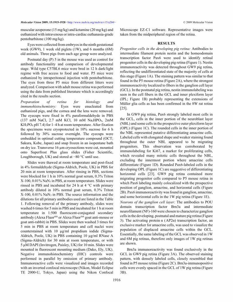
METHODSAnimal models: All animal procedures were performed incompliance with the UK Animals (Scientific procedures) Act1986. Mixed sex white Landrance pigs were obtained fromAgri-Food and Biosciences Institute, Northern Ireland (LargePark, Hillsborough, Co. Down, Northern Ireland, UK) whereanimals were commercially bred on slats and fed withgranulated pig food. The pigs were anaesthetised with intra-
Molecular Vision 2009; 15:1915-1928 <http://www.molvis.org/molvis/v15/a204>Received 9 April 2009 | Accepted 14 September 2009 | Published 21 September 2009
© 2009 Molecular Vision
1915
Correspondence to: Jasenka Guduric-Fuchs, QUB-Centre for Visionand Vascular Science, RVH-Institute of Clinical Science, BelfastBT12 6BA, Northern Ireland, UK; Phone: +44-28-9063-2729; FAX:+44-28-9063-2699; email: [email protected]. Cogliati is now at Neurobiology-Neurodegeneration & RepairLaboratory, National Eye Institute, NIH, Bethesda, MD 20892.
muscular azoperone (15 mg/kg) and ketamine (20 mg/kg) andeuthanized with intravenous or intra-cardiac euthanasia-gradepentobarbitone (100 mg/kg).
Eyes were collected from embryos in the ninth gestationalweek (GW9), 1 week old piglets (1W), and 6 months (6M)old animals. Three pigs from each age group were analyzed.
Postnatal day (P) 5 in the mouse was used as control forantibody functionality and comparison of developmentalstage. Wild type C57BL6 mice were bred in 12 h dark/lightregime with free access to food and water. P5 mice wereeuthanized by intraperitoneal injection with pentobarbitone.The eyes from three P5 mice from different litters wereanalyzed. Comparison with adult mouse retina was performedusing the data from published literature which is accordinglycited in the results section.Preparation of retina for histology andimmunohistochemistry: Eyes were enucleated fromeuthanized pigs, and the cornea and the lens were removed.The eyecups were fixed in 4% paraformaldehyde in PBS(137 mM NaCl, 2.7 mM KCl, 10 mM Na2HPO4, 2mMKH2PO4 pH 7.4) for 1–4 h at room temperature. After fixationthe specimens were cryoprotected in 10% sucrose for 6 hfollowed by 30% sucrose overnight. The eyecups wereembedded in optimal cutting temperature compound (OCT;Sakura, Kobe, Japan) and snap frozen in an isopentane bathon dry ice. Transverse 16 µm cryosections were cut, mountedonto Superfrost Plus glass slides (Fisher Scientific,Loughborough, UK) and stored at −80 °C until use.
Slides were thawed at room temperature and post-fixedin 4% formaldehyde (Sigma-Aldrich, Poole, UK) in PBS for20 min at room temperature. After rinsing in PBS, sectionswere blocked for 1 h in 10% normal goat serum, 0.3% TritonX-100, 0.01% NaN3 in PBS, at room temperature. Slides wererinsed in PBS and incubated for 24 h at 4 °C with primaryantibody diluted in 10% normal goat serum, 0.3% TritonX-100, 0.01% NaN3 in PBS. The source information and thedilutions for all primary antibodies used are listed in the Table1. Following removal of the primary antibody, slides werewashed 6 times for 5 min in PBS and incubated for 1 h at roomtemperature in 1:500 fluorescent-conjugated secondaryantibody (Alexa Fluor488 or Alexa Fluor568 goat anti-mouse orgoat anti-rabbit) in PBS. Slides were then washed 3 times for5 min in PBS at room temperature and cell nuclei werecounterstained with 10 µg/ml propidium iodide (Sigma-Aldrich, Poole, UK) in PBS containing 10 µg/ml RNase A(Sigma-Aldrich) for 30 min at room temperature, or with5 μM DAPI (Invitrogen, Paisley, UK) for 10 min. Slides weremounted in fluorescent mounting medium (Dako, Ely, UK).Negative immunohistochemistry (IHC) controls wereperformed in parallel by omission of primary antibody.Immunoreactive cells were visualized and images recordedwith an inverted confocal microscope (Nikon, Model EclipseTE 2000-U, Tokyo, Japan) using the Nikon Confocal
Microscope EZ-C1 software. Representative images weretaken from the midperipheral region of the retina.
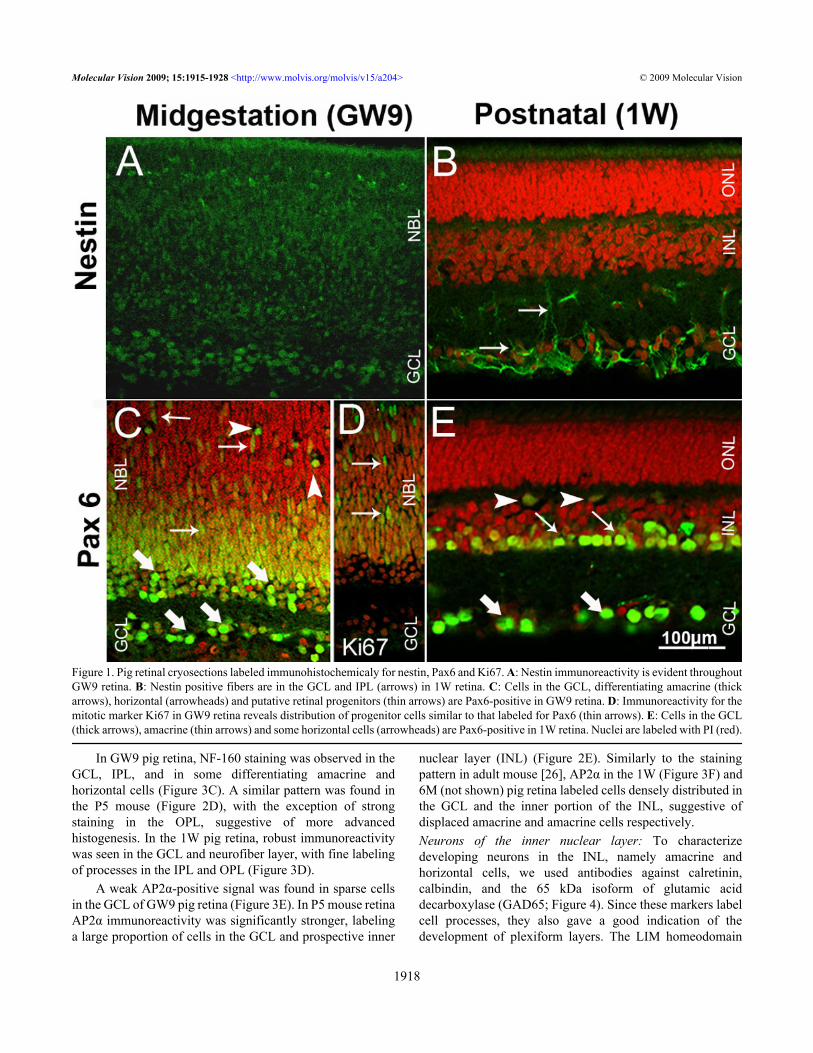
RESULTSProgenitor cells in the developing pig retina: Antibodies tointermediate filament protein nestin and the homeodomaintranscription factor Pax6 were used to identify retinalprogenitor cells in the developing pig retina (Figure 1). Nestinimmunoreactivity was detected throughout GW9 pig retina,reflecting the undifferentiated state of the majority of cells atthis stage (Figure 1A). The staining pattern was similar to thatfound in the P5 mouse retina (Figure 2A), where the strongestimmunoreactivity localized to fibers in the ganglion cell layer(GCL). In the postnatal pig retina, nestin immunolabeling wasseen in the cell fibers in the GCL and inner plexiform layer(IPL; Figure 1B) probably representing the extensions ofMüller glia cells as has been confirmed in the 8W rat retina[25].
In GW9 pig retina, Pax6 strongly labeled most cells inthe GCL, cells in the inner portion of the neuroblast layer(NBL) and some cells in the prospective outer plexiform layer(OPL) (Figure 1C). The rounded cells in the inner portion ofthe NBL represented putative differentiating amacrine cells.Labeled cells with elongated shape and weaker staining foundthroughout the outer NBL appeared to be migratingprogenitors. This observation was corroborated byimmunolabeling for Ki67, a marker for proliferating cells,which revealed many mitotic cells throughout the NBL,excluding the innermost portion where amacrine cellsdifferentiate (Figure 1D). Rounded Pax6-labeled cells in thedeveloping OPL (Figure 1C) are suggestive of differentiatinghorizontal cells [23]. GW9 pig retina contained moremigrating progenitor cells compared to P5 mouse retina inwhich Pax6 labeling mainly colocalized with the prospectiveposition of ganglion, amacrine, and horizontal cells (Figure2B). Pax6 immunoreactivity was found in ganglion, amacrine,and some horizontal cells in the 1W pig retina (Figure 1E).Neurons of the ganglion cell layer: The antibodies to POUdomain transcription factor Brn3a and intermediateneurofilament (NF)-160 were chosen to characterize ganglioncells in the developing, postnatal and mature pig retina (Figure3). The activating protein α (AP2α) transcription factor, anexclusive marker for amacrine cells, was used to visualize thepopulation of displaced amacrine cells within the GCL.Essentially, the same labeling of the GCL was observed in 1Wand 6M pig retinas, therefore only images of 1W pig retinasare shown.
Brn3a immunoreactivity was found exclusively in theGCL in GW9 pig retina (Figure 3A). The observed stainingpattern, with densely labeled cells, closely resembled thatfound in P5 mouse retina (Figure 2C). Brn3a-immunopositivecells were evenly spaced in the GCL of 1W pig retina (Figure3B).
Molecular Vision 2009; 15:1915-1928 <http://www.molvis.org/molvis/v15/a204> © 2009 Molecular Vision
1916
T AB
LE 1
. PR
IMA
RY
AN
TIB
OD
IES U
SED
FOR IM
MU
NO
HIS
TOC
HEM
ICA
L A
NA
LYSI
S OF T
HE
PIG
RET
INA
.
Ant
ibod
yC
ell s
peci
ficity
Hos
tD
ilutio
nSo
urce
Ref
eren
ceN
estin
neur
al st
em/p
roge
nito
r cel
ls, M
ülle
r glia
mou
se1:
400
BD
Bio
scie
nces
[25,
41,4
4]Pa
x6re
tinal
pro
geni
tors
, gan
glio
n, a
mac
rine,
and
horiz
onta
l cel
lsra
bbit
1:1,
000
Che
mic
on[2
8,45
]
Ki6
7pr
olife
ratin
g ce
llsm
ouse
1:30
0B
D B
iosc
ienc
es[4
6]B
rn3a
gang
lion
cells
mou
se1:
40C
hem
icon
[47]
Neu
rofil
amen
t(N
F)-1
60ga
nglio
n, h
oriz
onta
l, an
d bi
pola
r cel
lsm
ouse
1:35
0Si
gma
[22,
38,4
8,49
]
AP2α
amac
rine
cells
mou
se1:
50D
evel
opm
enta
l Stu
dies
Hyb
ridom
a B
ank
[26]
Cal
retin
inga
nglio
n, a
mac
rine,
hor
izon
tal,
cone
pho
tore
cept
or,
and
bipo
lar c
ells
mou
se1:
500
Che
mic
on[3
3,50
]
Cal
bind
inho
rizon
tal,
gang
lion,
am
acrin
e, b
ipol
ar, a
nd c
one
phot
orec
epto
r cel
lsra
bbit
1:1,
500
Che
mic
on[2
7,51
,52]
GA
D65
GA
BA
ergi
c am
acrin
e, ra
re h
oriz
onta
l gan
glio
n ce
llsm
ouse
1:1,
000
Dev
elop
men
tal S
tudi
esH
ybrid
oma
Ban
k[2
7,28
]
Isle
t1am
acrin
e, b
ipol
ar, g
angl
ion,
and
hor
izon
tal c
ells
mou
se1:
500
Dev
elop
men
tal S
tudi
esH
ybrid
oma
Ban
k[2
9,37
]
PKCα
rod
bipo
lar,
amac
rine,
and
gan
glio
n ce
lls, c
ones
mou
se1:
400
Sigm
a[2
7,30
,53]
Rec
over
inph
otor
ecep
tors
, con
e bip
olar
cells
, rar
e gan
glio
n ce
llsra
bbit
1:1,
000
K. K
och
[42,
51,5
4]R
hodo
psin
(Rho
4D2)
rods
mou
se1:
100
R. M
olda
y[2
,36,
43]
GN
AT2
cone
sra
bbit
1:50
0Sa
nta
Cru
zB
iote
chno
logy
[55-
57]
P75
neur
otro
phin
rece
ptor
Mül
ler g
lia a
nd g
angl
ion
cells
rabb
it1:
350
Prom
ega
[31,
32,5
8]
Glu
tam
ine
synt
heta
seM
ülle
r glia
mou
se1:
500
BD
Bio
scie
nces
[35,
59]
GFA
Pas
trocy
tes
rabb
it1:
500
DA
KO
[60-
62]
The
cell
spec
ifici
ty is
des
crib
ed a
ccor
ding
to th
e pu
blis
hed
liter
atur
e (li
sted
in th
e re
fere
nce
colu
mn)
on
the
antib
odie
s to
the
sam
e pr
otei
ns. D
etai
ls fo
r the
prim
ary
antib
odie
s (ho
st, d
ilutio
n us
ed a
nd th
e so
urce
) use
d in
our
stud
y ar
e pr
esen
ted.
Molecular Vision 2009; 15:1915-1928 <http://www.molvis.org/molvis/v15/a204> © 2009 Molecular Vision
1917
In GW9 pig retina, NF-160 staining was observed in theGCL, IPL, and in some differentiating amacrine andhorizontal cells (Figure 3C). A similar pattern was found inthe P5 mouse (Figure 2D), with the exception of strongstaining in the OPL, suggestive of more advancedhistogenesis. In the 1W pig retina, robust immunoreactivitywas seen in the GCL and neurofiber layer, with fine labelingof processes in the IPL and OPL (Figure 3D).
A weak AP2α-positive signal was found in sparse cellsin the GCL of GW9 pig retina (Figure 3E). In P5 mouse retinaAP2α immunoreactivity was significantly stronger, labelinga large proportion of cells in the GCL and prospective inner
nuclear layer (INL) (Figure 2E). Similarly to the stainingpattern in adult mouse [26], AP2α in the 1W (Figure 3F) and6M (not shown) pig retina labeled cells densely distributed inthe GCL and the inner portion of the INL, suggestive ofdisplaced amacrine and amacrine cells respectively.Neurons of the inner nuclear layer: To characterizedeveloping neurons in the INL, namely amacrine andhorizontal cells, we used antibodies against calretinin,calbindin, and the 65 kDa isoform of glutamic aciddecarboxylase (GAD65; Figure 4). Since these markers labelcell processes, they also gave a good indication of thedevelopment of plexiform layers. The LIM homeodomain
Figure 1. Pig retinal cryosections labeled immunohistochemicaly for nestin, Pax6 and Ki67. A: Nestin immunoreactivity is evident throughoutGW9 retina. B: Nestin positive fibers are in the GCL and IPL (arrows) in 1W retina. C: Cells in the GCL, differentiating amacrine (thickarrows), horizontal (arrowheads) and putative retinal progenitors (thin arrows) are Pax6-positive in GW9 retina. D: Immunoreactivity for themitotic marker Ki67 in GW9 retina reveals distribution of progenitor cells similar to that labeled for Pax6 (thin arrows). E: Cells in the GCL(thick arrows), amacrine (thin arrows) and some horizontal cells (arrowheads) are Pax6-positive in 1W retina. Nuclei are labeled with PI (red).
Molecular Vision 2009; 15:1915-1928 <http://www.molvis.org/molvis/v15/a204> © 2009 Molecular Vision
1918
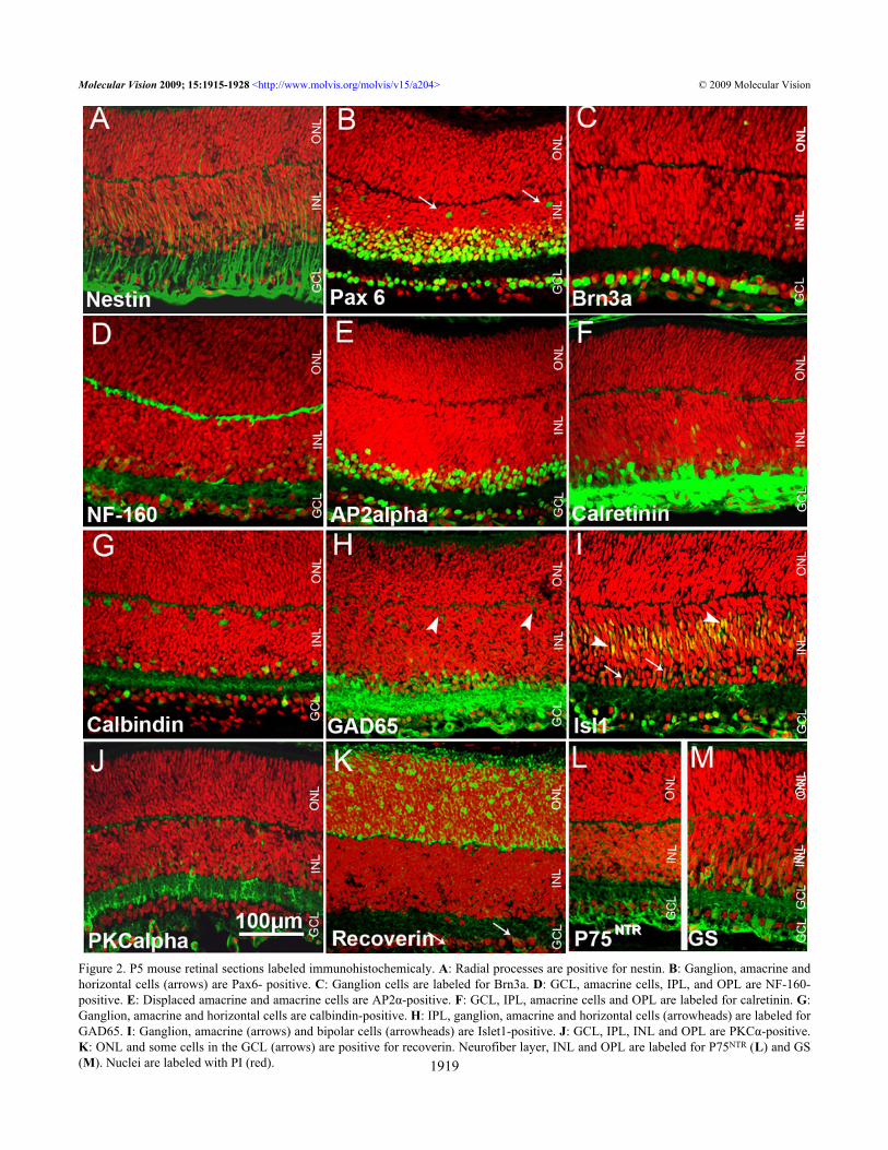
Figure 2. P5 mouse retinal sections labeled immunohistochemicaly. A: Radial processes are positive for nestin. B: Ganglion, amacrine andhorizontal cells (arrows) are Pax6- positive. C: Ganglion cells are labeled for Brn3a. D: GCL, amacrine cells, IPL, and OPL are NF-160-positive. E: Displaced amacrine and amacrine cells are AP2α-positive. F: GCL, IPL, amacrine cells and OPL are labeled for calretinin. G:Ganglion, amacrine and horizontal cells are calbindin-positive. H: IPL, ganglion, amacrine and horizontal cells (arrowheads) are labeled forGAD65. I: Ganglion, amacrine (arrows) and bipolar cells (arrowheads) are Islet1-positive. J: GCL, IPL, INL and OPL are PKCα-positive.K: ONL and some cells in the GCL (arrows) are positive for recoverin. Neurofiber layer, INL and OPL are labeled for P75NTR (L) and GS(M). Nuclei are labeled with PI (red).
Molecular Vision 2009; 15:1915-1928 <http://www.molvis.org/molvis/v15/a204> © 2009 Molecular Vision
1919
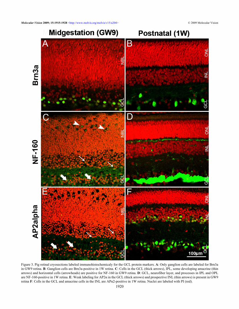
Figure 3. Pig retinal cryosections labeled immunohistochemicaly for the GCL protein markers. A: Only ganglion cells are labeled for Brn3ain GW9 retina. B: Ganglion cells are Brn3a-positive in 1W retina. C: Cells in the GCL (thick arrows), IPL, some developing amacrine (thinarrows) and horizontal cells (arrowheads) are positive for NF-160 in GW9 retina. D: GCL, neurofiber layer, and processes in IPL and OPLare NF-160-positive in 1W retina. E: Weak labeling for AP2α in the GCL (thick arrows) and prospective INL (thin arrows) is present in GW9retina F: Cells in the GCL and amacrine cells in the INL are APα2-positive in 1W retina. Nuclei are labeled with PI (red).
Molecular Vision 2009; 15:1915-1928 <http://www.molvis.org/molvis/v15/a204> © 2009 Molecular Vision
1920
transcription factor Islet1 and protein kinase C α isoform(PKCα) were primarily used to visualize bipolar cells (Figure5).
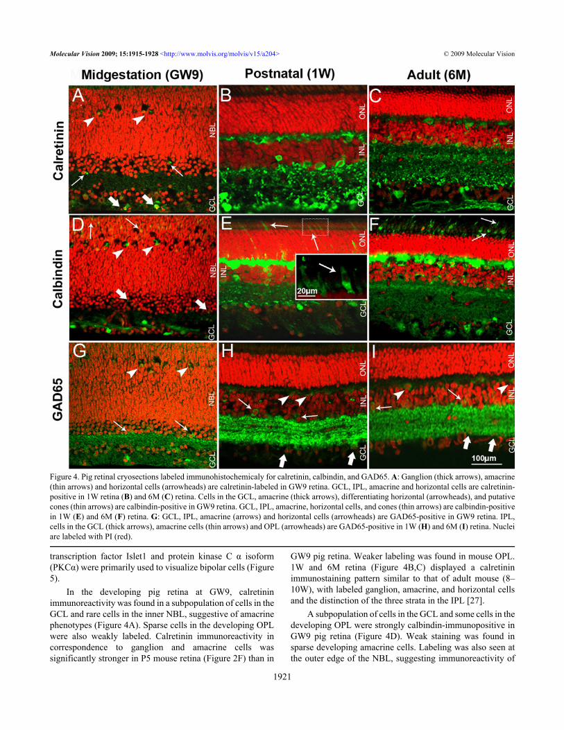
In the developing pig retina at GW9, calretininimmunoreactivity was found in a subpopulation of cells in theGCL and rare cells in the inner NBL, suggestive of amacrinephenotypes (Figure 4A). Sparse cells in the developing OPLwere also weakly labeled. Calretinin immunoreactivity incorrespondence to ganglion and amacrine cells wassignificantly stronger in P5 mouse retina (Figure 2F) than in
GW9 pig retina. Weaker labeling was found in mouse OPL.1W and 6M retina (Figure 4B,C) displayed a calretininimmunostaining pattern similar to that of adult mouse (8–10W), with labeled ganglion, amacrine, and horizontal cellsand the distinction of the three strata in the IPL [27].
A subpopulation of cells in the GCL and some cells in thedeveloping OPL were strongly calbindin-immunopositive inGW9 pig retina (Figure 4D). Weak staining was found insparse developing amacrine cells. Labeling was also seen atthe outer edge of the NBL, suggesting immunoreactivity of
Figure 4. Pig retinal cryosections labeled immunohistochemicaly for calretinin, calbindin, and GAD65. A: Ganglion (thick arrows), amacrine(thin arrows) and horizontal cells (arrowheads) are calretinin-labeled in GW9 retina. GCL, IPL, amacrine and horizontal cells are calretinin-positive in 1W retina (B) and 6M (C) retina. Cells in the GCL, amacrine (thick arrows), differentiating horizontal (arrowheads), and putativecones (thin arrows) are calbindin-positive in GW9 retina. GCL, IPL, amacrine, horizontal cells, and cones (thin arrows) are calbindin-positivein 1W (E) and 6M (F) retina. G: GCL, IPL, amacrine (arrows) and horizontal cells (arrowheads) are GAD65-positive in GW9 retina. IPL,cells in the GCL (thick arrows), amacrine cells (thin arrows) and OPL (arrowheads) are GAD65-positive in 1W (H) and 6M (I) retina. Nucleiare labeled with PI (red).
Molecular Vision 2009; 15:1915-1928 <http://www.molvis.org/molvis/v15/a204> © 2009 Molecular Vision
1921
some developing photoreceptors. Labeling of amacrine cellsand of the IPL was more prominent in P5 mouse retina (Figure2G) compared to GW9 pig, where the INL and IPL onlyoccasionally displayed weak positive signal. The labelingpattern of horizontal, amacrine, and ganglion cells and theirprocesses in the OPL and IPL in both 1W and 6M pig retinas(Figure 4E,F) closely resembled that described for the adultmouse retina. In the pig (1W and 6M) retina, calbindin alsolabeled sparse photoreceptors, which had a typical wide andtapered cone-like shape (Figure 4E, inset), and did not stainfor rhodopsin in double labeling experiments (not shown).Cones appeared immature and mostly inner segments werevisualized in 1W pig retina (Figure 4E, inset). At 6M, coneswere intensely labeled for calbindin, with inner and outersegments clearly visible (Figure 4F).
In the GW9 pig retina, GAD65 immunostaining wasfound in amacrine and ganglion cell bodies, with strongersignal localized to the IPL (Figure 4G). Some GAD65-positive cells with immature labeled processes, suggestive ofdeveloping horizontal cells, were also highlighted. Thisstaining pattern was similar to P5 mouse retina (Figure 2H),albeit less intense. In mouse, weak signal was also seen in the
developing horizontal cells and OPL. In postnatal and adultpig retinas, strong labeling was found in processes layeringthe IPL, with a characteristic striated appearance (Figure 4H,I)as seen in adult mouse retina [27,28]. Some labeled horizontalcell bodies were visible in 6M pig retina (Figure 4I).
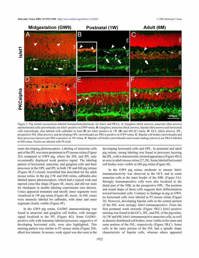
In the GW9 pig retina, moderate to intense Islet1immunoreactivity was observed in the GCL and in someamacrine cells at the inner border of the NBL (Figure 5A).Strongly immunopositive cells were also localized to thedistal part of the NBL in the prospective OPL. The positionand round shape of these cells suggests their differentiationtoward horizontal cells. Contrary to findings in pig at GW9,no horizontal cells were labeled in P5 mouse retina (Figure2I). However, developing bipolar cells in the central portionof the INL were strongly Islet1-immunopositive. From thefirst postnatal week onwards (Figure 5B,C) Islet1-positivestaining was found in the GCL, IPL, and INL of the pig retina.At 1W and 6M, Islet1-immunoreactive amacrine cells, as wellas densely distributed cell bodies, were visible in the inner andouter portion of the INL, respectively (Figure 5B,C). Somecells in the outer portion of the INL had a spindle shapecharacteristic of bipolar cells, whereas others appeared
Figure 5. Pig retinal cryosections labeled immunohistochemicaly for Islet1 and PKCα. A: Ganglion (thick arrows), amacrine (thin arrows)and horizontal cells (arrowheads) are Islet1-positive in GW9 retina. B: Ganglion, amacrine (thick arrows), bipolar (thin arrows) and horizontalcells (arrowheads, also labeled with calbindin in inset B) are Islet1-positive in 1W (B) and 6M (C) retina. D: GCL (thick arrows), IPL,prospective INL (thin arrows), and developing OPL (arrowheads) are PKCα-positive in GW9 retina. E: Bipolar cell bodies (arrowheads) andtheir processes (arrows) are PKCα-positive in 1W retina. F: Bipolar cell bodies (arrowheads) and axonal endings (arrows) are PKCα-labeledin 6M retina. Nuclei are labeled with PI (red).
Molecular Vision 2009; 15:1915-1928 <http://www.molvis.org/molvis/v15/a204> © 2009 Molecular Vision
1922
rounded, suggestive of horizontal cell phenotypes. Double-labeling experiments with calbindin (Figure 5B, inset)confirmed the presence of Islet1-immunoreactive horizontalcells in 1W pig retina, a feature also described for humanretina (12–85 years-old) [29].
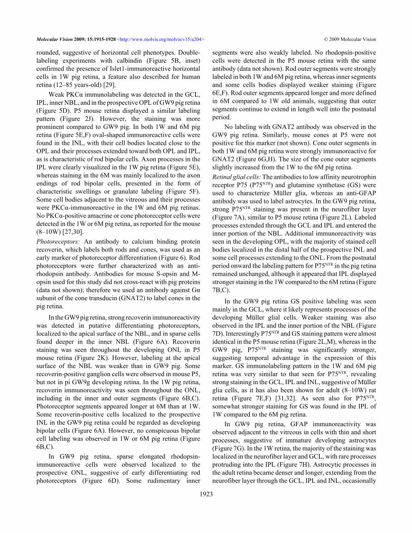
Weak PKCα immunolabeling was detected in the GCL,IPL, inner NBL, and in the prospective OPL of GW9 pig retina(Figure 5D). P5 mouse retina displayed a similar labelingpattern (Figure 2J). However, the staining was moreprominent compared to GW9 pig. In both 1W and 6M pigretina (Figure 5E,F) oval-shaped immunoreactive cells werefound in the INL, with their cell bodies located close to theOPL and their processes extended toward both OPL and IPL,as is characteristic of rod bipolar cells. Axon processes in theIPL were clearly visualized in the 1W pig retina (Figure 5E),whereas staining in the 6M was mainly localized to the axonendings of rod bipolar cells, presented in the form ofcharacteristic swellings or granulate labeling (Figure 5F).Some cell bodies adjacent to the vitreous and their processeswere PKCα-immunoreactive in the 1W and 6M pig retinas.No PKCα-positive amacrine or cone photoreceptor cells weredetected in the 1W or 6M pig retina, as reported for the mouse(8–10W) [27,30].Photoreceptors: An antibody to calcium binding proteinrecoverin, which labels both rods and cones, was used as anearly marker of photoreceptor differentiation (Figure 6). Rodphotoreceptors were further characterized with an anti-rhodopsin antibody. Antibodies for mouse S-opsin and M-opsin used for this study did not cross-react with pig proteins(data not shown); therefore we used an antibody against Gαsubunit of the cone transducin (GNAT2) to label cones in thepig retina.
In the GW9 pig retina, strong recoverin immunoreactivitywas detected in putative differentiating photoreceptors,localized to the apical surface of the NBL, and in sparse cellsfound deeper in the inner NBL (Figure 6A). Recoverinstaining was seen throughout the developing ONL in P5mouse retina (Figure 2K). However, labeling at the apicalsurface of the NBL was weaker than in GW9 pig. Somerecoverin-positive ganglion cells were observed in mouse P5,but not in pi GW9g developing retina. In the 1W pig retina,recoverin immunoreactivity was seen throughout the ONL,including in the inner and outer segments (Figure 6B,C).Photoreceptor segments appeared longer at 6M than at 1W.Some recoverin-positive cells localized to the prospectiveINL in the GW9 pig retina could be regarded as developingbipolar cells (Figure 6A). However, no conspicuous bipolarcell labeling was observed in 1W or 6M pig retina (Figure6B,C).
In GW9 pig retina, sparse elongated rhodopsin-immunoreactive cells were observed localized to theprospective ONL, suggestive of early differentiating rodphotoreceptors (Figure 6D). Some rudimentary inner
segments were also weakly labeled. No rhodopsin-positivecells were detected in the P5 mouse retina with the sameantibody (data not shown). Rod outer segments were stronglylabeled in both 1W and 6M pig retina, whereas inner segmentsand some cells bodies displayed weaker staining (Figure6E,F). Rod outer segments appeared longer and more definedin 6M compared to 1W old animals, suggesting that outersegments continue to extend in length well into the postnatalperiod.
No labeling with GNAT2 antibody was observed in theGW9 pig retina. Similarly, mouse cones at P5 were notpositive for this marker (not shown). Cone outer segments inboth 1W and 6M pig retina were strongly immunoreactive forGNAT2 (Figure 6G,H). The size of the cone outer segmentsslightly increased from the 1W to the 6M pig retina.Retinal glial cells: The antibodies to low affinity neurotrophinreceptor P75 (P75NTR) and glutamine synthetase (GS) wereused to characterize Müller glia, whereas an anti-GFAPantibody was used to label astrocytes. In the GW9 pig retina,strong P75NTR staining was present in the neurofiber layer(Figure 7A), similar to P5 mouse retina (Figure 2L). Labeledprocesses extended through the GCL and IPL and entered theinner portion of the NBL. Additional immunoreactivity wasseen in the developing OPL, with the majority of stained cellbodies localized in the distal half of the prospective INL andsome cell processes extending to the ONL. From the postnatalperiod onward the labeling pattern for P75NTR in the pig retinaremained unchanged, although it appeared that IPL displayedstronger staining in the 1W compared to the 6M retina (Figure7B,C).
In the GW9 pig retina GS positive labeling was seenmainly in the GCL, where it likely represents processes of thedeveloping Müller glial cells. Weaker staining was alsoobserved in the IPL and the inner portion of the NBL (Figure7D). Interestingly P75NTR and GS staining pattern were almostidentical in the P5 mouse retina (Figure 2L,M), whereas in theGW9 pig, P75NTR staining was significantly stronger,suggesting temporal advantage in the expression of thismarker. GS immunolabeling pattern in the 1W and 6M pigretina was very similar to that seen for P75NTR, revealingstrong staining in the GCL, IPL and INL, suggestive of Müllerglia cells, as it has also been shown for adult (8–10W) ratretina (Figure 7E,F) [31,32]. As seen also for P75NTR,somewhat stronger staining for GS was found in the IPL of1W compared to the 6M pig retina.
In GW9 pig retina, GFAP immunoreactivity wasobserved adjacent to the vitreous in cells with thin and shortprocesses, suggestive of immature developing astrocytes(Figure 7G). In the 1W retina, the majority of the staining waslocalized in the neurofiber layer and GCL, with rare processesprotruding into the IPL (Figure 7H). Astrocytic processes inthe adult retina became denser and longer, extending from theneurofiber layer through the GCL, IPL and INL, occasionally
Molecular Vision 2009; 15:1915-1928 <http://www.molvis.org/molvis/v15/a204> © 2009 Molecular Vision
1923
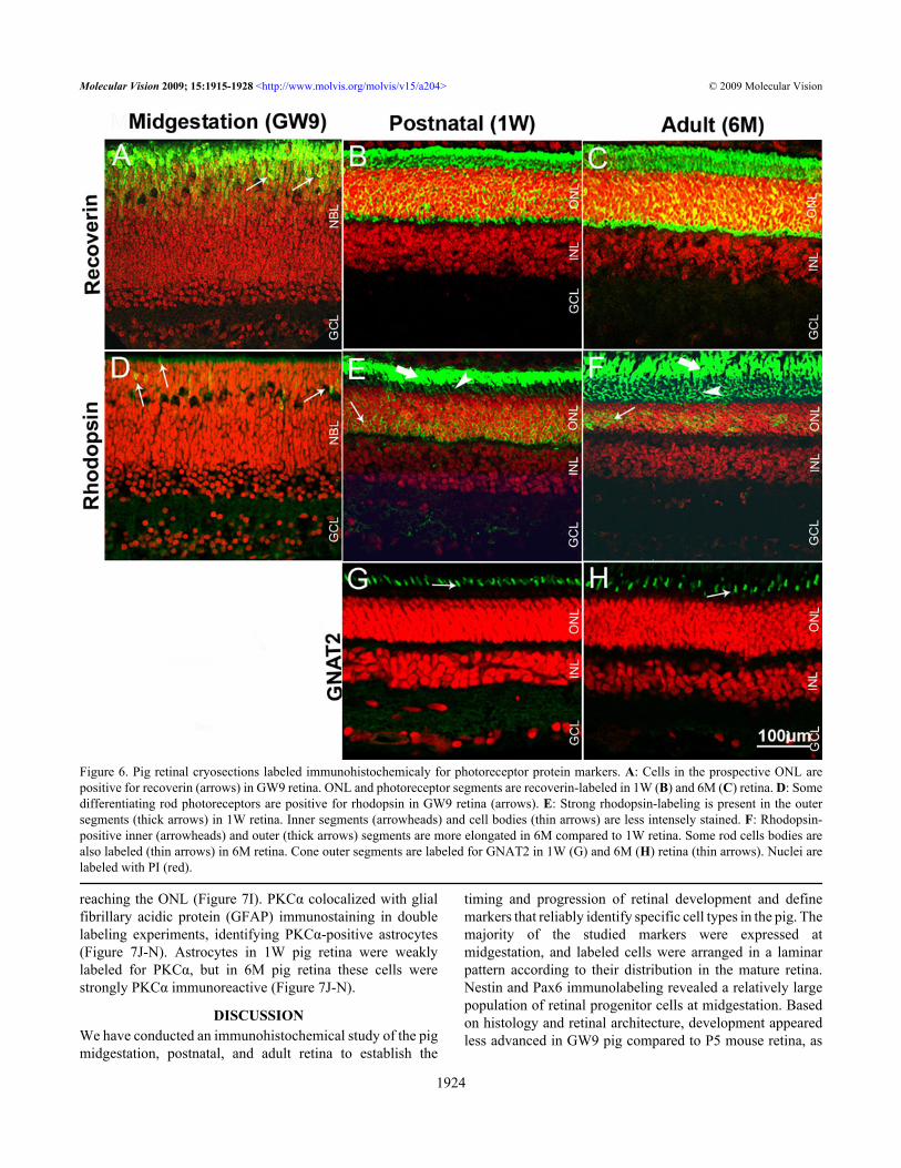
reaching the ONL (Figure 7I). PKCα colocalized with glialfibrillary acidic protein (GFAP) immunostaining in doublelabeling experiments, identifying PKCα-positive astrocytes(Figure 7J-N). Astrocytes in 1W pig retina were weaklylabeled for PKCα, but in 6M pig retina these cells werestrongly PKCα immunoreactive (Figure 7J-N).
DISCUSSIONWe have conducted an immunohistochemical study of the pigmidgestation, postnatal, and adult retina to establish the
timing and progression of retinal development and definemarkers that reliably identify specific cell types in the pig. Themajority of the studied markers were expressed atmidgestation, and labeled cells were arranged in a laminarpattern according to their distribution in the mature retina.Nestin and Pax6 immunolabeling revealed a relatively largepopulation of retinal progenitor cells at midgestation. Basedon histology and retinal architecture, development appearedless advanced in GW9 pig compared to P5 mouse retina, as
Figure 6. Pig retinal cryosections labeled immunohistochemicaly for photoreceptor protein markers. A: Cells in the prospective ONL arepositive for recoverin (arrows) in GW9 retina. ONL and photoreceptor segments are recoverin-labeled in 1W (B) and 6M (C) retina. D: Somedifferentiating rod photoreceptors are positive for rhodopsin in GW9 retina (arrows). E: Strong rhodopsin-labeling is present in the outersegments (thick arrows) in 1W retina. Inner segments (arrowheads) and cell bodies (thin arrows) are less intensely stained. F: Rhodopsin-positive inner (arrowheads) and outer (thick arrows) segments are more elongated in 6M compared to 1W retina. Some rod cells bodies arealso labeled (thin arrows) in 6M retina. Cone outer segments are labeled for GNAT2 in 1W (G) and 6M (H) retina (thin arrows). Nuclei arelabeled with PI (red).
Molecular Vision 2009; 15:1915-1928 <http://www.molvis.org/molvis/v15/a204> © 2009 Molecular Vision
1924
Figure 7. Pig retinal cryosections labeled for glial protein markers. A: Processes in the neurofiber layer, GCL, OPL, inner NBL, and developingOPL are P75NTR-positive in GW9 retina. Processes in the GCL, IPL, and INL are P75NTR-positive in 1W (B) and 6M (C) retina. D: Processesin the GCL (thick arrows), NBL (thin arrows), and developing OPL (arrowheads) are labeled for GS in GW9 retina. GS immunolabeling ispresent in the GCL, IPL, and INL in 1W (E) and 6M (F) retina. G: Immature astrocytes are GFAP-positive in GW9 retina. H: Processes inneurofiber and GCL are GFAP- positive in 1W retina. I: Elongated GFAP-positive processes in 6M retina. J-M: GFAP-immunopositiveastrocytes are PKCα-positive, weakly in 1W (J, K, arrows) but strongly in 6M retina (L, M, arrows). N: A Z-stack image shows extensivedouble GFAP-PKCα labeling at 6M (arrows). X- and Y- planes are shown to bottom and to the right of the image.
Molecular Vision 2009; 15:1915-1928 <http://www.molvis.org/molvis/v15/a204> © 2009 Molecular Vision
1925
suggested by the well demarcated developing OPL present inmouse but not visible in the pig retina. Histogenesis, asdetected by immunohistochemistry, appeared to follow adifferent sequential order in pig compared to mouse.Immunoreactivity of ganglion cells in GW9 pig was similarto that in P5 mouse. Horizontal cells were strongly calbindin-and NF-160-immunoreactive in both GW9 pig and P5 mouseretina. However, AP2α, calretinin, calbindin, Islet1, andPKCα immunolabeling indicated that development ofamacrine and bipolar cells was significantly less advanced inGW9 pig compared to P5 mouse. On the contrary, sparsedeveloping rod photoreceptors were identified in GW9 pigretina, while P5 mouse retina did not show any rhodopsinimmunoreactivity. These observations suggest that roddifferentiation occurs earlier in pig than in mouse, relative tothe development of the inner retina.
Bearing in mind the potential of the pig as a preclinicalanimal model for retinal disease, it is important to highlightthe similarities and differences between human and pig retina.Similar to the human, the pig retina is relatively mature at birthwith all layers and retinal cell types present in one-week-oldanimals. GW9 pig retinas appeared to be at an earlierdevelopmental stage than GW20–21 human retina, based onpublished literature [33]. At GW9 in pig only early signs ofOPL development were evident, whereas in human GW9retina separation between ONL and INL is fully accomplished[33]. Pax6 labeling in GW9 pig retina revealed cells ofelongated shape distributed throughout the NBL, likelymigrating retinal progenitors, whereas Pax6 immunostaininghas been described in the human retina at GW21 only indifferentiated ganglion cells and differentiating amacrine andhorizontal cells [34]. Similarly, expression of the calciumbinding proteins, calretinin and calbindin, appeared to belagging behind in GW9 pig compared to the GW20–21 humanretina. In the GW21 human retina, both calretinin andcalbindin robustly label ganglion and amacrine cells, withweaker staining of horizontal cells and additional labeling ofcones [33]. However, only sparse cells in the GCL and NBLwere calretinin- or calbindin-immunoreactive at GW9 in pig.Additionally, Müller glial cells are strongly GS positive in theGW20 human retina [35], but only weak labeling of theprocesses in the GCL was seen in the GW9 pig. Earlyimmunolabeling of rhodopsin at GW9 in the pig retina iscomparable to that in human, where rhodopsinimmunoreactivity has been shown at GW15 in rod innersegments [36]. Although recoverin has been detected inbipolar cells in both mouse and human [27,29], onlyphotoreceptors were labeled with anti-recoverin antibody inthe pig retina.
Several antibodies labeled the same cell types in pig andhuman retina, but displayed different cell specificity in mouse.As in adult human retina (12–85 years old) [29], but contraryto adult mouse (10W) [37], horizontal cells in the pig retinawere immunoreactive for Islet1 [29]. Moreover, as in
postnatal (4 months-old) and adult (35 years-old) humanretina [33], calbindin immunoreactive cones were found in1W and 6M pig, but not in adult 8–10 week-old mouse retina[26]. However, the antibody against NF-160 subunit labeledhorizontal cells in pig (GW9, 1W, 6M) as well as in mouseretina [38], but it has only been reported in retinal ganglioncells in the human retina [22,39,40].
The majority of investigated markers showed the samepattern of immunolabeling in both 1W and 6M pig retina.However, differences were found suggesting that some degreeof retinal maturation occurs during the postnatal period. Thesame is true for the human retina, which reaches maturity by5 years of age [2]. For example, photoreceptor outer segmentscontinue to grow in length well into the postnatal period in pigas in human retina. Furthermore, GFAP immunolabelingrevealed a significant increase in the length of astrocyticprocesses from 1W to the 6M pig retina. PKCαimmunostaining was distributed along the axons of bipolarcells in the 1W pig retina and concentrated at the synaptic siteadjacent to the GCL in the 6M retina. Finally, expression ofPKCα in astrocytes was stronger in adult compared to 1W pigretina.
The data presented herein should prove useful in futureinvestigations that use pigs as a preclinical model system forretinal disease and repair. Similarity with human retina makessuch studies a viable alternative to expensive and ethicallyproblematic experimentation on nonhuman primates.
ACKNOWLEDGMENTSWe wish to thank Alan Stitt for fruitful discussion and criticalreview of the manuscript, Mildred Wylie for assistance withcollection of embryonic and adult tissue, Karl-Wilhelm Kochand Robert Molday for kindly donating some of theantibodies, David Beattie, Lorraine Hanna and Stephen Lloydfor technical support. This work was supported in part by funding generously provided by Fighting Blindness, ROI and TheFraser Homes Foundation for Ophthalmic Research, UK.
REFERENCES1. Chandler MJ, Smith PJ, Samuelson DA, MacKay EO.
Photoreceptor density of the domestic pig retina. VetOphthalmol 1999; 2:179-84. [PMID: 11397262]
2. Hendrickson A, Bumsted-O'Brien K, Natoli R, Ramamurthy V,Possin D, Provis J. Rod photoreceptor differentiation in fetaland infant human retina. Exp Eye Res 2008; 87:415-26.[PMID: 18778702]
3. Hendrickson A, Hicks D. Distribution and density of medium-and short-wavelength selective cones in the domestic pigretina. Exp Eye Res 2002; 74:435-44. [PMID: 12076087]
4. Petters RM, Alexander CA, Wells KD, Collins EB, Sommer JR,Blanton MR, Rojas G, Hao Y, Flowers WL, Banin E,Cideciyan AV, Jacobson SG, Wong F. Geneticallyengineered large animal model for studying conephotoreceptor survival and degeneration in retinitis
Molecular Vision 2009; 15:1915-1928 <http://www.molvis.org/molvis/v15/a204> © 2009 Molecular Vision
1926
pigmentosa. Nat Biotechnol 1997; 15:965-70. [PMID:9335046]
5. Ruiz-Ederra J, García M, Hernández M, Urcola H, Hernández-Barbáchano E, Araiz J, Vecino E. The pig eye as a novelmodel of glaucoma. Exp Eye Res 2005; 81:561-9. [PMID:15949799]
6. Iandiev I, Uckermann O, Pannicke T, Wurm A, Tenckhoff S,Pietsch UC, Reichenbach A, Wiedemann P, Bringmann A,Uhlmann S. Glial cell reactivity in a porcine model of retinaldetachment. Invest Ophthalmol Vis Sci 2006; 47:2161-71.[PMID: 16639028]
7. Park KW, Kühholzer B, Lai L, Macháty Z, Sun QY, Day BN,Prather RS. Development and expression of the greenfluorescent protein in porcine embryos derived from nucleartransfer of transgenic granulosa-derived cells. Anim ReprodSci 2001; 68:111-20. [PMID: 11600279]
8. Matsunari H, Onodera M, Tada N, Mochizuki H, Karasawa S,Haruyama E, Nakayama N, Saito H, Ueno S, Kurome M,Miyawaki A, Nagashima H. Transgenic-cloned pigssystemically expressing red fluorescent protein, kusabira-orange. Cloning Stem Cells 2008; 10:313-23. [PMID:18729767]
9. Del Priore LV, Kaplan HJ, Hornbeck R, Jones Z, Swinn M.Retinal pigment epithelial debridement as a model for thepathogenesis and treatment of macular degeneration. Am JOphthalmol 1996; 122:629-43. [PMID: 8909202]
10. Nicolini J, Kiilgaard JF, Wiencke AK, Heegaard S, Scherfig E,Prause JU, la Cour M. The anterior lens capsule used assupport material in RPE cell-transplantation. ActaOphthalmol Scand 2000; 78:527-31. [PMID: 11037908]
11. Ghosh F, Wong F, Johansson K, Bruun A, Petters RM.Transplantation of full-thickness retina in the rhodopsintransgenic pig. Retina 2004; 24:98-109. [PMID: 15076950]
12. Klassen H, Kiilgaard JF, Zahir T, Ziaeian B, Kirov I, ScherfigE, Warfvinge K, Young MJ. Progenitor cells from the porcineneural retina express photoreceptor markers aftertransplantation to the subretinal space of allorecipients. StemCells 2007; 25:1222-30. [PMID: 17218397]
13. Klassen H, Warfvinge K, Schwartz PH, Kiilgaard JF, ShamieN, Jiang C, Samuel M, Scherfig E, Prather RS, Young MJ.Isolation of progenitor cells from GFP-transgenic pigs andtransplantation to the retina of allorecipients. Cloning StemCells 2008; 10:391-402. [PMID: 18729769]
14. Gu P, Harwood LJ, Zhang X, Wylie M, Curry WJ, Cogliati T.Isolation of retinal progenitor and stem cells from the porcineeye. Mol Vis 2007; 13:1045-57. [PMID: 17653049]
15. MacNeil A, Pearson RA, MacLaren RE, Smith AJ, Sowden JC,Ali RR. Comparative analysis of progenitor cells isolatedfrom the iris, pars plana, and ciliary body of the adult porcineeye. Stem Cells 2007; 25:2430-8. [PMID: 17600111]
16. Gaudin C, Forster V, Sahel J, Dreyfus H, Hicks D. Survival andregeneration of adult human and other mammalianphotoreceptors in culture. Invest Ophthalmol Vis Sci 1996;37:2258-68. [PMID: 8843922]
17. Picaud S, Pattnaik B, Hicks D, Forster V, Fontaine V, Sahel J,Dreyfus H. GABAA and GABAC receptors in adult porcinecones: evidence from a photoreceptor-glia co-culture model.J Physiol 1998; 513:33-42. [PMID: 9782157]
18. Luo X, Heidinger V, Picaud S, Lambrou G, Dreyfus H, SahelJ, Hicks D. Selective excitotoxic degeneration of adult pig
retinal ganglion cells in vitro. Invest Ophthalmol Vis Sci2001; 42:1096-106. [PMID: 11274091]
19. Traverso V, Kinkl N, Grimm L, Sahel J, Hicks D. Basicfibroblast and epidermal growth factors stimulate survival inadult porcine photoreceptor cell cultures. Invest OphthalmolVis Sci 2003; 44:4550-8. [PMID: 14507904]
20. Jeon MH, Jeon CJ. Immunocytochemical localization ofcalretinin containing neurons in retina from rabbit, cat, anddog. Neurosci Res 1998; 32:75-84. [PMID: 9831254]
21. Jeon YK, Kim SY, Jeon CJ. Morphology of calretinin andtyrosine hydroxylase-immunoreactive neurons in the pigretina. Mol Cells 2001; 11:250-6. [PMID: 11355708]
22. Ruiz-Ederra J, Garcia M, Hicks D, Vecino E. Comparativestudy of the three neurofilament subunits within pig andhuman retinal ganglion cells. Mol Vis 2004; 10:83-92.[PMID: 14961007]
23. De Schaepdrijver L, Lauwers H, Simoens P, de Geest JP.Development of the retina in the porcine fetus. A lightmicroscopic study. Anat Histol Embryol 1990; 19:222-35.[PMID: 2260772]
24. Engelsberg K, Johansson K, Ghosh F. Development of theembryonic porcine neuroretina in vitro. Ophthalmic Res2005; 37:104-11. [PMID: 15746566]
25. Walcott JC, Provis JM. Muller cells express the neuronalprogenitor cell marker nestin in both differentiated andundifferentiated human foetal retina. Clin ExperimentOphthalmol 2003; 31:246-9. [PMID: 12786777]
26. Bassett EA, Pontoriero GF, Feng W, Marquardt T, Fini ME,Williams T, West-Mays JA. Conditional deletion ofactivating protein 2alpha (AP-2alpha) in the developing retinademonstrates non-cell-autonomous roles for AP-2alpha inoptic cup development. Mol Cell Biol 2007; 27:7497-510.[PMID: 17724084]
27. Haverkamp S, Wassle H. Immunocytochemical analysis of themouse retina. J Comp Neurol 2000; 424:1-23. [PMID:10888735]
28. de Melo J, Qiu X, Du G, Cristante L, Eisenstat DD. Dlx1, Dlx2,Pax6, Brn3b, and Chx10 homeobox gene expression definesthe retinal ganglion and inner nuclear layers of the developingand adult mouse retina. J Comp Neurol 2003; 461:187-204.[PMID: 12724837]
29. Haverkamp S, Haeseleer F, Hendrickson A. A comparison ofimmunocytochemical markers to identify bipolar cell types inhuman and monkey retina. Vis Neurosci 2003; 20:589-600.[PMID: 15088712]
30. Haverkamp S, Ghosh KK, Hirano AA, Wassle H.Immunocytochemical description of five bipolar cell types ofthe mouse retina. J Comp Neurol 2003; 455:463-76. [PMID:12508320]
31. Hu B, Yip HK, So KF. Localization of p75 neurotrophinreceptor in the retina of the adult SD rat: Animmunocytochemical study at light and electron microscopiclevels. Glia 1998; 24:187-97. [PMID: 9728765]
32. Wei Y, Wang N, Lu Q, Zhang N, Zheng D, Li J. Enhancedprotein expressions of sortilin and p75NTR in retina of ratfollowing elevated intraocular pressure-induced retinalischemia. Neurosci Lett 2007; 429:169-74. [PMID:17997040]
33. Nag TC, Wadhwa S. Developmental expression of calretininimmunoreactivity in the human retina and a comparison with
Molecular Vision 2009; 15:1915-1928 <http://www.molvis.org/molvis/v15/a204> © 2009 Molecular Vision
1927
http://www.ncbi.nlm.nih.gov/entrez/query.fcgi?cmd=Retrieve&db=PubMed&dopt=abstract&list_uids=9335046
http://www.ncbi.nlm.nih.gov/entrez/query.fcgi?cmd=Retrieve&db=PubMed&dopt=abstract&list_uids=9335046
http://www.ncbi.nlm.nih.gov/entrez/query.fcgi?cmd=Retrieve&db=PubMed&dopt=abstract&list_uids=8909202
http://www.ncbi.nlm.nih.gov/entrez/query.fcgi?cmd=Retrieve&db=PubMed&dopt=abstract&list_uids=8843922
http://www.ncbi.nlm.nih.gov/entrez/query.fcgi?cmd=Retrieve&db=PubMed&dopt=abstract&list_uids=9782157
http://www.ncbi.nlm.nih.gov/entrez/query.fcgi?cmd=Retrieve&db=PubMed&dopt=abstract&list_uids=9831254
http://www.ncbi.nlm.nih.gov/entrez/query.fcgi?cmd=Retrieve&db=PubMed&dopt=abstract&list_uids=2260772
http://www.ncbi.nlm.nih.gov/entrez/query.fcgi?cmd=Retrieve&db=PubMed&dopt=abstract&list_uids=2260772
two other EF-hand calcium binding proteins. Neuroscience1999; 91:41-50. [PMID: 10336058]
34. Nishina S, Kohsaka S, Yamaguchi Y, Handa H, Kawakami A,Fujisawa H, Azuma N. PAX6 expression in the developinghuman eye. Br J Ophthalmol 1999; 83:723-7. [PMID:10340984]
35. Georges P, Cornish EE, Provis JM, Madigan MC. Muller cellexpression of glutamate cycle related proteins and anti-apoptotic proteins in early human retinal development. Br JOphthalmol 2006; 90:223-8. [PMID: 16424538]
36. Bumsted O'Brien KM. Cheng H, Jiang Y, Schulte D, SwaroopA, Hendrickson AE. Expression of photoreceptor-specificnuclear receptor NR2E3 in rod photoreceptors of fetal humanretina. Invest Ophthalmol Vis Sci 2004; 45:2807-12. [PMID:15277507]
37. Elshatory Y, Deng M, Xie X, Gan L. Expression of the LIM-homeodomain protein Isl1 in the developing and maturemouse retina. J Comp Neurol 2007; 503:182-97. [PMID:17480014]
38. Drager UC, Edwards DL, Barnstable CJ. Antibodies againstfilamentous components in discrete cell types of the mouseretina. J Neurosci 1984; 4:2025-42. [PMID: 6381660]
39. Straznicky C, Vickers JC, Gabriel R, Costa M. A neurofilamentprotein antibody selectively labels a large ganglion cell typein the human retina. Brain Res 1992; 582:123-8. [PMID:1498675]
40. Chevez P, Font RL. Practical applications of some antibodieslabelling the human retina. Histol Histopathol 1993;8:437-42. [PMID: 7689369]
41. Xue LP, Lu J, Cao Q, Hu S, Ding P, Ling EA. Muller glial cellsexpress nestin coupled with glial fibrillary acidic protein inexperimentally induced glaucoma in the rat retina.Neuroscience 2006; 139:723-32. [PMID: 16458441]
42. Lambrecht HG, Koch KW. Recoverin, a novel calcium-bindingprotein from vertebrate photoreceptors. Biochim BiophysActa 1992; 1160:63-6. [PMID: 1358206]
43. Hicks D, Barnstable CJ. Lectin and antibody labelling ofdeveloping rat photoreceptor cells: An electron microscopeimmunocytochemical study. J Neurocytol 1986; 15:219-30.[PMID: 3755163]
44. Ahmad I, Dooley CM, Thoreson WB, Rogers JA, Afiat S. Invitro analysis of a mammalian retinal progenitor that givesrise to neurons and glia. Brain Res 1999; 831:1-10. [PMID:10411978]
45. Marquardt T, Ashery-Padan R, Andrejewski N, Scardigli R,Guillemot F, Gruss P. Pax6 is required for the multipotentstate of retinal progenitor cells. Cell 2001; 105:43-55. [PMID:11301001]
46. Sawhney N, Hall PA. Ki67–structure, function, and newantibodies. J Pathol 1992; 168:161-2. [PMID: 1460534]
47. Xiang M, Zhou L, Macke JP, Yoshioka T, Hendry SH, EddyRL, Shows TB, Nathans J. The brn-3 family of POU-domainfactors: Primary structure, binding specificity, and expressionin subsets of retinal ganglion cells and somatosensoryneurons. J Neurosci 1995; 15:4762-85. [PMID: 7623109]
48. Shaw G, Weber K. The intermediate filament complement ofthe retina: A comparison between different mammalianspecies. Eur J Cell Biol 1984; 33:95-104. [PMID: 6538136]
49. Volgyi B, Bloomfield SA. Axonal neurofilament-Himmunolabeling in the rabbit retina. J Comp Neurol 2002;453:269-79. [PMID: 12378587]
50. Bastianelli E, Takamatsu K, Okazaki K, Hidaka H, Pochet R.Hippocalcin in rat retina. comparison with calbindin-D28k,calretinin and neurocalcin. Exp Eye Res 1995; 60:257-66.[PMID: 7789406]
51. Sharma RK, O'Leary TE, Fields CM, Johnson DA.Development of the outer retina in the mouse. Brain Res DevBrain Res 2003; 145:93-105. [PMID: 14519497]
52. Hendrickson A, Yan YH, Erickson A, Possin D, Pow D.Expression patterns of calretinin, calbindin and parvalbuminand their colocalization in neurons during development ofmacaca monkey retina. Exp Eye Res 2007; 85:587-601.[PMID: 17845803]
53. Kosaka J, Suzuki A, Morii E, Nomura S. Differentiallocalization and expression of alpha and beta isoenzymes ofprotein kinase C in the rat retina. J Neurosci Res 1998;54:655-63. [PMID: 9843156]
54. Yan XX, Wiechmann AF. Early expression of recoverin in aunique population of neurons in the human retina. AnatEmbryol (Berl) 1997; 195:51-63. [PMID: 9006715]
55. Ying S, Jansen HT, Lehman MN, Fong SL, Kao WW. Retinaldegeneration in cone photoreceptor cell-ablated transgenicmice. Mol Vis 2000; 6:101-8. [PMID: 10869099]
56. Elias RV, Sezate SS, Cao W, McGinnis JF. Temporal kineticsof the light/dark translocation and compartmentation ofarrestin and alpha-transducin in mouse photoreceptor cells.Mol Vis 2004; 10:672-81. [PMID: 15467522]
57. Rosenzweig DH, Nair KS, Wei J, Wang Q, Garwin G, Saari JC,Chen CK, Smrcka AV, Swaroop A, Lem J, Hurley JB, SlepakVZ. Subunit dissociation and diffusion determine thesubcellular localization of rod and cone transducins. JNeurosci 2007; 27:5484-94. [PMID: 17507570]
58. Butowt R, von Bartheld CS. Anterograde axonal transport ofBDNF and NT-3 by retinal ganglion cells: Roles ofneurotrophin receptors. Mol Cell Neurosci 2005; 29:11-25.[PMID: 15866043]
59. Rich KA, Figueroa SL, Zhan Y, Blanks JC. Effects of mullercell disruption on mouse photoreceptor cell development. ExpEye Res 1995; 61:235-48. [PMID: 7556487]
60. Sarthy PV, Fu M, Huang J. Developmental expression of theglial fibrillary acidic protein (GFAP) gene in the mouse retina.Cell Mol Neurobiol 1991; 11:623-37. [PMID: 1723659]
61. Verderber L, Johnson W, Mucke L, Sarthy V. Differentialregulation of a glial fibrillary acidic protein-LacZ transgenein retinal astrocytes and muller cells. Invest Ophthalmol VisSci 1995; 36:1137-43. [PMID: 7730023]
62. Chu Y, Hughes S, Chan-Ling T. Differentiation and migrationof astrocyte precursor cells and astrocytes in human fetalretina: Relevance to optic nerve coloboma. FASEB J 2001;15:2013-5. [PMID: 11511521]
Molecular Vision 2009; 15:1915-1928 <http://www.molvis.org/molvis/v15/a204> © 2009 Molecular Vision
The print version of this article was created on 16 September 2009. This reflects all typographical corrections and errata to thearticle through that date. Details of any changes may be found in the online version of the article.
1928
http://www.ncbi.nlm.nih.gov/entrez/query.fcgi?cmd=Retrieve&db=PubMed&dopt=abstract&list_uids=6381660
http://www.ncbi.nlm.nih.gov/entrez/query.fcgi?cmd=Retrieve&db=PubMed&dopt=abstract&list_uids=1498675
http://www.ncbi.nlm.nih.gov/entrez/query.fcgi?cmd=Retrieve&db=PubMed&dopt=abstract&list_uids=1498675
http://www.ncbi.nlm.nih.gov/entrez/query.fcgi?cmd=Retrieve&db=PubMed&dopt=abstract&list_uids=7689369
http://www.ncbi.nlm.nih.gov/entrez/query.fcgi?cmd=Retrieve&db=PubMed&dopt=abstract&list_uids=1358206
http://www.ncbi.nlm.nih.gov/entrez/query.fcgi?cmd=Retrieve&db=PubMed&dopt=abstract&list_uids=3755163
http://www.ncbi.nlm.nih.gov/entrez/query.fcgi?cmd=Retrieve&db=PubMed&dopt=abstract&list_uids=3755163
http://www.ncbi.nlm.nih.gov/entrez/query.fcgi?cmd=Retrieve&db=PubMed&dopt=abstract&list_uids=1460534
http://www.ncbi.nlm.nih.gov/entrez/query.fcgi?cmd=Retrieve&db=PubMed&dopt=abstract&list_uids=7623109
http://www.ncbi.nlm.nih.gov/entrez/query.fcgi?cmd=Retrieve&db=PubMed&dopt=abstract&list_uids=6538136
http://www.ncbi.nlm.nih.gov/entrez/query.fcgi?cmd=Retrieve&db=PubMed&dopt=abstract&list_uids=7789406
http://www.ncbi.nlm.nih.gov/entrez/query.fcgi?cmd=Retrieve&db=PubMed&dopt=abstract&list_uids=7789406
http://www.ncbi.nlm.nih.gov/entrez/query.fcgi?cmd=Retrieve&db=PubMed&dopt=abstract&list_uids=9843156
http://www.ncbi.nlm.nih.gov/entrez/query.fcgi?cmd=Retrieve&db=PubMed&dopt=abstract&list_uids=9006715
http://www.ncbi.nlm.nih.gov/entrez/query.fcgi?cmd=Retrieve&db=PubMed&dopt=abstract&list_uids=7556487
http://www.ncbi.nlm.nih.gov/entrez/query.fcgi?cmd=Retrieve&db=PubMed&dopt=abstract&list_uids=1723659