Immunofluorescence Handbook 1601971
80
Thermo Scientific Pierce Antibody Immunostaining Guide Design, visualize and detect Immunostaining Primary Antibodies • Secondary Antibodies • Staining Dyes • Kits
Transcript of Immunofluorescence Handbook 1601971
Thermo Scientific Pierce Antibody Immunostaining Guide
Design, visualize and detectImmunostainingPrimary Antibodies • Secondary Antibodies • Staining Dyes • Kits

Table of Contents
Page
Introduction 1-3
TheCell 4-5
MammalianCellTypeChoices 6-8
Immunohistochemistry 9-12
Immunofluorescence 13-22
SecondaryAntibodies 23-27
PrimaryAntibodies
byCellularStructures 28-31
byResearchAreas 32-43
byCellSignaling 44-59
byBiologicalProcesses 60-76
Thermo Scientific Pierce Antibody Immunostaining Guide
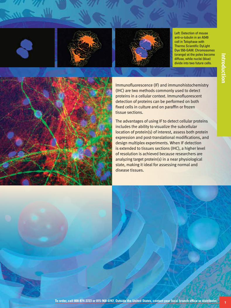
Left: Detection of mouse anti-α-tubulin in an A549 cell in Telophase with Thermo Scientific DyLight Dye 550-GAM. Chromosomes (orange) at the poles become diffuse, while nuclei (blue) divide into two future cells.
Immunofluorescence (IF) and immunohistochemistry (IHC) are two methods commonly used to detect proteins in a cellular context. Immunofluorescent detection of proteins can be performed on both fixed cells in culture and on paraffin or frozen tissue sections.
The advantages of using IF to detect cellular proteins includes the ability to visualize the subcellular location of protein(s) of interest, assess both protein expression and post-translational modifications, and design multiplex experiments. When IF detection is extended to tissues sections (IHC), a higher level of resolution is achieved because researchers are analyzing target protein(s) in a near physiological state, making it ideal for assessing normal and disease tissues.
To order, call 800-874-3723 or 815-968-0747. Outside the United States, contact your local branch office or distributor. 1
Introduction

2 For more information and complete antibody listings, visit www.thermoscientific.com/pierce-abs
Need Antibodies?
We have over 30,000 antibodies in 42 research areas.
Thermo Scientific Pierce Antibodies are developed for a wide variety of application needs. Our website enables you to easily search by protein target and then filter by the specific assays that interest you. All of our antibodies are validated in the stated applications and are guaranteed to perform.
Applications
• Agglutination • Immunodiffusion• Competition Assay • Immunofluorescence• ChIP Assay • Immunohistochemistry
(Frozen)• Cytotoxicity Assay• Control • Immunohistochemistry
(Paraffin)• ELISA• Electron Microscopy • Immunohistochemistry
(Paraffin, Frozen)• FACS• Functional Assay • Infection• Gel Shift • Immunoprecipitation• Hemagglutination Assay • Immunoradiometric Assay• Inhibition Assay • Radioimmunoassay• Immunocytochemistry • Western Blot
Build a Better Antibody
Use our custom services to produce antibodies you can trust.
The Thermo Scientific Custom Antibody Development Service leverages our experience in making more than 18,500 antibodies to peptides and recombinant proteins. Our proprietary antigen design tools, including the Thermo Scientific Antigen Profiler Software and targeted antigen display produces more robust antibodies that perform better in your targeted assays.
When you initiate a custom antibody project with us we provide you access to our online project management tool. This secure account gives you easy access to project information and allows you to provide specific instructions for your projects.
Intr
oduc
tion

To order, call 800-874-3723 or 815-968-0747. Outside the United States, contact your local branch office or distributor. 3
IntroductionIntroduction
With both immunofluorescence (IF) and
immunohistochemistry (IHC), a fluorophore
conjugated to an antibody is used for detection
(see Figure 1). Fluorophores can be conjugated to
a primary antibody (direct), secondary antibody
(indirect), or tertiary-labeled antibodies (e.g., biotin/
avidin system) for signal amplification. However,
with multiple amplification steps the researcher may
observe an increase in background, making it critical
to determine the best conjugation method for each
detection antibody and to include both positive and
negative controls.
Figure 1. Detection of antigens in immunofluorescence (Panel A) and immunohistochemistry applications (Panel B).
Antigen
Primaryantibody
Secondaryantibody
HRP
Signal detected
AvidinBiotin
Antigen
Primary antibody
Secondary antibody
FluorophoreSignal detected
Antigen
Primaryantibody
Secondaryantibody
HRP
Signal detected
AvidinBiotin
Antigen
Primary antibody
Secondary antibody
FluorophoreSignal detected
Panel B
Panel A

4
Immunofluorescence and ImmunohistochemistryIn
trod
uctio
n
For more information and complete antibody listings, visit www.thermoscientific.com/pierce-abs
Plasma Membrane
Glucose Transporter 1 Monoclonal Antibody (SPM498) #MA1-37783
Mitochondria
Mitochondrial Heat Shock Protein 70 Monoclonal Antibody (JG1) #MA3-028
Lysosome
LAMP1 Polyclonal Antibody #PA1-654A
Centrosome
g Tubulin Monoclonal Antibody (TU-30) #MA1-19421
Actin
Filamin (FLM NO1(PM6/317)) Monoclonal Antibody #MA5-11705
Microtubules
Tubulin b (TBN06(Tub2.5)) Monoclonal Antibody #MA5-117332
Nucleus
Phospho-c-Jun (Ser73) Polyclonal Antibody #PA5-17879
To order, call 800-874-3723 or 815-968-0747. Outside the United States, contact your local branch office or distributor. 5
Introduction
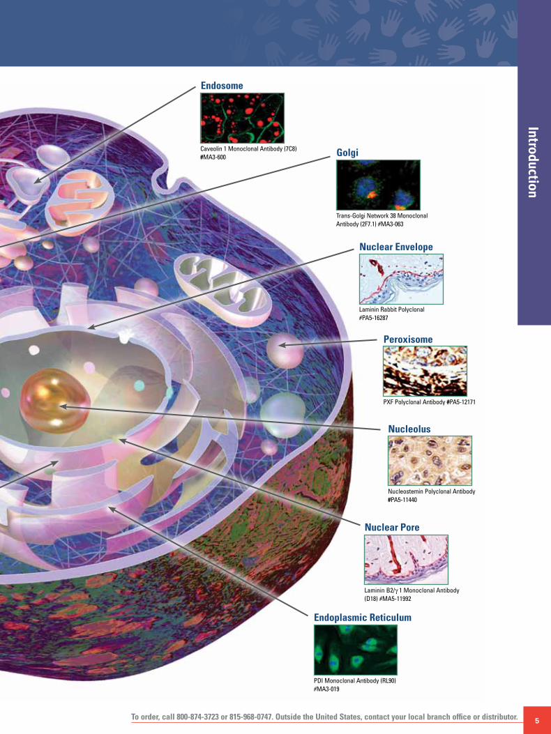
Golgi
Trans-Golgi Network 38 Monoclonal Antibody (2F7.1) #MA3-063
Endosome
Caveolin 1 Monoclonal Antibody (7C8) #MA3-600
Nuclear Envelope
Laminin Rabbit Polyclonal #PA5-16287
Peroxisome
PXF Polyclonal Antibody #PA5-12171
Nucleolus
Nucleostemin Polyclonal Antibody #PA5-11440
Endoplasmic Reticulum
PDI Monoclonal Antibody (RL90) #MA3-019
Nuclear Pore
Laminin B2/g 1 Monoclonal Antibody (D18) #MA5-11992

6
Immunofluorescence and ImmunohistochemistryIn
trod
uctio
n
For more information and complete antibody listings, visit www.thermoscientific.com/pierce-abs
Mammalian Cell Type Choices
Target protein expression levels should be considered when choosing a cell type because expression levels may vary significantly between cell lines. Additionally, the cell line should closely match the desired in vivo model. For example, researchers interested in understanding cellular mechanisms involved in liver development and disease most commonly choose a hepatic-derived cell line such as HepG2. Stem cells have become a growing trend in the lab. The advantage of using pluripotent stem cells is their ability to differentiate into virtually any cell type, which also lays the foundation to understand the initial stages of development and disease. The National Cancer Institutes has a panel of 60 markers to identify stem cells and to highly characterize multiple cell lines (NCI 60 panel), which is a common starting point for choosing an appropriate cell line (see Table 1).
Another consideration in cell line choice is whether to use primary or immortalized/transformed cell lines. Primary cells are isolated directly from tissue and represent the closest genotype to the intact organism under study. Primary cells closely mimic a “normal” state of protein expression. Consequently, they can maintain integrity of critical signaling pathways which mediate all biological processes. On the downside, primary cells proliferate in culture for a limited amount of time (~15-20 passages) before reaching the “Hayflick limit” or “crisis.” A cell which reaches the Hayflick limit will enter into either a reversible state of quiescence or an irreversible but metabolically active state of senescence. Primary cells are also more difficult to transfect with exogenous DNA or siRNA.
The use of immortalized or transformed cell lines bypasses the limitations of primary cells. Transformed cells arise when primary cells circumvent replicative senescence by exposure to oncogenic insults such as overexpression of oncoproteins (Ras) introduced by viruses, loss of tumor suppressor protein function (p53), or through DNA damaging events caused by UV, radiation or chemicals. Immortalized cells are generated primarily through the deregulation of telomere maintenance. Typically transformed or immortalized cells are easier to culture and have the potential to proliferate indefinitely. Transformed and immortalized cells are also more efficiently transfected with DNA vectors or siRNA than primary cells, making them ideal for characterizing a protein’s function in a cellular context. The primary disadvantage of using an immortalized cell line is that it does not truly recapitulate the normal cell state.
The use of mammalian cells in culture is very beneficial in terms of cost and time. Cell culture enables researchers to model near physiological events and address basic biological questions without the need of constructing time-consuming animal models. Although the maintenance of cells in culture is a highly efficient process, it does have its limitations with regards to mimicking the three-dimensional microenvironment present in an intact organism. Cell culture models typically represent the first stage of understanding protein or DNA function before generating a more expensive animal model.
Table 1. Markers commonly used to identify stem cells and to characterize differentiated cell types.
Page citation: From Appendix E: Stem Cell Markers. In Stem Cell Information [World Wide Web site]. Bethesda, MD: National Institutes of Health, U.S. Department of Health and Human Services, 2009 [cited Friday, July 09, 2010] Available at <http://stemcells.nih.gov/info/scireport/appendixe>
Marker Name Cell Type Significance
Blood Vessel
Fetal liver kinase-1 (Flk1)
Endothelial Cell-surface receptor protein that identifies endothelial cell progenitor; marker of cell-cell contacts
Smooth muscle cell-specific myo-sin heavy chain
Smooth muscle Identifies smooth muscle cells in the wall of blood vessels
Vascular endothe-lial cell cadherin
Smooth muscle Identifies smooth muscle cells in the wall of blood vessels
Bone
Bone-specific alkaline phospha-tase (BAP)
Osteoblast Enzyme expressed in osteoblast; activity indicates bone formation
Hydroxyapatite Osteoblast Mineralized bone matrix that provides structural integrity; marker of bone formation
Osteocalcin (OC) Osteoblast Mineral-binding protein uniquely synthesized by osteoblast; marker of bone formation
Bone Marrow and Blood
Bone morphoge-netic protein receptor (BMPR)
Mesenchymal stem and progenitor cells
Important for the differentiation of committed mesenchymal cell types from mesenchymal stem and progenitor cells; BMPR identifies early mesenchymal lineages (stem and progenitor cells)
CD4 and CD8 White blood cell (WBC)
Cell-surface protein markers specific for mature T lymphocyte (WBC subtype)
CD34 Hematopoietic stem cell (HSC), satellite, endothelial progenitor
Cell-surface protein on bone marrow cell, indicative of a HSC and endothelial progenitor; CD34 also identifies muscle satellite, a muscle stem cell
CD34+Sca1+ Lin- profile
Mesenchymal stem cell (MSC)
Identifies MSCs, which can dif-ferentiate into adipocyte, osteo-cyte, chondrocyte, and myocyte
CD38 Absent on HSC Present on WBC lineages
Cell-surface molecule that iden-tifies WBC lineages. Selection of CD34+/CD38- cells allows for purification of HSC populations
CD44 Mesenchymal A type of cell-adhesion molecule used to identify specific types of mesenchymal cells
c-Kit HSC, MSC Cell-surface receptor on BM cell types that identifies HSC and MSC; binding by fetal calf serum (FCS) enhances proliferation of ES cells, HSCs, MSCs, and hematopoietic progenitor cells
Colony-forming unit (CFU)
HSC, MSC progenitor
CFU assay detects the ability of a single stem cell or progenitor cell to give rise to one or more cell lineages, such as red blood cell (RBC) and/or white blood cell (WBC) lineages

To order, call 800-874-3723 or 815-968-0747. Outside the United States, contact your local branch office or distributor. 7
Introduction
Marker Name Cell Type Significance
Bone Marrow and Blood (cont’d.)
Fibroblast colony-forming unit (CFU-F)
Bone marrow fibroblast
An individual bone marrow cell that has given rise to a colony of multipotent fibroblastic cells; such identified cells are precursors of differentiated mesenchymal lineages
Hoechst® dye Absent on HSC Fluorescent dye that binds DNA; HSC extrudes the dye and stains lightly compared with other cell types
Leukocyte common antigen (CD45)
WBC Cell-surface protein on WBC progenitor
Lineage surface antigen (Lin)
HSC, MSC Differentiated RBC and WBC lineages
Thirteen to 14 different cell-sur-face proteins that are markers of mature blood cell lineages; detection of Lin-negative cells assists in the purification of HSC and hematopoietic progenitor populations
Mac-1 WBC Cell-surface protein specific for mature granulocyte and macro-phage (WBC subtypes)
Muc-18 (CD146) Bone marrow fibro-blasts, endothelial
Cell-surface protein (immuno-globulin superfamily) found on bone marrow fibroblasts, which may be important in hematopoie-sis; a subpopulation of Muc-18+ cells are mesenchymal precursors
Stem cell antigen (Sca-1)
HSC, MSC Cell-surface protein on bone marrow (BM) cell, indicative of HSC and MSC Bone Marrow and Blood
Stro-1 antigen Stromal (mesen-chymal) precursor cells, hematopoietic cells
Cell-surface glycoprotein on subsets of bone marrow stromal (mesenchymal) cells; selection of Stro-1+ cells assists in isolat-ing mesenchymal precursor cells, which are multipotent cells that give rise to adipocytes, osteocytes, smooth myocytes, fibroblasts, chondrocytes, and blood cells
Thy-1 HSC, MSC Cell-surface protein; negative or low detection is suggestive of HSC
Cartilage
Collagen types II and IV
Chondrocyte Structural proteins produced specifically by chondrocyte
Keratin Keratinocyte Principal protein of skin; identi-fies differentiated keratinocyte
Sulfated proteoglycan
Chondrocyte Molecule found in connective tissues; synthesized by chondrocyte
Fat
Adipocyte lipid-binding protein (ALBP)
Adipocyte Lipid-binding protein located specifically in adipocyte
Fatty acid trans-porter (FAT)
Adipocyte Transport molecule located specifically in adipocyte
Adipocyte lipid-binding protein (ALBP)
Adipocyte Lipid-binding protein located specifically in adipocyte
Marker Name Cell Type SignificanceGeneral
Y chromosome Male cells Male-specific chromosome used in labeling and detecting donor cells in female transplant recipients
Karyotype Most cell types Analysis of chromosome structure and number in a cell
Liver
Albumin Hepatocyte Principal protein produced by the liver; indicates functioning of maturing and fully differentiated hepatocytes
B-1 integrin Hepatocyte Cell-adhesion molecule important in cell-cell interac-tions; marker expressed during development of liver
Nervous System
CD133 Neural stem cell, HSC
Cell-surface protein that identi-fies neural stem cells, which give rise to neurons and glial cells
Glial fibrillary acidic protein (GFAP)
Astrocyte Protein specifically produced by astrocyte
Microtubule-associated pro-tein-2 (MAP-2)
Neuron Dendrite-specific MAP; protein found specifically in dendritic branching of neuron
Myelin basic protein (MPB)
Oligodendrocyte Protein produced by mature oligodendrocytes; located in the myelin sheath surrounding neuronal structures
Nestin Neural progenitor Intermediate filament structural protein expressed in primitive neural tissue
Neural tubulin Neuron Important structural protein for neuron; identifies differentiated neuron
Neurofilament (NF)
Neuron Important structural protein for neuron; identifies differentiated neuron
Neurosphere Embryoid body (EB), ES
Cluster of primitive neural cells in culture of differentiating ES cells; indicates presence of early neurons and glia
Noggin Neuron A neuron-specific gene expressed during the development of neurons
O4 Oligodendrocyte Cell-surface marker on immature, developing oligodendrocyte
O1 Oligodendrocyte Cell-surface marker that characterizes mature oligodendrocyte
Synaptophysin Neuron Neuronal protein located in synapses; indicates connections between neurons
Tau Neuron Type of MAP; helps maintain structure of the axon

8
Immunofluorescence and ImmunohistochemistryIn
trod
uctio
n
For more information and complete antibody listings, visit www.thermoscientific.com/pierce-abs
Marker Name Cell Type SignificancePancreas
Cytokeratin 19 (CK19)
Pancreatic epithelium
CK19 identifies specific pancreatic epithelial cells that are progenitors for islet cells and ductal cells
Glucagon Pancreatic islet Expressed by alpha-islet cell of pancreas
Insulin Pancreatic islet Expressed by beta-islet cell of pancreas Pancreas
Insulin-promoting factor-1 (PDX-1)
Pancreatic islet Transcription factor expressed by beta-islet cell of pancreas
Nestin Pancreatic progenitor
Structural filament protein indicative of progenitor cell lines including pancreatic
Pancreatic poly-peptide
Pancreatic islet Expressed by gamma-islet cell of pancreas
Somatostatin Pancreatic islet Expressed by delta-islet cell of pancreas
Pluripotent Stem Cells
Alkaline phosphatase
Embryonic stem (ES), embryonal carcinoma (EC)
Elevated expression of this enzyme is associated with undifferentiated pluripotent stem cell (PSC)
α-fetoprotein (AFP)
Endoderm Protein expressed during devel-opment of primitive endoderm; reflects endodermal differentia-tion Pluripotent Stem Cells
Bone morphoge-netic protein-4
Mesoderm Growth and differentiation factor expressed during early mesoderm formation and differentiation
Brachyury Mesoderm Transcription factor important in the earliest phases of mesoderm formation and differentiation; used as the earliest indicator of mesoderm formation
Cluster designa-tion 30 (CD30)
ES, EC Surface receptor molecule found specifically on PSC
Cripto (TDGF-1) ES, cardiomyocyte Gene for growth factor expressed by ES cells, primitive ectoderm, and developing cardiomyocyte
GATA-4 gene Endoderm Expression increases as ES differentiates into endoderm
GCTM-2 ES, EC Antibody to a specific extracellu-lar-matrix molecule that is synthesized by undifferentiated PSCs
Genesis ES, EC Transcription factor uniquely expressed by ES cells either in or during the undifferentiated state of PSCs
Germ cell nuclear factor
ES, EC Transcription factor expressed by PSCs
Hepatocyte nucle-ar factor-4 (HNF-4)
Endoderm Transcription factor expressed early in endoderm formation
Nestin Ectoderm, neural and pancreatic progenitor
Intermediate filaments within cells; characteristic of primitive neuroectoderm formation
Neuronal cell-adhesion mole-cule (N-CAM)
Ectoderm Cell-surface molecule that promotes cell-cell interaction; indicates primitive neuroecto-derm formation
Marker Name Cell Type Significance
OCT4/POU5F1 ES, EC Transcription factor unique to PSCs; essential for establish-ment and maintenance of undifferentiated PSCs
Pax6 Ectoderm Transcription factor expressed as ES cell differentiates into neuroepithelium
Stage-specific embryonic anti-gen-3 (SSEA-3)
ES, EC Glycoprotein specifically expressed in early embryonic development and by undifferenti-ated PSCs
Stage-specific embryonic antigen-4 (SSEA-4)
ES, EC Glycoprotein specifically expressed in early embryonic development and by undifferenti-ated PSCs
Stem cell factor (SCF or c-Kit ligand)
ES, EC, HSC, MSC Membrane protein that enhances proliferation of ES and EC cells, hematopoietic stem cell (HSCs), and mesenchymal stem cells (MSCs); binds the receptor c-Kit
Telomerase ES, EC An enzyme uniquely associated with immortal cell lines; useful for identifying undifferentiated PSCs
TRA-1-60 ES, EC Antibody to a specific extracellu-lar matrix molecule is synthe-sized by undifferentiated PSCs
TRA-1-81 ES, EC Antibody to a specific extracellu-lar matrix molecule normally synthesized by undifferentiated PSCs
Vimentin Ectoderm, neural and pancreatic progenitor
Intermediate filaments within cells; characteristic of primitive neuroectoderm formation
Skeletal Muscle/Cardiac/Smooth Muscle
MyoD and Pax7 Myoblast, myocyte Transcription factors that direct differentiation of myoblasts into mature myocytes
Myogenin and MR4
Skeletal myocyte Secondary transcription factors required for differentiation of myoblasts from muscle stem cells
Myosin heavy chain
Cardiomyocyte A component of structural and contractile protein found in cardiomyocyte
Myosin light chain Skeletal myocyte A component of structural and contractile protein found in skeletal myocyte
To order, call 800-874-3723 or 815-968-0747. Outside the United States, contact your local branch office or distributor. 9
Introduction
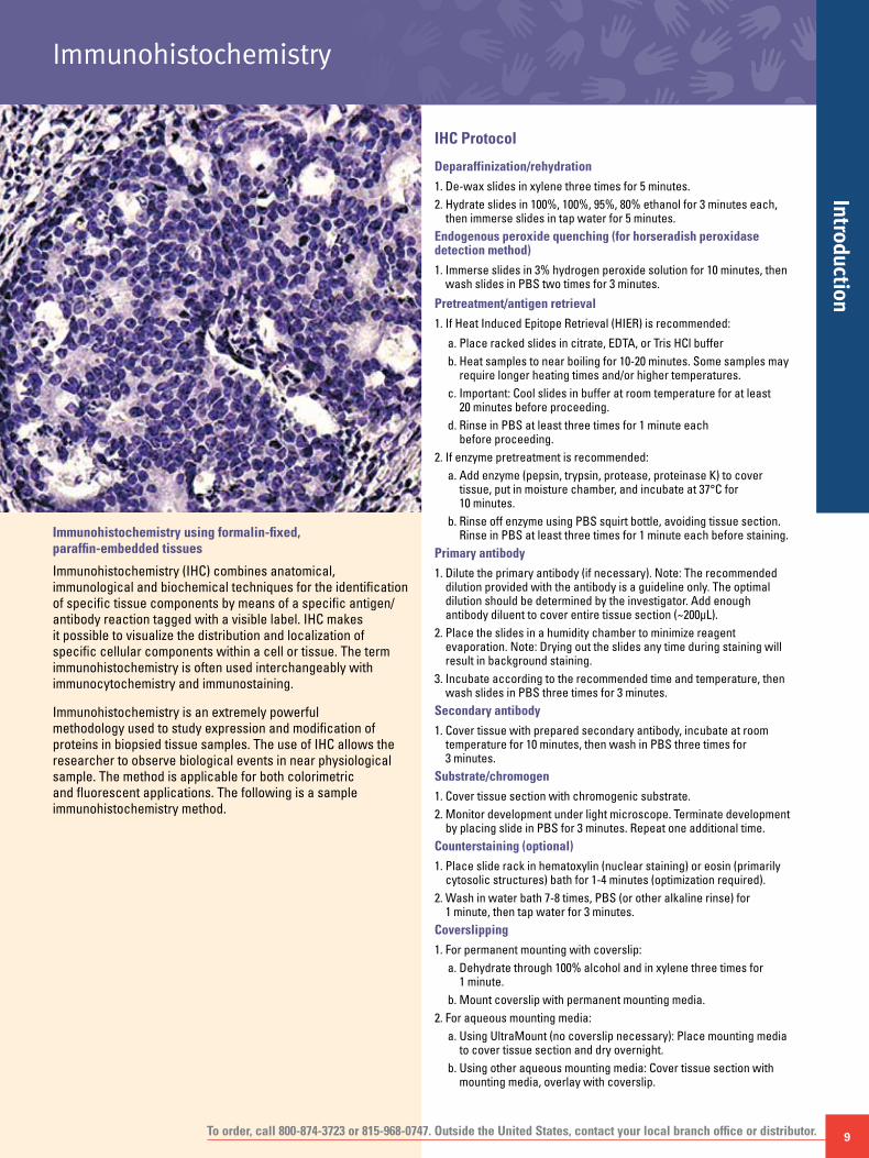
Immunohistochemistry using formalin-fixed, paraffin-embedded tissues
Immunohistochemistry (IHC) combines anatomical, immunological and biochemical techniques for the identification of specific tissue components by means of a specific antigen/antibody reaction tagged with a visible label. IHC makes it possible to visualize the distribution and localization of specific cellular components within a cell or tissue. The term immunohistochemistry is often used interchangeably with immunocytochemistry and immunostaining.
Immunohistochemistry is an extremely powerful methodology used to study expression and modification of proteins in biopsied tissue samples. The use of IHC allows the researcher to observe biological events in near physiological sample. The method is applicable for both colorimetric and fluorescent applications. The following is a sample immunohistochemistry method.
IHC Protocol
Deparaffinization/rehydration
1. De-wax slides in xylene three times for 5 minutes.2. Hydrate slides in 100%, 100%, 95%, 80% ethanol for 3 minutes each,
then immerse slides in tap water for 5 minutes.Endogenous peroxide quenching (for horseradish peroxidase detection method)
1. Immerse slides in 3% hydrogen peroxide solution for 10 minutes, then wash slides in PBS two times for 3 minutes.
Pretreatment/antigen retrieval
1. If Heat Induced Epitope Retrieval (HIER) is recommended:
a. Place racked slides in citrate, EDTA, or Tris HCl bufferb. Heat samples to near boiling for 10-20 minutes. Some samples may
require longer heating times and/or higher temperatures.c. Important: Cool slides in buffer at room temperature for at least
20 minutes before proceeding.d. Rinse in PBS at least three times for 1 minute each
before proceeding.2. If enzyme pretreatment is recommended:
a. Add enzyme (pepsin, trypsin, protease, proteinase K) to cover tissue, put in moisture chamber, and incubate at 37°C for 10 minutes.
b. Rinse off enzyme using PBS squirt bottle, avoiding tissue section. Rinse in PBS at least three times for 1 minute each before staining.
Primary antibody
1. Dilute the primary antibody (if necessary). Note: The recommended dilution provided with the antibody is a guideline only. The optimal dilution should be determined by the investigator. Add enough antibody diluent to cover entire tissue section (~200μL).
2. Place the slides in a humidity chamber to minimize reagent evaporation. Note: Drying out the slides any time during staining will result in background staining.
3. Incubate according to the recommended time and temperature, then wash slides in PBS three times for 3 minutes.
Secondary antibody
1. Cover tissue with prepared secondary antibody, incubate at room temperature for 10 minutes, then wash in PBS three times for 3 minutes.
Substrate/chromogen
1. Cover tissue section with chromogenic substrate.2. Monitor development under light microscope. Terminate development
by placing slide in PBS for 3 minutes. Repeat one additional time. Counterstaining (optional)
1. Place slide rack in hematoxylin (nuclear staining) or eosin (primarily cytosolic structures) bath for 1-4 minutes (optimization required).
2. Wash in water bath 7-8 times, PBS (or other alkaline rinse) for 1 minute, then tap water for 3 minutes.
Coverslipping
1. For permanent mounting with coverslip:a. Dehydrate through 100% alcohol and in xylene three times for
1 minute.b. Mount coverslip with permanent mounting media.
2. For aqueous mounting media: a. Using UltraMount (no coverslip necessary): Place mounting media
to cover tissue section and dry overnight.b. Using other aqueous mounting media: Cover tissue section with
mounting media, overlay with coverslip.
Immunohistochemistry

10
Stai
ning
and
Det
ectio
nImmunohistochemistry
For more information and complete antibody listings, visit www.thermoscientific.com/pierce-abs
Thermo Scientific ABC Staining Kits
Systems that offer highly sensitive and rapid detection.
The ABC Staining Kits for the avidin-biotin complex (ABC) technique are highly sensitive, produce very low background staining and have rapid avidin-biotin interactions. Highly diluted primary antibodies can be used with ABC Staining Kits, producing stain intensity comparable to other methods that require higher concentrations of antibody.
The Ultra-Sensitive ABC Peroxidase Staining Kits are more sensitive than the ABC Peroxidase Staining Kits, without exhibiting increased background staining. These kits supply the extra sensitivity needed for localizing antigens present in very small quantities. An expensive primary antibody may be diluted approximately five-fold further than with the standard ABC Peroxidase Kit – while producing equal staining intensity.1
Reference1. Bayer, E.A., et al. (1988). Anal. Biochem. 170, 271-281.
Ordering InformationDescription Pkg. Size Product #
Ultra-Sensitive ABC Peroxidase Mouse IgG Staining KitIncludes: Biotinylated Anti-Mouse IgG Antibody
Blocking Buffer Avidin Biotinylated HRP
Kit 32052
Ultra-Sensitive ABC Standard Peroxidase Rabbit IgG Staining KitIncludes: Biotinylated Anti-Rabbit IgG Antibody
Blocking Buffer Avidin Biotinylated HRP
Kit 32054
ABC Standard Peroxidase Staining KitIncludes: Avidin
Biotinylated HRP
Kit 32020
Ultra-Sensitive ABC Standard Peroxidase Staining KitIncludes: Avidin
Biotinylated HRP
Kit 32050
B
B
B
B
A
B
B
B
B
B
A
E
E
E
E
Substrate
Substrate
Avidin-biotin complex (ABC) for signal amplification.

To order, call 800-874-3723 or 815-968-0747. Outside the United States, contact your local branch office or distributor. 11
Staining and Detection
Thermo Scientific Pierce Metal Enhanced DAB Substrate Kit
The most sensitive DAB substrate formulation available.
The Pierce® Metal Enhanced DAB Substrate Kit optimizes this intensifying chemistry, producing an exceptionally sensitive colorimetric detection system for immunohistochemistry applications.
Highlights:• Incredible sensitivity – 50 times more sensitive than the
traditional DAB and 30 times more sensitive than other metal-intensified versions
• Low background, high intensity – crisp dark brown-black precipitate, almost no background
• Six-hour stability – solution is stable for > six hours
Superior staining performance with Thermo Scientific Pierce Metal Enhanced DAB Substrate Kit. Specific staining of prostatic acid phospha-tase detected with the Pierce Kit (left panel) exhibits higher intensity and greater resolution than staining detected by non-enhanced DAB method (right panel).
Reference1. Graham, R.C. and Karnovsky, M.J. (1966). J. Histochem. Cytochem. 14, 291-302.
Ordering InformationDescription Pkg. Size Product #
Metal Enhanced DAB Substrate KitIncludes: 10X Metal Enhanced DAB
Stable Peroxide Buffer
Kit25mL 250mL
34065
Thermo Scientific Pierce Peroxidase Detection Kit
The benefits of metal enhanced DAB in a complete kit.
This immunohistochemical staining kit bundles the Pierce Metal Enhanced DAB Substrate Kit with all necessary components to stain, counter-stain and preserve experimental results with frozen or paraffin tissue section.
Staining of glial fibrillary acidic protein (GFAP) using the Thermo Scientific Pierce Peroxidase Detection Kit. Panel A. Staining was performed as indicated in the protocol provided with the kit. Anti-GFAP and goat anti-mouse IgG biotin conjugate were used as the primary and secondary anti-bodies, respectively. Streptavidin-HRP was the detection conjugate. Panel B. Same as Panel A except no anti-GFAP primary antibody was used.
Ordering InformationDescription Pkg. Size Product #
Pierce Peroxidase Detection KitIncludes: DAB/Metal Concentrate (10X)
Peroxidase Detection Reagent Pack: Stable Peroxide Substrate Buffer (1X) Universal Blocker in TBS Peroxidase Suppressor BupH™ Tris Buffered Saline Surfact-Amps 20 (10% Tween®-20) Harris Modified Hematoxylin (without Acetic Acid, Hg free) Mounting Medium (dropper bottle)
Kit25mL 250mL 250mL 2 x 100mL 4 packs 10mL 100mL 60mL
36000
Thermo Scientific Pierce Metal Enhanced DAB
Ordinary DAB Method (Graham and Karnovsky, 1966)
A. Metal enhanced DAB substrate staining of GFAP
B. Negative control (no primary antibody)

12
Stai
ning
and
Det
ectio
nImmunohistochemistry
For more information and complete antibody listings, visit www.thermoscientific.com/pierce-abs
Other Common Substrates and CounterstainNBT/BCIP Substrate Solutions
The combination of NBT and BCIP is an ideal system for blotting or staining applications with alkaline phosphatase (AP). Together, they yield an intense, black-purple precipitate that provides much greater sensitivity than either substrate alone.
• Ready-to-use, single-component solutions• Regular formulation ideal for Western blotting• Suppressor formulation contains levamisole for inhibition of
endogenous enzyme, making it ideal for immunohistochemis-try applications
Colorimetric b-Galactosidase Precipitating Substrates
For detection of b-Galactosidase in immunohistochemical and clone expression applications.
X-Gal (5-bromo-4-chloro-3-indolyl-b-D-galactopyranoside), a chromogenic substrate for b-Galactosidase, yields a blue precipitate. IPTG (isopropyl-b-D-thiogalactopyranoside) maximizes the expression of b-gal.
Peroxidase Suppressor
A stable, easy-to-use endogenous peroxidase inhibitor.
The Thermo Scientific Peroxidase Suppressor inhibits endogenous peroxidase activity even more effectively than the method using hydrogen peroxide in methanol.
DAPI Counterstaining Reagents
Compatible blue color with fluorescent staining.
Ordering InformationDescription Pkg. Size Product #
NBT/BCIP Substrate Solution 250mL 34042
NBT/BCIP Plus Suppressor Substrate Solution 100mL ❄ 34070
X-Gal 100mg powder
34050
IPTG 1g powder 34060
Peroxidase SuppressorSupplied in methanol solution.
100mL 35000
4′,6-Diamidino-2-phenylindole, hydrochloride (DAPI)
10mg 46190
❄ Additional dry ice and/or freight charges
Thermo Scientific Immunohistochemistry ReagentsNormal Sera for Blocking
The most popular blocking agent for immunohistochemical staining; great for use as blocking reagents or negative controls.
Formaldehyde Ampules, Methanol-free
High-purity, 16% (w/v) formaldehyde for use as crosslinker and fixative.
Formaldehyde is a highly reactive, cell-permeable agent that is used by researchers as a reversible crosslinking agent for proteins and nucleic acids within the cell or as a general cell-fixing agent for imaging-based applications.
Ordering InformationDescription Pkg. Size Product #
Normal Goat Serum 2mL 31872
Normal Goat Serum 10mL 31873
Normal Horse Serum 2mL 31874
Normal Human Serum 2mL 31876
Normal Mouse Serum 2mL 31880
Normal Mouse Serum 5mL 31881
Normal Rabbit Serum 2mL 31884
Normal Rabbit Serum 5mL 31883
Normal Rat Serum 2mL 31888
Normal Swine Serum 2mL 31890
16% Formaldehyde (w/v), Methanol-free 10 x 1mL 28906
16% Formaldehyde (w/v), Methanol-free 10 x 10mL 28908
Related ProductsPierce Immunostain EnhancerSufficient for 100 large (~3cm²) tissue section slides.
20mL 46644
Pierce Immunostain EnhancerSufficient for 10 large (~3cm²) tissue section slides.
2mL 46645

To order, call 800-874-3723 or 815-968-0747. Outside the United States, contact your local branch office or distributor. 13
Introduction
IF Protocol (Fixed Cells in Culture)
Buffer preparation
Make fresh before each use.
Wash buffer: Prepare 500mL of a 1X PBST/0.1% Tween-20 solution by adding 1 pack of PBS BupH (Product # 28372) pack to 500mL of Milli-Q® water. Add 500μL of 100% Tween-20 to make 0.1%.
Fixation buffer: Add 3mL 16% paraformaldehyde (PFA) (Product # 28908) to 9mL 1X Wash buffer. Use appropriate personal protective equipment, fume hood. Collect PFA waste in a separate container in fume hood. Pre-warm fixation solution to 37°C prior to adding.
Permeabilization buffer: Add 1.5mL 10X stock solution (Product # 8408400) to 13.5mL Milli-Q water.
Blocking buffer: Add 7.5mL 10X stock solution (Product # 8408500) to 67.5mL 1X wash buffer to make a 0.3% blocking solution.
Note: Normal serum may be substituted in place of 10X blocking solution.
Primary antibody: Dilute primary antibody 1:100-1:1000 in 2mL blocking buffer. Prepare just before use.
Secondary antibody dilution buffer: Prepare a 1:500-1:1000 dilution of fluorescent secondary antibody in 6mL of 1X blocking buffer. Add 3μL of DAPI dye (Product # 62248).
Protocol
1. Aspirate media and add 100μL of warm fixation solution to each well. Incubate at room temperature for 15 minutes in fume hood.
2. Remove fixation solution and discard into PFA waste container (fume hood). Note: Remaining steps are performed at room temperature.
3. Add 100μL wash buffer to each well. Aspirate buffer.4. Repeat step 3 one additional time.5. Add 100μL of 1X permeabilization buffer to each well. Incubate for 15
minutes. Aspirate 1X permeabilization buffer.6. Add 100μL blocking buffer to each well. Aspirate buffer.7. Repeat step 6 one additional time.8. Add 100μL blocking buffer to each well. Incubate for 15 minutes.
Aspirate blocking buffer.9. Add primary antibody solution to primary antibody positive wells. To
negative control wells add blocking buffer. Incubate for 1 hour at 37°C. Aspirate primary antibody solution.
10. Add 100μL 1X blocking buffer to each well. Aspirate buffer.11. Repeat step 10 one additional time.12. Add secondary antibody/DAPI solution to all wells. Incubate at
room temperature for 30 minutes in the dark. Aspirate secondary antibody solution.
13. Add 100μL wash buffer to each well. Aspirate buffer.14. Repeat step 13 one additional time.15. Add 150μL wash buffer to each well, avoiding bubbles.16. Seal plate with thin adhesive plate seal. 17. Store plates of 4°C until ready to process. Plate can be stored for up
to one week before processing. Do not allow plate to dry out.
Immunofluorescence using fixed cells in culture
Traditional immunohistochemical staining methods are rapidly being replaced with fluorescence techniques. The development of high-performance fluorescent dyes that have complementary (non-overlapping) emission spectra enables simultaneous detection and analysis of multiple parameters in whole cells.
Immunofluroescent staining of fixed cells in culture is a powerful technique to study protein expression in an intact cell. This method is also commonly used to track protein localization in response to various cellular treatments. In-cell fluorescent technologies have also been adapted to study biological events occurring on DNA. Following is a sample method for the immunofluorescent staining of fixed cells.
Immunofluorescence

14
Fluo
resc
ent D
yes
Immunofluorescence
For more information and complete antibody listings, visit www.thermoscientific.com/pierce-abs
Bright New Alternatives to Alexa Fluor®, CyDye® and LI-COR® Fluorescent Dyes
Thermo Scientific DyLight Fluorescent Dyes are a complete family of high-intensity, photostable fluorescent tags for labeling antibodies and other molecular probes. The DyLight® Fluors are available as reactive labeling agents and as conjugates of secondary antibodies, biotin-binding proteins and molecular weight markers for use in fluorescence microscopy, flow cytometry, Western blotting, ELISA, high-content screening and other array platforms.
Properties of DyLight FluorophoresDyLight Fluors have absorption maxima ranging from 350nm to 777nm (Table 2), covering the entire visible light spectrum and several key near-infared and infrared wavelengths. Both the absorption and emission properties of the DyLight Fluors match the output (excitation) and detection wavelengths of common fluorescence instrumentation.
The DyLight Dyes exhibit higher fluorescence intensity and photostability than Alexa Fluor, CyDye and LI-COR Dyes in many applications and remain highly fluorescent over a broad pH range (pH 4-9). Additionally, the water solubility of the DyLight Dyes allows a high dye-to-protein ratio to be achieved without causing precipitation of conjugates.
Thermo Scientific DyLight 488 and DyLight 633 Dyes exhibit outstanding fluorescence in structured illumination. The uniform fluorescence intensity throughout the images demonstrates the outstanding brightness and photostability of DyLight 488 and 633 Dyes. Red: α-tubulin detected in HeLa cells with anti-tubulin monoclonal antibody and DyLight 633 Dye-conjugated secondary antibody (highly cross-adsorbed). Green: Histone H4 detected with anti-histone monoclonal antibody and DyLight 488 Dye-conjugated secondary antibody (highly cross-adsorbed). Blue: Nucleus counter-stained with fluorescent mounting media containing DAPI. Images were acquired with the Axio Imager™ Z1 and ApoTome™ Slider (Zeiss MicroImaging, Inc). The ApoTome Module provides confocal-like resolution allowing optical sectioning without using a pinhole (e.g., confocal). No image enhancement was performed.
Emission DyLight Dye Ex/Em* ε† Spectrally Similar Dyes
Blue 350 353/432 15,000 AMCA, Alexa Fluor 350 Dye
Blue 405 400/420 30,000 Alexa Fluor 405 and Cascade Blue® Dyes
Green 488 493/518 70,000 Alexa Fluor 488, fluorescein and FITC Dyes
Yellow 550 556/576 150,000 Fluor 546, Alexa Fluor 555, Cy®3 and TRITC Dyes
Red 594 593/618 80,000 Alexa Fluor 594 and Texas Red® Dyes
Red 633 638/658 170,000 Alexa Fluor 633 Dye
Red 650 652/677 250,000 Alexa Fluor 647 and Cy5 Dyes
Near-IR 680 692/712 140,000 Alexa Fluor 680 and Cy5.5 Dyes
Near-IR 800 777/790 270,000 IRDye® 800 Dye*Excitation and emission maxima in nanometers (± 4nm) † Molar extinction coefficient (M-1 cm-1)
Table 2. Spectral properties of Thermo Scientific DyLight Fluorescent Dyes.

To order, call 800-874-3723 or 815-968-0747. Outside the United States, contact your local branch office or distributor. 15
Fluorescent Dyes
Ordering InformationDescription Pkg. Size Product #
Amine-Reactive Dyes
DyLight 350 NHS Ester 1mg 46426
DyLight 350 NHS Ester 5 x 65μg 46427
DyLight 405 NHS Ester 1mg 46400
DyLight 405 NHS Ester 5 x 50μg 46401
DyLight 488 NHS Ester 1mg 46402
DyLight 488 NHS Ester 5 x 50μg 46403
DyLight 550 NHS Ester 1mg 62262
DyLight 550 NHS Ester 5 x 50μg 62263
DyLight 594 NHS Ester 1mg 46412
DyLight 594 NHS Ester 5 x 65μg 46413
DyLight 633 NHS Ester 1mg 46414
DyLight 633 NHS Ester 5 x 50μg 46417
DyLight 650 NHS Ester 1mg 62265
DyLight 650 NHS Ester 5 x 50μg 62266
DyLight 680 NHS Ester 1mg 46418
DyLight 680 NHS Ester 5 x 50μg 46419
DyLight 800 NHS Ester 1mg 46421
DyLight 800 NHS Ester 5 x 50μg 46422
Thermo Scientific DyLight Amine-Reactive and Sulfhydryl-Reactive Dyes
Excellent photostability make these dyes the clear alternative.
Highlights:• Available in both amine- and sulfhydryl-reactive chemistries
for fast and efficient labeling of IgG or other proteins• High water solubility• Excellent photostability• Compatible with common fluorescence instrumentation
Applications:• Fluorescence microscopy• Western blot detection • Protein arrays• Flow cytometry • ELISA • FRET-based technology• And many more
Ordering InformationDescription Pkg. Size Product #
Sulfhydryl-Reactive Dyes
DyLight 350 Maleimide 1mg 46622
DyLight 405 Maleimide 1mg 46600
DyLight 488 Maleimide 1mg 46602
DyLight 550 Maleimide 1mg 62290
DyLight 594 Maleimide 1mg 46608
DyLight 633 Maleimide 1mg 46613
DyLight 650 Maleimide 1mg 62295
DyLight 680 Maleimide 1mg 46618
DyLight 800 Maleimide 1mg 46621
Related ProductsDye Removal Columns Kit 22858Includes: Purification Resin
Spin Columns Microcentrifuge Collection Tubes
5mL 10 each 20 each

16
Fluo
resc
ent D
yes
Immunofluorescence
For more information and complete antibody listings, visit www.thermoscientific.com/pierce-abs
Thermo Scientific DyLight Antibody Labeling Kits
Label and purify antibodies in one hour.
The DyLight Antibody Labeling Kits for fast, efficient labeling of antibodies are supplied in two convenient kit formats to accommodate varied labeling requirements. The Antibody Labeling Kits contain all necessary components to perform three separate labeling reactions using 1mg of IgG or similar quantities of other proteins. The DyLight Microscale Antibody Labeling Kits contain all the necessary components to perform five separate labeling reactions using 100μg of IgG. The labeling kits use high-performance spin desalting columns to provide exceptional dye removal and antibody recovery.
Highlights: • Fast – fluorescently label and purify protein in approximately
one hour• Amine-reactive dyes – label virtually any protein• Pre-measured fluorescent dye – eliminate the time, waste and
hassle associated with weighing dye• Efficient non-reacted dye removal• Minimal sample dilution• Spin column format eliminates the need for column
preparation, fraction screening and waiting for protein to emerge from column
• Easy protocol
Microscale KitsContain sufficient reagents to label and purify 5 x 100µg of IgG.
In addition to contents listed below, all Microscale Kits include: Reaction Buffer, 1mLSpin Columns, 5 eachMicrocentrifuge Collection Tubes, 10 eachPurification Resin, 5mL
Ordering InformationDescription Pkg. Size Product #
DyLight 350 Microscale Antibody Labeling KitDyLight 350 NHS Ester
Kit 5 vials
62276
DyLight 405 Microscale Antibody Labeling Kit DyLight 405 NHS Ester
Kit 5 vials
53021
DyLight 488 Microscale Antibody Labeling KitDyLight 488 NHS Ester
Kit 5 vials
53025
DyLight 550 Microscale Antibody Labeling Kit DyLight 550 NHS Ester
Kit 5 vials
84531
DyLight 594 Microscale Antibody Labeling Kit DyLight 594 NHS Ester
Kit 5 vials
53045
DyLight 633 Microscale Antibody Labeling KitDyLight 633 NHS Ester
Kit 5 vials
53047
DyLight 650 Microscale Antibody Labeling Kit DyLight 650 NHS Ester
Kit 5 vials
84536
DyLight 680 Microscale Antibody Labeling KitDyLight 680 NHS Ester
Kit 5 vials
53057
DyLight 800 Microscale Antibody Labeling KitDyLight 800 NHS Ester
Kit 5 vials
53063
Antibody Labeling KitsContain sufficient reagents to label and purify 3 x 1mg of IgG or similar quantities of other proteins.
In addition to contents listed below, all Antibody Labeling Kits include:Reaction Buffer, 1mLSpin Columns, 6 eachMicrocentrifuge Collection Tubes, 12 eachPurification Resin, 5mL
Ordering InformationDescription Pkg. Size Product #
DyLight 350 Antibody Labeling KitDyLight 350 NHS Ester
Kit 3 vials
62275
DyLight 405 Antibody Labeling KitDyLight 405 NHS Ester
Kit 3 vials
53020
DyLight 488 Antibody Labeling KitDyLight 488 NHS Ester
Kit 3 vials
53024
DyLight 550 Antibody Labeling Kit DyLight 550 NHS Ester
Kit 3 vials
84530
DyLight 594 Antibody Labeling KitDyLight 594 NHS Ester
Kit 3 vials
53044
DyLight 633 Antibody Labeling KitDyLight 633 NHS Ester
Kit 3 vials
53046
DyLight 650 Antibody Labeling Kit DyLight 650 NHS Ester
Kit 3 vials
84535
DyLight 680 Antibody Labeling KitDyLight 680 NHS Ester
Kit 3 vials
53056
DyLight 800 Antibody Labeling KitDyLight 800 NHS Ester
Kit 3 vials
53062
Step 1. Labeling reaction
Add antibody to vialcontaining pre-measured
dye. Incubate 1 hour at room temperature.
Apply labelingreaction to Spin
Desalting Column.
Recoverlabeled antibody.
Centrifuge
30 seconds
Step 2. Removal of excess fluorescent dye

To order, call 800-874-3723 or 815-968-0747. Outside the United States, contact your local branch office or distributor. 17
Fluorescent Dyes
Thermo Scientific Pierce Fluorescein
Amine-reactive derivatives of fluorescein dye.
NHS-fluorescein and fluorescein isothiocyanate (FITC), two reactive derivatives of fluorescein dye, are used in wide-ranging applications including fluorescence microscopy, flow cytometry and immunofluorescence-based assays such as Western blotting and ELISA. FITC is the base fluorescein molecule functionalized with an isothiocyanate reactive group (-N=C=S), replacing a hydrogen atom on the bottom ring of the structure. This derivative is reactive toward primary amine groups on proteins, peptides and other biomolecules. A succinimidyl-ester functional group attached to the fluorescein core, creating NHS-fluorescein, forms another common derivative that has much greater specificity toward primary amines in the presence of other nucleophiles and a more stable linkage following labeling.
Pierce Fluorescein is a mixture of isomers with reactive groups attached at the five and six positions of the bottom ring (See Structure). The properties of these isomers are indistinguishable in terms of excitation and emission spectra and for protein applications there is no need to isolate a specific isomer.
Fluorescein-5-maleimide and 5-Iodoacetamidofluorescein (5-IAF) are sulfhydryl-reactive derivatives of fluorescein dye. Fluorescein-5-maleimide is the base fluorescein molecule functionalized with a maleimide reactive group by replacing a hydrogen atom on the bottom ring of the structure. 5-IAF is the core fluorescein molecule functionalized with an iodoacetamide group. Both fluorescein derivatives are reactive toward sulfhydryl groups (e.g., reduced cysteine residues) on proteins, peptides and other biomolecules.
A derivative of fluorescein, DyLight 488 Fluor, has been tailored for various chemical and biological applications where greater photostability and fluorescence intensity, pH independence, and a narrower emission spectrum are required.
Properties of amine-reactive fluorescein dyes.
NHS-Fluorescein FITC
Structure
NHS-FluoresceinMW 473.39
O
ON
O
OH
O
OOHO
OFITC
MW 389.38
OHO O
O
OH
NCS
Alternative names 5/6-FAM SE 5/6-FITC
Chemical name 5/6-carboxyfluorescein succinimidyl ester 5(6)-fluorescein isothiocyanate mixed isomer
Molecular weight 473.4 389.2
Excitation source 488nm spectral line, argon-ion laser 488nm spectral line, argon-ion laser
Excitation wavelength 494nm 494nm
Emission wavelength 518nm 518nm
Extinction coefficient > 70,000/M-1 cm-1 > 70,000/M-1 cm-1
CAS # 117548-22-8 27072-45-3
Purity > 90% by HPLC > 95% by HPLC
Solubility Soluble in DMF or DMSO Soluble in aqueous buffers at pH > 6
Storage Desiccated at -20°C, protect from moisture, use only fresh solutions
Desiccated at -20°C, protect from moisture, use only fresh solutions
Reactive groups NHS ester, reacts with primary amines at pH 7.0 to 9.0
Isothiocyanate, reacts with primary amines at pH 7.0 to 9.0

18
Fluo
resc
ent D
yes
Immunofluorescence
For more information and complete antibody listings, visit www.thermoscientific.com/pierce-abs
Properties of sulfhydryl-reactive dyes.
Fluorescein-5-maleimide 5-Iodoacetamido-fluorescein
Structure
Fluorescein-5-MaleimideMW 427.36
O N O
OH
O
OOHO
5-IAF5-Iodoacetamido-fluorescein
MW 515.25
OH
O
OOHO
NH
O
I
Alternative names 5-MF, 5-maleimido-fluorescein 5-IAF, 5-iodoacetamidofluorescein
Chemical name 1H-Pyrrole-2,5-dione, 1-(3’,6’-dihydroxy-3-oxospiro(isobenzofuran-1(3H),9’-(9H)xanthen-5-yl)-
Acetamide, N-(3’,6’-dihydroxy-3-oxospiro(isobenzofuran-1(3H), 9’-(9H)xanthen)-5-yl)-2-iodo
Molecular weight 427.36 ±3 515.26 ±3
Excitation source 488nm spectral line, argon-ion laser 488nm spectral line, argon-ion laser
Excitation wavelength 494nm 494nm
Emission wavelength 518nm 518nm
Extinction coefficient ~ 68,000/M-1 cm-1 > 80,000/M-1 cm-1
CAS # 75350-46-8 63368-54-7
Solubility Soluble in DMF or DMSO Soluble in DMF; aqueous buffers at pH > 6
Storage Desiccated at -20°C, protect from moisture, use only fresh solutions
Desiccated at -20°C, protect from moisture, use only fresh solutions
Reactive groups Maleimide, reacts with sulfhydryls at pH 6.5 to 7.5 Iodoacetamide, reacts with sulfhydryls at pH 7.0 to 7.5
Ordering InformationDescription Pkg. Size Product #
FITC (Fluorescein Isothiocyanate) 1g 46424
FITC (Fluorescein Isothiocyanate) 100mg 46425
NHS-Fluorescein 1g 46409
NHS-Fluorescein 100mg 46410
FITC Antibody Labeling KitEfficiently labels and purifies 3 x 1mg of IgG or other protein in about 1 hour.
Kit 53027
Includes: FITC Borate Buffer Spin Columns Microcentrifuge Collection Tubes Purification Resin
3 vials1mL 6 each 12 each 5mL
Ordering InformationDescription Pkg. Size Product #
Fluorescein Antibody Labeling KitEfficiently labels and purifies 3 x 1mg of IgG or other protein in about 1 hour.
Kit 53029
Includes: NHS Fluorescein Borate Buffer Spin Columns Microcentrifuge Collection Tubes Purification Resin
3 vials1mL 6 each 12 each 5mL
Fluorescein-5-Maleimide 25mg 62245
5-Iodoacetamido-fluorescein (5-IAF) 25mg 62246

To order, call 800-874-3723 or 815-968-0747. Outside the United States, contact your local branch office or distributor. 19
Fluorescent Dyes
Thermo Scientific Pierce Rhodamine
Amine-reactive derivatives of rhodamine dye.
NHS-rhodamine and tetramethylrhodamine isothiocyanate (TRITC), two reactive derivatives of rhodamine dye, are used in wide-ranging applications including fluorescence microscopy, flow cytometry and immunofluorescence-based assays such as Western blotting and ELISA.
TRITC is the base tetramethylrhodamine molecule functionalized with an isothiocyanate reactive group (-N=C=S), replacing a hydrogen atom on the bottom ring of the structure. This derivative is reactive toward amine and sulfhydryl groups on proteins, peptides and other biomolecules. A succinimidyl-ester functional group attached to the tetramethylrhodamine core, creating NHS-fluorescein, forms another common derivative that has much greater specificity toward primary
amines in the presence of other nucleophiles and a more stable linkage following labeling. Texas Red Sulfonyl Chloride is a long-wavelength derivative of rhodamine that is modified with sulfonyl chloride for reaction to primary amines.
Pierce Rhodamine Dyes are a mixture of isomers with reactive groups attached at the five and six positions of the bottom ring (See Structure). The properties of these isomers are indistinguishable in terms of excitation and emission spectra and for protein applications there is no need to isolate a specific isomer.
The Thermo Scientific NHS-Rhodamine Antibody Labeling Kit (Product # 53031) produces ideal conjugates for immunofluorescence. A549 cells were fixed with 4% paraformaldehyde (Product # 28906) and permeabilized with 0.1% Surfact-Amps® X-100 (Product # 28314). The cells were then probed with a 0.4μg/mL mouse anti-α-tubulin antibody and 2μg/mL rhodamine-goat anti-mouse secondary antibody. Nuclei were labeled with Hoechst 33342. Images were acquired on Nikon Eclipse TS100 fluorescent microscope using Zeiss AxioCam® camera and AxioVision® software.
Ordering InformationDescription Pkg. Size Product #
TRITC (Tetramethylrhodamine Isothiocyanate)
10mg 46112
NHS-Rhodamine 25mg 46406
Rhodamine Antibody Labeling KitEfficiently labels and purifies 3 x 1mg of IgG or other protein in about 1 hour.
Kit 53031
Includes: NHS Rhodamine Borate Buffer Spin Columns Microcentrifuge Collection Tubes Purification Resin
3 vials 1mL6 each12 each 5mL
TRITCMW 478.97
Em/Ex 544/572
ON N+
O
O-
NCS
Cl-
NHS-RhodamineMW 527.52
Em/Ex 552/575
O N+N
O
O-
OO
N
O
O
20
Fluo
resc
ent S
tain
sImmunofluorescence
For more information and complete antibody listings, visit www.thermoscientific.com/pierce-abs
Thermo Scientific DAPI Stain
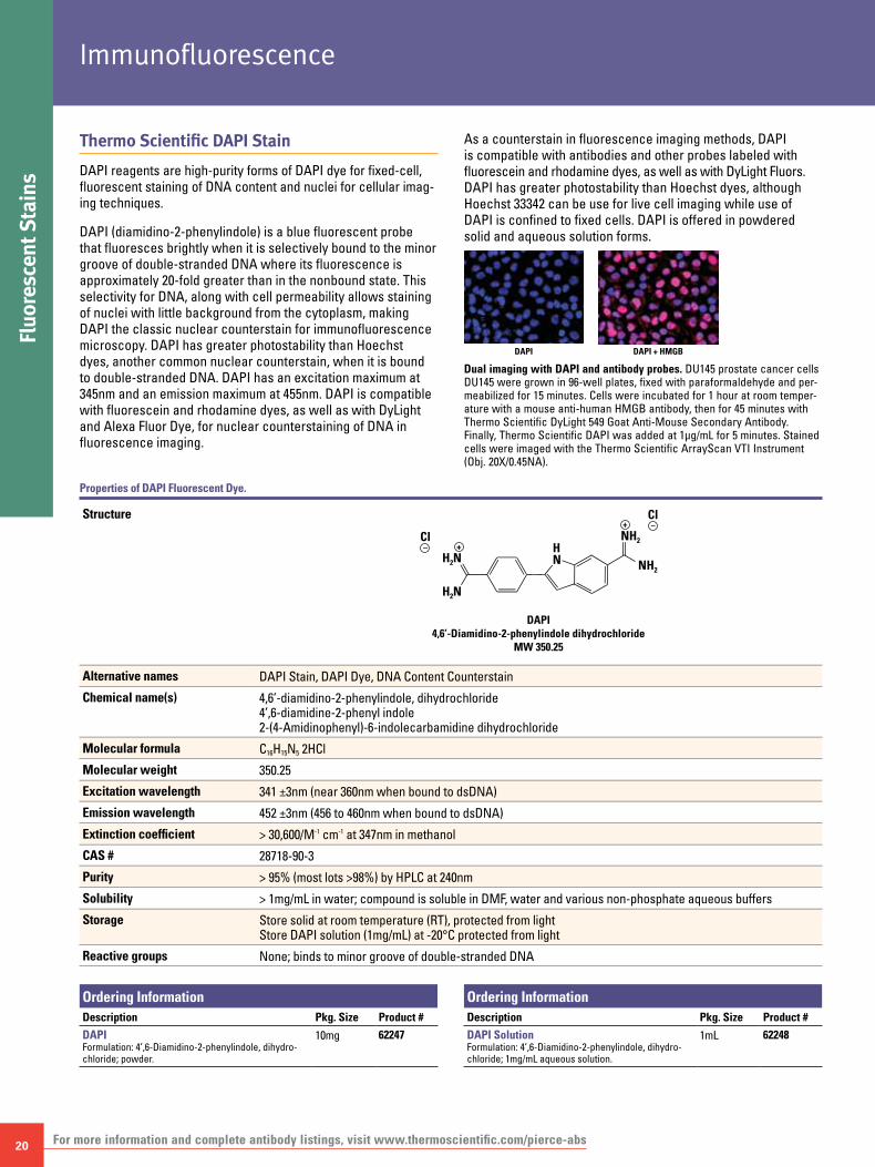
DAPI reagents are high-purity forms of DAPI dye for fixed-cell, fluorescent staining of DNA content and nuclei for cellular imag-ing techniques.
DAPI (diamidino-2-phenylindole) is a blue fluorescent probe that fluoresces brightly when it is selectively bound to the minor groove of double-stranded DNA where its fluorescence is approximately 20-fold greater than in the nonbound state. This selectivity for DNA, along with cell permeability allows staining of nuclei with little background from the cytoplasm, making DAPI the classic nuclear counterstain for immunofluorescence microscopy. DAPI has greater photostability than Hoechst dyes, another common nuclear counterstain, when it is bound to double-stranded DNA. DAPI has an excitation maximum at 345nm and an emission maximum at 455nm. DAPI is compatible with fluorescein and rhodamine dyes, as well as with DyLight and Alexa Fluor Dye, for nuclear counterstaining of DNA in fluorescence imaging.
As a counterstain in fluorescence imaging methods, DAPI is compatible with antibodies and other probes labeled with fluorescein and rhodamine dyes, as well as with DyLight Fluors. DAPI has greater photostability than Hoechst dyes, although Hoechst 33342 can be use for live cell imaging while use of DAPI is confined to fixed cells. DAPI is offered in powdered solid and aqueous solution forms.
Dual imaging with DAPI and antibody probes. DU145 prostate cancer cells DU145 were grown in 96-well plates, fixed with paraformaldehyde and per-meabilized for 15 minutes. Cells were incubated for 1 hour at room temper-ature with a mouse anti-human HMGB antibody, then for 45 minutes with Thermo Scientific DyLight 549 Goat Anti-Mouse Secondary Antibody. Finally, Thermo Scientific DAPI was added at 1μg/mL for 5 minutes. Stained cells were imaged with the Thermo Scientific ArrayScan VTI Instrument (Obj. 20X/0.45NA).
Structure
DAPI4,6’-Diamidino-2-phenylindole dihydrochloride
MW 350.25
H2N
H2N
NH2
NH2Cl– +
Cl
HN
–+
Alternative names DAPI Stain, DAPI Dye, DNA Content Counterstain
Chemical name(s) 4,6’-diamidino-2-phenylindole, dihydrochloride 4’,6-diamidine-2-phenyl indole 2-(4-Amidinophenyl)-6-indolecarbamidine dihydrochloride
Molecular formula C16H15N5 2HCl
Molecular weight 350.25
Excitation wavelength 341 ±3nm (near 360nm when bound to dsDNA)
Emission wavelength 452 ±3nm (456 to 460nm when bound to dsDNA)
Extinction coefficient > 30,600/M-1 cm-1 at 347nm in methanol
CAS # 28718-90-3
Purity > 95% (most lots >98%) by HPLC at 240nm
Solubility > 1mg/mL in water; compound is soluble in DMF, water and various non-phosphate aqueous buffers
Storage Store solid at room temperature (RT), protected from light Store DAPI solution (1mg/mL) at -20°C protected from light
Reactive groups None; binds to minor groove of double-stranded DNA
Ordering InformationDescription Pkg. Size Product #
DAPIFormulation: 4’,6-Diamidino-2-phenylindole, dihydro-chloride; powder.
10mg 62247
Ordering InformationDescription Pkg. Size Product #
DAPI SolutionFormulation: 4’,6-Diamidino-2-phenylindole, dihydro-chloride; 1mg/mL aqueous solution.
1mL 62248
Properties of DAPI Fluorescent Dye.
DAPI + HMGBDAPI

To order, call 800-874-3723 or 815-968-0747. Outside the United States, contact your local branch office or distributor. 21
Fluorescent Stains
Thermo Scientific Hoechst 33342 Stain
Hoechst 33342 Solution is a high-quality form of Hoechst dye for fixed- and live-cell fluorescent staining of DNA and nuclei in cellular imaging techniques
Hoechst 33342 (2’-[4-ethoxyphenyl]-5-[4-methyl-1-piperazinyl]-2,5’-bi-1H-benzimidazole trihydrochloride trihydrate) is a cell-permeable DNA stain that is excited by ultraviolet light and emits blue fluorescence at 460-490nm. Hoechst 33342 binds preferentially to adenine-thymine (A-T) regions of DNA. This stain binds into the minor groove of DNA and exhibits distinct fluorescence emission spectra that are dependent on dye:base pair ratios.
Hoechst 33342 is used for specifically staining the nuclei of living or fixed cells and tissues. This stain is commonly used in combination with 5-bromo-2’-deoxyuridine (BrdU) labeling to distinguish the compact chromatin of apoptotic nuclei, to identify replicating cells and to sort cells based on their DNA content. A combination of Hoechst 33342 and propidium iodide have been extensively used for simultaneous flow cytometric and fluorescence imaging analysis of the stages of apoptosis and cell-cycle distribution.
As a counterstain in fluorescent imaging, Hoechst dye is compatible with antibodies and other probes labeled with fluorescein and rhodamine dyes, as well as with Thermo Scientific DyLight Fluors. The stable 20mM aqueous stock solution is essentially ready for use.
Properties of Hoescht 33342 Dye.
Structure
Hoechst 33342MW 615.99
•3 HCl•3 H2O
N
O
NH
N
NH N
N
Alternative names Hoechst Stain, Hoechst Dye, DNA Content Counterstain
Chemical name 2’-(4-Ethoxyphenyl)-5-(4-methyl-1-piperazinyl)-2,5’-bi-1H-benzimidazole trihydrochloride trihydrate
Molecular formula C27H28N6O •3HCl •3H2O
Molecular weight 615.99
Excitation wavelength 346 ±3nm (361nm when bound to dsDNA)
Emission wavelength 497 ±3nm
Extinction coefficient Source compound ~47,000/M-1 cm-1 (> 45,000) at 343nm in methanol
CAS # 28491-52-3
Purity > 95% (most lots >98%) by HPLC at 240nm
Solubility Product is supplied at 20mM (12.3mg/mL) in water; Hoechst dye is soluble in DMF, water and various non-phosphate aqueous buffers
Storage Store supplied solution at 2 to 8°C protected from light
Reactive groups None; dye binds to minor groove of double-stranded DNA
Cellular imaging with Hoechst 33342. A549 human lung cancer cells were grown on 96-well plates, fixed with paraformaldehyde and permeabilized for 15 minutes. Cells were incubated for 30 minutes at room temperature with mouse anti-human α-tubulin antibody, then for 45 minutes with Thermo Scientific DyLight 549 Goat Anti-Mouse Secondary Antibody. Finally, Thermo Scientific Hoechst 33342 Solution was added at 1μg/mL for 5 minutes. Stained cells were imaged with the ArrayScan® VTI Instrument (Obj. 20X/0.45NA).
Ordering InformationDescription Pkg. Size Product #
Hoechst 33342 SolutionFormulation: 12.3mg/mL (20mM) aqueous solution.
5mL 62249
Hoechst 33342 Hoechst + α-Tubulin
22
Fluo
resc
ent S
tain
sImmunofluorescence
For more information and complete antibody listings, visit www.thermoscientific.com/pierce-abs
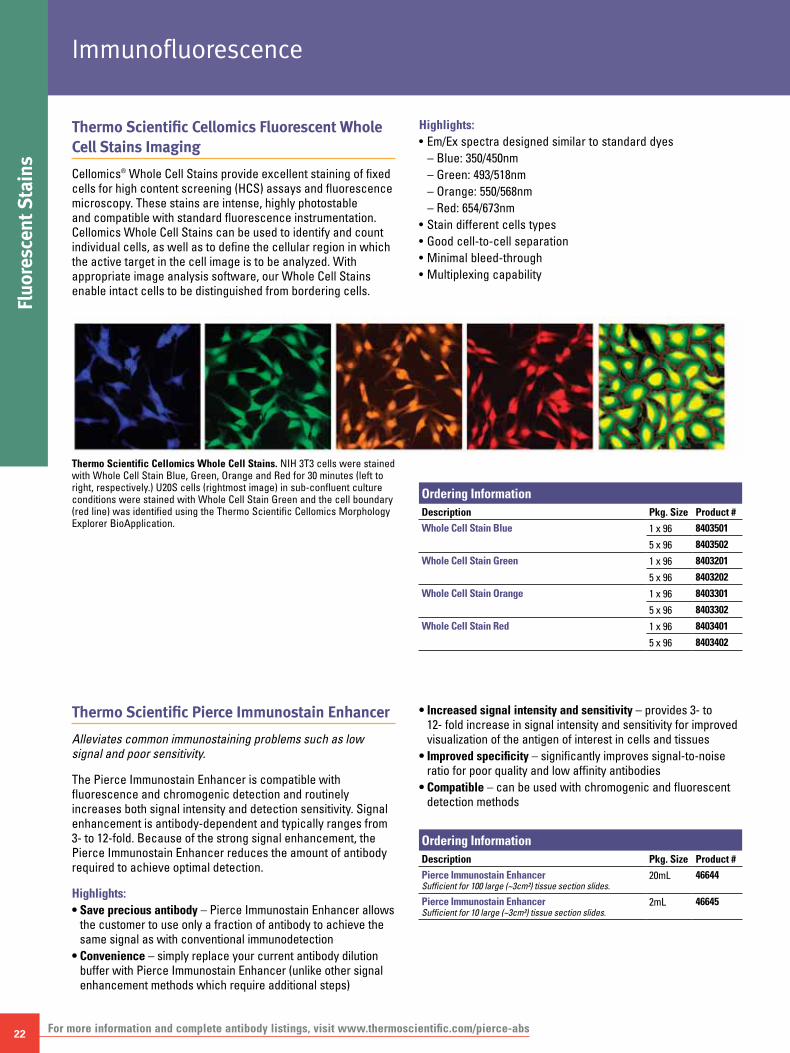
Thermo Scientific Cellomics Fluorescent Whole Cell Stains Imaging
Cellomics® Whole Cell Stains provide excellent staining of fixed cells for high content screening (HCS) assays and fluorescence microscopy. These stains are intense, highly photostable and compatible with standard fluorescence instrumentation. Cellomics Whole Cell Stains can be used to identify and count individual cells, as well as to define the cellular region in which the active target in the cell image is to be analyzed. With appropriate image analysis software, our Whole Cell Stains enable intact cells to be distinguished from bordering cells.
Highlights:• Em/Ex spectra designed similar to standard dyes
– Blue: 350/450nm– Green: 493/518nm– Orange: 550/568nm– Red: 654/673nm
• Stain different cells types• Good cell-to-cell separation• Minimal bleed-through• Multiplexing capability
Thermo Scientific Cellomics Whole Cell Stains. NIH 3T3 cells were stained with Whole Cell Stain Blue, Green, Orange and Red for 30 minutes (left to right, respectively.) U20S cells (rightmost image) in sub-confluent culture conditions were stained with Whole Cell Stain Green and the cell boundary (red line) was identified using the Thermo Scientific Cellomics Morphology Explorer BioApplication.
Ordering InformationDescription Pkg. Size Product #
Whole Cell Stain Blue 1 x 96 8403501
5 x 96 8403502
Whole Cell Stain Green 1 x 96 8403201
5 x 96 8403202
Whole Cell Stain Orange 1 x 96 8403301
5 x 96 8403302
Whole Cell Stain Red 1 x 96 8403401
5 x 96 8403402
Thermo Scientific Pierce Immunostain Enhancer
Alleviates common immunostaining problems such as low signal and poor sensitivity.
The Pierce Immunostain Enhancer is compatible with fluorescence and chromogenic detection and routinely increases both signal intensity and detection sensitivity. Signal enhancement is antibody-dependent and typically ranges from 3- to 12-fold. Because of the strong signal enhancement, the Pierce Immunostain Enhancer reduces the amount of antibody required to achieve optimal detection.
Highlights:• Save precious antibody – Pierce Immunostain Enhancer allows
the customer to use only a fraction of antibody to achieve the same signal as with conventional immunodetection
• Convenience – simply replace your current antibody dilution buffer with Pierce Immunostain Enhancer (unlike other signal enhancement methods which require additional steps)
• Increased signal intensity and sensitivity – provides 3- to 12- fold increase in signal intensity and sensitivity for improved visualization of the antigen of interest in cells and tissues
• Improved specificity – significantly improves signal-to-noise ratio for poor quality and low affinity antibodies
• Compatible – can be used with chromogenic and fluorescent detection methods
Ordering InformationDescription Pkg. Size Product #
Pierce Immunostain EnhancerSufficient for 100 large (~3cm²) tissue section slides.
20mL 46644
Pierce Immunostain EnhancerSufficient for 10 large (~3cm²) tissue section slides.
2mL 46645

To order, call 800-874-3723 or 815-968-0747. Outside the United States, contact your local branch office or distributor. 23
Introduction
A secondary antibody indirectly detects a target antigen to which a specific primary antibody is first bound. The secondary antibody has a tag or other label to facilitate detection or purification. It also requires that the secondary antibody has the same specificity for the primary antibody species and isotypes.
Although indirect detection using a secondary antibody requires more steps, it offers increased sensitivity through the signal amplification of multiple secondary antibodies binding to a single primary antibody. Secondary antibodies are also a more versatile reagent than individually labeled primary antibodies. A given secondary antibody can be used with any primary antibody of the same type and host species.
Because the vast majority of primary antibodies are produced in just a few host animal species and most are of the IgG class, it is easy and economical for manufacturers to produce and supply ready-to-use secondary antibodies for many methods and detection systems. From a relatively small number of secondary antibodies, many options are available for purity level, specificity and label type required for a given application.
Secondary Antibody Fragments
Secondary antibodies may be provided in three formats: whole IgG, divalent F(ab’)2 fragments and monovalent Fab fragments.
Whole IgGSecondary antibodies are typically affinity purified from the pooled serum of immunized hosts. In this first round of purification, whole immunoglobulins binding to the immunizing antibody are recovered and mainly consist of the ~150-kDa IgG class. Further affinity purification with Protein A or G removes all immunoglobulin classes except IgG. Whole IgG secondary antibodies produced in the manner are widely applicable, easiest to produce and least expensive.
F(ab’)2 antibody fragmentsWhile whole immunoglobulins are compatible with most assays, certain methods benefit from removing the Fc portion of the antibody by either reducing the mass of the antibody or reduc-ing cross-reactivity with probed samples containing active Fc-binding proteins (e.g., Fc receptors, Protein A, Protein G). The Fc portion can be removed from several species of IgG by digestion with pepsin, leaving the divalent F(ab’)2 fragment (~100 kDa) of the antibody intact.
Fab antibody fragmentsSome species of IgG can be enzymatically digested with papain to cleave the antibody between the antigen binding domain and hinge region to produce two Fab fragments and an Fc fragment. The monovalent Fab antibody fragments are useful in blocking applications and other special circumstances where controlled binding ratios and/or the elimination of Fc interactions is required. The small size (~50 kDa) of Fab fragments may improve antigen detection by penetrating deeper than whole IgG into tissue sections and other complex samples.
Specificity of Secondary Antibodies
Secondary antibodies are generated by immunizing a host animal with the antibody(s) from a different species. For example, anti-mouse antibodies are raised by injecting mouse antibodies into an animal other than a mouse. Goat, donkey and rabbit are the most commonly used host species for raising secondary antibodies, but others may be available from individual suppliers.
The most common types of secondary antibodies are those generated against a pooled population of immunoglobulins from a target species. For example, immunizing a goat with purified mouse IgG will generate goat anti-mouse IgG antibodies that will bind to all classes, heavy and light chains (H&L) and fragments of mouse IgG as well as any other molecules sharing the same conserved domains (e.g., IgM shares the same light chains as IgG). In contrast, immunizing a goat with only mouse IgG1 anti-bodies will only generate antibodies specific for mouse IgG1 antibodies and molecules sharing the same conserved domains.
Because of the high degree of conservation in the structure of many immunoglobulin domains, class-specific secondary anti-bodies must be affinity purified and cross-adsorbed to achieve minimal cross-reactivity with other immunoglobulins. Using the
Secondary Antibodies
24
Intr
oduc
tion
Secondary Antibodies
For more information and complete antibody listings, visit www.thermoscientific.com/pierce-abs
example described above, immobilized mouse IgG1 antibodies would be used to affinity purify all goat antibodies that bind to mouse IgG1. These anti-mouse IgG1 antibodies would then be further purified by passage through a chromatography column(s) containing mouse IgG2a, IgG2b, IgG3, IgM, etc., to remove any antibodies that cross-react with non-IgG1 isotypes.
Additionally, secondary antibodies can be further purified by passage through columns containing the immobilized serum proteins from species other than those used to immunize the host. This method of cross-adsorption (often referred to as “Highly Cross-Adsorbed”) is an additional purification step recommended for applications where primary antibodies from multiple species will be used and when immunoglobulins or other serum proteins may be present in the samples being probed.
Highly Cross-Adsorbed Thermo Scientific Pierce Secondary Antibodies are purified for minimal cross-reactivity with the serum proteins of specific species. These antibodies are indi-cated by the code “min x Sp” where Sp is an abbreviation for the one or more species of serum proteins against which the sec-ondary antibody was cross-adsorbed. A list of Sp abbreviations used with Pierce Antibodies follows in Table 3. Other companies often use similar codes.
Table 3. Common Sp abbreviations for Thermo Scientific Pierce Secondary Antibodies.
Bv = Bovine Hs = Horse
Ch = Chicken Ms = Mouse
Gt = Goat Rb = Rabbit
Gu = Guinea pig Rt = Rat
Ha = Hamster Sh = Sheep
Hn = Human Sw = Swine
In addition to class and species specificity, secondary antibodies can be generated against specific antibody fragments [F(ab’)1, Fab] or individual antibody chains (mu, gamma, kappa) and domains. Table 3 contains a list of commonly used notations that indicate the specificity of secondary antibodies.
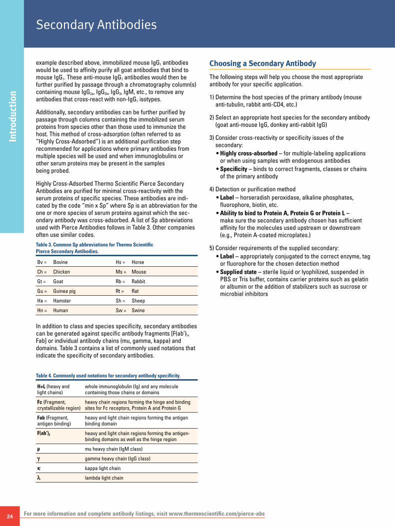
Table 4. Commonly used notations for secondary antibody specificity.
H+L (heavy and light chains)
whole immunoglobulin (Ig) and any molecule containing those chains or domains
Fc (Fragment, crystallizable region)
heavy chain regions forming the hinge and binding sites for Fc receptors, Protein A and Protein G
Fab (Fragment, antigen binding)
heavy and light chain regions forming the antigen binding domain
F(ab’)2 heavy and light chain regions forming the antigen-binding domains as well as the hinge region
µ mu heavy chain (IgM class)
g gamma heavy chain (IgG class)
k kappa light chain
l lambda light chain
Choosing a Secondary Antibody
The following steps will help you choose the most appropriate antibody for your specific application.
1) Determine the host species of the primary antibody (mouse anti-tubulin, rabbit anti-CD4, etc.)
2) Select an appropriate host species for the secondary antibody (goat anti-mouse IgG, donkey anti-rabbit IgG)
3) Consider cross-reactivity or specificity issues of the secondary:• Highly cross-absorbed – for multiple-labeling applications
or when using samples with endogenous antibodies• Specificity – binds to correct fragments, classes or chains
of the primary antibody
4) Detection or purification method• Label – horseradish peroxidase, alkaline phosphates,
fluorophore, biotin, etc.• Ability to bind to Protein A, Protein G or Protein L –
make sure the secondary antibody chosen has sufficient affinity for the molecules used upstream or downstream (e.g., Protein A-coated microplates.)
5) Consider requirements of the supplied secondary:• Label – appropriately conjugated to the correct enzyme, tag
or fluorophore for the chosen detection method• Supplied state – sterile liquid or lyophilized, suspended in
PBS or Tris buffer, contains carrier proteins such as gelatin or albumin or the addition of stabilizers such as sucrose or microbial inhibitors

To order, call 800-874-3723 or 815-968-0747. Outside the United States, contact your local branch office or distributor. 25
Conjugates
Ordering InformationProduct #
Specificity Source Unconjugated Biotin Peroxidase Alk. Phos. Fluorescein Rhodamine Texas Red
Chicken IgY (H+L) Rabbit 31104 31720 31401 31501Goat IgG (H+L) (min x HnMsRb Sr Prot)† Mouse 31107 31730 31400 31512 31620 31940Goat IgG (H+L) Rabbit 31105 31732 31402 31300 31509 31650 31492Goat IgG [F(ab’)2] Rabbit 31214 31753 31403 31553Goat IgG (Fc) Rabbit 31133 31433 31337 31533Goat IgG (H+L) (min x Hn Sr Prot)† Rabbit F(ab´)2 31109 31302Hamster IgG (H+L) Goat 31115 31750Hamster IgG (H+L) Rabbit 31120 31587Horse IgG (H+L) Goat 31760Human IgG (H+L) Goat 31130 31770 31410 31310 31529 31656 31943Human IgG g Chain Specific Goat 31118Human IgG (H+L) (min x BvHsMs Sr Prot)† Goat 31119 31774 31412 31531 31683 31944Human IgG [F(ab’)2] Goat 31122 31482 31312 31628Human IgG [F(ab’)2] (min x BvHsMs Sr Prot)† Goat 31132 31414Human IgG (Fc) (min x BvHsMs Sr Prot)† Goat 31125 31413Human IgM (Fc5μ) Goat 31136 31415 31575Human IgM (μ) Goat 31124 31778Human IgA (α) Goat 31140 31417 31314 31577Human IgA + IgG + IgM (H+L) Goat 31128 31782 31418 31316Human k Chain Goat 31129 31780Human l Chain Goat 31131Human IgG (H+L) (min x Ms Sr Prot)† Mouse 31135 31420Human IgG (H+L) (min x BvHsMs Sr Prot)† Mouse 31137 31784Human IgG (H+L) Rabbit 31143 31786Human IgG (Fc) Rabbit 31142 31789 31423 31318 31535Human IgG (Fc) Goat F(ab´)2 31163Human IgG (H+L) Goat F(ab´)2 31165Human IgA + IgG + IgM (H+L) Goat F(ab´)2 31539Mouse IgA (α) (min x Hn Sr Prot)† Goat 31169Mouse IgA + IgG + IgM (H+L) Goat 31171Mouse IgG (H+L) Goat 31160 31800 31430†† 31320 31569 31660 31498Mouse IgG (H+L), Highly Cross-adsorbed GoatMouse IgG (H+L) (min x BvHnHs Sr Prot)† Goat 31164 31802 31432 31322 31541 31661 31500Mouse IgG [F(ab’)2] Goat 31166 31803 31436 31324 31543Mouse IgG (Fc) Goat 31168 31805 31437 31325 31547 31663Mouse IgG (Fc) (min x BvHnHs Sr Prot)† Goat 31170 31439 31327 31632Mouse IgM (μ) Goat 31172 31804 31440 31326 31992Mouse IgG + IgM (H+L) Goat 31182 31807 31444 31328Mouse IgG + IgM (H+L) (min x BvHnHs Sr Prot)† Goat 31446 31330Mouse IgG (Fcg) (subclasses 1+2a+2b+3) (min x BvHnRb Sr Prot)† Goat 31232 31630Mouse IgG (Fcg) subclass 1 specific (min x BvHnRb Sr Prot)† Goat 31236Mouse IgG (Fcg) subclass 2a specific (min x BvHnRb Sr Prot)† Goat 31237 31634Mouse IgG (H+L) Horse 31181 31806Mouse IgG (H+L) Rabbit 31188 31450 31329 31561 31665 31610Mouse IgG (H+L) (min x Hn Sr Prot)† Rabbit 31190 31452 31334Mouse IgG [F(ab’)2] Rabbit 31192 31451 31331 31559Mouse IgG (Fc) Rabbit 31194 31813 31455 31332 31555Mouse IgM (μ) Rabbit 31196 31456 31333 31557Mouse IgG + IgM (H+L) Rabbit 31198 31457 31335 31558Mouse IgG (H+L) (min x BvHnHs Sr Prot)† Goat F(ab´)2 31185 31438 31565Mouse IgM (μ) Goat F(ab´)2 31178Mouse IgM (μ) (min x BvHnHs Sr Prot)† Goat F(ab´)2 31186Mouse IgG + IgM (H+L) (min x BvHnHs Sr Prot)† Goat F(ab´)2 31448Rabbit IgG (H+L) (min x BvChGtGuHaHnHsMsRtSh Sr Prot)† Donkey 31238 31821 31458 31345 31568 31685 31504Rabbit IgG (H+L) Goat 31210 31820 31460†† 31340 31635 31670 31506Rabbit IgG (H+L), Highly Cross-Adsorbed GoatRabbit IgG (H+L) (min x Hn Sr Prot)† Goat 31212 31822 31462 31342 31583 31686 31507Rabbit IgG [F(ab’)2] Goat 31234 31823 31461 31343 31573Rabbit IgG (Fc) Goat 31216 31463 31341Rabbit IgG (H+L) (min x GtHnMsSh Sr Prot)† Mouse 31213 31824 31464 31584Rabbit IgG (H+L) Goat F(ab´)2 31579Rabbit IgG (H+L) (min x HnMsRt Sr Prot)† Goat F(ab´)2 31239 31636Rat IgG (H+L) Goat 31220 31830 31470 31350 31629 31680 31508Rat IgG [F(ab’)2] Goat 31474Rat IgG (Fc) Goat 31226 31475 31621Rat IgM (μ) Goat 31832 31476 31354 31631Rat IgG (H+L) Rabbit 31218 31834Rat IgG (H+L) (min x Ms Sr Prot)† Rabbit 31219Sheep IgG (H+L) Rabbit 31240 31840 31480 31360 31627Streptavidin Recombinant 21125 21127 21323 21224 21724 21726NeutrAvidin Protein Hen Egg 31000 31001 31002 31006
† See Tables on page 24 for the Key to Abbreviations. †† Stabilized, pre-diluted format also available; see our website. See page 26 for DyLight Dye fluorescent-conjugated secondary antibody.

26
Fluo
resc
ent C
onju
gate
sSecondary Antibodies
For more information and complete antibody listings, visit www.thermoscientific.com/pierce-abs
Thermo Scientific DyLight Conjugates
Excellent brightness make these conjugates a clear alternative.
Highlights:• Available conjugated to commonly used secondary
antibodies, Streptavidin and Thermo Scientific NeutrAvidin Biotin-Binding Protein
• Molar ratio (dye:protein) optimized to provide excellent fluorescent intensity
• Stable for 1 year at 4°C• Antibody conjugates are affinity-purified to minimize
cross-reactivity
1 2 3 4 5
Western Blotting
Two-color infrared Western blot detection of p53 and cyclophilin B knockdown using Thermo Scientific DyLight 680- and Thermo Scientific DyLight 800-labeled secondary antibodies. Protein lysate from transfected A549 cells was separated using SDS-PAGE and transferred to PVDF membrane. Lane 1: MW marker, Lane 2: mock transfected sample, Lane 3: negative control siRNA, Lane 4: siRNA targeted against p53 and Lane 5: siRNA targeted against cyclophilin. The membranes were imaged with the Odyssey® Infrared Imaging System using the 700 and 800 channels.
DyLight 800 StreptavidinIRDye 800 Streptavidin
DyLight 680 StreptavidinAlexa Fluor 680 Streptavidin
Rela
tive
Fluo
resc
ent I
nten
sity
4,000,000
8,000,000
12,000,000
16,000,000
0 2 4 86 1210Biotinylated BSA (ng per well)
Thermo Scientific DyLight 680 and Thermo Scientific DyLight 800 Streptavidin Conjugates are brighter than Alexa Fluor 680 or IRDye 800 Conjugates in microplate-based assays. Microplates were coated with biotinylated BSA at the indicated concentrations. Conjugates were diluted to 10μg/mL in PBS and 100μL was applied to each plate. Fluorescent intensity was measured with the Odyssey Infrared Imaging System using the 700 and 800 channels.
Ordering InformationConjugates: Package size for these items is 1mg at 1mg/mL.
Description Product #
DyLight 405 Dye
DyLight 488 Dye
DyLight 550 Dye
DyLight 594 Dye
DyLight 633 Dye
DyLight 650 Dye
DyLight 680 Dye
DyLight 800 Dye
Goat Anti-Mouse IgG (H+L) 35502 84540 84545 35518 35521
Goat Anti-Mouse IgG Highly Cross-Adsorbed 35500 35503 35511 35513 35519
Goat Anti-Rabbit IgG (H+L) 35552 84541 84546 35568 35571
Goat Anti-Rabbit IgG Highly Cross-Adsorbed 35550 35553 35561 35563 35569
Streptavidin 21832 84542 84547 21848 21851
NeutrAvidin® Biotin-Binding Protein 22832 84606 84607 22848 22853

To order, call 800-874-3723 or 815-968-0747. Outside the United States, contact your local branch office or distributor. 27
Other Conjugates
Thermo Scientific High Sensitivity HRP Conjugates
High-performance horseradish peroxidase conjugates for the most demanding immunoassays.
Our High Sensitivity HRP Conjugates meet the demands of today’s scientists for utmost sensitivity in ELISA and Western blotting applications. Our Thermo Scientific Streptavidin and NeutrAvidin Protein Conjugates are ideal for immunohistochemistry (IHC), Western blotting and ELISA applications using chemiluminescence, chemifluorescence or colorimetric substrates. Each High Sensitivity HRP Conjugate is packaged in an easy-to-use stabilized solution that enables convenient storage at 4°C for up to 1 year.
NeutrAvidin-HRPdetection of
GAPDH
Streptavidin-HRPdetection of
cytokeratin 18
Biotinylated Primary Ab Negative Control
Excellent IHC staining of GAPDH and cytokeratin 18 in human colon carcinoma with Thermo Scientific High Sensitivity HRP Conjugates. Formalin-fixed paraffin embedded human colon carcinoma tissues were stained using Metal Enhanced DAB Substrate (Product # 36000). The tissues were incubated with biotinylated mouse anti-GAPDH at 4.6μg/mL or rabbit anti-cytokeratin 18 at 0.1μg/mL or blocking buffer alone (right panels). The sections were incubated with High Sensitivity Neutravidin HRP (4μg/mL, top panels) or High Sensitivity Streptavidin HRP (4μg/mL bottom panels). Tissues were counterstained using Harris-modified hematoxylin solution (blue staining in all panels).
Thermo Scientific Pierce Poly-HRP Conjugates
Get the ultimate sensitivity in immunodetection techniques with Pierce Poly-HRP Conjugates.
The Pierce Poly-HRP Conjugates deliver the highest sensitivity and low background in immunoassays where sample volume is limited or when the target molecule is present at low levels. Pierce Poly-HRP Conjugates are purified to remove unconjugated probe molecules that reduce signal intensity by competing for binding sites with HRP-conjugated molecules. In addition, these conjugates are free of HRP monomers that may lead to increased background signal. Together, these features provide consistent and reliable sensitivity and deliver higher sensitivity than conventional HRP and poly-HRP conjugates without the need for additional signal amplification steps. These conjugates are compatible with chromogenic, fluorogenic and chemiluminescent HRP substrates used in ELISA, Western blotting, IHC and nucleic acid hybridization assays.
Streptavidinpoly-HRPdetectionof GAPDH
Positive Negative
GAR poly-HRP detection of
cytokeratin 18
GAM poly-HRP detection of vimentin
Obtain immunohistochemical images with outstanding clarity using Thermo Scientific Pierce Poly-HRP Conjugates. Formalin-fixed paraffin embedded human colon carcinoma tissues were stained using Metal Enhanced DAB Substrate (Product # 36000). The tissues were incubated with biotinylated mouse anti-GAPDH at 4.6μg/mL or rabbit anti-cytokeratin 18 at 1μg/mL or mouse anti-vimentin at 2μg/mL or blocking buffer alone (right panels). The sections were incubated with Streptavidin Poly-HRP (4μg/mL, top panels) or Goat Anti-Rabbit Poly-HRP (2μg/mL, middle panels) or Goat Anti-Mouse Poly-HRP (2μg/mL, bottom panels). Tissues were counterstained using Harris-modified hematoxylin solution (blue staining in all panels).
Ordering InformationDescription Pkg. Size Product #
High Sensitivity Streptavidin-HRP (1mg/mL) 0.5mL 21130
High Sensitivity Streptavidin-HRP (1mg/mL) 5mL 21132
High Sensitivity Streptavidin-HRP, pre-diluted (10µg/mL)
2mL 21134
High Sensitivity NeutrAvidin-HRP (1mg/mL) 0.5mL 31030
High Sensitivity NeutrAvidin-HRP (1mg/mL) 5mL 31032 Ordering InformationDescription Pkg. Size Product #
Pierce Streptavidin Poly-HRP (0.5mg/mL) 0.5mL 21140
Pierce Goat Anti-Mouse Poly-HRP (0.5mg/mL) 0.5mL 32230
Pierce Goat Anti-Rabbit Poly-HRP (0.5mg/mL) 0.5mL 32260

28
Org
anel
les
& S
truc
ture
Mar
kers
Primary Antibodies by Cellular Structures
For more information and complete antibody listings, visit www.thermoscientific.com/pierce-abs
The maintenance and control of cell shape, cell movement, cytokinesis and the organization of organelles are some of the most basic functions of the cell. Because changes in these features are often the consequence of cellular differentiation, cellular toxicity, pathology or other critical cellular event, their measurement against potential therapeutic targets can be critical.
The cytoskeleton is typically composed of microfilaments, microtubules and intermediate filaments in the cell and plays a key role in the morphological change of cells. The cytoskeleton also facilitates proper function of other cellular proteins through direct binding, transporting, repositioning and sequestering these proteins. For certain cell types such as neurons, cellular morphological changes during development are necessary for the proper function in the tissue.
Fluorescence microscopy provides both intensity as well as spatial information of the fluorescently labeled constituents that are imaged and analyzed. Spatial information, such as cell shape and morphology, sizes and arrangements of intracellular constituents, and the arrangement and location of cells relative to each other, can all be obtained from fluorescence microscopy that other methods may not.
We offer a broad array of antibodies that can be used solely or in combination with one another to help visualize cellular organelles and structures and orient the stains of your primary antibody target. Primary antibodies from different species hosts can be used to multiplex antibodies together and provide multiple colors in the same test. To do this standard fluorescent dyes such as FITC and Rhodamine (see pages 17-19) or more diverse brighter
dyes such as DyLight Dyes are combined with species-specific secondary antibodies (see Secondary Antibodies, pages 23-27) for detection after probing with the variable host antibodies.
Primary antibodies can also be directly conjugated to dyes and used to visualize up to six different colors at one time (see DyLight Dye table, page 14). It is best to pick dyes that are distal from each other on the color wheel for adjacent stains to maximize the degree of spectral differentiation and minimize the amount of emission cross-reactivity from the dyes.
Keratin 14 Rabbit Polyclonal #PA1-38001
Annexin VI Rabbit Polyclonal #PA1-37086
Tubulin b Mouse Monoclonal (TBN06 (Tub 2.5)) #MA5-11732

To order, call 800-874-3723 or 815-968-0747. Outside the United States, contact your local branch office or distributor. 29
Organelles &
Structure Markers
Keratin 18 Mouse Monoclonal (SPM510) #MA1-38011
Skeletal Muscle Actin Rabbit Polyclonal #PA1-37020
Dysferlin Rabbit Polyclonal #PA1-37585
S100 α-4 Rabbit Polyclonal #PA1-38589 CD105 Rabbit Polyclonal #PA1-37372 Villin Rabbit Polyclonal #PA1-38853
Smooth Muscle Actin Rabbit Polyclonal #PA1-37024
Retinoic Acid Receptor b Rabbit Polyclonal #PA1-38562
Keratin 5 Rabbit Polyclonal #PA1-37974
Ordering Information for Select Thermo Scientific Pierce AntibodiesProduct Description Target Species Applications Size Product #
α-1 Antichymotrypsin Polyclonal Antibody Hu IHC (P) 1mL PA1-37070
Annexin VI Polyclonal Antibody Hu, Ms, Rt IHC (P), WB 1mL PA1-37086
Cadherin P Monoclonal Antibody (6A9) Hu IF, IP, WB 100μg MA1-2003
Calcitonin Polyclonal Antibody Ca, Eq, Hu, Ms, Nhp, Ov, Po, Rt IHC (P) 1mL PA1-37211
Calreticulin Polyclonal Antibody Ca, Hm, Hu, Ms, Rb, Rt ICC, IF, IP, WB 100μL PA1-903
Calreticulin Polyclonal Antibody Ca, Hu, Ms, Nhp, Rb, Rt FACS, ICC, IF, IP, WB 100μL PA3-900
Caveolin 1 Monoclonal Antibody (7C8) Hm, Rt IF, WB 100μL MA3-600
CD105 Polyclonal Antibody Hu IHC (P) 1mL PA1-37372
CD9 Polyclonal Antibody Hu IHC (P) 1mL PA1-37299
CD99 Polyclonal Antibody Hu IHC (P) 1mL PA1-37370
CDX2 Polyclonal Antibody Hu IHC (P) 1mL PA1-39430
Chromogranin A Polyclonal Antibody Hu IHC (P) 1mL PA1-37445
Clathrin, Heavy Chain Monoclonal Antibody (X22) Bv, Hm, Hu, Ms, Nhp, Rt ICC, IF, IP, WB 100μL MA1-065
Dysferlin Polyclonal Antibody Hu IHC (P), WB 1mL PA1-37585
Epithelial Specific Antigen Monoclonal Antibody (SPM491) Hu IHC (P) 1mL MA1-37643
For species and application abbreviations, see page 77. More antibodies on next page Designates product with photo.

30
Org
anel
les
& S
truc
ture
Mar
kers
Primary Antibodies by Cellular Structures
For more information and complete antibody listings, visit www.thermoscientific.com/pierce-abs
α-1 Antichymotrypsin Rabbit Polyclonal #PA1-37070
Calcitonin Rabbit Polyclonal #PA1-37211 CD9 Rabbit Polyclonal #PA1-37299
GAD65 Rabbit Polyclonal #PA1-37732 Gastrin 1 Rabbit Polyclonal #PA1-37749Epithelial Specific Antigen Mouse Monoclonal (SPM491) #MA1-37643
Ordering Information for Select Thermo Scientific Pierce AntibodiesProduct Description Target Species Applications Size Product #
ERp29 Polyclonal Antibody Bv, Ca, GP, Hm, Hu, Ms, Nhp, Rt IF, IP, WB 100μL PA3-011
Filamin Monoclonal Antibody (FLMN01 (PM6/317)) Hu, Ms, Rt, Ck, Gp, Rb ELISA, IHC (P), WB 500μL MA5-11705
GAD65 Polyclonal Antibody Hu, Rt IHC (P), IP 1mL PA1-37732
g Tubulin Monoclonal Antibody (4D11) Hu, Ms, Rt IF, IP, WB 100μg MA1-850
g Tubulin Monoclonal Antibody (TU-30) Ck, Hu, Ms, Pl, Po, Pz, Rt ICC, WB 100μg MA1-19421
Gastrin 1 Polyclonal Antibody Hu IHC (P) 1mL PA1-37749
Glucose Transporter 1 Monoclonal Antibody (SPM498) Hu IHC (P) 1mL MA1-37783
Glucose-regulated Protein 94 Polyclonal Antibody Hu IHC (P), IP 1mL PA1-37807
Keratin 14 Polyclonal Antibody Hu IHC (P) 1mL PA1-38001
Keratin 18 Monoclonal Antibody (SPM510) Hu IHC (P) 1mL MA1-38011
Keratin 5 Polyclonal Antibody Hu IHC (P) 1mL PA1-37974
Lamin A/C Monoclonal Antibody (mab636) Bv, Hu, Po IF, IHC (F), WB 200μL MA3-1000
Laminin B2/g1 Monoclonal Antibody (D18) Hu, Rt, Gp IHC (P) 500μL MA5-11992
Laminin Polyclonal Antibody Hu, Rt IHC (P) 500μL PA5-16287
LAMP1 Polyclonal Antibody Hu IF 100μg PA1-654A
LAMP2 Polyclonal Antibody Hu, Ms, Rt IF, WB 100μL PA1-655
LAP1 Monoclonal Antibody (RL13) Rt IF, IP, WB 200μL MA1-074
Mannose 6-Phosphate Receptor Monoclonal Antibody (2G11) Bv, Hu, Nhp, Rt ICC, IF, IP, WB 100μg MA1-066
Methyl CpG Binding Protein 2 Polyclonal Antibody Hu, Ms, Rt IF, IP, WB 100μg PA1-887
Mitochondrial Heat Shock Protein 70 Monoclonal Antibody (JG1) Ca, Hu, Ms, Nhp ICC, IP, WB 100μL MA3-028
Mitofilin Polyclonal Antibody Ca, Fe, Hu, Ms, Rt IF, WB 100μL PA3-870
NCK2 Polyclonal Antibody Hu IHC (P) 1mL PA1-38262
Nucleoporin p62 Monoclonal Antibody (RL31) Rt IF, WB 200μL MA1-073
Nucleostemin Polyclonal Antibody Hu WB, IHC 100μg PA5-11440
Paxillin Polyclonal Antibody Hu IHC (P) 1mL PA1-38419
PDI Monoclonal Antibody (RL90) Hm, Hu, Ms, Po, Rt FACS, IF, IHC (P, F), IP, WB 100μL MA3-019
PGP9.5 Polyclonal Antibody Rt, Hu IHC (P) 1mL PA1-38446
Phospho-c-Jun (Ser73) Polyclonal Antibody Hu, Ms, Nhp, Rt ICC, WB 100μL PA5-17879
For species and application abbreviations, see page 77. Designates product with photo.

To order, call 800-874-3723 or 815-968-0747. Outside the United States, contact your local branch office or distributor. 31
Organelles &
Structure Markers
CD99 Rabbit Polyclonal #PA1-37370 CDX2 Rabbit Polyclonal #PA1-39430 Chromogranin A Rabbit Polyclonal #PA1-37445
Paxillin Rabbit Polyclonal #PA1-38419
Prolactin Rabbit Polyclonal #PA1-39446
Retinoid X Receptor g Rabbit Polyclonal #PA1-38583
Glucose-Regulated Protein 94 Rabbit Polyclonal #PA1-37807
PGP9.5 Rabbit Polyclonal #PA1-38446
Ordering Information for Select Thermo Scientific Pierce AntibodiesProduct Description Target Species Applications Size Product #
Progestin Receptor b Polyclonal Antibody Hu, Ms, Po, Rt ICC, WB 100μL PA3-881
Prolactin Polyclonal Antibody Hu IHC, (P) 1mL PA1-39446
RAB4 Polyclonal Antibody Hu IF, WB 100μL PA3-912
RAB5 Polyclonal Antibody Hu IF, WB 100μL PA3-915
Renal Cell Carcinoma Marker Monoclonal Antibody (PN-15) Hu, Rt IHC (P), WB 500μL MA5-13365
Retinoic Acid Receptor b Polyclonal Antibody Hu IHC (P) 1mL PA1-38562
Retinoid X Receptor g Polyclonal Antibody Hu, Ms, Rt IHC (P), WB 1mL PA1-38583
S100 α-4 Polyclonal Antibody Hu IHC (P) 1mL PA1-38589
Skeletal Muscle Actin Polyclonal Antibody Gp, Hu, Rb, Rt IHC (P) 1mL PA1-37020
Smooth Muscle Actin Polyclonal Antibody Bv, Ck, Hu, Ms, Nhp, Rb, Rt IHC (P) 1mL PA1-37024
Sodium/Potassium ATPase Monoclonal Antibody (9-A5) Ca, Ck, Hu, Rt ELISA, ICC, IF, IHC, IP 100μL MA3-924
Trans-Golgi Network 38 Monoclonal Antibody (2F7.1) Hu, Rt FACS, ICC, IF, WB 100μL MA3-063
Tubulin b Monoclonal Antibody (TBN06 (Tub 2.5)) Hu, Ms, Rt, Am, Bv, Ck, Rb IF, IHC (P), WB 500μL MA5-11732
Villin Polyclonal Antibody Hu IHC (P) 1mL PA1-38853
Vimentin Monoclonal Antibody (J144) Hu, Ms, Rt IF, IHC (P), WB 200μL MA3-745
For species and application abbreviations, see page 77. Designates product with photo.

32
Canc
erPrimary Antibodies by Research Areas
For more information and complete antibody listings, visit www.thermoscientific.com/pierce-abs
At the molecular level cancer development is highly complex, arising from deregulation of numerous biological processes that manage the balance between tumor suppressor and proto-oncogenes. When this balance is disrupted, uncontrolled cell proliferation and ultimately tumorigenesis results.
The “Two-Hit Hypothesis” proposed by Nordling in the 1950s and experimentally demonstrated by Knudson in the 1970s is the central dogma to the mechanistic study of human cancer formation. In short, tumor formation requires an initial “hit” or gain of function mutation in a deregulated proto-oncogene such as K-Ras, followed by a loss of function mutation in a tumor suppressor protein such as p53, Rb and/or PTEN. Common oncogenic insults that may trigger these genetic mutations include viruses, toxic chemicals, UV and g-irradiation. Recent research on stem cells suggests that humans may be predisposed to cancer from the initial stages of development.
Central to tumor formation is the deregulation of multiple cellular processes including cell proliferation, cell survival, transcription/translation control and angiogenesis. Depending on the cell type, normal somatic adult cells enter into one of two different resting stages referred to as quiescence or senescence. During quiescence cells proceed into a reversible, metabolically active stationary phase and await cellular cues to re-enter the cell cycle. Senescence, however, is an irreversible metabolically active state. The integrity of both proliferating and resting cells are maintained through a highly regulated circuitry of cellular checkpoints. Two essential proteins involved in this process are the tumor suppressor proteins p53 and Rb.
The transcription factor p53, often referred to as the gatekeeper of the genome, regulates gene expression of myriad key players involved in cell proliferation, cell survival, DNA repair, senescence and angiogenesis. In response to genotoxic stress, p53 functions to determine the level of the damage signal and to make a cellular decision to either arrest the cell cycle or trigger a cell death program termed apoptosis. p53-dependent cell cycle arrest and apoptosis is stimulated primarily through up-regulation and expression of the cyclin-dependent kinase inhibitor p21CIP1/WAF1 and the pro-apoptotic protein Bax, respectively. Other key genes regulated by p53 include MDM2, HIF1α, GADD45, BRCA1, FAS and PTEN. Interestingly, the E3 ubiquitin ligase MDM2 participates in a negative feedback loop stimulating degradation of p53 via the 26S proteosome. Overexpression of MDM2 has been observed in numerous human tumors.
The retinoblastoma protein, Rb, functions as a major tumor suppressor protein to control cell cycle progression during the G1/S phase checkpoint by controlling the transcriptional activity of E2F proteins. Protein complex formation between Rb and E2F inhibits E2F-dependent gene expression of numerous genes involved in cell cycle progression. Rb is regulated primarily through phosphorylation by cyclin:cdk complexes which results in release of E2F and subsequent transcriptional activation.
Loss of cell cycle checkpoint control is a hallmark of nearly all human cancers. Genetic mutations resulting in loss of heterzygosity and subsequent decreased protein expression of both p53 and Rb are observed in over 50% of all human cancers. In addition to deregulation in cell cycle checkpoint control, tumors also display an imbalance in the level of anti-apoptotic (pro-survival) proteins as compared to pro-apoptotic proteins.
Tumors also require a nutrient supply for cells to grow and divide. These nutrients are delivered to tumors through the formation of new vasculature, termed angiogenesis. This process is aided by an increase in activity in tissue remodeling proteins such as the matrix metalloprotease family (MMPs) of enzymes which help guide new blood vessel formation into the tumor. Current research is focused on developing a combinatorial therapeutic approach to target multiple integration points along cancer pathways.

To order, call 800-874-3723 or 815-968-0747. Outside the United States, contact your local branch office or distributor. 33
Cancer
Phospho-CENP-A Rabbit Polyclonal (Ser7) #PA5-17195
INCENP Rabbit Polyclonal #PA5-17201
TIF1b Mouse Monoclonal (20C1) #MA1-2023
HIF-1α Mouse Monoclonal (mgc3) #MA1-516
c-Abl Mouse Monoclonal (8E9) #MA5-14398CDK9 Rabbit Monoclonal (K.513.1) #MA5-14912
Aurora A-AIK Rabbit Monoclonal (J.458.1) #MA5-15075
Ordering Information for Select Thermo Scientific Pierce AntibodiesProduct Description Target Species Applications Size Product #
Androgen Receptor Monoclonal Antibody (AN1-15) Hu, Ms, Nhp, Rt GS, ICC, IHC (P, F), IP, WB 50μg MA1-150
Androgen Receptor Monoclonal Antibody (AR 441) Hu, Ca GS, IF, IHC (P), IP, WB 500μL MA5-13426
Aurora A/AIK Monoclonal Antibody (J.458.1) Hu, Nhp ICC, IP, WB 100μL MA5-15075
c-Abl Monoclonal Antibody (8E9) Hu, Ms IF, IHC (P), IP, WB 500μL MA5-14398
CDK9 Monoclonal Antibody (K.513.1) Bv, Ca, Hm, Hu, Ms, Nhp, Rt FACS, ICC, IHC (P), IP, WB 100μL MA5-14912
Cyclin A Polyclonal Antibody Hu, Ms, Rt IHC (P), IP, WB 500μL PA5-16519
Cyclin B1 Polyclonal Antibody Hm, Hu, Ms, Nhp, Rt ICC, WB 100μL PA5-17220
Cyclin D1 Monoclonal Antibody (S738.0) Hu, Ms, Rt FACS, IHC (P), WB 100μL MA5-15057
EGF Receptor Polyclonal Antibody Hu IF, IHC, IP, WB 100μg PA1-1110
FAK Polyclonal Antibody Bv, Ck, Hu, Ms, Nhp, Po, Rt IHC (P), IP, WB 100μL PA5-17591
HIF-1 α Monoclonal Antibody (mgc3) Bv, Hu, Ms, Nhp, Po GS, ICC, IF, IP, WB 100μL MA1-516
HIF-1 b Monoclonal Antibody (2B10) Hu, Ms, Nhp, Rt GS, ICC, IF, IP, WB 100μL MA1-515
INCENP Polyclonal Antibody Hu FACS, ICC, WB 100μL PA5-17201
iNOS Polyclonal Antibody Hu, Ms, Rt IF, WB 200μL PA1-036
MMP-10 Polyclonal Antibody Hu IHC 100μg PA5-13179
For species and application abbreviations, see page 77. More antibodies on next page Designates product with photo.

34
Canc
erPrimary Antibodies by Research Areas
For more information and complete antibody listings, visit www.thermoscientific.com/pierce-abs
Androgen Receptor Monoclonal (AN1-15) # MA1-150
Cyclin B1 Rabbit Polyclonal #PA5-17220
Cyclin D1 Mouse Monoclonal (S.738.0) #MA5-15057
MMP-10 Polyclonal #PA5-13179
PLK4 Rabbit Polyclonal #PA5-17272
Ordering Information for Select Thermo Scientific Pierce AntibodiesProduct Description Target Species Applications Size Product #
p53 Monoclonal Antibody (PAb 240) Hu FACS, IHC (P), IP, WB 500μL MA5-15244
p53 Monoclonal Antibody (S.18.9) Hu, Nhp FACS, ICC, IHC (P), WB 100μL MA5-15152
Phospho-ATM (Ser1981) Monoclonal Antibody (10H11) Hu, Ms IF, IP, WB 200μg MA1-2020
Phospho-ATM (Ser1981) Monoclonal Antibody (10H11) Hu, Ms IF, IP, WB 200μg MA1-2020
Phospho-Bad (Ser112) Monoclonal Antibody (G.445.9) Hu, Ms, Nhp, Rt FACS, IHC (P), WB 100μL MA5-15085
Phospho-CaM Kinase II (Thr286) Monoclonal Antibody (22B1) Ms, Rt ELISA, ICC, IF, IHC, IP, WB 100μL MA1-047
Phospho-Caveolin 2 (Tyr19) Polyclonal Antibody Ms IF, WB 100μg PA1-060
Phospho-CENP-A (Ser7) Polyclonal Antibody Hu, Nhp ICC, IP, WB 100μL PA5-17195
Phospho-Chk2 (Thr68) Polyclonal Antibody Hu, Nhp FACS, ICC, IP, WB 100μL PA5-17818
Phospho-EGF Receptor (Tyr1173) Monoclonal Antibody (S.331.5) Hu, Ms, Rt IHC (P), IP, WB 100μL MA5-15158
Phospho-eIF2-α (Ser51) Monoclonal Antibody (S.674.5) Dm, Hu, Ms, Nhp, Rt IHC (P), IP, WB 100μL MA5-15133
Phospho-FAK (Tyr397) Polyclonal Antibody Hm, Hu, Ms, Po, Rt FACS, ICC, WB 100μL PA5-17084
Phospho-H2AX (Ser140) Monoclonal Antibody (3F2) Hu, Ms ELISA, IF, WB 100μg MA1-2022
Phospho-Histone H3 (Ser10) Monoclonal Antibody (K.872.3) Hu, Ms, Rt FACS, ICC, IF, WB 100μL MA5-15220
Phospho-JAK1 (Tyr1022) Polyclonal Antibody Hu, Ms, Rt IHC, WB 200μg PA1-14228
Phospho-JAK2 (Tyr1007) Polyclonal Antibody Hu, Ms, Rt IHC (P), WB 200μg PA1-14232
For species and application abbreviations, see page 77. Designates product with photo.

To order, call 800-874-3723 or 815-968-0747. Outside the United States, contact your local branch office or distributor. 35
Cancer
Cyclin A Rabbit Polyclonal #PA5-16519
Phospho-Histone H3 Mouse Monoclonal (Ser10) (K.872.3) #MA5-15220
Phospho-p53 Rabbit Polyclonal (Ser37) #PA5-17866
VEGF Receptor 2 Rabbit Monoclonal (B.309.4) #MA5-15157
Phospho-FAK (Tyr397) Polyclonal #PA5-17084
Ordering Information for Select Thermo Scientific Pierce AntibodiesProduct Description Target Species Applications Size Product #
Phospho-MAPK1/3 (Thr202/Tyr204) Polyclonal Antibody Hu, Rt DB, IHC, WB 100μL PA1-4607
Phospho-p53 (Ser15) Monoclonal Antibody (C.381.0) Hu FACS, ICC, WB 100μL MA5-15229
Phospho-p53 (Ser33) Polyclonal Antibody Hu, Nhp IHC (P), WB 100μL PA5-17596
Phospho-p53 (Ser37) Polyclonal Antibody Hu, Nhp FACS, ICC, IP, WB 100μL PA5-17866
Phospho-p53 (Ser46) Polyclonal Antibody Hu, Nhp FACS, ICC, IP, WB 100μL PA5-17817
Phospho-p53 (Thr81) Polyclonal Antibody Hu, Nhp IHC (P), WB 100μL PA5-17545
Phospho-SAPK/JNK (Thr183/Tyr185) Monoclonal Antibody (E.665.10) Hm, Hu, Ms, Rt, Ys FACS, ICC, IP, WB 200μL MA5-15228
Phospho-Stat1 (Ser727) Polyclonal Antibody Bv, Hu, Ms, Rt ChIP, FACS, ICC, WB 100μL PA5-17635
Phospho-Stat1 (Tyr701) Monoclonal Antibody (S.213.5) Hu, Ms ChIP, FACS, ICC, IHC (F), IHC (P), IP, WB 100μL MA5-15071
Phospho-Stat3 (Tyr705) Monoclonal Antibody (R.263.6) Bv, Hu, Ms, Rt FACS, IP, WB 100μL MA5-15193
Phospho-STAT5a (Ser780) Polyclonal Antibody Hu, Ms, Rt IHC, WB 200μg PA1-14399
Phospho-STAT6 (Tyr641) Polyclonal Antibody Hu IHC, WB 200μg PA1-14401
PLK4 Polyclonal Antibody Hu, Ms, Nhp, Rt ICC, WB 100μL PA5-17272
RB1 Monoclonal Antibody (R.232.9) Bv, Hu, Nhp, Po FACS, IF, IHC (P), IP, WB 100μL MA5-15203
TIF1 b Monoclonal Antibody (20C1) Hu, Ms IF, WB 100μg MA1-2023
VEGF Receptor 2 Monoclonal Antibody (B.309.4) Hu, Ms FACS, ICC, IF, IHC (P), IP, WB 100μL MA5-15157
For species and application abbreviations, see page 77. Designates product with photo.

36
Neu
robi
olog
yPrimary Antibodies by Research Areas
For more information and complete antibody listings, visit www.thermoscientific.com/pierce-abs
Neurobiology has rapidly become one of the most important and exciting areas of life science research. The field of neurobiology involves studying how cells of the nervous system process information and mediate behavioral changes. The nervous system is composed of neurons and other supportive cells, such as glial cells. These cells compose the functional circuits that sense and respond to biological signals. Understanding the molecular mechanisms of nerve function, including how neurotransmitters and electrical signals are processed by neurons (e.g., dendrites, axons) continues to be a major focus of neuroscience research. Neuronal differentiation, growth, survival and regeneration are also key areas of investigation. Neurobiology research has far reaching implications into human health, including development, memory, mood disorders, aging and disease.
Neurons represent a highly specialized cell type. A neuron’s genetic expression profile is designed to generate cells capable of transducing electrochemical impulses originating from external stimuli to the central nervous system and from there to the various organs of the body. Understanding the spatial and temporal molecular cues that regulate the differentiation of ectodermal stem cells to neuron cells is a central area of study in neurobiology and mammalian development.
An integral part to neural cell differentiation is the movement of sheets of epithelial cells during early stages of development. Neural tube formation involves invagination of the neural ectoderm to generate the central nervous system while neural crest cells migrate throughout the embryo to form what will become the peripheral nervous system. In each case, regulation of cell motility is critical for proper development and is dependent on the function of key regulatory proteins, such as the neural-specific members of the Rho-family of GTPases.
The increasing rate of occurrence of neurodegenerative disorders, such as Alzheimer’s and Parkinson’s diseases, have highlighted the need to better understand the cellular mechanisms and pathways involved in neural function. A common thought suggests that a phenotype of every major neurodegenerative disease centers on the accumulation of insoluble filamentous aggregates of normally soluble proteins. Much of this research focuses on Amyloid-b (Ab) peptide and Tau protein.
The ability to disrupt the regulation of key proteins involved in neural development and function has enabled researchers to identify hallmark events in the progression of neurodegenerative diseases. For example, altered phosphorylation of the microtubule stabilizing protein Tau as well as modifications in the ratio of splice variants that compose Tau aggregates, have provided insight into its role in Alzheimer’s and Parkinson’s diseases. Mapping the cleavage pathways of amyloid precursor protein (APP) using different secretases has increased understanding of b-amyloid peptide formation and amyloidosis, which is often found in Alzheimer’s, Creutzfeldt–Jakob disease and other neurodegenerative diseases. Detection of gg enolase (enolase 2) in cerebral spinal fluid has become a useful marker for neural damage after injury, stroke and the presence of tumors.
Despite major advances, neurobiology research has been hindered by various technical limitations. For example, primary neuronal cells are difficult to culture in vitro, traditional transfection reagents have been relatively ineffective and toxic for primary neurons, and neurite outgrowth measurements are laborious and time-consuming. Powerful new techniques have been developed to address these technical challenges. For example, the introduction of RNA interference (RNAi) for modulating expression levels of key neurobiology targets, high-efficiency and low-toxicity transfection reagents for neuronal cells, novel stem cell reagents, optimized neurite outgrowth assays and new antibodies specific for neurobiology and signal transduction pathway studies should accelerate neurobiology research forward.

To order, call 800-874-3723 or 815-968-0747. Outside the United States, contact your local branch office or distributor. 37
Neurobiology
S100 A4 Rabbit Polyclonal #PA5-16586
NMDA Receptor 1 Rabbit Polyclonal #PA3-102
SynGAP Rabbit Polyclonal #PA1-046
NMDA Receptor 2B Rabbit Polyclonal #PA3-104
Calmodulin Mouse Monoclonal (2D1) #MA3-917
PSD93 Rabbit Polyclonal #PA1-043
NHERF1 Rabbit Polyclonal #PA5-17045
Ordering Information for Select Thermo Scientific Pierce AntibodiesProduct Description Target Species Applications Size Product #
Acetylcholinesterase Monoclonal Antibody (HR2) Hu, Rb, Bv, Fe ELISA, IHC (F), IP 200μL MA3-042
Acetylcholinesterase Monoclonal Antibody (ZR3) Fe, GP, Rb, Rt IHC, IP 100μL MA3-041
Amyloid b Polyclonal Antibody Hu IHC (P) 1mL PA1-37077
Ataxin 7 Polyclonal Antibody Dm, Hu IHC (F), WB 100μg PA1-749
b Amyloid Monoclonal Antibody (10H3) Hu ELISA, IHC, Neu, WB 100μg MN1150
b Synuclein Polyclonal Antibody Hu, Rt IHC (P) 1mL PA1-38703
Calmodulin Monoclonal Antibody (2D1) Ba, Bv, Ck, Rt ELISA, ICC, WB 200μL MA3-917
Calpastatin Monoclonal Antibody (1F7E3D10) Bv, Hu, Po, Rt ICC, WB 100μL MA3-944
Cannabinoid Receptor I Polyclonal Antibody Hu, Ms, Nhp, Rt FACS, ICC, IHC (P), WB 100μL PA1-745
Cannabinoid Receptor II Polyclonal Antibody Hu, Rt FACS, ICC, IF, IHC (P, F), WB 100μL PA1-744
CD44 Monoclonal Antibody (E.649.3) Hu FACS, ICC, IHC (P), IP, WB 100μL MA5-14983
CREB Monoclonal Antibody (C.12.2) Hu, Ms, Nhp, Rt FACS, ICC, IF, WB 100μL MA5-15114
CREB Monoclonal Antibody (LB9) Hu, Ms, Rt ELISA, WB 100μg MA1-083
Cystic Fibrosis Transmembrane Regulator Monoclonal Antibody (M3A7) Hu, Ms, Rt IF, IP, WB 500μL MA5-11768
Dihydropyridine Receptor α 2 Monoclonal Antibody (20A) GP, Hu, Ms, Rb, Rt ICC, IF, IHC (P, F), WB 100μL MA3-921
DISC1 Polyclonal Antibody Hu, Ms, Rt IHC (P) 100μL PA1-46163
For species and application abbreviations, see page 77. More antibodies on next page
CD44 Mouse Monoclonal (E.649.3) #MA5-14983
Designates product with photo.

38
Neu
robi
olog
yPrimary Antibodies by Research Areas
For more information and complete antibody listings, visit www.thermoscientific.com/pierce-abs
S100 Protein Mouse Monoclonal (4C4.9) #MA5-12969
Cystic Fibrosis Transmembrane Regulator Mouse Monoclonal (M3A7) #MA5-11768
Phospho-NMDAR1 Rabbit Polyclonal (Ser890) #PA5-17750
GLUT-1 Mouse Monoclonal (SPM498) #MA5-11315
Phospho-GSK-3b Rabbit Monoclonal (Ser9) (C.367.3) #MA5-14873
Amyloid b Rabbit Polyclonal #PA1-37077
Ordering Information for Select Thermo Scientific Pierce AntibodiesProduct Description Target Species Applications Size Product #
Dopamine b Hydroxylase Polyclonal Antibody Hu, Ms, Rt ICC, IF, IHC (F), WB 100μL PA3-925
FGF Receptor 3 Monoclonal Antibody (T.994.9) Hu ICC, IHC (P), IP, WB 100μL MA5-14843
Glial Fibrillary Monoclonal Antibody (MIG-G2) Hu IHC staining 500μL MN1180
Glial Fibrillary Acidic Protein Monoclonal Antibody (ASTRO6) Hu, Rt, Ck, Po IF, IHC (P), WB 500μL MA5-12023
GLUT-1 Mouse Monoclonal (SPM498) Hu, Rt IHC (P) 500μL MA5-11315
iNOS Polyclonal Antibody Hu, Ms, Rt IF, WB 200μL PA1-036
Laminin Monoclonal Antibody (A5) Hu, Ms ICC, IHC (F) 100μg MA1-06100
LAP1 Monoclonal Antibody (RL13) Rt IF, IP, WB 200μL MA1-074
Munc18 Polyclonal Antibody Ms, Rt IHC (F), IP, WB 100μg PA1-742
Nestin Monoclonal Antibody (10C2) Hu ICC, IHC (P, F), WB 100μg MA1-06802
Neurofibromin Monoclonal Antibody (McNFn27b) Hu, Ms, Rt IHC, WB 200μg MA1-085
Neurofilament Monoclonal Antibody (MIG-N18) Hu IHC 500μL MN1190
Neurofilament, Heavy Chain Monoclonal Antibody (3G3) Hu, Rt IF, IHC, WB 100μg MA1-2012
Neurofilament, Light Chain Monoclonal Antibody (DA2) Hu, Rt ELISA, ICC, IF, WB 100μg MA1-2010
Neurofilament, Medium Chain Monoclonal Antibody (3H11) Hu, Rt IF, IP, WB 100μg MA1-2011
Neuropeptide S (NPS) Polyclonal Antibody Hu , Ms , Rt ICC, IHC (P) 100μL PA3-212
Neurotensin Receptor 1 (NTSR1) Polyclonal Antibody Hu ICC, WB 100μL PA3-214
NHERF1 Polyclonal Antibody Hu ICC, WB 100μL PA5-17045
NMDA Receptor 1 Polyclonal Antibody Hu, Ms, Rt ELISA, ICC, IHC, IP, WB 200μL PA3-102
NMDA Receptor 1 Polyclonal Antibody Hu, Ms, Rt ELISA, ICC, IHC, IP, WB 200μL PA3-103
NMDA Receptor 2B Monoclonal Antibody (NR2B) Hu, Ms, Rt ICC, WB 100μg MA1-2014
NMDA Receptor 2B Polyclonal Antibody Hu, Ms, Rt ELISA, ICC, IHC, IP, WB 200μL PA3-104
nNOS Polyclonal Antibody Bv, Ms, Rb, Rt IHC (F), IP, WB 100μL PA3-032A
Parkin Polyclonal Antibody Hu, Rt IHC, WB 100μg PA1-751
Parkin Polyclonal Antibody Hu, Ms, Rt IHC (P) 1mL PA1-38412
Parvalbumin Polyclonal Antibody Hu, Rt ELISA, IHC (P), IP, WB 100μg PA1-933
PHF-tau (Ser202/Thr205)a Monoclonal Antibody (AT8) Hu ELISA, IF, IHC(P), WB 100μg MN1020
For species and application abbreviations, see page 77. Designates product with photo.
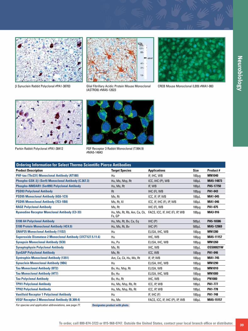
To order, call 800-874-3723 or 815-968-0747. Outside the United States, contact your local branch office or distributor. 39
NeurobiologyGlial Fibrillary Acidic Protein Mouse Monoclonal
(ASTRO6) #MA5-12023b Synuclein Rabbit Polyclonal #PA1-38703
Parkin Rabbit Polyclonal #PA1-38412 FGF Receptor 3 Rabbit Monoclonal (T.994.9) #MA5-14843
Ordering Information for Select Thermo Scientific Pierce AntibodiesProduct Description Target Species Applications Size Product #
PHF-tau (Thr231) Monoclonal Antibody (AT180) Hu IF, IHC, WB 100μg MN1040
Phospho-GSK-3b (Ser9) Monoclonal Antibody (C.367.3) Hu, Ms, Nhp, Rt ICC, IHC (P), WB 100μL MA5-14873
Phospho-NMDAR1 (Ser890) Polyclonal Antibody Hu, Ms, Rt IF, WB 100μL PA5-17750
PSD93 Polyclonal Antibody Rt IHC (F), WB 100μg PA1-043
PSD95 Monoclonal Antibody (6G6-1C9) Ms, Rt ICC, IF, IP, WB 100μL MA1-045
PSD95 Monoclonal Antibody (7E3-1B8) Ms, Rt, Xl ICC, IF, IHC (F), IP, WB 100μL MA1-046
RAGE Polyclonal Antibody Ms, Rt IHC (F), WB 100μg PA1-075
Ryanodine Receptor Monclonal Antibody (C3-33) Hu, Ms, Rt, Rb, Am, Ca, Ck, Fs, GP
FACS, ICC, IF, IHC (F), IP, WB 100μg MA3-916
S100 A4 Polyclonal Antibody Hu, Ms, Rt, Bv, Ca, Eq IHC (P) 500μL PA5-16586
S100 Protein Monoclonal Antibody (4C4.9) Hu, Ms, Rt, Bv IHC (P) 500μL MA5-12969
SNAP25 Monoclonal Antibody (11D2) Hu ELISA, IHC, WB 100μg MN1280
Superoxide Dismutase 2 Monoclonal Antibody (37CT127.5.11.6) Hu IHC, WB 100μg MA5-11157
Synapsin Monoclonal Antibody (5C8) Hu, Po ELISA, IHC, WB 100μg MN1260
Synaptophysin Polyclonal Antibody Ms, Rt IHC, WB 100μL OSS00021W
SynGAP Polyclonal Antibody Ms, Rt ICC, WB 100μg PA1-046
Syntrophin Monoclonal Antibody (1351) Am, Ca, Ck, Hu, Ms, Rt IF, IP, WB 100μg MA1-745
Synuclein Monoclonal Antibody (9B6) Hu ELISA, IHC, WB 100μg MN1290
Tau Monoclonal Antibody (BT2) Bv, Hu, Nhp, Rt ELISA, WB 100μg MN1010
Tau Monoclonal Antibody (HT7) Bv, Hu ELISA, IHC, WB 100μg MN1000
Tau Polyclonal Antibody Bv, Hu, Rt IHC, WB 500μg PN1000
TPH1 Polyclonal Antibody Hu, Ms, Nhp, Rb, Rt ICC, IP, WB 100μL PA1-777
TPH2 Polyclonal Antibody Hu, Ms, Nhp, Rb, Rt ICC, IP, WB 100μL PA1-778
Vanilloid Receptor 1 Polyclonal Antibody Hu IF, IHC (F) 100μg PA1-748
VEGF Receptor 2 Monoclonal Antibody (B.309.4) Hu, Ms FACS, ICC, IF, IHC (P), IP, WB 100μL MA5-15157
For species and application abbreviations, see page 77.
CREB Mouse Monoclonal (LB9) #MA1-083
Designates product with photo.

40 For more information and complete antibody listings, visit www.thermoscientific.com/pierce-abs
Stem
Cel
l Bio
logy
Primary Antibodies by Research Areas
Stem cells are characterized by their ability to self-renew an initial, undifferentiated state. Stem cells also have the ability to give rise to more specialized or differentiated cells, referred to as potency. There are different degrees of stem cell potency. Totipotent or pluripotent stem cells can give rise to cells from all three germ layers (endoderm, ectoderm and mesoderm), while multi- and oligopotent cells can give rise to only a small number of closely related cells. In addition to normal stem cells, recent work has established the existence of cancer stem cells. Cancer stem cells maintain many of the normal stem cell markers; however, in contrast to their normal counterparts, cancer stem cells are able to give rise to tumors once transplanted.
Various pluripotent stem cells can be guided to differentiate in vitro into hematopoietic progenitors, muscle cells or neuronal cells. In reverse, differentiated somatic cells can be re-programmed to become pluripotent stem cells. Pluripotent cells can be generated from somatic cells in one of three different ways: nuclear transfer involving the transplantation of a somatic nucleus into an enucleated oocyte, cell fusion of a somatic cell and an embryonic stem cell resulting in pluripotent hybrids, or induced pluripotent stem cells (iPS). Induction of pluripotency requires transduction of somatic cells with the transcription factors c-Myc, Sox2, Klf4 and Oct4. Bottlenecks to iPS generation include the low frequency and asynchronous generation of iPS cells. In addition to the above-mentioned transcription factors, activation-induced cytidine deaminase (AID)-dependent DNA demethylation is also required for efficient iPS generation. AID was found to be necessary for promoter demethylation and induction of the Oct4 and NANOG genes. The efficient generation of iPS cells has great potential in the study of human disease, regenerative medicine and individualized, patient-specific targeted therapy. Oct-4A Monoclonal Antibody (T.631.9) #MA5-14845
SMAD2 Rabbit Monoclonal (R.542.9) #MA5-14996
MAP2 Rabbit Polyclonal #PA5-17646

To order, call 800-874-3723 or 815-968-0747. Outside the United States, contact your local branch office or distributor. 41
Stem Cell Biology
CD31 Mouse Monoclonal (1A10) #MA5-12564
Vimentin Mouse Monoclonal (V9) #MA5-11883
PDX-1 Polyclonal #PA3-830
Cadherin E Rabbit Polyclonal #PA1-37204 CD45 Mouse Monoclonal (SPM496) #MA1-37337
CD44 Mouse Monoclonal (SPM521) #MA1-39422 SOX2 Polyclonal #PA1-16968
CD15 Mouse Monoclonal (SPM119) #MA1-37308 CD56 Mouse Monoclonal (123C3) #MA1-37354
Ordering Information for Select Thermo Scientific Pierce AntibodiesProduct Description Target Species Applications Size Product #
Cadherin E Polyclonal Antibody Hu IHC (P) 1mL PA1-37204
CD15 Monoclonal Antibody (SPM119) Hu IHC (P) 1mL MA1-37308
CD31 Monoclonal Antibody (1A10) Hu IHC (P) 500μL MA5-12564
CD44 Monoclonal Antibody (SPM521) Hu IHC (P) 1mL MA1-39422
CD45 Monoclonal Antibody (SPM496) Hu IHC (P) 1mL MA1-37337
CD56 Monoclonal Antibody (123C3) Hu IHC (P) 1mL MA1-37354
c-KIT Polyclonal Antibody Hu, Ms WB, IHC, IF 100μg PA5-14695
c-Myc Monoclonal Antibody (9E10) Hu, Ms IHC, IP, WB 100μg MA1-980
MAP2 Polyclonal Antibody Hu, Ms, Nhp, Rt IF, WB 100μL PA5-17646
Oct-4A Monoclonal Antibody (T.631.9) Hu, Ms FACS, ICC, WB 100μL MA5-14845
PDX-1 Polyclonal Antibody Hu, Ms IF, IHC, WB 100μL PA3-830
PRDM14 Polyclonal Antibody Hu WB, IHC, IF 100μg PA5-11303
SMAD2 Monclonal Antibody (R.542.9) Hu, Nhp ICC, IP, WB 100μL MA5-14996
SOX2 Polyclonal Antibody Hu, Ms, Ov FACS, ICC, IF, IHC (P), WB 100μL PA1-16968
Vimentin Monoclonal Antibody (APD) Hu, Ms, Rt, Bv, Ca, Ck, Eq, Gb, Hm, Nhp, Po, Rb IHC (P) 500μL MA5-11883
For species and application abbreviations, see page 77. Designates product with photo.

42
Infla
mm
atio
n an
d Ce
ll St
ress
Primary Antibodies by Research Areas
For more information and complete antibody listings, visit www.thermoscientific.com/pierce-abs
The process of inflammation provides affected cells, tissues or the entire organism an opportunity to regain homeostasis. Inflammation is triggered by a variety of stimuli such as infection, tissue stress, malfunction and injury. Pathological consequences of inflammation include autoimmunity, fibrosis, tissue damage and tumor microenvironment establishment.
The normal inflammatory pathway consists of inducing stimuli, sensors, mediators and effectors. The source of the stimulus determines whether inflammation inducers are endogenous or exogenous. For example, in the case of bacterial infection, lipopolysaccharides (LPS) serve as exogenous inducers; toll-like receptors (TLRs) on the surface of resident macrophages serve as sensors; cytokines (e.g., TNFα) are mediators; and hepatocytes, epithelial cells and neutrophils are the inflammatory pathway effectors. Neutrophils are normally restricted to blood vessels and are recruited to the infection site by selectins which are expressed on the surface of infected epithelial cells. Upon selectin binding, integrins become activated and then bind to intercellular adhesion molecules (ICAMs), which are expressed on the surface of the infected epithelial cells. The neutrophils are now able to penetrate the epithelium into the affected tissue where they unload toxic contents (ROS, neutrophil elastase, cathepsin G and proteinase 3) to fight the source of the infection. If the neutrophils fail to clear the infection, additional cells, including macrophages and T-cells, are recruited to the inflammation site. Finally, if all attempts fail to degrade or clear the source of inflammation, the foreign body is gradually engulfed by layers of macrophages to form a granuloma.
Endogenous inducers generate inflammation signals often by sensing “abnormal states” arising from the inappropriate cellular or tissue localization of various factors. For example, purinoreceptors on the surface of macrophages detect ATP released from necrotic cells as a result of membrane disruption.
ATP binding to the purinoreceptors results in increased potassium ion (K+) influx and successive activation of the NALP inflammasome. The activated NALP (via endogenous or exogenous inducers) in turn stimulates caspase-1, which then promotes the processing and secretion of pro-inflammatory cytokines.
Several different types of inflammation mediators exist. Pro-inflammatory cytokines are one of several mediators of inflammation. Other well known mediators of inflammation are vasoactive amines (histamine, serotonin), vasoactive peptides which can be generated by the Hageman factor, lipid mediators (eicosanoids), chemokines, and proteases (elastin, cathepsin, MMP). Negative regulators of inflammation include lipoxins, resolvins and protectins. Other negative regulators include inhibitors of pro-inflammatory cytokine production and function such as the p38 MAPK pathway, antagonists of COX-1/COX-2 and TNFα signaling.
Phospho-p38 MAPK Rabbit Monoclonal (Thr180-Tyr182) (S.417.1) #MA5-15182
Phospho-p53 Rabbit Polyclonal (Ser37) #PA5-17866

To order, call 800-874-3723 or 815-968-0747. Outside the United States, contact your local branch office or distributor. 43
Inflamm
ation and Cell Stress
Heat Shock Protein 90 α Rabbit Polyclonal #PA5-16341
NFkB-p65 Rabbit Polyclonal #PA5-16545
p21WAF11 Mouse Monoclonal (CP74) #MA5-14353Phospho-ATF-2 Rabbit Polyclonal (Thr71) #PA5-17885
FoxO3a Rabbit Monoclonal (K.115.9) #MA5-14932
Ordering Information for Select Thermo Scientific Pierce AntibodiesProduct Description Target Species Applications Size Product #COX-1 Polyclonal Antibody Hu IHC (P) 500μL PA5-16318
COX2 Polyclonal Antibody Hu, Ms, Rt IHC (P), IP, WB 500μL PA5-16817COX2 Polyclonal Antibody Hu, Ms, Rt IHC (P), WB 100μL PA5-17614
FoxO3a Monoclonal Antibody (K.115.9) Hu, Ms, Nhp, Rt ICC, WB 100μL MA5-14932Heat Shock Factor 1 Monoclonal Antibody (4B4) Hu, Ms, Rt GS, IF, IP, WB 500μL MA5-14622Heat Shock Protein 27 Monoclonal Antibody (G3.1) Ca, Hu, Rt ELISA, ICC, IF, IHC (P), IP, WB 100μL MA3-015Heat Shock Protein 60 Monoclonal Antibody (A57-B9) Ba ICC, IP, WB 100μL MA3-023
Heat Shock Protein 70 Monoclonal Antibody (3A3) Am, Ar, Av, Dm, Fs, Hu, Ms, Pl, Po, Rt, Ys GS, ICC, IF, IHC (P), IP, WB 50μL MA3-006Heat Shock Protein 84 Polyclonal Antibody Hu, Ms, Rt IF, IHC (F), IP, WB 100μg PA3-012
Heat Shock Protein 90 α Polyclonal Antibody Hu, Ms, Rt IF, IHC (P), IP, WB 500μL PA5-16341
NFkB/p65 Polyclonal Antibody Hu, Ms, Rt, Bv, Ca, Dm, Hm, Po, Rb, Xl IHC (P), WB 500μL PA5-16545
p21WAF11 (CP74) Hu, Rt FACS, IHC (P), IP, WB 500μL MA5-14353
Phospho-ATF-2 (Thr71) Polyclonal Antibody Hu, Ms, Nhp, Rt FACS, ICC, IHC (F), IHC (P), IP, WB 100μL PA5-17885Phospho-p38 MAPK (Thr180/Tyr182) Monoclonal Antibody (S.417.1)
Bv, Dm, Hm, Hu, Mn, Ms, Nhp, Po, Rt, Ys, Zf
FACS, ICC, WB 200μL MA5-15182
Phospho-p53 (Ser37) Polyclonal Antibody Hu, Nhp FACS, ICC, IP, WB 100μL PA5-17866
SOD2 Monoclonal Antibody (37CT127.5.11.6) Hu WB, IHC 100μg MA5-11157
For species and application abbreviations, see page 77. Designates product with photo.

44
Rece
ptor
Tyr
osin
e Ki
nase
sPrimary Antibodies by Cell Signaling
For more information and complete antibody listings, visit www.thermoscientific.com/pierce-abs
Receptor Tyrosine Kinases (RTKs) are essential cell surface receptors which mediate signal transmission from external ligands such as growth factors, hormones and cytokines. RTKs are key regulators of multiple biological processes including cell proliferation, cell growth, cell survival, development, vascular-genesis, transcription and protein synthesis. Over 50 RTKs have been identified (e.g., EGFR, PDGFR, VEGFR, c-Kit, InsR and c-Met).
The primary mode of RTK activation involves binding of external ligand, receptor dimerization and autophosphorylation of tyrosine residues located within the cytoplasmic tail of the receptor. Tyrosine phosphorylation of RTKs results in docking of Src Homology domain (SH2)-containing proteins which transmit the signal through a cascade of downstream phosphorylation events, ultimately resulting in target protein functionality. Deregulation of RTKs have been implicated in human disease and is a hallmark of a myriad of human cancers. Current research on RTKs focuses on development of both chemical- and antibody-based inhibitors of receptor tyrosine kinase activation including SH2 domain inhibitor mimetics.
IGF-I Receptor b Rabbit Polyclonal #PA5-17530
FGF Receptor 3 Rabbit Monoclonal (T.994.9) #MA5-14843
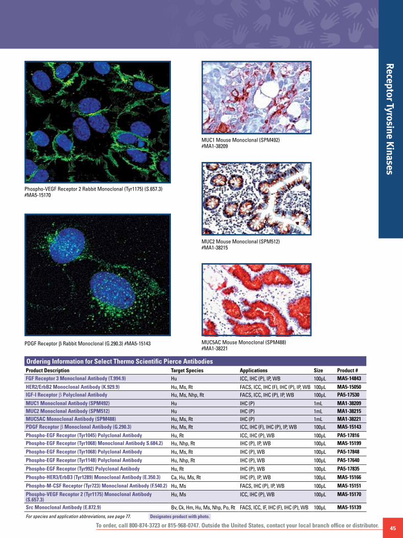
To order, call 800-874-3723 or 815-968-0747. Outside the United States, contact your local branch office or distributor. 45
Receptor Tyrosine Kinases
PDGF Receptor b Rabbit Monoclonal (G.290.3) #MA5-15143
Phospho-VEGF Receptor 2 Rabbit Monoclonal (Tyr1175) (S.657.3) #MA5-15170
MUC1 Mouse Monoclonal (SPM492) #MA1-38209
MUC5AC Mouse Monoclonal (SPM488) #MA1-38221
MUC2 Mouse Monoclonal (SPM512) #MA1-38215
Ordering Information for Select Thermo Scientific Pierce AntibodiesProduct Description Target Species Applications Size Product #FGF Receptor 3 Monoclonal Antibody (T.994.9) Hu ICC, IHC (P), IP, WB 100μL MA5-14843
HER2/ErbB2 Monoclonal Antibody (K.929.9) Hu, Ms, Rt FACS, ICC, IHC (F), IHC (P), IP, WB 100μL MA5-15050IGF-I Receptor b Polyclonal Antibody Hu, Ms, Nhp, Rt FACS, ICC, IHC (P), IP, WB 100μL PA5-17530
MUC1 Monoclonal Antibody (SPM492) Hu IHC (P) 1mL MA1-38209MUC2 Monoclonal Antibody (SPM512) Hu IHC (P) 1mL MA1-38215MUC5AC Monoclonal Antibody (SPM488) Hu, Ms, Rt IHC (P) 1mL MA1-38221PDGF Receptor b Monoclonal Antibody (G.290.3) Hu, Ms, Rt ICC, IHC (F), IHC (P), IP, WB 100μL MA5-15143
Phospho-EGF Receptor (Tyr1045) Polyclonal Antibody Hu, Rt ICC, IHC (P), WB 100μL PA5-17816Phospho-EGF Receptor (Tyr1068) Monoclonal Antibody S.684.2) Hu, Nhp, Rt IHC (P), IP, WB 100μL MA5-15199
Phospho-EGF Receptor (Tyr1068) Polyclonal Antibody Hu, Ms, Rt IHC (P), WB 100μL PA5-17848
Phospho-EGF Receptor (Tyr1148) Polyclonal Antibody Hu, Nhp, Rt IHC (P), WB 100μL PA5-17640
Phospho-EGF Receptor (Tyr992) Polyclonal Antibody Hu, Rt IHC (P), WB 100μL PA5-17835
Phospho-HER3/ErbB3 (Tyr1289) Monoclonal Antibody (E.350.3) Ca, Hu, Ms, Rt IHC (P), IP, WB 100μL MA5-15166
Phospho-M-CSF Receptor (Tyr723) Monoclonal Antibody (F.540.2) Hu, Ms FACS, IHC (P), IP, WB 100μL MA5-15151
Phospho-VEGF Receptor 2 (Tyr1175) Monoclonal Antibody (S.657.3)
Hu, Ms ICC, IHC (P), WB 100μL MA5-15170
Src Monoclonal Antibody (E.872.9) Bv, Ck, Hm, Hu, Ms, Nhp, Po, Rt FACS, ICC, IF, IHC (F), IHC (P), WB 100μL MA5-15139
For species and application abbreviations, see page 77. Designates product with photo.

46
Prot
ein
Phos
phat
ases
Primary Antibodies by Cell Signaling
For more information and complete antibody listings, visit www.thermoscientific.com/pierce-abs
Protein phosphatases are a class of enzymes which catalyze the dephosphorylation of target proteins. At the most basic level, protein phosphatases counteract protein kinases, enzymes that catalyze the addition of phosphate groups to target proteins, thereby influencing their activity. The most common form of reversible protein post-translational modification (PTM) is phosphorylation.
Protein phosphorylation is an essential cellular event which mediates nearly all biological processes and is a central circuit for transmission of extracellular stimuli throughout the cell via signal transduction pathways. Protein phosphorylation occurs primarily on four amino acids, Serine (Ser), Threonine (Thr), Tyrosine (Tyr), and Histidine (His), with a frequency of approximately 80%/15%/5%/0.1%, respectively. Phosphorylation also occurs on lipid moieties essential for activation of the PI3K/Akt signaling cascade. More recent analysis of the phosphoproteome using mass spectrometry suggests that greater than 60% of the human proteome is phosphorylated at any given time. The exact level of protein phosphorylation is a complicated question to resolve, as any protein containing kinase consensus motifs with the amino acids Ser, Thr or Tyr have the potential to become phosphorylated at any given time in response to biological signals.
As is the case for protein phosphorylation, dephosphorylation can either influence protein function in a positive or negative manner. Five main classes of protein phosphatases exist including tyrosine-specific (PTP1B), serine/threonine-specific (PP2C), dual-specific (DUSP1), histidine (PHP) and lipid phosphatases (PTEN). Deregulation of protein phosphatases has been implicated in many human diseases including cancer, neurodegeneration and metabolic disorders. A classic example of a deregulated phosphatase in human disease is the Phosphatase and Tensin homolog, PTEN. PTEN is a critical tumor suppressor protein which mediates signaling through the PI3K/Akt pathway by counteracting PI3K-directed phosphorylation of membrane lipids. Epigenetic downregulation and loss of heterozygosity mutations at the PTEN locus account for aberrant PI3K/Akt signaling contributing to the formation of numerous human cancers.
PP2A A Subunit Rabbit Polyclonal #PA5-17516
RhoA Rabbit Polyclonal #OSR00266W
Akt1 Mouse Monoclonal (G.668.2) #MA5-15187
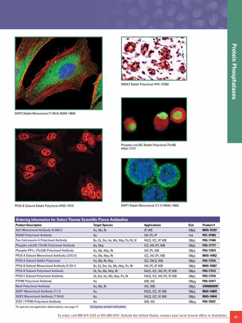
To order, call 800-874-3723 or 815-968-0747. Outside the United States, contact your local branch office or distributor. 47
Protein Phosphatases
PP2A B Subunit Rabbit Polyclonal #PA5-17515
SHIP2 Rabbit Monoclonal (T.194.8) #MA5-14844
DRAK2 Rabbit Polyclonal #PA1-37583
SHIP1 Rabbit Monoclonal (T.7.7) #MA5-14893
Phospho-cdc25C Rabbit Polyclonal (Thr48) #PA5-17777
Ordering Information for Select Thermo Scientific Pierce AntibodiesProduct Description Target Species Applications Size Product #
Akt1 Monoclonal Antibody (G.668.2) Hu, Ms, Rt IP, WB 100μL MA5-15187
DRAK2 Polyclonal Antibody Hu IHC (P), IP 1mL PA1-37583
Pan-Calcineurin A Polyclonal Antibody Bv, Ck, Dm, Hu, Ms, Nhp, Po, Rt, Xl FACS, ICC, IP, WB 100μL PA5-17446
Phospho-cdc25C (Thr48) Polyclonal Antibody Hu, Nhp ICC, IHC (P), WB 100μL PA5-17777
Phospho-PP1α (Thr320) Polyclonal Antibody Hu, Ms, Nhp, Rt IHC (P), WB 100μL PA5-17819
PP2A A Subunit Monoclonal Antibody (J.613.4) Hu, Ms, Nhp, Rt ICC, IHC (P), WB 100μL MA5-14952
PP2A A Subunit Rabbit Polyclonal Hu, Ms, Rt, Nhp ICC, FACS, WB 100μL PA5-17516
PP2A B Subunit Monoclonal Antibody (F.722.1) Bv, Ck, Dm, Hu, Ms, Nhp, Po, Rt IHC (P), IP, WB 100μL MA5-15007
PP2A B Subunit Polyclonal Antibody Ck, Hu, Ms, Nhp, Rt FACS, ICC, IHC (P), IP, WB 100μL PA5-17515
PP2A C Subunit Polyclonal Antibody Ck, Dm, Hu, Ms, Nhp, Po, Rt FACS, ICC, IHC (P), IP, WB 100μL PA5-17510
PTPN6 Polyclonal Antibody Hu WB, IHC 100μg PA5-12417
RhoA Polyclonal Antibody Hu, Ms, Rt IHC, WB 100μL OSR00266W
SHIP1 Monoclonal Antibody (T.7.7) Hu FACS, ICC, IP, WB 100μL MA5-14893
SHIP2 Monoclonal Antibody (T.194.8) Hu FACS, ICC, IP, WB 100μL MA5-14844
STEP / PTPN5 Polyclonal Antibody Hu WB, IHC 100μg PA5-15531
For species and application abbreviations, see page 77. Designates product with photo.
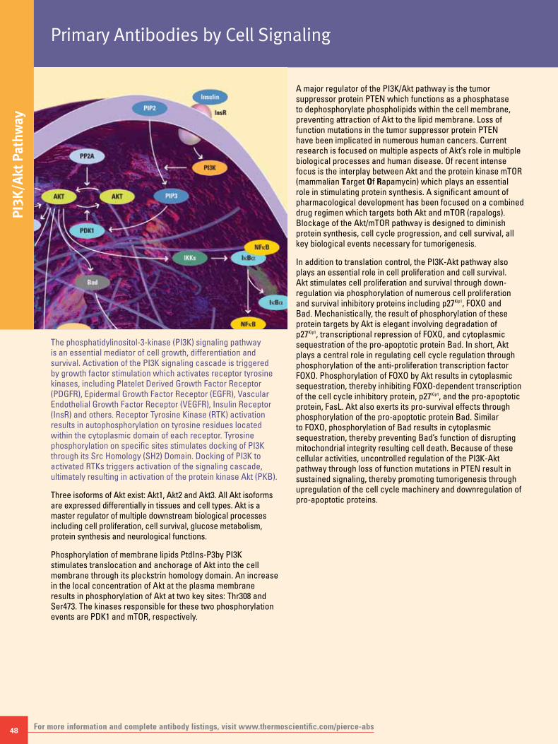
48
PI3K
/Akt
Pat
hway
Primary Antibodies by Cell Signaling
For more information and complete antibody listings, visit www.thermoscientific.com/pierce-abs
The phosphatidylinositol-3-kinase (PI3K) signaling pathway is an essential mediator of cell growth, differentiation and survival. Activation of the PI3K signaling cascade is triggered by growth factor stimulation which activates receptor tyrosine kinases, including Platelet Derived Growth Factor Receptor (PDGFR), Epidermal Growth Factor Receptor (EGFR), Vascular Endothelial Growth Factor Receptor (VEGFR), Insulin Receptor (InsR) and others. Receptor Tyrosine Kinase (RTK) activation results in autophosphorylation on tyrosine residues located within the cytoplasmic domain of each receptor. Tyrosine phosphorylation on specific sites stimulates docking of PI3K through its Src Homology (SH2) Domain. Docking of PI3K to activated RTKs triggers activation of the signaling cascade, ultimately resulting in activation of the protein kinase Akt (PKB).
Three isoforms of Akt exist: Akt1, Akt2 and Akt3. All Akt isoforms are expressed differentially in tissues and cell types. Akt is a master regulator of multiple downstream biological processes including cell proliferation, cell survival, glucose metabolism, protein synthesis and neurological functions.
Phosphorylation of membrane lipids PtdIns-P3by PI3K stimulates translocation and anchorage of Akt into the cell membrane through its pleckstrin homology domain. An increase in the local concentration of Akt at the plasma membrane results in phosphorylation of Akt at two key sites: Thr308 and Ser473. The kinases responsible for these two phosphorylation events are PDK1 and mTOR, respectively.
A major regulator of the PI3K/Akt pathway is the tumor suppressor protein PTEN which functions as a phosphatase to dephosphorylate phospholipids within the cell membrane, preventing attraction of Akt to the lipid membrane. Loss of function mutations in the tumor suppressor protein PTEN have been implicated in numerous human cancers. Current research is focused on multiple aspects of Akt’s role in multiple biological processes and human disease. Of recent intense focus is the interplay between Akt and the protein kinase mTOR (mammalian Target Of Rapamycin) which plays an essential role in stimulating protein synthesis. A significant amount of pharmacological development has been focused on a combined drug regimen which targets both Akt and mTOR (rapalogs). Blockage of the Akt/mTOR pathway is designed to diminish protein synthesis, cell cycle progression, and cell survival, all key biological events necessary for tumorigenesis.
In addition to translation control, the PI3K-Akt pathway also plays an essential role in cell proliferation and cell survival. Akt stimulates cell proliferation and survival through down-regulation via phosphorylation of numerous cell proliferation and survival inhibitory proteins including p27Kip1, FOXO and Bad. Mechanistically, the result of phosphorylation of these protein targets by Akt is elegant involving degradation of p27Kip1, transcriptional repression of FOXO, and cytoplasmic sequestration of the pro-apoptotic protein Bad. In short, Akt plays a central role in regulating cell cycle regulation through phosphorylation of the anti-proliferation transcription factor FOXO. Phosphorylation of FOXO by Akt results in cytoplasmic sequestration, thereby inhibiting FOXO-dependent transcription of the cell cycle inhibitory protein, p27Kip1, and the pro-apoptotic protein, FasL. Akt also exerts its pro-survival effects through phosphorylation of the pro-apoptotic protein Bad. Similar to FOXO, phosphorylation of Bad results in cytoplasmic sequestration, thereby preventing Bad’s function of disrupting mitochondrial integrity resulting cell death. Because of these cellular activities, uncontrolled regulation of the PI3K-Akt pathway through loss of function mutations in PTEN result in sustained signaling, thereby promoting tumorigenesis through upregulation of the cell cycle machinery and downregulation of pro-apoptotic proteins.

To order, call 800-874-3723 or 815-968-0747. Outside the United States, contact your local branch office or distributor. 49
PI3K/Akt Pathway
EGF Receptor Mouse Monoclonal (SPM341) #MA1-37605
ErbB2 Rabbit Polyclonal #PA1-37426 FoxO3a Rabbit Monoclonal (K.115.9) #MA5-14932
FoxO1 Rabbit Monoclonal (S.502.4) #MA5-14846
Phospho-GSK-3b Rabbit Monoclonal (Ser9) (C.367.3) #MA5-14873
Phospho-Akt Rabbit Polyclonal (Ser473) #PA1-37046
Akt Rabbit Monoclonal (pan) (E.32.10) #MA5-14999
Ordering Information for Select Thermo Scientific Pierce AntibodiesProduct Description Target Species Applications Size Product #
Akt (pan) Monoclonal Antibody (E.32.10) Hu, Ms, Nhp, Rt FACS, ICC, IHC (P), IP, WB 100μL MA5-14999
Akt1 Monoclonal Antibody (G.145.7) Hu, Ms, Nhp, Rt IHC (P), IP, WB 100μL MA5-14898
EGF Receptor Monoclonal Antibody (SPM341) Hu IHC (P) 1mL MA1-37605
eNOS Polyclonal Antibody Bv, Ca, Hu, Ms, Po, Rt IHC (P, F), WB 100μL PA1-037
ErbB2 Polyclonal Antibody Hu, Ms, Nhp, Rt IHC (P), WB 1mL PA1-37426
FoxO1 Monoclonal Antibody (S.502.4) Hu, Ms, Nhp, Rt ICC, IHC (P), WB 100μL MA5-14846
FoxO3a Monoclonal Antibody (K.115.9) Hu, Ms, Nhp, Rt ICC, WB 100μL MA5-14932
Phospho-AKT (Ser473) Monoclonal Antibody (14-6) Hu, Ms ICC, IF, WB 100μL OMA1-03061
Phospho-AKT (Ser473) Polyclonal Antibody Hu IHC (P) 1mL PA1-37046
Phospho-AKT (Thr308) Polyclonal Antibody Hu, Ms, Rt IF, IHC, WB 200μg PA1-14030
Phospho-AKT2 (Ser474) Polyclonal Antibody Hu, Ms, Rt IHC, WB 200μg PA1-14034
Phospho-GSK3 α (Ser21) Polyclonal Antibody Hu, Ms, Rt IHC, WB 200μg PA1-14180
Phospho-GSK3 b (Ser9) Polyclonal Antibody Hu, Ms, Rt IF, IHC, WB 200μg PA1-14182
Phospho-GSK-3b (Ser9) Monoclonal Antibody (C.367.3) Hu, Ms, Nhp, Rt ICC, IHC (P), WB 100μL MA5-14873
PI3 Kinase p110-α Monoclonal Antibody (H.843.0) Bv, Hu, Ms, Rt IHC (P), IP, WB 100μL MA5-14870
PTEN Monoclonal Antibody (17.A) Hu IHC 500μL MA5-12278
For species and application abbreviations, see page 77. Designates product with photo.

50
MAP
K Si
gnal
ing
Path
way
sPrimary Antibodies by Cell Signaling
For more information and complete antibody listings, visit www.thermoscientific.com/pierce-abs
The Mitogen Activated Protein Kinases (MAPK) family of serine/threonine protein kinases regulates a diverse range of biological processes including cell proliferation, development, differentiation, cell survival and protein synthesis. Three classical MAPK pathways have been identified in mammalian cells. These pathways include the growth factor signaling ERK1/2 pathway and two stress-activated pathways, SAPK/JNK and p38. More recently another MAPK pathway, ERK5/BMK1, has been discovered and has been shown to be responsive to both growth factor and stress signals. More recent evidence suggests that these pathways are nonlinear and that extensive crosstalk exists between neighboring signaling cascades including parallel MAPK and PI3K/Akt pathways.
MAPK (ERK1/2) Signaling Pathway The first characterized MAPK was ERK1/2 also known as p42/p44. This kinase cascade responds primarily to growth factor stimulation which then activates receptor tyrosine kinases and GPCRs. Receptor activation results in downstream signaling through small G-proteins, such as the proto-oncoprotein p21Ras which stimulates a conformational change and subsequent activation of the serine/threonine kinase family Raf (A-Raf, B-Raf, c-Raf). Activated Raf phosphorylates the downstream dual-specific serine/threonine/tyrosine kinase MEK1/2, which in turn phosphorylates the activation loop of ERK1/2 on residues Thr202/Tyr204. Activated MAPK translocates into the nucleus where it phosphorylates multiple transcription factors and key regulatory proteins involved in cell proliferation, cell growth, cell survival and differentiation. Because of the role that the ERK1/2 pathway plays in tumorigenesis, intense research is focused on the development of pharmacological inhibitors to ameliorate this disease.
JNK/SAPK Signaling Pathway The Jun N-terminal Kinase (JNK), Stress Activated Protein Kinases (SAPK) families of MAP kinases play crucial roles in regulating responses to cell stress, inflammation and apoptosis. The JNK/SAPK pathway is activated by UV, gamma radiation, ceramides, inflammatory cytokines and, in rare cases, growth factors signaling through GPCRs. The small GTPases belonging to the Rho family (Rho, Rac, cdc42) mediate the signaling process by activating downstream MAPKKKs which activate the dual specific serine/threonine/tyrosine kinases MKK4/7 and result in downstream activation of SAPK/JNK protein complexes. Dimerized SAPK/JNK translocate to the nucleus where they stimulate activity of multiple transcription factors, including c-Jun, ATF-2, SMAD4, p53, STAT3 and NFAT4.
p38 MAPK Pathway Similar to the JNK/SAPK pathway, the p38 MAPK cascade also responds to cell stress, including UV irradiation, heat shock, high osmotic stress, lipopolysaccharide and proinflammatory cytokines such as IL-1 and TNF-α. Although both pathways target many of the same downstream effector proteins, the primary difference between these two pathways is the fate of the cell. Signaling through the JNK/SAPK pathway often triggers apoptosis whereas the p38 pathway favors cell cycle arrest. Factors which influence these decisions are the nature of the signal, signal strength and signal duration. Understanding the mechanisms of differential signaling through these two pathways remains a central focus for many research labs.
Phospho-c-Jun Rabbit Polyclonal (Ser73) #PA5-17879
MEK1/2 Monoclonal (J.653.9) #MA5-15135
JunB Rabbit Polyclonal #PA5-17263

To order, call 800-874-3723 or 815-968-0747. Outside the United States, contact your local branch office or distributor. 51
MAPK Signaling Pathw
ays
p44/42 MAPK Rabbit Monoclonal (Erk1-2) (K.913.4) #MA5-15134
Phospho-ATF-2 Rabbit Polyclonal (Thr71) #PA5-17885
Phospho-MEK1 Rabbit Polyclonal (Thr286) #PA5-17664
Phospho-p90RSK Rabbit Polyclonal (Thr573) #PA5-17841
RSK1-RSK2-RSK3 Rabbit Monoclonal (F.940.7) #MA5-15040
Phospho-p44/42 MAPK Rabbit Monoclonal (Erk1-2) (Thr202-Tyr204) (B.742.5) #MA5-15174
Ordering Information for Select Thermo Scientific Pierce AntibodiesProduct Description Target Species Applications Size Product #14-3-3 Tau Polyclonal Antibody Hu, Ms, Nhp, Rt IHC (P), WB 100μL PA5-17426ATF-2 Monoclonal Antibody (S.613.5) Hu, Ms, Nhp, Rt IHC (P), IP, WB 100μL MA5-15169
c-Jun Polyclonal Antibody Hu, Ms, Rt IHC, WB 200μg PA1-14099
ERK1 Polyclonal Antibody Hu WB, IHC 100μg PA5-14093ERK2 Polyclonal Antibody Hu WB, IHC, FACS 100μg PA5-14414JunB Polyclonal Antibody Hu ICC, IP, WB 100μL PA5-17263JunD Polyclonal Antibody Hu, Ms, Rt IHC, WB 200μg PA1-14238
MEK1/2 Monoclonal Antibody (J.653.9) Hu, Ms, Nhp, Rt FACS, ICC, IHC (P), WB 100μL MA5-15135
p38-α MAPK Polyclonal Antibody Hu, Ms, Rt, Nhp IHC (P), IP, WB 100μL PA5-17713
p42 MAP Kinase (Erk2) Polyclonal Antibody Hu, Mn, Ms, Nhp, Rt IHC (P), WB 100μL PA5-17710
p44/42 MAPK (Erk1/2) Monoclonal Antibody (K.913.4) Bv, Ca, Ce, Ck, Dm, Hm, Hu, Mn, Ms, Nhp, Po, Rt, Ys, Zf
FACS, ICC, IHC (P), IP, WB 200μL MA5-15134
Phospho-ATF-2 (Thr69/71) Polyclonal Antibody Hu, Ms, Nhp, Rt IHC (P), WB 100μL PA5-17886
Phospho-ATF-2 (Thr71) Polyclonal Antibody Hu, Ms, Nhp, Rt FACS, ICC, IHC (F), IHC (P), IP, WB 100μL PA5-17885
Phospho-c-Jun (Ser243) Polyclonal Antibody Hu, Ms, Rt IHC, WB 200μg PA1-14100
Phospho-c-Jun (Ser63) Monoclonal Antibody Hu, Ms, Rt IHC (P), WB 100μL MA5-15115
Phospho-c-Jun (Ser73) Polyclonal Antibody Hu, Ms, Nhp, Rt ICC, WB 100μL PA5-17879
Phospho-c-Jun (Thr239) Polyclonal Antibody Hu, Ms, Rt IHC, WB 200μg PA1-14098
Phospho-c-Jun (Thr91) Polyclonal Antibody Hu, Ms, Rt IHC, WB 200μg PA1-14106
Phospho-c-Jun (Thr93) Polyclonal Antibody Hu, Ms, Rt IHC, WB 200μg PA1-14108
Phospho-JunB (Ser259) Polyclonal Antibody Hu, Ms, Rt IHC, WB 200μg PA1-14233
Phospho-JunB (Ser79) Polyclonal Antibody Hu, Ms, Rt IHC 200μg PA1-14235
Phospho-JunD (Ser255) Polyclonal Antibody Hu, Ms, Rt IHC, WB 200μg PA1-14237
Phospho-MAPKAPK-2 (Thr334) Polyclonal Antibody Hu, Ms, Nhp, Rt IHC (P), WB 100μL PA5-17791
Phospho-MEK1 (Thr286) Polyclonal Antibody Hu, Ms, Nhp, Rt FACS, ICC, IP, WB 100μL PA5-17664
Phospho-MEK1/2 (Ser221) Monoclonal Antibody (K.742.1) Ca, Hu, Ms, Nhp, Rt FACS, IHC (P), WB 100μL MA5-15118
Phospho-p44/42 MAPK (Erk1/2) (Thr202/Tyr204) Monoclonal Antibody (B.742.5)
Hu, Ms, Rt, Dm, Mn, Nhp, Po, Zf FACS, ICC, WB 200μL MA5-15174
Phospho-p90RSK (Thr573) Polyclonal Antibody Hu, Ms, Rt, Ck, Hm, Xl, Zf ICC, WB 100μL PA5-17841
RSK1/RSK2/RSK3 Monoclonal Antibody (F.940.7) Hu, Ms, Rt, GP, Nhp ICC, IP, WB 100μL MA5-15040
For species and application abbreviations, see page 77. Designates product with photo.

52
JAK/
STAT
Pat
hway
Primary Antibodies by Cell Signaling
For more information and complete antibody listings, visit www.thermoscientific.com/pierce-abs
The JAK-STAT pathway is a major regulatory element in cytokine signaling. When cytokines, such as interferons and IL-6, are activated, they bind to their respective receptors, resulting in the activation of receptor-associated JAK tyrosine kinases. Several receptors exist that are capable of inducing various JAK kinases. With IL-6, the receptor consists of IL-6R α chain and gp130 subunits. IL-6 binding results in the activation of Jak1, Jak2 and Tyk2 tyrosine kinases, which in turn phosphorylate STAT1 and STAT3 transcription factors. Tyrosine phosphorylated STATs dimerize via binding to their respective SH2 domains and accumulate in the nucleus where they mediate IL-6-induced gene transcription.
Negative regulators of the JAK-STAT pathway include SOCS, SHP1 and PIAS proteins. Recent work suggests a more complex image of the JAK/STAT pathway, which involves regulation of gene transcription via unphosphorylated STATs, glycosylated STATs and crosstalk with other signaling pathways, such as the PKC-delta, p38 MAPK and NFkB pathways.
Phospho-STAT3 Rabbit Polyclonal (Ser727) #PA5-17876
Phospho-STAT1 Rabbit Monoclonal (Tyr701) (S.213.5) #MA5-15071

To order, call 800-874-3723 or 815-968-0747. Outside the United States, contact your local branch office or distributor. 53
JAK/STAT Pathway
Phospho-STAT5 Rabbit Monoclonal (Tyr694) (J.536.2) #MA5-14822
STAT3 Mouse Monoclonal (G.659.4) #MA5-15179
Phospho-JAK2 (Tyr1007) Rabbit Polyclonal #PA1-14232
JAK2 Polyclonal #PA1-14231
Phospho-JAK1 (Tyr1022) Rabbit Polyclonal #PA1-14228
Ordering Information for Select Thermo Scientific Pierce AntibodiesProduct Description Target Species Applications Size Product #
JAK1 Polyclonal Antibody Hu, Ms, Rt IHC, WB 200μg PA1-14229
JAK2 Polyclonal Antibody Hu, Ms, Rt IHC 200μg PA1-14231
Phospho-JAK1 (Tyr1022) Polyclonal Antibody Hu, Ms, Rt IHC, WB 200μg PA1-14228
Phospho-JAK2 (Tyr1007) Polyclonal Antibody Hu, Ms, Rt IHC (P), WB 200μg PA1-14232
Phospho-JAK2 (Tyr221) Polyclonal Antibody Hu, Ms, Rt IHC 200μg PA1-14230
Phospho-Stat1 (Ser727) Polyclonal Antibody Bv, Hu, Ms, Rt ChIP, FACS, ICC, WB 100μL PA5-17635
Phospho-Stat1 (Tyr701) Monoclonal Antibody (S.213.5) Hu, Ms ChIP, FACS, ICC, IHC (F), IHC (P), IP, WB 100μL MA5-15071
Phospho-Stat3 (Ser727) Polyclonal Antibody Bv, Hu, Ms, Rt ChIP, ICC, IP, WB 100μL PA5-17876
Phospho-Stat5 (Tyr694) Monoclonal Antibody (J.536.2) Bv, Hu, Ms, Nhp, Rt FACS, ICC, IHC (P), WB 100μL MA5-14822
Phospho-Stat6 (Tyr641) Polyclonal Antibody Bv, Hu FACS, ICC, IP, WB 100μL PA5-17902
PRMT5 Polyclonal Antibody Hu WB, IHC 100μg PA5-11121
Stat1 Monoclonal Antibody (C.156.9) Hu, Nhp IHC (P), WB 100μL MA5-15129
STAT2 Polyclonal Antibody Hu WB, IHC 100μg PA5-11629
Stat3 Monoclonal Antibody (F.424.5) Hu, Ms, Nhp, Rt ChIP, IHC (P), IP, WB 100μL MA5-15147
Stat3 Monoclonal Antibody (G.659.4) Hu, Ms, Nhp, Rt ChIP, FACS, ICC, IHC (P), IP, WB 100μL MA5-15179
TYK2 Polyclonal Antibody Hu, Ms, Rt IHC 200μg PA1-14430
For species and application abbreviations, see page 77. Designates product with photo.

54
TNF/
NFk
B Pa
thw
ayPrimary Antibodies by Cell Signaling
For more information and complete antibody listings, visit www.thermoscientific.com/pierce-abs
Signaling through the NFkB pathway is a fairly complex and strictly regulated process in mammalian cells. The NFkB pathway is crucial to the function of the immune system because it modulates the expression of various cytokines and growth factors in response to activation of various receptors involved in immunity, such as the TNF receptor, TLR/IL-1 receptor, T-cell and B-cell receptors. For example, in the case of TNFα, it has been shown that NFkB activation counters uncontrolled TNF-induced apoptosis by providing a balance of pro-apoptotic and pro-proliferative signaling events.
The NFkB family of transcription factors consists of 5 members: RelA (p65), RelB, c-Rel, NFkB1 (p50, p105) and NFkB2 (p52, p100). Under non-activating conditions, NFkB members form a complex with IkB, which prevents translocation to the nucleus. TNFα binding to the TNF receptor triggers NFkB activation through the canonical pathway, while TCR activation can also induce an alternative NFkB pathway. In the canonical pathway, TNF-receptor binding leads to phosphorylation of the IKK complex, which consists of IKKα, IKKb and NEMO and subsequent phosphorylation of IkB. Phosphorylated IkB is then targeted for b-TrCP-mediated proteasomal degradation thereby allowing Rel-NFkB dimers to translocate to the nucleus and to regulate gene transcription.
In the alternative pathway, IKKα is activated via NIK kinase resulting in phosphorylation and subsequent proteasomal processing of RelB-bound p100 protein. After proteasomal processing of p100, active RelB-p52 heterodimers are now able to translocate to the nucleus and regulate gene transcription. A well known mechanism of feedback inhibition of the NFkB pathway is through export of nuclear NFkB to the cytosol by newly synthesized IkB.
Phospho-NFkB p65 Rabbit Monoclonal (Ser536) (T.894.2)#MA5-15160
TRAF1 Rabbit Monoclonal (K.915.10) #MA5-15043
Mouse Monoclonal IkB-α (T.937.7) #MA5-15132

To order, call 800-874-3723 or 815-968-0747. Outside the United States, contact your local branch office or distributor. 55
TNF/N
FkB Pathway
TRADD Rabbit Polyclonal #PA1-38791NFkB-p65 Rabbit Polyclonal #PA5-16545
TNF Receptor II Rabbit Polyclonal #PA1-38784c-Rel Rabbit Polyclonal #PA5-17452
COX-1 Rabbit Polyclonal #PA5-16318
COX2 Rabbit Monoclonal (SP21) #MA5-14568
Ordering Information for Select Thermo Scientific Pierce AntibodiesProduct Description Target Species Applications Size Product #Cox-1 Polyclonal Antibody Hu IHC(P) 500μL PA5-16318
COX2 Monoclonal Antibody (SP21) Hu, Ms, Rt IHC (P) 500μL MA5-14568
c-Rel Polyclonal Antibody Hu, Nhp FACS, ICC, IHC (P), IP, WB 100μL PA5-17452
I-k-B-α Monoclonal Antibody (T.937.7) Bv, GP, Hu, Ms, Nhp, Po, Rt FACS, ICC, IHC (P), IP, WB 100μL MA5-15132
IRAK1 Polyclonal Antibody Hu, Nhp ICC, WB 100μL PA5-17490
IRAK2 Polyclonal Antibody Hu, Ms, Nhp, Rt ICC, WB 100μL PA5-17448NFkB/p65 Polyclonal Antibody Hu, Ms, Rt, Bv, Ca, Dm, Hm, Po, Rb, Xl IHC (P), WB 500μL PA5-16545
Phospho-I-k-B-α (Ser32/36) Monoclonal Antibody (H.709.9) Bv, Ca, Hu, Ms, Nhp, Po, Rt IHC (P), IP, WB 100μL MA5-15224
Phospho-NF-kB p65 (Ser276) Polyclonal Antibody Bv, Ca, Hu, Ms, Po, Rt IHC (P), WB 100μL PA5-17643Phospho-NF-kB p65 (Ser536) Monoclonal Antibody (T.849.2) Ca, Hm, Hu, Ms, Nhp, Po, Rt FACS, ICC, IP, WB 100μL MA5-15160Phospho-NFkB p100/p52 (Ser865) Polyclonal Antibody Hu, Ms, Rt IF, IHC, WB 200μg PA1-14273
Phospho-NFkB p100/p52 (Ser869) Polyclonal Antibody Hu, Ms, Rt IHC, WB 200μg PA1-14275Phospho-NFkB p105/p50 (Ser337) Polyclonal Antibody Hu, Ms, Rt IF, IHC, WB 200μg PA1-14277
Phospho-NFkB p105/p50 (Ser893) Polyclonal Antibody Hu IHC, WB 200μg PA1-14279
Phospho-NFkB p105/p50 (Ser907) Polyclonal Antibody Hu IHC, WB 200μg PA1-14281
Phospho-NFkB p105/p50 (Ser932) Polyclonal Antibody Hu, Ms, Rt IHC, WB 200μg PA1-14283
Phospho-NFkB p65 (Ser311) Polyclonal Antibody Hu, Ms, Rt IF, IHC, WB 200μg PA1-14289
Phospho-NFkB p65 (Ser468) Polyclonal Antibody Hu, Ms, Rt IF, WB 200μg PA1-14293
Phospho-NFkB p65 (Ser529) Polyclonal Antibody Hu, Ms, Rt IHC, WB 200μg PA1-14295
Phospho-NFkB p65 (Thr254) Polyclonal Antibody Hu, Ms, Rt IHC 200μg PA1-14285
Phospho-NFkB p65 (Thr435) Polyclonal Antibody Hu, Ms, Rt IHC, WB 200μg PA1-14291
RAGE Polyclonal Antibody Ms, Rt IHC (F), WB 100μg PA1-075
TLR2 Polyclonal Antibody Hu, Ms WB, IHC 100μg PA5-11592
TLR4 Polyclonal Antibody Ms WB, IHC 100μg PA5-11597
TLR5 Polyclonal Antibody Hu, Ms WB, IHC 100μg PA5-11599
TNF Receptor II Polyclonal Antibody Hu, Ms, Rt IHC (P) 1mL PA1-38784
TRADD Polyclonal Antibody Hu IHC (P) 1mL PA1-38791
TRAF1 Monoclonal Antibody (K.915.10) Hu, Nhp FACS, ICC, IHC (P), IP, WB 100μL MA5-15043
TRAF2 Polyclonal Antibody Hu, Ms, Nhp ICC, IP, WB 100μL PA5-17500
For species and application abbreviations, see page 77. Designates product with photo.

56
TGF-
b/SM
AD P
athw
ayPrimary Antibodies by Cell Signaling
For more information and complete antibody listings, visit www.thermoscientific.com/pierce-abs
Transforming growth factor beta (TGF-b) signaling is implicated in a variety of biological processes, including embryogenesis, cell differentiation and adhesion. Members of the TGF-b family of ligands include activins, nodals, bone morphogenetic proteins (BMPs) and TGF-b 1-3. Ligand-receptor binding and subunit oligomerization results in SARA (SMAD Anchor for Receptor Activation)-assisted binding and serine phosphorylation of the receptor-bound R-SMADs, such as SMAD2 and SMAD3. Once phosphorylated, R-SMADs dissociate from SARA and the TGF-b receptor. Active R-SMADs can then form dimers via MH2 (MAD Homology 2) domains or dimerize with the co-SMAD SMAD4, translocate to nucleus and bind to the promoters of TGF-b regulated genes.
In addition to the SMADs, TGF-b signaling can also modulate accessory pathways such as the JNK/p38/MAPK cascades and Rho/ROCK pathways. The TGF-b pathway is strictly regulated by a series of negative regulators, such as the ligand antagonists Noggin and Lefty; the inhibitory SMADS, I-SMAD6 and I-SMAD7; and the E3 ubiquitin ligases SMURF1 (SMAD ubiquitin regulatory factor 1) and SMURF2.
SMAD3 Rabbit Monoclonal (E.980.9) #MA5-14939
SMAD2 Rabbit Monoclonal (R.542.9) #MA5-14996
TAB1 Rabbit Polyclonal #PA5-17483

To order, call 800-874-3723 or 815-968-0747. Outside the United States, contact your local branch office or distributor. 57
TGF-b/SM
AD Pathw
ay
TGF b Receptor 1 Rabbit Polyclonal #PA5-14959
Akt (pan) Monoclonal (E.32.10) #MA5-14999ERK1 Monoclonal (12D11) #MA1-13041
TGF b Receptor 1 Rabbit Polyclonal #PA1-38737
mTOR Polyclonal #PA1-518
EGF Receptor Polyclonal #PA1-1110
Ordering Information for Select Thermo Scientific Pierce AntibodiesProduct Description Target Species Applications Size Product #Akt (pan) Monoclonal Antibody (E.32.10) Hu, Ms, Nhp, Rt FACS, ICC, IHC (P), IP, WB 100μL MA5-14999b-IG-H3 Polyclonal Antibody Hu, Nhp ICC, IP, WB 100μL PA5-17189
BMP10 Polyclonal Antibody Hu WB, IHC 100μg PA5-11711
BMP15 Polyclonal Antibody Hu WB, IHC 100μg PA5-11713BMP3 Polyclonal Antibody Hu WB, IHC 100μg PA5-11716BMP-4 Polyclonal Antibody Hu IHC (P) 1mL PA1-37175
BMP9 Polyclonal Antibody Hu, Ms WB, IHC 100μg PA5-11931EGF Receptor Polyclonal Antibody Hu IF, IHC, IP, WB 100μg PA1-1110
ERK1 Monoclonal Antibody (12D11) Hu, Ms, Nhp IF, IHC, WB 100μL MA1-13041
ERK2 Polyclonal Antibody Hu WB, IHC, FACS 100μg PA5-14414
GREMLIN Polyclonal Antibody Hu, Ms WB, IHC 100μg PA5-13123
Lefty Polyclonal Antibody Hu WB, IHC 100μg PA5-14182
Mic-1 Polyclonal Antibody Hu, Nhp IHC (P), WB 100μL PA5-17066
mTOR Polyclonal Antibody Hu, Nhp, Rt, Ms IF, IHC, IP, WB 100μg PA1-518
NEDD4 Polyclonal Antibody Hu, Ms, Nhp, Rt FACS, ICC, WB 100μL PA5-17463
NEDD8 Monoclonal Antibody (B.226.2) Bv, Hu, Ms, Nhp, Rt, Xl, Zf FACS, ICC, IHC (P), IP, WB 100μL MA5-15028
Phospho-mTOR (Ser2448) Polyclonal Antibody Hu IHC (P) 200μg PA1-14263
Phospho-SMAD1/5 (Ser463/465) Monoclonal Antibody (E.239.4) Hu, Ms, Rt FACS, ICC, WB 100μL MA5-15124
PI3 Kinase p110-α Monoclonal Antibody (H.843.0) Bv, Hu, Ms, Rt IHC (P), IP, WB 100μL MA5-14870
SMAD2 Monoclonal Antibody (R.542.9) Hu, Nhp ICC, IP, WB 100μL MA5-14996
SMAD3 Monoclonal Antibody (E.980.9) Bv, Hu, Ms, Nhp, Rt, Xl, Zf ChIP, FACS, ICC, IP, WB 100μL MA5-14939
SMAD3 Monoclonal Antibody (F.701.10) Hu , Ms, Rt WB, IP, IF, FACS, ChIP 100μL MA5-11194
SMAD4 Monoclonal Antibody H IF, IP, WB 500μL MA5-14297
SMURF1 Polyclonal Antibody Hu, Ms WB, IHC 100μg PA5-11943
SMURF2 Polyclonal Antibody Hu WB, IHC, IF 100μg PA5-11946
TAB1 Polyclonal Antibody Hu, Ms, Nhp, Rt FACS, ICC, WB 100μL PA5-17483
TAK1 Polyclonal Antibody Hu WB, IHC 100μg PA5-15149
TGF b Receptor I Polyclonal Antibody Hu, Ms WB, IHC 100μg PA5-14959
TGF b Receptor 1 Polyclonal Antibody Hu, Ms, Rt IHC (P) 1mL PA1-38737
For species and application abbreviations, see page 77. Designates product with photo.

58
Wnt
Sig
nalin
gPrimary Antibodies by Cell Signaling
For more information and complete antibody listings, visit www.thermoscientific.com/pierce-abs
Wnt signalingWnt proteins are secreted glycoproteins crucial for normal embryonic development. Their effects include dorso-ventral patterning, which is essential for neural tube formation, regulation of cell polarity, morphogenic cell migration and proliferation. In the wnt pathway, Wnt-ligand binds to the LRP (lipoprotein receptor-related protein) and Frizzled receptors, activating Dishevelled (Dsh). Dsh activation prompts the recruitment of a protein complex containing the adapter protein axin, GSK3-b and b-catenin to LRP. LRP is then phosphorylated by GSK3-b and casein kinase 1-g, allowing axin binding to LRP, b-catenin stabilization and nuclear translocation. In the absence of Wnt ligand, b-catenin is bound to axin and is phosphorylated by GSK3-b. Then b-catenin is continuously targeted for destruction via the b-catenin destruction complex.
Negative regulators of wnt signaling include secreted factors, which directly interact with wnt, such as the Wnt inhibitory factor (Wif), Frizzled related protein (Frp) and Cerberus as well as LRP binding factors, such as Dickkopf (Dkk) and the Wnt-modulator-in-surface-ectoderm (Wise) protein. Mutations in wnt genes and pathway have been linked to various genetic diseases and cancer.
Hedgehog signalingHedgehog signaling is essential for areas of development including neural tube pattern formation and limb development. Three Hedgehog (Hh) proteins exist: Sonic Hh, Desert Hh and Indian Hh. In this pathway, a modified Hh is released from the cell and binds the 12-transmembrane receptor Patched1 (Ptch1), resulting in transcription of Cyclins D and E, Myc, and a series of Hh regulatory genes. The Ptch1-Hh interaction also
triggers movement of the transmembrane protein Smoothened (Smo) to the cytoplasmic membrane, inducing the glioma-associated (Gli) zinc-finger transcription factors 1-3. Gli1/2 proteins are activators of Hh-mediated transcription, while Gli3 is a repressor.
Deregulated Hh signaling is found in various cancers and mutations of Hh signaling components are associated with the development of basal cell carcinomas, lung cancer, pancreatic cancer, breast cancer and liver cancer.
Notch signalingNotch proteins are transmembrane receptors involved in the regulation of embryonic developmental signaling events and play a major role in pattern formation, including boundary formation, lateral inhibition and asymmetric cell fate determination. Notch proteins can function as transmembrane receptors and as transcription factors.
Four Notch proteins (Notch 1-4) and five Notch ligands (Delta 1, Delta 3, Delta 4, Jagged 1 and Jagged 2) exist, all harboring conserved epidermal growth factor (EGF)-like and Delta-Serrate-Lag2 (DSL) motifs. The intracellular region of Notch consists of nuclear localization motifs, transactivation domain, Ankyrin repeats and proline-glutamine-serine-threonine (PEST) domain.
Notch ligands expressed on a cell’s surface interact and activate Notch receptors expressed on the surface of neighboring cells. In the signaling pathway, Notch ligands associate with Notch which leads to a series of site-specific cleavages to Notch resulting in the nuclear translocation of the Notch intracellular domain (NCID). In the nucleus, NCID binds to transcriptional co-activator CBF1/Su(H)/LAG1 (CSL). Next, CSL and NCID form a complex with Mastermind (Mam), which promotes the transcription of Notch target genes.
Notch signaling can be inhibited through ligand-ligand interaction, decreasing free ligand-notch binding. At the receptor level, Notch can be attenuated by the Delta-like 3 ligand. Intracellular Notch is degraded by several ubiquitin ligases, while in the nucleus the CSL-Mam complex-bound NCID can be phosphorylated and ubiquitinated, marking it for degradation. Notch mutations have been associated with developmental disorders and T-cell acute lymphoblastic leukemia.

To order, call 800-874-3723 or 815-968-0747. Outside the United States, contact your local branch office or distributor. 59
Wnt Signaling
Phospho-GSK-3b Rabbit Monoclonal (Ser9) (C.363.3) #MA5-14873 b-Catenin Rabbit Polyclonal #PA5-17469
Wnt1 Rabbit Polyclonal #PA1-38869
APC Rabbit Polyclonal (Adenomateous Polyposis Coli) #PA5-16883
Ordering Information for Select Thermo Scientific Pierce AntibodiesProduct Description Target Species Applications Size Product #
APC (Adenomateous Polyposis Coli) Polyclonal Antibody Hu, Ms, Rt IHC (P) 500μL PA5-16883
Axin1 Monoclonal Antibody (K.53.7) Hu, Ms IHC (P), IP, WB 100μL MA5-14854
b-Catenin Polyclonal Antibody Hu, Ms, Nhp, Rt IF, IP, WB 100μL PA5-17469
CK1 α Polyclonal Antibody Hu, Ms, Rt ELISA, IF, IHC, IP, WB 100μL PA1-10006
Frizzled 2 Polyclonal Antibody Hu IHC (P) 25μg PA1-32857
Frizzled 5 Polyclonal Antibody Hu IHC (P) 25μg PA1-32554
Frizzled 6 Polyclonal Antibody Hu ICC, IHC (P) 25μg PA1-32780
Frizzled 7 Polyclonal Antibody Hu IHC (P) 25μg PA1-32790
Frizzled 9 Polyclonal Antibody Hu IHC (P) 25μg PA1-32796
FZD1 Polyclonal Antibody Hu WB, IHC 100μg PA5-12341
GSK-3b Monoclonal Antibody (E.948.2) Hu, Ms, Nhp, Rt IHC (P), IP, WB 100μL MA5-15109
LRP5 Polyclonal Antibody Hu, Ms WB, IHC 100μg PA5-13144
Phospho-Catenin Sigma-1 (Tyr228) Polyclonal Antibody Hu ICC, WB 100μL PA5-17171
Phospho-GSK-3b (Ser9) Monoclonal Antibody (C.367.3) Hu, Ms, Nhp, Rt ICC, IHC (P), WB 100μL MA5-14873
Phospho-LPR1-S452 Polyclonal Antibody Hu IHC, DB 100μg PA5-12617
Wnt1 Polyclonal Antibody Hu, Ms, Rt IHC (P) 1mL PA1-38869
For species and application abbreviations, see page 77. Designates product with photo.
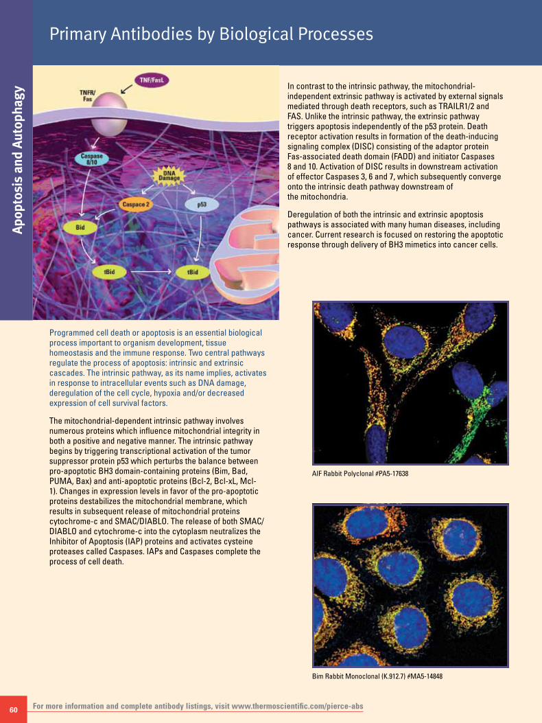
60
Apop
tosi
s an
d Au
toph
agy
Primary Antibodies by Biological Processes
For more information and complete antibody listings, visit www.thermoscientific.com/pierce-abs
Programmed cell death or apoptosis is an essential biological process important to organism development, tissue homeostasis and the immune response. Two central pathways regulate the process of apoptosis: intrinsic and extrinsic cascades. The intrinsic pathway, as its name implies, activates in response to intracellular events such as DNA damage, deregulation of the cell cycle, hypoxia and/or decreased expression of cell survival factors.
The mitochondrial-dependent intrinsic pathway involves numerous proteins which influence mitochondrial integrity in both a positive and negative manner. The intrinsic pathway begins by triggering transcriptional activation of the tumor suppressor protein p53 which perturbs the balance between pro-apoptotic BH3 domain-containing proteins (Bim, Bad, PUMA, Bax) and anti-apoptotic proteins (Bcl-2, Bcl-xL, Mcl-1). Changes in expression levels in favor of the pro-apoptotic proteins destabilizes the mitochondrial membrane, which results in subsequent release of mitochondrial proteins cytochrome-c and SMAC/DIABLO. The release of both SMAC/DIABLO and cytochrome-c into the cytoplasm neutralizes the Inhibitor of Apoptosis (IAP) proteins and activates cysteine proteases called Caspases. IAPs and Caspases complete the process of cell death.
In contrast to the intrinsic pathway, the mitochondrial-independent extrinsic pathway is activated by external signals mediated through death receptors, such as TRAILR1/2 and FAS. Unlike the intrinsic pathway, the extrinsic pathway triggers apoptosis independently of the p53 protein. Death receptor activation results in formation of the death-inducing signaling complex (DISC) consisting of the adaptor protein Fas-associated death domain (FADD) and initiator Caspases 8 and 10. Activation of DISC results in downstream activation of effector Caspases 3, 6 and 7, which subsequently converge onto the intrinsic death pathway downstream of the mitochondria.
Deregulation of both the intrinsic and extrinsic apoptosis pathways is associated with many human diseases, including cancer. Current research is focused on restoring the apoptotic response through delivery of BH3 mimetics into cancer cells.
Bim Rabbit Monoclonal (K.912.7) #MA5-14848
AIF Rabbit Polyclonal #PA5-17638
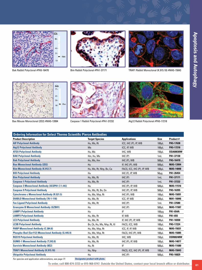
To order, call 800-874-3723 or 815-968-0747. Outside the United States, contact your local branch office or distributor. 61
Apoptosis and Autophagy
Bim Rabbit Polyclonal #PA1-37171 TRAF1 Rabbit Monoclonal (K.915.10) #MA5-15043
Atg12 Rabbit Polyclonal #PA5-17218Caspase 1 Rabbit Polyclonal #PA1-37232
Bak Rabbit Polyclonal #PA5-16470
Bax Mouse Monoclonal (2D2) #MA5-13994
Ordering Information for Select Thermo Scientific Pierce AntibodiesProduct Description Target Species Applications Size Product #
AIF Polyclonal Antibody Hu, Ms, Rt ICC, IHC (P), IP, WB 100μL PA5-17638
Atg12 Polyclonal Antibody Ms ICC, IP, WB 100μL PA5-17218
ATG5 Polyclonal Antibody Hu, Ms IHC, WB 150μL OSA00030W
BAK Polyclonal Antibody Hm, Hu, Ms IHC (P) 1mL PA1-37130
Bak Polyclonal Antibody Hu, Ms, Hm IHC (P), WB 500μL PA5-16470
Bax Monoclonal Antibody (2D2) Hu IF, IHC (P), WB 500μL MA5-13994
Bim Monoclonal Antibody (K.912.7) Hu, Ms, Rt, Nhp, Bv, Ca FACS, ICC, IHC (P), IP, WB 100μL MA5-14848
BID Polyclonal Antibody Hu IHC (F), IP, WB 50μg PA1-26454
Bim Polyclonal Antibody Hu, Ms, Rt IHC (P) 1mL PA1-37171
Caspase 1 Polyclonal Antibody Hu, Ms, Rt IHC (P) 1mL PA1-37232
Caspase 3 Monoclonal Antibody (3CSP01 (7.1.44)) Hu IHC (P), IP, WB 500μL MA5-11516
Caspase 9 Polyclonal Antibody Hu, Ms, Rt, Bv, Ov IHC (P), IP, WB 500μL PA5-16355
Cytochrome c Monoclonal Antibody (R.521.5) Hu, Ms, Nhp, Rt IHC (P), WB 100μL MA5-15091
DIABLO Monoclonal Antibody (78-1-118) Hu, Ms, Rt ICC, IP, WB 200μL MA1-16846
Fas Ligand Polyclonal Antibody Hu, Ms, Rt IHC (P) 1mL PA1-37680
Granzyme B Monoclonal Antibody (GZB01) Hu IHC (P) 500μL MA5-11587
LAMP1 Polyclonal Antibody Hu IF 100μg PA1-654A
LAMP2 Polyclonal Antibody Hu, Ms, Rt IF, WB 100μL PA1-655
LC3 Polyclonal Antibody Hu, Ms, Rt IF, IHC (P), IP, WB 100μL PA1-16930
LC3B Polyclonal Antibody Bv, Ck, Hu, Ms, Nhp, Rt, Xl FACS, ICC, WB 100μL PA5-17224
PARP Monoclonal Antibody (C.384.8) Hu, Ms, Nhp, Rt ICC, IF, IP, WB 100μL MA5-15031
Phospho-Bad (Ser112) Monoclonal Antibody (G.445.9) Hu, Ms, Nhp, Rt FACS, IHC (P), WB 100μL MA5-15085
RGS19 Polyclonal Antibody Hu, Ms, Rt IHC, WB 150μL OSR00248W
SUMO-1 Monoclonal Antibody (T.243.0) Hu, Ms, Rt IHC (P), IP, WB 100μL MA5-14877
Survivin Monoclonal Antibody (8E2) Hu, Rt IF 500μL MA5-11680
TRAF1 Monoclonal Antibody (K.915.10) Hu, Nhp FACS, ICC, IHC (P), IP, WB 100μL MA5-15043
Ubiquitin Polyclonal Antibody Hu IHC (P) 500μL PA5-16829
For species and application abbreviations, see page 77. Designates product with photo.

62
Cell
Prol
ifera
tion
Primary Antibodies by Biological Processes
For more information and complete antibody listings, visit www.thermoscientific.com/pierce-abs
The cell cycle is a highly orchestrated event in which the DNA content of the cell is duplicated and segregated into two daughter cells. The cell cycle can be divided into five distinct phases: Gap0 (G0), Gap1 (G1), Synthesis (S), Gap2 (G2) and Mitosis (M). Multiple cellular events coordinate the proper transitioning through each phase of the cell cycle, including temporal expression of cyclins, degradation of positive and negative checkpoint regulators, nuclear-cytoplasmic shuttling of proteins, protein-protein interactions, and post-translational modifications.
G0 is marked by cells which have undergone growth factor withdrawal, placing them in a resting or quiescent state. Transition from G0 to G1 requires the cell to overcome a sequence of events termed the “restriction point.” Reentry is marked by strong signals emanating from two essential signal transduction cascades, Ras-MAPK and PI3K-Akt. Signaling from these pathways triggers accumulation of proteins required for entry into G1. One of the major classes of proteins expressed at this time are D-type cyclins. During early G1 D-type cyclins pair with cyclin-dependent-kinases 4/6 to form active cyclinD:cdk4/6 complexes. The goal of G1 is to prime the cell for entry into S-phase (DNA synthesis). cyclinD:cdk4/6 complexes achieve this goal by phosphorylating the Retinoblastoma protein Rb. Phosphorylation of the tumor suppressor protein Rb is a critical cellular event as hypophosphorylated Rb maintains the transcription factor E2F in a repressed state through formation of a heterotrimeric protein complex consisting of E2F, Rb and HDACs. CyclinD:cdk4/6 phosphorylation of Rb results in partial release of E2F. Accumulation of free E2F and binding to its transcriptional partner DP1 promotes entry into late G1/S by transcriptionally regulating genes required for DNA synthesis. One of the major transcriptional targets of E2F-DP1 is cyclin E. E2F-DP1 induced expression of cyclin E results in the formation of active cyclinE:cdk2 complexes which function to complete the phosphorylation of Rb resulting in complete liberation of E2F.
Regulation of the G1/S transition is carried out by multiple proteins including the cyclin dependent kinase inhibitors (CKIs) p21WAF1/Cip1, p27KIP1, p16Ink4a, p15Ink4b, p18Ink4c, and p19Ink4d (ARF). The CKI’s p21 and p27 function by blocking cyclin:cdk complex kinase activity while Ink4 members p15, p16, and p18 block formation of active cyclinD:cdk4/6 complexes. Additional regulation occurs at the level of the G1/S CDK checkpoint. Proteins involved at this level include the Cdc25 protein phosphatase family. The primary Cdc25 family member involved during the G1/S transition is Cdc25A which functions to activate cdk2 through removal of inhibitory phosphates on amino acids Thr14 and Tyr15 (Wee1 and Myt1 targets) of cdks. Furthermore, Cdc25 activity is regulated by both activating and inhibitory phosphorylations. TAK1 (TGF-b activating kinase-1) has been shown to inhibit Cdc25 function through phosphorylation of Ser216. Phosphorylation of Ser216 by TAK1 results in cytoplasmic sequestration of Cdc25 through interactions with 14-3-3 proteins. Inhibitory phosphorylation of Cdc25 is overcome by phosphorylation of neighboring Ser214 by PLK1 (polo-like-kinase-1). In addition to regulation by reversible phosphorylation timely degradation of G1/S cyclins and regulators are critical for proper cell cycle transitioning.
The payoff of a successful G1 is entry into S phase. At this time the DNA content doubles from 2N to 4N. Accumulation of A-type cyclins occurs during S phase and when paired with cdk2 primes the cell for entry into G2. During late synthesis B-type cyclin levels also rise. Together cyclinA:cdk1/2 and cyclinB:cdk1 complexes drive the cell into Gap 2 (G2). During early to mid G2, the appearance of active nuclear Cdc25B/C is also apparent and promotes cyclin:cdk activity. The major driving force for entry into mitosis is cyclinB:cdk1 complexes also know as MPF (M-phase promoting factor). CyclinB:cdk1 complexes trigger mitosis through phosphorylation of lamins and histones. These events promote progression from prophase through anaphase by stimulating spindle assembly, chromatin condensation and nuclear envelope breakdown. Successful mitosis also requires degradation of cyclin B. This event is coordinated through phosphorylation by the Anaphase Promoting Complex (APC)/cyclosome. A variety of stimuli control both the G1/S and G2/M checkpoints, including DNA damage, contact inhibition, replicative senescence and growth factor withdrawal. Deregulation in cell cycle transitioning and checkpoint control through an imbalance of tumor suppressor proteins (Rb, p53) with proto-oncoproteins (E2F, MDM2) is a common theme with nearly all human cancers. Development of therapeutics to restore tumor suppressor protein expression and/or function in combination with pharmacological agents targeting cell proliferation and survival pathways is under intense research focus.
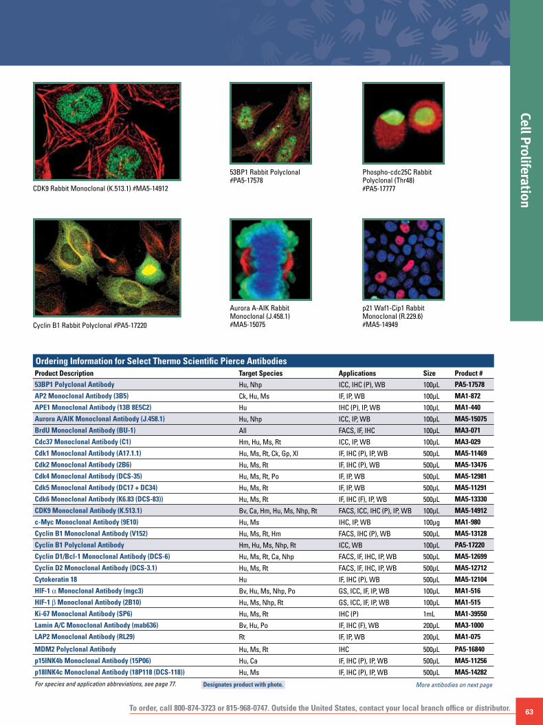
To order, call 800-874-3723 or 815-968-0747. Outside the United States, contact your local branch office or distributor. 63
Cell Proliferation
Cyclin B1 Rabbit Polyclonal #PA5-17220
CDK9 Rabbit Monoclonal (K.513.1) #MA5-14912
p21 Waf1-Cip1 Rabbit Monoclonal (R.229.6) #MA5-14949
Phospho-cdc25C Rabbit Polyclonal (Thr48) #PA5-17777
53BP1 Rabbit Polyclonal #PA5-17578
Aurora A-AIK Rabbit Monoclonal (J.458.1) #MA5-15075
Ordering Information for Select Thermo Scientific Pierce AntibodiesProduct Description Target Species Applications Size Product #
53BP1 Polyclonal Antibody Hu, Nhp ICC, IHC (P), WB 100μL PA5-17578
AP2 Monoclonal Antibody (3B5) Ck, Hu, Ms IF, IP, WB 100μL MA1-872
APE1 Monoclonal Antibody (13B 8E5C2) Hu IHC (P), IP, WB 100μL MA1-440
Aurora A/AIK Monoclonal Antibody (J.458.1) Hu, Nhp ICC, IP, WB 100μL MA5-15075
BrdU Monoclonal Antibody (BU-1) All FACS, IF, IHC 100μL MA3-071
Cdc37 Monoclonal Antibody (C1) Hm, Hu, Ms, Rt ICC, IP, WB 100μL MA3-029
Cdk1 Monoclonal Antibody (A17.1.1) Hu, Ms, Rt, Ck, Gp, Xl IF, IHC (P), IP, WB 500μL MA5-11469
Cdk2 Monoclonal Antibody (2B6) Hu, Ms, Rt IF, IHC (P), WB 500μL MA5-13476
Cdk4 Monoclonal Antibody (DCS-35) Hu, Ms, Rt, Po IF, IP, WB 500μL MA5-12981
Cdk5 Monoclonal Antibody (DC17 + DC34) Hu, Ms, Rt IF, IP, WB 500μL MA5-11291
Cdk6 Monoclonal Antibody (K6.83 (DCS-83)) Hu, Ms, Rt IF, IHC (F), IP, WB 500μL MA5-13330
CDK9 Monoclonal Antibody (K.513.1) Bv, Ca, Hm, Hu, Ms, Nhp, Rt FACS, ICC, IHC (P), IP, WB 100μL MA5-14912
c-Myc Monoclonal Antibody (9E10) Hu, Ms IHC, IP, WB 100μg MA1-980
Cyclin B1 Monoclonal Antibody (V152) Hu, Ms, Rt, Hm FACS, IHC (P), WB 500μL MA5-13128
Cyclin B1 Polyclonal Antibody Hm, Hu, Ms, Nhp, Rt ICC, WB 100μL PA5-17220
Cyclin D1/Bcl-1 Monoclonal Antibody (DCS-6) Hu, Ms, Rt, Ca, Nhp FACS, IF, IHC, IP, WB 500μL MA5-12699
Cyclin D2 Monoclonal Antibody (DCS-3.1) Hu, Ms, Rt FACS, IF, IHC, IP, WB 500μL MA5-12712
Cytokeratin 18 Hu IF, IHC (P), WB 500μL MA5-12104
HIF-1 α Monoclonal Antibody (mgc3) Bv, Hu, Ms, Nhp, Po GS, ICC, IF, IP, WB 100μL MA1-516
HIF-1 b Monoclonal Antibody (2B10) Hu, Ms, Nhp, Rt GS, ICC, IF, IP, WB 100μL MA1-515
Ki-67 Monoclonal Antibody (SP6) Hu, Ms, Rt IHC (P) 1mL MA1-39550
Lamin A/C Monoclonal Antibody (mab636) Bv, Hu, Po IF, IHC (F), WB 200μL MA3-1000
LAP2 Monoclonal Antibody (RL29) Rt IF, IP, WB 200μL MA1-075
MDM2 Polyclonal Antibody Hu, Ms, Rt IHC 500μL PA5-16840
p15INK4b Monoclonal Antibody (15P06) Hu, Ca IF, IHC (P), IP, WB 500μL MA5-11256
p18INK4c Monoclonal Antibody (18P118 (DCS-118)) Hu, Ms IF, IHC (P), IP, WB 500μL MA5-14282
For species and application abbreviations, see page 77. More antibodies on next page Designates product with photo.

64
Cell
Prol
ifera
tion
Primary Antibodies by Biological Processes
For more information and complete antibody listings, visit www.thermoscientific.com/pierce-abs
p53 Rabbit Monoclonal (S.18.9) #MA5-15152
Phospho-cdc2 Rabbit Monoclonal (Tyr15) (E.658.6) #MA5-15062
Phospho-p53 Rabbit Polyclonal (Ser46) #PA5-17817
Skp2 Rabbit Polyclonal #PA5-17009
PLK4 Rabbit Polyclonal #PA5-17272
Ordering Information for Select Thermo Scientific Pierce AntibodiesProduct Description Target Species Applications Size Product #
p21 Waf1/Cip1 Monoclonal Antibody (R.229.6) Hu, Nhp FACS, ICC, IHC (P), IP, WB 100μL MA5-14949
p27Kip1 Monoclonal Antibody (DCS-72.F6) Hu, Ms, Rt, Ca FACS, IF, IP, WB 500μL MA5-12832
p53 Monoclonal Antibody (S.18.9) Hu, Nhp FACS, ICC, IHC (P), WB 100μL MA5-15152
p57Kip2 Monoclonal Antibody (57P06 (KP10)) Hu, Ms IHC (P), IP 500μL MA5-11309
PDGF Receptor b Monoclonal Antibody (R.140.4) Hu, Ms, Rt FACS, IHC (P), WB 100μL MA5-14851
Peroxiredoxin 1 Polyclonal Antibody Hu, Ms, Rt ICC, WB 100μL PA3-750
Peroxiredoxin 2 Polyclonal Antibody Hu ICC, WB 100μL PA3-751
Peroxiredoxin 3 Polyclonal Antibody Hu ICC, WB 100μL PA3-752
Peroxiredoxin 4 Polyclonal Antibody Hu ICC, WB 100μL PA3-753
Phospho-Aurora A (Thr288) Monoclonal Antibody (F.131.2) Hu ICC, WB 100μL MA5-14904
Phospho-cdc2 (Tyr15) Monoclonal Antibody (E.658.6) Hu, Ms, Nhp, Rt FACS, ICC, IP, WB 100μL MA5-15062
Phospho-cdc25C (Thr48) Polyclonal Antibody Hu, Nhp ICC, IHC (P), WB 100μL PA5-17777
Phospho-Chk2 (Thr68) Polyclonal Antibody Hu, Nhp FACS, ICC, IP, WB 100μL PA5-17818
Phospho-EGF Receptor (Tyr1173) Monoclonal Antibody (S.331.5) Hu, Ms, Rt IHC (P), IP, WB 100μL MA5-15158
Phospho-H2AX (Ser140) Monoclonal Antibody (3F2) Hu, Ms ELISA, IF, WB 100μg MA1-2022
For species and application abbreviations, see page 77. Designates product with photo.

To order, call 800-874-3723 or 815-968-0747. Outside the United States, contact your local branch office or distributor. 65
Cell Proliferation
BrdU Mouse Monoclonal (BU-1) #MA3-071
Phospho-Chk2 (Thr68) Rabbit Polyclonal #PA5-17818Phospho-p53 Mouse Monoclonal (Ser15) (C.381.0) #MA5-15229
Ordering Information for Select Thermo Scientific Pierce AntibodiesProduct Description Target Species Applications Size Product #
Phospho-p53 (Ser15) Monoclonal Antibody (C.381.0) Hu FACS, ICC, WB 100μL MA5-15229
Phospho-p53 (Ser46) Polyclonal Antibody Hu, Nhp FACS, ICC, IP, WB 100μL PA5-17817
Phospho-Rb (Ser608) Polyclonal Antibody Ca, Hu, Nhp, Rt IHC (P), IP, WB 100μL PA5-17585
Phospho-Rb (Ser807/811) Polyclonal Antibody Hu, Nhp, Rt IHC (P), IP, WB 100μL PA5-17897
Phospho-Stat3 (Tyr705) Monoclonal Antibody (R.263.6) Bv, Hu, Ms, Rt FACS, IP, WB 100μL MA5-15193
Phospho-Wee1 (Ser642) Monoclonal Antibody (T.870.10) Bv, Hu, Ms, Nhp, Rt, Xl, Zf IP, WB 100μL MA5-14806
PLK1 Monoclonal Antibody (13E8) Hu, Ms, Rt IF, IP, WB 100μg MA1-848
PLK4 Polyclonal Antibody Hu, Ms, Nhp, Rt ICC, WB 100μL PA5-17272
RB1 Monoclonal Antibody (R.232.9) Bv, Hu, Nhp, Po FACS, IF, IHC (P), IP, WB 100μL MA5-15203
SCD1 Monoclonal Antibody (G.545.10) Ms ICC, IHC (P), IP, WB 100μL MA5-14885
Skp2 Polyclonal Antibody Hu, Ms, Nhp, Rt ICC, WB 100μL PA5-17009
TIF1 b Monoclonal Antibody (20C1) Hu, Ms IF, WB 100μg MA1-2023
TIF1 g Monoclonal Antibody (16G9) Hu, Ms, Rt IF, IP, WB 100μg MA1-801
TIF1 b Monoclonal Antibody (F.373.5) Hu, Ms, Nhp, Rt FACS, ICC, IHC (P), WB 100μL MA5-14824
UBC3B Polyclonal Antibody Hu, Ms, Rt FACS, IHC (P), WB 100μL PA5-17579
UBE1a Polyclonal Antibody Hu, Ms, Rt FACS, ICC, IHC (P), IP, WB 100μL PA5-17262
For species and application abbreviations, see page 77. Designates product with photo.
DAPI MergeBrdU

66
Tran
slat
ion
Cont
rol
Primary Antibodies by Biological Processes
For more information and complete antibody listings, visit www.thermoscientific.com/pierce-abs
Translation is the intricate process of decoding messenger RNA (mRNA) into an amino acid polypeptide which will fold into a functional protein. Translation occurs in the cytoplasm with four distinct phases: activation, initiation, elongation and termination.
During the activation stage, amino acids are linked to transfer RNA (tRNA) through peptide bonds. Initiation begins when newly transcribed mRNA is transported to ribosomes located on the rough endoplasmic reticulum. With the aid of translation initiation factors (eIFs), the ribosome scans the mRNA for the start site of translation, identified by a unique triplet ribonucleotide sequence, AUG, coding for the amino acid methionine. Elongation proceeds with the formation of amino acid linkages based on the complementary mRNA codon sequence. When the ribosome reaches a triplet code which the tRNA does not recognize (UAA, UAG, UGA), the polypeptide chain is terminated and released from the ribosome.
The entire process of protein synthesis requires numerous regulatory factors and upstream signaling events. Of intense research focus is the role the PI3K-Akt-mTOR kinase cascade plays in the control of eukaryotic translation. In response to growth factors, the PI3K-Akt-mTOR pathway becomes activated. Kinase activation via phosphorylation of Akt at Thr308 and Ser473 triggers downstream phosphorylation and subsequent activation of the mammalian target of rapamycin (mTOR). Activated mTOR integrates extracellular signals with amino acid availability to control the rate of protein synthesis. mTOR then stimulates translation through phosphorylation of two key translation components, the eIF4E inhibitory binding protein 4E-BP1 and the S6 ribosomal protein p70S6 kinase. Deregulation in mTOR signaling via aberrant activation of the PI3K-Akt cascade has been observed in numerous human cancers. Recent studies using a combinatorial drug approach targeting at the levels of PI3K and mTOR (rapalogs) may be a viable therapeutic option to combat cancer.
eIF4A1 Rabbit Polyclonal #PA5-17313
eIF4G Rabbit Monoclonal (G.689.9) #MA5-14971
eIF3A Rabbit Polyclonal #PA5-17212

To order, call 800-874-3723 or 815-968-0747. Outside the United States, contact your local branch office or distributor. 67
Translation Control
eIF4GI Rabbit Polyclonal #PA5-17140 eIF4H Rabbit Polyclonal #PA5-17211 Phospho-eIF2-α Rabbit Monoclonal (Ser51) (S.674.5) #MA5-15133
Phospho-S6 Ribosomal Protein Rabbit Monoclonal (Ser235-236) (G.22.6) #MA5-15140
Phospho-S6 Ribosomal Protein Rabbit Monoclonal (Ser240-244) (T.540.2) #MA5-15034
Phospho-eIF4G Rabbit Polyclonal (Ser1108) #PA5-17824
Ordering Information for Select Thermo Scientific Pierce AntibodiesProduct Description Target Species Applications Size Product #
Acetyl Lysine Monoclonal Antibody (1C6) Hu, Ms ICC, IF, WB 100μg MA1-2021
Calnexin Monoclonal Antibody (AF18) Hu, Ms ICC, IF, IP, WB 100μL MA3-027
CHOP Monoclonal Antibody (B.84.2) Hu, Ms, Rt ICC, IP, WB 100μL MA5-14819
CREB Monoclonal Antibody (LB9) Hu, Ms, Rt ELISA, WB 100μg MA1-083
eEF1A Polyclonal Antibody Hu, Ms, Nhp, Rt ICC, WB 100μL PA5-17213
eEF2k Polyclonal Antibody Hu, Nhp, Rt ICC, IP, WB 100μL PA5-17720
eIF2-α Monoclonal Antibody (J.633.8) Hu, Ms, Nhp, Rt IHC (P), WB 100μL MA5-14938
eIF3A Polyclonal Antibody Hu, Ms, Nhp, Rt ICC, IP, WB 100μL PA5-17212
eIF3j Polyclonal Antibody Hu, Ms, Rt IP, WB 100μL PA5-17062
eIF4A1 Polyclonal Antibody Hu, Ms, Nhp, Rt ICC, WB 100μL PA5-17313
eIF4G Monoclonal Antibody (G.689.9) Hu, Ms, Nhp, Rt FACS, ICC, IHC (P), WB 100μL MA5-14971
eIF4G Polyclonal Antibody Hu, Ms, Nhp, Rt FACS, ICC, IHC (P), WB 100μL PA5-17422
eIF4GI Polyclonal Antibody Hu, Ms, Rt ICC, IHC (P), WB 100μL PA5-17140
eIF4H Polyclonal Antibody Hu, Ms, Nhp, Rt ICC, WB 100μL PA5-17211
ERp29 Polyclonal Antibody Bv, Ca, GP, Hm, Hu, Ms, Nhp, Rt IF, IP, WB 100μL PA3-011
HIF-1 α Monoclonal Antibody (mgc3) Bv, Hu, Ms, Nhp, Po GS, ICC, IF, IP, WB 100μL MA1-516
HIF-1 b Monoclonal Antibody (2B10) Hu, Ms, Nhp, Rt GS, ICC, IF, IP, WB 100μL MA1-515
Mist1 Monoclonal Antibody (6E8/A12/C11P1) Hu, Ms IHC (P, F), WB 100μg MA1-517
PDI Monoclonal Antibody (RL90) Hm, Hu, Ms, Po, Rt FACS, IF, IHC (P, F), IP, WB 100μL MA3-019
Phospho-eIF2-α (Ser51) Monoclonal Antibody (S.674.5) Dm, Hu, Ms, Nhp, Rt IHC (P), IP, WB 100μL MA5-15133
Phospho-eIF4G (Ser1108) Polyclonal Antibody Bv, Hm, Hu, Ms, Nhp, Rt ICC, IP, WB 100μL PA5-17824
Phospho-S6 Ribosomal Protein (Ser235/236) Monoclonal Antibody (G.22.6)
Hu, Ms, Rt ICC, IHC (F), IHC (P), WB 100μL MA5-15140
Phospho-S6 Ribosomal Protein (Ser240/244) Monoclonal Antibody (T.540.2)
Hu, Ms, Nhp, Rt FACS, ICC, WB 100μL MA5-15034
SERCA1 ATPase Monoclonal Antibody (IIH11) Ca, GP, Ms, Rb, Rt IF, IHC (F), WB 100μL MA3-911
SERCA2 ATPase Monoclonal Antibody (2A7-A1) Ca, GP, Hu, Ms, Po, Rb, Rt ICC, IP, WB 100μL MA3-919
SERCA2 ATPase Monoclonal Antibody (IID8) Bv, Ca, Hu, Po, Rb, Rt ICC, IF, IHC (F), WB 100μL MA3-910
For species and application abbreviations, see page 77. Designates product with photo.

68
Gen
otox
ic S
tres
s an
d D
NA
Dam
age
Primary Antibodies by Biological Processes
For more information and complete antibody listings, visit www.thermoscientific.com/pierce-abs
DNA damage caused by extracellular signals such as ultraviolet (UV) irradiation and chemotherapeutic drugs are a major cause of genomic instability. This damage results in the formation of single- and double-strand DNA breaks. To control for these genomic insults, the cell has evolved elaborate mechanisms to sense the level of DNA damage and decide whether to repair or activate a cellular program leading to cell death. During DNA damage, multiple key cell cycle checkpoint proteins become activated, including Histone H2AX, ATM/ATR kinases, Chk1/2 kinases, Ku 70/80 complexes, BRCA1 and the tumor suppressor protein p53. The transcription factor p53, considered the “gatekeeper” of the genome, controls responses to genotoxic stress by inducing either DNA repair processes or apoptosis. Increased p53 levels in stressed cells induce both p21Waf1/Cip1-mediated cell cycle arrest and DNA repair. If the level of genotoxic stress is too severe, p53 may trigger a transcriptional program resulting in an increase in pro-apoptotic proteins Bax, PUMA and NOXA which function to disrupt mitochondrial integrity. Release of cytochrome-c in response to mitochondrial perturbations activates the caspase cascade, ultimately resulting in protein degradation and cell death. Deregulation in cell cycle checkpoints, DNA repair and/or apoptotic pathways is a hallmark of nearly all human cancers. A major contributor to genomic instability is the loss of p53 expression and/or functionality in many tumors. Cells which harbor mutations in p53 have evolved a mechanism to elude the effects of chemotherapeutic agents which target the p53 pathway to induce their anti-tumor effects.
Phospho-Chk2 Rabbit Polyclonal (Thr68) #PA5-17818
Phospho-Chk1 Rabbit Monoclonal (Ser 345) (S.48.4) #MA5-15145

To order, call 800-874-3723 or 815-968-0747. Outside the United States, contact your local branch office or distributor. 69
Genotoxic Stress and D
NA D
amagePhospho-p53 Mouse Monoclonal (Ser15) (C.381.0) #MA5-15229
p53 Rabbit Monoclonal (S.18.9) #MA5-15152Phospho-p53 Rabbit Polyclonal (Ser46) #PA5-17817
Phospho-p53 Rabbit Polyclonal (Ser37) #PA5-17866
Ordering Information for Select Thermo Scientific Pierce AntibodiesProduct Description Target Species Applications Size Product #
APE1 Monoclonal Antibody (13B 8E5C2) Hu IHC (P), IP, WB 100μL MA1-440
Ku80 Monoclonal Antibody (S.669.4) Hu, Nhp ICC, IHC (F), IHC (P), IP, WB 100μL MA5-14953
Mre11 Monoclonal Antibody (F.519.4) Hu FACS, IHC (F), IHC (P), IP, WB 100μL MA5-15081
p21WAF1 Monoclonal Antibody (DCS-60.2) Hu IF, IHC (P), IP 500μL MA5-12764
p53 Monoclonal Antibody (S.18.9) Hu, Nhp FACS, ICC, IHC (P), WB 100μL MA5-15152
Phospho-53BP1 (Ser1778) Polyclonal Antibody Hu, Nhp FACS, ICC, WB 100μL PA5-17462
Phospho-ATM (Ser1981) Monoclonal Antibody (10H11) Hu, Ms IF, IP, WB 200μg MA1-2020
Phospho-Chk1 (Ser345) Monoclonal Antibody (S.48.4) Hu, Ms, Nhp, Rt FACS, ICC, WB 100μL MA5-15145
Phospho-Chk2 (Ser19) Polyclonal Antibody Hu IHC (P), WB 100μL PA5-17731
Phospho-Chk2 (Thr68) Polyclonal Antibody Hu, Nhp FACS, ICC, IP, WB 100μL PA5-17818
Phospho-H2AX (Ser140) Monoclonal Antibody (3F2) Hu, Ms ELISA, IF, WB 100μg MA1-2022
Phospho-p53 (Ser15) Monoclonal Antibody (C.381.0) Hu FACS, ICC, WB 100μL MA5-15229
Phospho-p53 (Ser37) Polyclonal Antibody Hu, Nhp FACS, ICC, IP, WB 100μL PA5-17866
Phospho-p53 (Ser46) Polyclonal Antibody Hu, Nhp FACS, ICC, IP, WB 100μL PA5-17817
Phospho-TLK1 (Ser695) Polyclonal Antibody Hu, Ms, Rt IHC (P), IP, WB 100μL PA5-17572
RPA70 Polyclonal Antibody Hu, Nhp, Rt FACS, ICC, IP, WB 100μL PA5-17377
For species and application abbreviations, see page 77. Designates product with photo.

70
Ion
Tran
spor
tPrimary Antibodies by Biological Processes
For more information and complete antibody listings, visit www.thermoscientific.com/pierce-abs
The transport of ions across biological membranes requires the presence of integral membrane proteins that form channels across the membrane’s phospholipid bilayer. Ion channels allow for the transfer of specific ions from the cytosol to the extracellular matrix or cell compartment space and vice versa. The transfer of ions to and from the cellular matrix can be done through either active transport channels (ion transporters) or passive diffusion channels (passive ion channels).
Ion transporters are essential for many biological processes, including ATP generation across the electrochemical gradient and maintenance of the cell’s membrane potential. Ion transporters are also necessary for the normal function of the nervous system, muscle contraction, nutrient transport, immunity (e.g., via activation of the inflammasome), and cell integrity.
The three common types of ion transporters are uniporters, symporters and antiporters. Uniporters allow for unidirectional ion transport following an ion gradient or are ATP-driven transporters transporting ions against a concentration gradient. Typical ion uniporters include P-Class, F-Class and V-Class ATP-powered ion pumps.
A well known symporter is the sodium-glucose symporter in the microvilli membranes of epithelial cells. The sodium-glucose symporter actively transports glucose against a concentration gradient from the intestinal lumen into epithelial cells by coupling “uphill” glucose transport with “downhill” sodium transport.
Many symporters and antiporters are involved in cytosolic pH regulation. In parietal cells of the stomach, protons are actively exported into the acidic lumen against a concentration gradient via potassium-antiport and ATP hydrolysis. Another important example for ATP-driven ion transport is the regulation of calcium uptake by cardiac muscle cells. Cardiac sodium-calcium antiporters require the import of three sodium ions to power the export one calcium ion against a steep concentration gradient. This process is tightly regulated since the timely increase and release of intracellular calcium results in the periodic contraction and relaxation of heart muscle cells.
Ion channels can also be grouped based on the nature of the species or signal needed for their induction, a process known as “gating.” For example, ligand-gated channels are active upon ligand binding and voltage-gated channels are active in response to changes in the membrane potential. Other gated channels include mechanosensitive, light-gated and temperature-gated ion channels.
Several human conditions exist and are associated with mutations affecting ion transport. These include cystic fibrosis (chloride channel defect), episodic ataxia (voltage-gated potassium channel) and Timothy syndrome (calcium channel defect). Finally, since ion channel function is so crucial for homeostasis, defensive ion channel-targeting toxins have evolved in several venomous species. For example, tetrodotoxin is a Na+-channel targeting toxin used by puffer fish. K+-channel targeting toxins include dendrotoxin (mamba snake), iberiotoxin (Indian red scorpion) and heteropodatoxin (huntsman spider).
TRPA1 Rabbit Polyclonal #PA1-46159
Calcium Sensing Receptor Mouse Monoclonal (5C10, ADD) #MA1-934

To order, call 800-874-3723 or 815-968-0747. Outside the United States, contact your local branch office or distributor. 71
Ion Transport
Claudin 3 Rabbit Polyclonal #PA5-16867
Claudin 4 Rabbit Polyclonal #PA5-16875
Cystic Fibrosis Transmembrane Regulator Mouse Monoclonal (M3A7) #MA5-11768
Pan Sodium Channel Rabbit Polyclonal #PA1-38631
GnRH Receptor Mouse Monoclonal (GNRH03) #MA5-11538
GLUT-1 Mouse Monoclonal (SPM498) #MA5-11315
Ordering Information for Select Thermo Scientific Pierce AntibodiesProduct Description Target Species Applications Size Product #
Calcium Sensing Receptor Monoclonal Antibody (5C10, ADD) Bv, Hu, Rt ELISA, IF, IHC (F), WB 100μg MA1-934
Claudin 3 Polyclonal Antibody Hu, Ms, Rt IHC (P) 500μL PA5-16867
Claudin 4 Polyclonal Antibody Hu, Ms IHC (P) 500μL PA5-16875
CNG Channel Monoclonal Antibody (CNC 9C1) Hu, Ms, Rt IHC, WB 100μL MA3-901
CREB Monoclonal Antibody (LB9) Hu, Ms, Rt ELISA, WB 100μg MA1-083
Cystic Fibrosis Transmembrane Regulator Monoclonal Antibody (M3A7) Hu, Ms, Rt IF, IHC (P), IP, WB 500μL MA5-11768
Glucocorticoid Receptor Monoclonal Antibody (BuGR2) GP, Hu, Ms, Ov, Rb, Rt, Ys FACS, GS, ICC, IF, IHC (P), IP, WB
100μg MA1-510
Glucocorticoid Receptor Polyclonal Antibody Hu, Rt GS, ICC, IP, WB 200μL PA1-510A
GLUT-1 Monoclonal Antibody (SPM498) Hu, Rt IHC (P) 500μL MA5-11315
GnRH Receptor Monoclonal Antibody (GNRH03) Hu FACS, IF, IHC (P) 500μL MA5-11538
HCN3 Monoclonal Antibody (TLL6C5) Hu, Ms, Rt IHC, WB 100μL MA3-902
HCN4 Monoclonal Antibody (SHG 1E5) Hu, Ms, Rt IHC, WB 100μL MA3-903
KCNQ2 Polyclonal Antibody Hu, Ms, Rt ICC, IF, IHC (F), WB 100μg PA1-929
KCNQ3 Polyclonal Antibody Hu, Ms, Rt ICC, IF, IHC (F) 100μg PA1-930
Pan Sodium Channel Polyclonal Antibody Hu IHC (P) 1mL PA1-38631
Potassium Channel kv3.2 Polyclonal Antibody Hu, Ms, Rt IHC (P) 1mL PA1-38480
Renal Cell Carcinoma Marker Monoclonal Antibody (PN-15) Hu, Rt IHC (P), WB 500μL MA5-13365
Sodium/Calcium Exchanger Monoclonal Antibody (C2C12) Ca, GP, Hu, Ms, Rb, Rt IF, IHC, IP, WB 100μL MA3-926
Sodium/Potassium ATPase α Monoclonal Antibody (M7-PB-E9) Bv, Ca, Ck, Hu, Ms, Ov, Po, Rt ELISA, ICC, IHC, IP, WB 400μL MA3-928
Sodium/Potassium ATPase α-1 Monoclonal Antibody (M8-P1-A3) Ca, Hu, Ov, Po, Rt IHC (F), WB 200μL MA3-929
Sodium/Potassium ATPase α-3 Monoclonal Antibody (XVIF9-G10) Bv, Ca, GP, Hu, Ms, Nhp, Ov, Rb, Rt
IF, IHC (F), WB 100μg MA3-915
Sodium/Potassium ATPase b Monoclonal Antibody (M17-P5-F11) Ca, Hu, Ms, Ov, Po ICC, IHC, IHC (P), IP, WB 200μL MA3-930
Sodium/Potassium ATPase Monoclonal Antibody (9-A5) Ca, Ck, Hu, Rt ELISA, ICC, IF, IHC, IP 100μL MA3-924
TRPA1 Polyclonal Antibody Hu, Ms IHC, IHC (P), WB 100μL PA1-46159
For species and application abbreviations, see page 77. Designates product with photo.
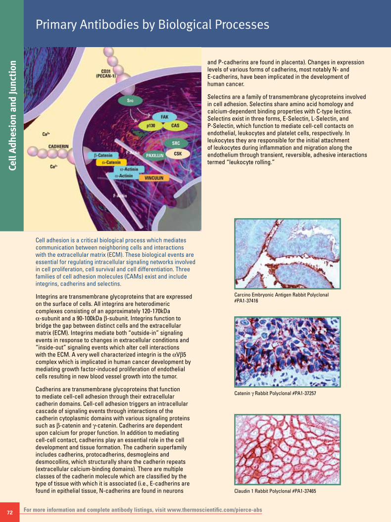
72
Cell
Adhe
sion
and
Junc
tion
Primary Antibodies by Biological Processes
For more information and complete antibody listings, visit www.thermoscientific.com/pierce-abs
Cell adhesion is a critical biological process which mediates communication between neighboring cells and interactions with the extracellular matrix (ECM). These biological events are essential for regulating intracellular signaling networks involved in cell proliferation, cell survival and cell differentiation. Three families of cell adhesion molecules (CAMs) exist and include integrins, cadherins and selectins.
Integrins are transmembrane glycoproteins that are expressed on the surface of cells. All integrins are heterodimeric complexes consisting of an approximately 120-170kDa α-subunit and a 90-100kDa b-subunit. Integrins function to bridge the gap between distinct cells and the extracellular matrix (ECM). Integrins mediate both “outside-in” signaling events in response to changes in extracellular conditions and “inside-out” signaling events which alter cell interactions with the ECM. A very well characterized integrin is the αVb5 complex which is implicated in human cancer development by mediating growth factor-induced proliferation of endothelial cells resulting in new blood vessel growth into the tumor.
Cadherins are transmembrane glycoproteins that function to mediate cell-cell adhesion through their extracellular cadherin domains. Cell-cell adhesion triggers an intracellular cascade of signaling events through interactions of the cadherin cytoplasmic domains with various signaling proteins such as b-catenin and g-catenin. Cadherins are dependent upon calcium for proper function. In addition to mediating cell-cell contact, cadherins play an essential role in the cell development and tissue formation. The cadherin superfamily includes cadherins, protocadherins, desmogleins and desmocollins, which structurally share the cadherin repeats (extracellular calcium-binding domains). There are multiple classes of the cadherin molecule which are classified by the type of tissue with which it is associated (i.e., E-cadherins are found in epithelial tissue, N-cadherins are found in neurons
and P-cadherins are found in placenta). Changes in expression levels of various forms of cadherins, most notably N- and E-cadherins, have been implicated in the development of human cancer.
Selectins are a family of transmembrane glycoproteins involved in cell adhesion. Selectins share amino acid homology and calcium-dependent binding properties with C-type lectins. Selectins exist in three forms, E-Selectin, L-Selectin, and P-Selectin, which function to mediate cell-cell contacts on endothelial, leukocytes and platelet cells, respectively. In leukocytes they are responsible for the initial attachment of leukocytes during inflammation and migration along the endothelium through transient, reversible, adhesive interactions termed “leukocyte rolling.”
Carcino Embryonic Antigen Rabbit Polyclonal #PA1-37416
Claudin 1 Rabbit Polyclonal #PA1-37465
Catenin g Rabbit Polyclonal #PA1-37257

To order, call 800-874-3723 or 815-968-0747. Outside the United States, contact your local branch office or distributor. 73
Cell Adhesion and Junction
Claudin 2 Rabbit Polyclonal #PA1-37467 Claudin 3 Rabbit Polyclonal #PA1-37469 Claudin 4 Rabbit Polyclonal #PA1-37471
Integrin b-5 Rabbit Polyclonal #PA1-37932 CA-125 Monoclonal (Ov185:1) #MA5-11579
Claudin 7 Rabbit Polyclonal #PA1-37474
Ordering Information for Select Thermo Scientific Pierce AntibodiesProduct Description Target Species Applications Size Product #CA-125 Monoclonal Antibody (Ov185:1) Hu IHC (P) 1mL MA5-11579Cadherin E Monoclonal Antibody (SPM471) Hu IHC (P) 1mL MA1-37201Carcino Embryonic Antigen Polyclonal Antibody Hu IHC (P) 1mL PA1-37416Catenin b Monoclonal Antibody (9F2) Hu, Ms IF, IP, WB 100μg MA1-2001Catenin g Polyclonal Antibody Hu, Ms, Rt IHC (P) 1mL PA1-37257Claudin 1 Polyclonal Antibody Hu IHC (P) 1mL PA1-37465Claudin 2 Polyclonal Antibody Hu, Ms IHC (P) 1mL PA1-37467Claudin 3 Polyclonal Antibody Hu, Ms, Rt IHC (P) 1mL PA1-37469Claudin 4 Polyclonal Antibody Hu, Ms IHC (P) 1mL PA1-37471Claudin 7 Polyclonal Antibody Hu, Ms, Rt IHC (P) 1mL PA1-37474Collagen II Monoclonal Antibody (2B1.5) Hu, Ms, Rt, Bv, Ck FACS, IF, IHC (P), WB 500μL MA5-12789Collagen IV Monoclonal Antibody (PHM-12) Hu IHC (P) 500μL MA5-13255Collagen Type X Monoclonal Antibody (X-AC9) Ck IF, IHC (P, F) 500μL MA5-14265Collagen VII Monoclonal Antibody (LH7.2) Hu, Bv, Gp, Gt, Nhp, Ov, Po IF, IHC (F), IM 500μL MA5-13432Cytokeratin 10 Monoclonal Antibody (DE-K10) Hu, Rt, Ca IF, IHC (P), WB 500μL MA5-13702Cytokeratin 14 Monoclonal Antibody (LL002) Hu, Rt IHC (P), WB 500μL MA5-11596Cytokeratin 15 Monoclonal Antibody (LHK15) Hu, Ms, Rt, Bv IF 500μL MA5-11344Cytokeratin 16 Monoclonal Antibody (LL025) Hu IF, IHC (P) 500μL MA5-13730Cytokeratin 17 Monoclonal Antibody (E3) Hu, Rt IF, IHC (P) 500μL MA5-13539Cytokeratin 18 Monoclonal Antibody (DC10) Hu IF, IHC (P), WB 500μL MA5-12101Cytokeratin 19 Monoclonal Antibody (BA17) Hu IHC (P), WB 500μL MA5-12319Cytokeratin 20 Monoclonal Antibody (Q2) Hu IHC (P), WB 500μL MA5-12514Cytokeratin 5 Monoclonal Antibody (XM26) Hu IHC (P) 500μL MA5-12596Cytokeratin 5/6 Monoclonal Antibody (D5/I6 B4) Hu IHC (P) 500μL MA5-12429Cytokeratin 7 Monoclonal Antibody (OV-TL 12/30) Hu IHC (P) 500μL MA5-11986Cytokeratin 8 Monoclonal Antibody (TS1) Hu IHC (P), IP 500μL MA5-14428Cytokeratin Pan Monoclonal Antibody (AE1/AE3) Hu, Ms, Rt, Bv, Ck, Nhp, Rb IF, IHC (P) 500μL MA5-13156Integrin b-5 Polyclonal Antibody Hm, Hu, Ms, Rt IF, IHC (P), IP, WB 1mL PA1-37932Pan Cadherin Polyclonal Antibody Hu IHC (P), IP 1mL PA1-37199Tenascin Monoclonal Antibody (T2H5) Hu IHC (P) 500μL MA5-11848
For species and application abbreviations, see page 77. Designates product with photo.

74
Chro
mat
in R
emod
elin
g/Tr
ansc
riptio
nPrimary Antibodies by Biological Processes
For more information and complete antibody listings, visit www.thermoscientific.com/pierce-abs
The processes of chromatin remodeling, transcription and translation are tightly linked biological processes essential for organism survival. The transmission of genetic information from DNA into protein involves multiple steps and numerous regulatory proteins. At the most basic level gene expression occurs when local chromatin structure relaxes allowing access of transcription factors and other regulatory proteins to the target gene’s promoter. Transcription proceeds through an enzymatic reaction involving RNA polymerase which transcribes the DNA sequence into a complementary product, termed messenger RNA (mRNA). Processed mRNA is transported into the cytoplasm where it is translated into protein.
Chromatin RemodelingA major epigenetic mechanism to control gene expression involves the modification of chromatin, a higher-ordered nucleoprotein-DNA complex. Chromatin serves three main biological purposes: to compact DNA within the nucleus, to regulate gene expression and to control DNA replication.
Compaction of genomic DNA occurs through a highly organized repeating nucleoprotein complex termed the nucleosome. Each nucleosome unit consists of approximately 200 base pairs of DNA wrapped around four pairs of core histone proteins: H2A, H2B, H3 and H4. The nucleosome also acts as a regulatory mechanism to control gene expression and DNA replication by creating a physical barrier between the DNA and regulatory proteins.
To overcome this regulatory barrier a series of post-translational modifications occur on the amino-terminal tails of the four core histone proteins, specifically at lysine, arginine, serine, threonine and tyrosine. Methylation of arginine and lysine residues is an important marker for transcriptionally active or inactive genomic regions. The protein arginine methyltransferase (PRMT) family of co-activators, including PRMT1 and PRMT4, play key roles in promoting transcription
through methylation of arginine residues on histone H3 and histone H4, whereas methylation of lysines can both stimulate (histone H3) and repress (histone H3 and H4) transcription. The DNA methyltransferases DNMT1, DNMT2 and DNMT3 counteract the PRMT methyltransferase activity. DNMT1, DNMT2 and DNMT3 are overexpressed in numerous human tumors. Interestingly, hypomethylation of promoters of essential tumor suppressor proteins has been implicated in tumor formation.
Histone acetylation on lysines also plays a critical role in modulating transcriptional activity. Histone acetylation through histone acetyltransferase (HAT) activity promotes a relaxed chromatin conformation, allowing access of transcription factors and other regulatory proteins to the DNA and stimulating transcription. Histone deacetylases (HDACs) counteract the effects of HATs on the local chromatin structure. Disruption of the balance between HATs and HDACs has been implicated in a variety of human diseases including cancer. More recently HDAC inhibitors have become an attractive target for development of anti-cancer therapeutics. Recent evidence suggests that HDAC inhibitors promote a state of hyperacetylation and subsequent transcriptional activation of genes involved in cell cycle arrest and apoptosis .
TranscriptionEukaryotic transcription is a highly evolved process involving numerous regulatory proteins called transcription factors that act in concert to initiate the production of RNA from DNA. These transcription factors bind to upstream promoter sequences which are non-protein coding regions of a specific gene.
Transcription begins with the binding of mega-protein complexes containing core transcription factors (TFIIA, TFIIB, TFIID, TFIIE, TFIIF, TFIIH), gene-specific transcription factors (e.g., c-myc, E2F, c-Fos, c-Jun, NFkB, STAT, SMAD, FOXM1), and transcriptional co-activators (CBP/p300, PCAF) to specific DNA consensus sequences within the promoter of a particular gene. Formation of the transcriptional activation complex guides RNA polymerase (I, II, III) to the DNA coding sequence where it enzymatically transcribes the DNA sequence into an intermediate RNA species [e.g., messenger RNA (mRNA) in the case of RNA polymerase II]. Newly synthesized RNA is then processed and exported into the cytoplasm where it interacts with ribosomes, initiating translation into protein.
Regulation of transcription occurs at multiple levels including DNA modification (methylation), post-translational modification of histones and post-translational modification of transcription factors. In many cases the level of regulation is a dictated by upstream signaling events in response to internal and external cellular cues.
Hypermethylation of CpG islands located within promoter sequences is a key regulatory mechanism to silence gene expression. Post-translational modifications to core histones are also an essential regulatory mechanism governing accessibility of transcriptional regulatory proteins to DNA. Likewise, post-translational modification of transcription factors regulates transcription through multiple effects, including cellular localization, DNA binding and protein-protein interactions.

To order, call 800-874-3723 or 815-968-0747. Outside the United States, contact your local branch office or distributor. 75
Chromatin Rem
odeling/Transcription
Phospho-Histone H3 Mouse Monoclonal (Ser10) (K.872.3) #MA5-15220
Phospho-Histone H3 Rabbit Polyclonal (Ser28) #PA5-17318
c-Jun Rabbit Monoclonal (C.238.2) #MA5-15172
SirT1 Rabbit Polyclonal #PA5-17074
Pan-Methyl-Histone H3 Rabbit Polyclonal (Lys9) #PA5-17367
Phospho-SMAD1-5 Rabbit Monoclonal (Ser463-465) (E.239.4) #MA5-15124
SMAD3 Rabbit Monoclonal (E.980.9) #MA5-14939Phospho-CENP-A (Ser7) Rabbit Polyclonal #PA5-17195
Histone H3 Rabbit Polyclonal #PA5-17697
Ordering Information for Select Thermo Scientific Pierce AntibodiesProduct Description Target Species Applications Size Product #
Acetyl Lysine Monoclonal Antibody (1C6) Hu, Ms ICC, IF, WB 100μg MA1-2021
Acetyl-CoA Carboxylase Monoclonal Antibody (B.800.8) Hm, Hu, Ms, Rt FACS, ICC, IHC (P), WB 100μL MA5-15025
Acetyl-Histone H2A (Lys5) Polyclonal Antibody Hu, Ms, Nhp, Rt IHC (P), IP, WB 100μL PA5-17763
Acetyl-Histone H2B (Lys20) Polyclonal Antibody Hu, Ms, Rt ICC, IHC (P), IP, WB 100μL PA5-17821
Acetyl-Histone H2B (Lys5) Polyclonal Antibody Hu, Ms, Nhp, Rt IHC (P), IP, WB 100μL PA5-17779
Acetyl-Histone H3 (Lys18) Polyclonal Antibody Hu, Ms, Rt ChIP, IHC (P), WB 100μL PA5-17801
Acetyl-Histone H3 (Lys9) Monoclonal Antibody (T.69.2) Hu, Ms, Nhp, Rt, Zf ChIP, FACS, ICC, IHC (P), WB 100μL MA5-14970
Acetyl-Histone H3 (Lys9) Polyclonal Antibody Dm, Hu, Ms, Nhp, Rt, Ys ChIP, IHC (P), IP, WB 100μL PA5-17868
Acetyl-Histone H4 (Lys12) Polyclonal Antibody Hu, Ms, Nhp, Rt FACS, ICC, IHC (P), WB 100μL PA5-17773
CENP-A Polyclonal Antibody Hu ICC, WB 100μL PA5-17194
c-Fos Polyclonal Antibody Hu IHC (P) 1mL PA1-37437
c-Jun Monoclonal Antibody (C.238.2) Hu, Ms, Nhp, Rt ICC, IHC (F), IHC (P), IP, WB 100μL MA5-15172
CREB Monoclonal Antibody (C.480.4) Hu, Ms, Rt, Nhp, Dm ChIP, FACS, ICC, IF, IHC (F), IHC (P), IP, WB 100μL MA5-15154
Di-Methyl-Histone H3 (Lys36) Monoclonal Antibody (T.571.7) Hu, Ms, Nhp, Rt ICC, IHC (P), WB 100μL MA5-14867
Di-Methyl-Histone H3 (Lys36) Polyclonal Antibody Hu, Ms, Nhp, Rt ICC, IP, WB 100μL PA5-17341
For species and application abbreviations, see page 77. More antibodies on next page Designates product with photo.

76
Chro
mat
in R
emod
elin
g/Tr
ansc
riptio
nPrimary Antibodies by Biological Processes
For more information and complete antibody listings, visit www.thermoscientific.com/pierce-abs
Ordering Information for Select Thermo Scientific Pierce AntibodiesProduct Description Target Species Applications Size Product #
Di-Methyl-Histone H3 (Lys4) Monoclonal Antibody (T.767.5) Hu, Ms, Nhp, Rt ChIP, ICC, IHC (P), IP, WB 100μL MA5-14977
Di-Methyl-Histone H3 (Lys4) Polyclonal Antibody Hu, Ms, Nhp, Rt, Xl, Zf ChIP, ICC, IHC (P), IP, WB 100μL PA5-17174
Di-Methyl-Histone H3 (Lys9) Polyclonal Antibody Dm, Hu, Ms, Nhp, Rt, Ys ChIP, ICC, IF, IHC (P), IP, WB 100μL PA5-17370
FoxO1 Monoclonal Antibody (S.502.4) Hu, Ms, Rt, Nhp ICC, IHC (P), WB 100μL MA5-14846
FOXP4 Polyclonal Antibody Hu IHC (P) 1mL PA1-37716
g Tubulin Monoclonal Antibody (4D11) Hu, Ms, Rt IF, IP, WB 100μg MA1-850
HDAC1 Polyclonal Antibody Ca, Hm, Hu, Ms, Rt ChIP, ICC, IF, IHC (F), IP, WB 100μg PA1-860
HDAC2 Polyclonal Antibody Ca, Hm, Hu, Ms, Rt ICC, IF, IHC, IP, WB 100μg PA1-861
Histone H3 Polyclonal Antibody Bv, Dm, Hu, Ms, Nhp, Po, Rt, Zf ICC, IHC (P), WB 100μL PA5-17697
Methyl CpG Binding Protein 2 Polyclonal Antibody Hu, Ms, Rt IF, IP, WB 100μg PA1-887
Mono-Methyl-Histone H3 (Lys4) Polyclonal Antibody Hu, Ms, Nhp, Rt, Xl, Zf ICC, IP, WB 100μL PA5-17418
Pan-Methyl-Histone H3 (Lys9) Polyclonal Antibody Hu, Ms, Nhp, Rt, Zf ChIP, ICC, IP, WB 100μL PA5-17367
Phospho-CENP-A (Ser7) Polyclonal Antibody Hu, Nhp ICC, IP, WB 100μL PA5-17195
Phospho-Histone H2A.X (Ser139) Monoclonal Antibody (G.327.3) Hu, Ms, Nhp, Rt FACS, ICC, IHC (P), WB 100μL MA5-15130
Phospho-Histone H3 (Ser10) Monoclonal Antibody (K.872.3) Hu, Ms, Rt FACS, ICC, IF, WB 100μL MA5-15220
Phospho-Histone H3 (Ser10) Polyclonal Antibody Ck, Dm, Hu, Ms, Nhp, Rt, Xl, Ys, Zf FACS, ICC, IHC (F), IHC (P), IP, WB 100μL PA5-17869
Phospho-Histone H3 (Ser28) Polyclonal Antibody Bv, Ck, Dm, Hm, Hu, Ms, Rt, Xl, Zf FACS, ICC, IF, IP, WB 100μL PA5-17318
Phospho-Histone H3 (Thr11) Polyclonal Antibody Hu, Ms, Rt, Xl FACS, ICC, IP, WB 100μL PA5-17360
Phospho-Histone H3 (Thr3) Polyclonal Antibody Hu, Ms, Rt IHC (P), WB 100μL PA5-17502
Phospho-p53 (Ser37) Polyclonal Antibody Hu, Nhp FACS, ICC, IP, WB 100μL PA5-17866
Phospho-SMAD1/5 (Ser463/465) Monoclonal Antibody (E.239.4) Hu, Ms, Rt FACS, ICC, WB 100μL MA5-15124
RbAp46 Polyclonal Antibody Hu IHC, WB 100μg PA1-868
SirT1 Polyclonal Antibody Ms ICC, IP, WB 100μL PA5-17074
SMAD3 Monoclonal Antibody (E.980.9) Bv, Hu, Ms, Nhp, Rt, Xl, Zf ChIP, FACS, ICC, IP, WB 100μL MA5-14939
Tri-Methyl-Histone H3 (Lys27) Polyclonal Antibody Hu, Ms, Nhp, Rt, Xl ChIP, ICC, IHC (P), IP, WB 100μL PA5-17173
Tri-Methyl-Histone H3 (Lys4) Monoclonal Antibody (S.840.3) Dm, Hu, Ms, Nhp, Rt, Xl, Ys, Zf ChIP, ICC, IHC (P), WB 100μL MA5-14933
Tri-Methyl-Histone H3 (Lys4) Polyclonal Antibody Hu, Ms, Nhp, Rt, Xl, Zf ChIP, ICC, IHC (P), IP, WB 100μL PA5-17420
For species and application abbreviations, see page 77.
HDAC2 Rabbit Polyclonal #PA1-861 CREB Rabbit Monoclonal (C.480.4) #MA5-15154 FoxO1 Rabbit Monoclonal (S.502.4) #MA5-14846
c-Fos Rabbit Polyclonal #PA1-37437 FOXP4 Rabbit Polyclonal #PA1-37716Phospho-p53 Rabbit Polyclonal (Ser37) #PA5-17866
Designates product with photo.

77To order, call 800-874-3723 or 815-968-0747. Outside the United States, contact your local branch office or distributor.
ThermoScientificPierceCytokineELISATechnologyHandbook
Includes a complete listing of our ELISA kits and reagents.
Thermo Scientific Pierce ELISA Kits are standard 96-well microplate assays for quantitative measurement of individual cytokine, chemokine and other markers using the enzyme-linked immunosorbent assay (ELISA) technique. The kits are optimized to provide the best possible performance for the testing range most often
needed for each analyte while requiring a minimal volume of sample (usually 50μL).
• Precision and accuracy – lot-specific testing ensures precision and accuracy
• Sensitive – kit matrices optimized for maximum sensitivity using minimal sample (50μL)
• Sample compatibility – kits are validated for multiple sample types with specific guidelines for each
• Quick results – easy-to-follow protocols generate results in only 3 hours for most kits
• Complete kit – from the pre-coated plate to the substrate, we provide all of the reagents you need to detect your target
To download or request a copy, visit www.thermoscientific.com/pierce or contact your local office or distributor.
Target Species Abbreviations
Am amphibian Ck chicken Hm hamster Po porcineAr antropod Dm Drosophila Hu human Pz protozoaAv avian Eq equine Mn mink Rb rabbitBa bacteria Fe feline Ms murine Rt ratBv bovine Fs fish Nhp non-human primate Xl Xenopus laevisCa canine GP guinea pig Ov ovine Ys yeastCe C. elegans Gt goat Pl plant Zf zebrafish
Validated Applications Abbreviations
DB Dot blot ICC Immunocytochemistry IHC(P) Immunohistochemistry (Paraffin)ELISA ELISA IF Immunofluorescence IP ImmunoprecipitationFACS Flow cytometry IHC Immunohistochemistry Neu NeutralizationGS Gel shift IHC(F) Immunohistochemistry (Frozen) WB Western blot
Also Available: Target-Specific ELISA Kits
Guide to Abbreviations

Life Science Research Contact Information
Belgium and Europe, the Middle East and Africa Distributors Tel: +32 53 85 71 84
France Tel: 0 800 50 82 15
The Netherlands Tel: 076 50 31 880
Germany Tel: 0228 9125650
United Kingdom Tel: 0800 252 185
Switzerland Tel: 0800 56 31 40
Email: [email protected] www.thermoscientific.com/perbio
United States Tel: 815-968-0747 or 800-874-3723 Customer Assistance E-mail: [email protected] www.thermoscientific.com/pierce
© 2011 Thermo Fisher Scientific Inc. All rights reserved. These products are supplied for laboratory or manufacturing applications only. Tween is a registered trademark of ICI Americas Inc. Milli-Q is a registered trademark of Millipore Corporation. CyDye and Cy are registered trademarks of GE Healthcare. Cascade Blue, Texas Red and Alexa Fluor are registered trademarks of Life Technologies Inc. Axio Imager, ApoTome, AxioCam, AxioVision and Zeiss are registered trademarks of Carl Zeiss AG Corporation. IRDye, Odyssey and LI-COR are registered trademarks of LI-COR Biosciences. Hoechst is a trademark of Hoechst GmbH Corporation. Facebook is a registered trademark of Facebook, Inc. All other trademarks are the property of Thermo Fisher Scientific Inc. and its subsidiaries.16
0197
1 0
8/11
Prin
ted
in th
e U.
S.