IMMUNE SYSTEM 6.3 Defense against infectious disease.
79
IMMUNE SYSTEM 6.3 Defense against infectious disease
-
Upload
anna-gallagher -
Category
Documents
-
view
220 -
download
2
Transcript of IMMUNE SYSTEM 6.3 Defense against infectious disease.

IMMUNE SYSTEM
6.3 Defense against infectious disease

• Define pathogen.• Explain why antibiotics are effective against bacteria but not against viruses.• Outline the role of skin and mucous membranes in defense against pathogens.• Outline how phagocytic leucocytes ingest pathogens in the blood and in body
tissues.• Distinguish between antigens and antibodies.• Explain antibody production• Outline the effects of HIV on the immune system.• Discuss the cause, transmission and social implications of AIDS

Pathogens and InfectiousInfectious disease (communicable disease) is caused when another organism or virus invades the body (host) and lives there parasitically.Pathogen: is an organism or a virus that causes disease. Usually they are microorganisms.Vector: an organism that transmits a disease causing organism, or a device for transferring genes during genetic engineering.
What are the non-communicable diseases?

PATHOGENS
1- Bacteria2- Fungi3-Viruses4-Protozoa5-Some invertebrate animals (flatworms, roundworm ……….)
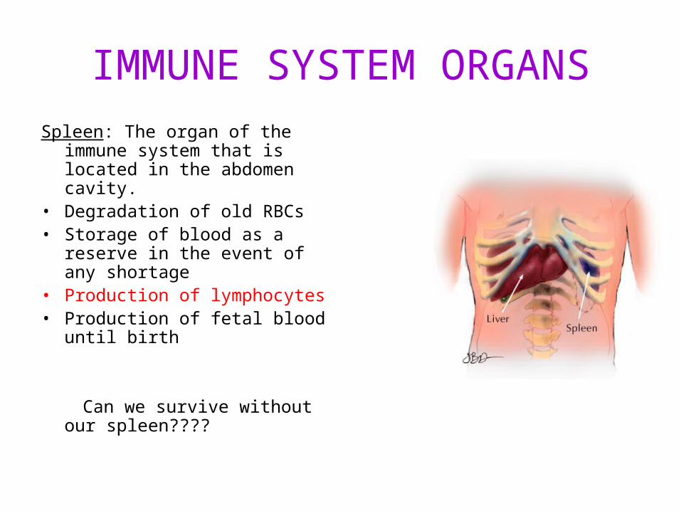
IMMUNE SYSTEM ORGANSSpleen: The organ of the immune
system that is located in the abdomen cavity.
• Degradation of old RBCs• Storage of blood as a reserve in
the event of any shortage• Production of lymphocytes• Production of fetal blood until
birth
Can we survive without our spleen????

Lymph nodes: Lymph nodes filter the lymphatic fluid and store special cells that can trap cancer cells or bacteria that are traveling through the body in the lymph fluid

Tonsils:
The tonsils trap bacteria and viruses entering through the throat and produce antibodies to help fight infections.

Thymus Gland:
• The site of T lymphocyte cell differentiation.
• The thymus increases gradually in size and activity until puberty, becoming vestigial (useless) thereafter.

What is immunity? It is the recognition and removal of molecules, cells
that foreign to the body. Immunity can be gained by two ways.
- Vaccine- Having infectious

There are two types of immunity;
1) Non-specific immunity (innate immunity)2) Specific immunity (acquired immunity)

1- Non-specific immunity
The basic resistance to disease that a species possesses - the first line of defense against infection.
• Skin barrier• HCl in the stomach• Lysozyme enzyme in sweat/saliva ( to digest
bacteria)• Mucous membranes protecting mouth, anus,
genitals.• Phagocytic leucocytes.

SKIN
• It is the first line of defense. External skin is covered by dry dead cells with keratinized proteins.
• Folds or moist parts of the skin are good habitat for pathogens.

Mucus
• Inner lining of the body (respiratory tract and digestive tract) are covered by mucosa layer that secrete mucus for protection.
Phagocytic WBS
• Granulocytes, Monocytes

• Interferons protein that is produced by body cells to fight with viruses.
Sythesized proteins inhibit the synthesis of some enzymes that are required for virus replication.
• Inflammation The local response to injury, involving small blood vessels, the cells circulating within these vessels, and nearby connective tissue. Fever occurs.

White blood cells are fighting with bacteria.

2) Specific Immunity (acquired-adaptive immunity)
It is provided bylypmhocytes which arespecific to the bacteria.
a) B lymphocytes: synthesize antibodies. Antibodies destroy bacteria (antigen). ….
b) T lymphocytes: they kill the infected body cells to prevent spread the infectious.
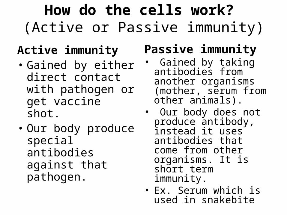
How do the cells work? (Active or Passive immunity)
Active immunity• Gained by either
direct contact with pathogen or get vaccine shot.
• Our body produce special antibodies against that pathogen.
Passive immunity• Gained by taking
antibodies from another organisms (mother, serum from other animals).
• Our body does not produce antibody, instead it uses antibodies that come from other organisms. It is short term immunity.
• Ex. Serum which is used in snakebite

ANTIBODIES ARE BIOLOGICAL WEAPONS. HOW DO THEY KILL THE PATHOGEN?

ANTIBODIES• Y-shaped proteins• Produced by B cells• Antibody recognizes the pathogen/antigen and
binds to it (tagging it)• This causes one of two things to happen: 1) Macrophages (killer cells) come and ‘eat’ the
bacteria (phagocytosis)Or 2) Other antibodies come and help out and kill the
bacteria directly (cell lysis)


ANTIBIOTICS or ANTIBACTERIAL• Antibiotics are used to treat infections caused by
bacteria. (ex. Penicillin)• A substance that kills or slows down the growth of
bacteria.• There is concern worldwide that antibiotics are
being overused. Antibiotic overuse is one of the factors that contributes towards antibiotic resistance as the growing number of bacterial infections which are becoming resistant to antibacterial medications.

How do antibiotics kill bacteria?
Remember bacterial life cycle!Antibiotics stop bacterial reproduction by blocking:1- DNA polymerase2- RNA polymerase2- Enzyme for cell wall synthesis
Are antibiotics effective against viruses?Think about viral life cycle!!

VIRUSES
• Viruses are known as ‘genes in a box’.• They are made of protein coat (capsid) and
and a nucleic acid either DNA or RNA but not both of them.
• They do not have metabolic reactions and can not reproduce themselves.
• They need a host cell to reproduce.

Viruses have two types of reproductive cycles.
1. In the lytic cycle,– viral particles are produced using host cell components,– the host cell lyses, and– viruses are released.
2. In the Lysogenic cycle– Viral DNA is inserted into the host – Viral DNA is duplicated along with the host chromosome during
each host cell division.– The inserted phage DNA is called a prophage. – Most prophage genes are inactive.– Environmental signals can cause a switch to the lytic cycle,
causing the viral DNA to be excised from the bacterial chromosome and leading to the death of the host cell.
© 2012 Pearson Education, Inc.

Figure 10.17_s2
Phage
Attachesto cell
Phage DNA Bacterialchromosome
The phage injects its DNA
Lytic cycle
The phage DNAcircularizes
1
2
The cell lyses,releasingphages
4
New phage DNA andproteins are synthesized
Phages assemble
3
OR
Environmentalstress
Lysogenic cycle
Many celldivisions
The lysogenic bacteriumreplicates normallyProphage
Phage DNA inserts into the bacterialchromosome by recombination
5
7
6

The cell lyses,releasingphages
Figure 10.17_1Phage
Attachesto cell
Phage DNA
The phage injects its DNA
Lytic cycle
The phage DNAcircularizes
1
New phage DNA andproteins are synthesized
Phages assemble 2
3
4
Bacterialchromosome

Figure 10.17_2
Phage
Attachesto cell
Phage DNA Bacterialchromosome
The phage injects its DNA
The phage DNAcircularizes
Environmentalstress
Many celldivisions
The lysogenic bacteriumreplicates normally, copying theprophage at each cell division
Prophage
Phage DNA inserts into the bacterialchromosome by recombination
Lysogenic cycle
1
2
7
6
5

Figure 10.20A
Envelope
Glycoprotein
Protein coat
RNA(two identicalstrands)
Reversetranscriptase(two copies)
HIV VIRUS (human immunodeficiency virus)

The AIDS virus makes DNA on an RNA template
AIDS (acquired immunodeficiency syndrome) is caused by HIV (human immunodeficiency virus).
HIV• is an RNA virus,• has two copies of its RNA genome,• carries molecules of reverse transcriptase, which causes
reverse transcription, producing DNA from an RNA template.
© 2012 Pearson Education, Inc.
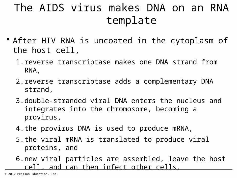
After HIV RNA is uncoated in the cytoplasm of the host cell,
1. reverse transcriptase makes one DNA strand from RNA,
2. reverse transcriptase adds a complementary DNA strand,
3. double-stranded viral DNA enters the nucleus and integrates into the chromosome, becoming a provirus,
4. the provirus DNA is used to produce mRNA,
5. the viral mRNA is translated to produce viral proteins, and
6. new viral particles are assembled, leave the host cell, and can then infect other cells.
The AIDS virus makes DNA on an RNA template
© 2012 Pearson Education, Inc.

Figure 10.20B
Viral RNA
DNAstrand
Reversetranscriptase
Double-strandedDNA
ViralRNAandproteins
1
2
3
4
5
6
CYTOPLASM
NUCLEUS
ChromosomalDNA
ProvirusDNA
RNA

Effect of HIV on the immune system: AIDS
• HIV host cells are antibody secreting lymphocytes.
• HIV takes control of lymphocytes, and their number decreases.
• So, patient becomes vulnerable to attack by opportunistic infectious (pneumonia, meningitis, cancers that would normally be resisted by a person with a healthy immune system.

Treatment and prevention of AIDS
• Until there is a vaccine or a cure, the best way to stop AIDS is to educate people about how the virus is transmitted.
• HIV mutates very quickly.• New strains are resistant to AIDS drugs.• Drug-resistant strains now infect new
patients.

The Social Consequence of AIDS
• Psychological• Economic • Child development and education• Child health. • Medical services provision• National factors

TOPIC 11.1 Defense against infectious disease• Describe the process of blood clotting• Outline the principle of challenge and response, clonal
selection and memory cells as the basis of immunity.• Define active and passive immunity.• Explain antibody production• Describe the production of monoclonal antibodies and their
use in diagnosis and in treatment.• Explain the principle of vaccination• Discuss the benefits and dangers of vaccination

Antigen versus our own cells
Antigen: ‘non-self’ invading macromolecules (protein, DNA, RNA) and microorganisms are known as antigen.
•How do lymphocytes recognize antigens and our own cells?
•Remember receptor molecules (Glycoproteins) on the cell membrane.

24.6 Antigens have specific regions where antibodies bind to them
• Antigens– are molecules that elicit the adaptive immune
response,
– usually do not belong to the host animal, and
– are proteins or large polysaccharides on the surfaces of viruses or foreign cells.
© 2012 Pearson Education, Inc.

Receptors on the cell membrane
• Glycoproteins (receptors) are unique for each individual.
• Glycoproteins are known as major histocompatibility complex (MHC) antigens.
• Each individual MHC is genetically determined.
• Our own Lymhocytes have have antigen receptors that recognize our own MHC.

The adaptive immune response counters specific invaders
• Infection or vaccination triggers active immunity.
• Vaccination, or immunization, exposes the immune system to a vaccine,– a harmless variant or – part of a disease-causing microbe.
• We can temporarily acquire passive immunity by receiving premade antibodies.
© 2012 Pearson Education, Inc.
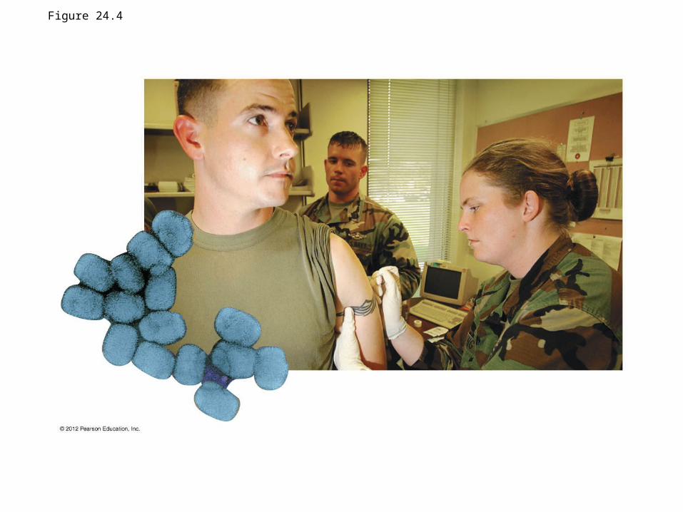
Figure 24.4

The role of lymphocytes
• Lymphocytes– are white blood cells that are found in the tissues and
organs of the lymphatic system,– are responsible for adaptive immunity, and– originate from stem cells in the bone marrow.
• B lymphocytes or B cells continue developing in bone marrow.
• T lymphocytes or T cells develop further in the thymus.
© 2012 Pearson Education, Inc.

Lymphocytes
• B cells– secrete antibodies (humoral immune responsecell-
mediated immune)
• T cells– attack cells infected with bacteria or viruses, (cell-mediated
immune)– promote phagocytosis by other white blood cells – stimulate B cells to produce antibodies.
© 2012 Pearson Education, Inc.

Figure 24.5A
Bonemarrow
Stem cell
Immature lymphocytes
Viablood
Antigenreceptors
Thymus
T cellB cellVia
blood
Final maturationof B and T cells in a
lymphatic organ
Lymphnodes,
spleen, andother
lymphaticorgans
Humoralimmune response
Cell-mediatedimmune response

Lymphocytes Millions of kinds of B cells and T cells
• each with different antigen receptors, capable of binding one specific type of antigen,• wait in the lymphatic system, • where they may respond to invaders.
© 2012 Pearson Education, Inc.

What is an antibody?
• Antibodies are secreted – by plasma (effector) B
cells, into the blood and lymph.
– Special protein called immunoglobulin
– is Y-shaped and– has two antigen-binding
sites specific to the antigenic determinants
© 2012 Pearson Education, Inc.

Figure 24.8B
Antigen
Lightchain
Heavychain
Antigen-bindingsites
C C
C CV
V
V
V

How do antibodies bind with antigen?
• Antigenic determinants are specific regions on an antigen where antibodies bind.
– An antigen usually has several different determinants.
– The antigen-binding
site of an antibody and
an antigenic determinant
have complementary shapes.
© 2012 Pearson Education, Inc.

Clonal Selection
When an antigen enters the body it activates only a small subset of lymphocytes that have complementary receptors.
In clonal selection, the selected lymphocyte cells• multiply into clones of short-lived effector cells, specialized
for defending against the antigen that triggered the response, and
• multiply into memory cells, which confer long-term immunity.
• Plasma cells are the effector cells produced during clonal selection of B cells.
© 2012 Pearson Education, Inc.

Clonal Selection
• The clonal selection of B cells occurs in two responses.– In the primary immune response, clonal selection
produces • effector cells and• memory cells that may confer lifelong immunity.
– In the secondary immune response, memory cells are activated by a second exposure to the same antigen.
© 2012 Pearson Education, Inc.
Animation: Role of B Cells

Figure 24.7A
Primary immune response
B cells withdifferentantigenreceptors
1
2
3
4 5 6
Antigen receptoron the cellsurface
Cell activation:growth, division,and differentiation
Antigenmolecules
First exposureto the antigen
Antibodymolecules
First clone
Endoplasmicreticulum
Plasma (effector) cells secreting antibodies Memory cells
Second exposureto the same antigen
Antigenmolecules
Second clone
Secondary immune response
Antibodymolecules
Clone of memory cells
Clone of plasma (effector) cellssecreting antibodies

Figure 24.7A_s1
1
Primary immune response
B cells withdifferentantigenreceptors
Antigen receptoron the cellsurface

Figure 24.7A_s2
1
2
Primary immune response
B cells withdifferentantigenreceptors
Antigen receptoron the cellsurface
Antigenmolecules

Figure 24.7A_s3
1
2
3
Primary immune response
B cells withdifferentantigenreceptors
Antigen receptoron the cellsurface
Cell activation:growth, division,and differentiation
Antigenmolecules
First exposureto the antigen

Figure 24.7A_s4
1
2
3
4 5
Primary immune response
B cells withdifferentantigenreceptors
Antigen receptoron the cellsurface
Cell activation:growth, division,and differentiation
Antigenmolecules
First exposureto the antigen
Antibodymolecules
First clone
Endoplasmicreticulum
Plasma (effector) cells secreting antibodies Memory cells

24.11 Helper T cells stimulate the humoral and cell-mediated immune responses
• In the cell-mediated immune response, an antigen-presenting cell displays – a foreign antigen (a nonself molecule) and– one of the body’s own self proteins– to a helper T cell.
© 2012 Pearson Education, Inc.

24.11 Helper T cells stimulate the humoral and cell-mediated immune responses
• The helper T cell’s receptors – recognize the self–nonself complexes and– the interaction activates the helper T cells.
• The helper T cell can then activate– cytotoxic T cells, which attack body cells that are
infected with pathogens, and– B cells.
© 2012 Pearson Education, Inc.
Animation: Helper T Cells
Video: T Cell Receptors

Figure 24.11
Antigen from the microbe(nonself molecule)
Antigen-presentingcell
Self protein
Microbe Macrophage
12
3
4
5 6
7
Self-nonselfcomplex
Phagocytic cell(yellow) engulfinga foreign cell T cell
receptor
Interleukin-1stimulates thehelper T cell
HelperT cell
Bindingsite for theantigen
Bindingsite for theself protein
Interleukin-2stimulatescell division
B cell
CytotoxicT cell
Interleukin-2activates B cellsand other T cells
Cell-mediatedimmune response(attack oninfected cells)
Humoralimmune response(secretion ofantibodies byplasma cells)

Figure 24.11_1
Antigen from the microbe(nonself molecule)
Antigen-presentingcell
Self protein
Microbe Macrophage
Self-nonselfcomplex
32
1

Figure 24.11_2
32
4
5 6
7
Antigen-presentingcell
Self-nonselfcomplex T cell
receptor
Interleukin-1stimulates thehelper T cell
HelperT cell
Bindingsite for theantigen
Bindingsite for theself protein
Interleukin-2stimulatescell division
B cell
CytotoxicT cell
Interleukin-2activates B cellsand other T cells

Figure 24.11_3
Phagocytic cell(yellow) engulfinga foreign cell

24.12 Cytotoxic T cells destroy infected body cells
• Cytotoxic T cells– are the only T cells that kill infected cells,– bind to infected body cells, and– destroy them.
• Cytotoxic T cells also play a role in protecting the body against the spread of some cancers.
© 2012 Pearson Education, Inc.
Animation: Cytotoxic T Cells

Figure 24.12_s1
1 A cytotoxic T cell bindsto an infected cell.
Self-nonselfcomplex
ForeignantigenInfected cell
Perforinmolecule
CytotoxicT cell

Figure 24.12_s2
21 A cytotoxic T cell bindsto an infected cell.
Perforin makes holes in theinfected cell’s membrane,and an enzyme thatpromotes apoptosis enters.Self-nonself
complex
ForeignantigenInfected cell
Perforinmolecule
CytotoxicT cell
Enzymes thatpromote apoptosis
A hole forming

Figure 24.12_s3
321 A cytotoxic T cell bindsto an infected cell.
Perforin makes holes in theinfected cell’s membrane,and an enzyme thatpromotes apoptosis enters.
The infected cellis destroyed.
Self-nonselfcomplex
ForeignantigenInfected cell
Perforinmolecule
CytotoxicT cell
Enzymes thatpromote apoptosis
A hole forming

CONNECTION: Monoclonal antibodies are powerful tools in the lab and clinic
• Monoclonal antibodies (mAb) are – identical antibodies
– produced by cells that are all descendants of a single, hybrid cell.
• To make the hybrid cell with desirable properties, two cells are fused.1. A cancerous tumor cell, able to multiply indefinitely,
is fused to
2. a normal antibody-producing B cell, which is producing the desired antibody.
© 2012 Pearson Education, Inc.

Antibodies mark antigens for elimination
• Antibodies promote antigen elimination through several mechanisms:1. neutralization, binding to surface proteins on a virus
or bacterium and blocking its ability to infect a host,2. agglutination, using both binding sites of an antibody
to join invading cells together into a clump,
© 2012 Pearson Education, Inc.

Antibodies mark antigens for elimination
3. precipitation, similar to agglutination, except that the antibody molecules link dissolved antigen molecules together, and
4. activation of the complement system by antigen-antibody complexes.
© 2012 Pearson Education, Inc.
Animation: Antibodies

Figure 24.9
Bacterium
Virus
Neutralization(blocks viral binding sites;
coats bacteria)
Binding of antibodies to antigensinactivates antigens by
Agglutinationof microbes
Precipitation ofdissolved antigens
Activation of thecomplement system
Bacteria
Antigenmolecules
Complementmolecule
Foreign cell Hole
Leads to
Cell lysis
Enhances
Phagocytosis
Macrophage
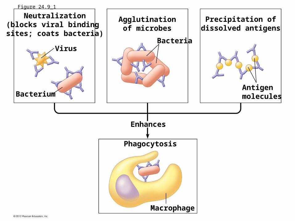
Figure 24.9_1
Bacterium
Virus
Neutralization(blocks viral binding sites; coats bacteria)
Agglutinationof microbes
Precipitation ofdissolved antigens
Bacteria
Antigenmolecules
Enhances
Phagocytosis
Macrophage

Figure 24.9_2Activation of the
complement systemComplementmolecule
Foreign cell Hole
Leads to
Cell lysis
Clonal selection
• Primary vs. secondary immune responses– The primary immune response
• occurs upon first exposure to an antigen and• is slower than the secondary immune response.
– The secondary immune response• occurs upon second exposure to an antigen and• is faster and stronger than the primary immune response.
© 2012 Pearson Education, Inc.

Figure 24.7B
Time (days)5649423528211470
An
tib
od
y c
on
cen
tra
tio
n
Antibodiesto X
Antibodiesto Y
Second exposureto antigen X,
first exposureto antigen Y
First exposure to antigen X
Primary immuneresponse to
antigen X
Secondary immuneresponse to
antigen X
Primary immuneresponse to
antigen Y

CONNECTION: Monoclonal antibodies are powerful tools in the lab and clinic
• Monoclonal antibodies are useful in – research,– diagnosis (such as home pregnancy tests), and– treatment of certain cancers.
© 2012 Pearson Education, Inc.

Figure 24.10 Early pregnancy(hCG in the blood and urine)
Urine is appliedto the strip
hCG/mAbcomplex
ControlmAb
hCG
hCG

Monoclonal antibody production and their usage
– Treatment of certain cancers.– Cancer cells have specific tumor associated antigens
(TAA). – Monoclonal antibodies to TAA produced. – Drugs to kill/ inhibit cancer cell have been attached to
antibody. – Cancer cells can be specifically targeted and killed.– Monoclonal antibodies could be developed by mouse
cells or genetically engineered methods.
© 2012 Pearson Education, Inc.

CONNECTION: HIV destroys helper T cells, compromising the body’s defenses
• AIDS (acquired immunodeficiency syndrome), results from infection by HIV, the human immunodeficiency virus.– Since 1981 AIDS has killed more than 27 million
people, and more than 33 million people live today with HIV.
– In 2008,• 2.7 million people were newly infected with HIV, and• over 2 million died, including 300,000 children under age 15.
– Most AIDS infections and deaths occur in nonindustrialized nations of southern Asia and sub-Saharan Africa.
© 2012 Pearson Education, Inc.

Figure 24.13

VACCINATION
– Discuss the benefits and danger of vaccines.
© 2012 Pearson Education, Inc.