Immune phenotypes predict survival in patients with ...
14
RESEARCH Open Access Immune phenotypes predict survival in patients with glioblastoma multiforme Haouraa Mostafa 1,2 , Andrej Pala 3 , Josef Högel 4 , Michal Hlavac 3 , Elvira Dietrich 1,2 , M. Andrew Westhoff 5 , Lisa Nonnenmacher 3 , Timo Burster 3 , Michael Georgieff 2 , C. Rainer Wirtz 3 and E. Marion Schneider 1,2* Abstract Background: Glioblastoma multiforme (GBM), a common primary malignant brain tumor, rarely disseminates beyond the central nervous system and has a very bad prognosis. The current study aimed at the analysis of immunological control in individual patients with GBM. Methods: Immune phenotypes and plasma biomarkers of GBM patients were determined at the time of diagnosis using flow cytometry and ELISA, respectively. Results: Using descriptive statistics, we found that immune anomalies were distinct in individual patients. Defined marker profiles proved highly relevant for survival. A remarkable relation between activated NK cells and improved survival in GBM patients was in contrast to increased CD39 and IL-10 in patients with a detrimental course and very short survival. Recursive partitioning analysis (RPA) and Cox proportional hazards models substantiated the relevance of absolute numbers of CD8 cells and low numbers of CD39 cells for better survival. Conclusions: Defined alterations of the immune system may guide the course of disease in patients with GBM and may be prognostically valuable for longitudinal studies or can be applied for immune intervention. Keywords: Glioblastoma multiforme, CD8+ lymphocytes, NK cells, CD39-ectonucleotidase, Recursive partitioning analysis, Survival Background Glioblastoma multiforme (GBM) is the most common pri- mary brain tumor in adults. Despite intensive research efforts and a multimodal management that consists of sur- gery, radiotherapy, and chemotherapy with temozolomide, the prognosis is very poor [1]. The hypoxic environment represents a very potent stimulus for angiogenesis [2], while molecular alterations, cell survival mechanisms, the acquisition of chemo- and radioresistance [3], and tumor heterogeneity [4] as well as multiple immune escape mechanisms are further important factors that contribute to the inferior prognosis in GBM [5, 6]. In addition to age, a number of stress factors, including chemical exposure, and demographical aspects influence the incidence of tumor manifestation [7]. At least one study demonstrated an increased risk for glioblastoma by alcohol consumption [8]. When compared with other malignancies, modulation of immune suppressor effector cells appears to be of major importance [9], and individual tumor patients may require an individual treatment regimen against im- mune suppressive elements. We addressed here im- mune phenotypes in patients with glioblastoma at the time of diagnosis. We applied flow cytometry and vali- dated enzyme-linked immunosorbent assay (ELISA) as a reliable technique to systematically analyze immune cells in peripheral blood as well as cytokines. In gen- eral, immune modulation initiated by the tumor may require direct cell-cell contact or may occur via soluble factors, including interleukins and chemokines. At present, malignancies manifesting distantly from the peripheral immune system have not been systematically described to influence immune phenotypes. However, an important work published by Kmiecik and colleagues [10], which was performed in a small group of GBM pa- tients, was highly promising in implying the sensitization * Correspondence: [email protected] 1 Sektion Experimentelle Anaesthesiologie, University Hospital Ulm, Albert Einstein Allee 23, 89081 Ulm, Germany 2 Klinik für Anaesthesiologie, University Hospital Ulm, Albert Einstein Allee 23, 89081 Ulm, Germany Full list of author information is available at the end of the article © 2016 Mostafa et al. Open Access This article is distributed under the terms of the Creative Commons Attribution 4.0 International License (http://creativecommons.org/licenses/by/4.0/), which permits unrestricted use, distribution, and reproduction in any medium, provided you give appropriate credit to the original author(s) and the source, provide a link to the Creative Commons license, and indicate if changes were made. The Creative Commons Public Domain Dedication waiver (http://creativecommons.org/publicdomain/zero/1.0/) applies to the data made available in this article, unless otherwise stated. Mostafa et al. Journal of Hematology & Oncology (2016) 9:77 DOI 10.1186/s13045-016-0272-3
Transcript of Immune phenotypes predict survival in patients with ...

RESEARCH Open Access
Immune phenotypes predict survival inpatients with glioblastoma multiformeHaouraa Mostafa1,2, Andrej Pala3, Josef Högel4, Michal Hlavac3, Elvira Dietrich1,2, M. Andrew Westhoff5,Lisa Nonnenmacher3, Timo Burster3, Michael Georgieff2, C. Rainer Wirtz3 and E. Marion Schneider1,2*
Abstract
Background: Glioblastoma multiforme (GBM), a common primary malignant brain tumor, rarely disseminates beyondthe central nervous system and has a very bad prognosis. The current study aimed at the analysis of immunologicalcontrol in individual patients with GBM.
Methods: Immune phenotypes and plasma biomarkers of GBM patients were determined at the time of diagnosisusing flow cytometry and ELISA, respectively.
Results: Using descriptive statistics, we found that immune anomalies were distinct in individual patients. Definedmarker profiles proved highly relevant for survival. A remarkable relation between activated NK cells and improvedsurvival in GBM patients was in contrast to increased CD39 and IL-10 in patients with a detrimental course and veryshort survival. Recursive partitioning analysis (RPA) and Cox proportional hazards models substantiated therelevance of absolute numbers of CD8 cells and low numbers of CD39 cells for better survival.
Conclusions: Defined alterations of the immune system may guide the course of disease in patients with GBMand may be prognostically valuable for longitudinal studies or can be applied for immune intervention.
Keywords: Glioblastoma multiforme, CD8+ lymphocytes, NK cells, CD39-ectonucleotidase, Recursive partitioninganalysis, Survival
BackgroundGlioblastoma multiforme (GBM) is the most common pri-mary brain tumor in adults. Despite intensive researchefforts and a multimodal management that consists of sur-gery, radiotherapy, and chemotherapy with temozolomide,the prognosis is very poor [1]. The hypoxic environmentrepresents a very potent stimulus for angiogenesis [2],while molecular alterations, cell survival mechanisms, theacquisition of chemo- and radioresistance [3], and tumorheterogeneity [4] as well as multiple immune escapemechanisms are further important factors that contributeto the inferior prognosis in GBM [5, 6]. In addition to age,a number of stress factors, including chemical exposure,and demographical aspects influence the incidence oftumor manifestation [7]. At least one study demonstrated
an increased risk for glioblastoma by alcohol consumption[8]. When compared with other malignancies, modulationof immune suppressor effector cells appears to be ofmajor importance [9], and individual tumor patients mayrequire an individual treatment regimen against im-mune suppressive elements. We addressed here im-mune phenotypes in patients with glioblastoma at thetime of diagnosis. We applied flow cytometry and vali-dated enzyme-linked immunosorbent assay (ELISA) asa reliable technique to systematically analyze immunecells in peripheral blood as well as cytokines. In gen-eral, immune modulation initiated by the tumor mayrequire direct cell-cell contact or may occur via solublefactors, including interleukins and chemokines. Atpresent, malignancies manifesting distantly from theperipheral immune system have not been systematicallydescribed to influence immune phenotypes. However,an important work published by Kmiecik and colleagues[10], which was performed in a small group of GBM pa-tients, was highly promising in implying the sensitization
* Correspondence: [email protected] Experimentelle Anaesthesiologie, University Hospital Ulm, AlbertEinstein Allee 23, 89081 Ulm, Germany2Klinik für Anaesthesiologie, University Hospital Ulm, Albert Einstein Allee 23,89081 Ulm, GermanyFull list of author information is available at the end of the article
© 2016 Mostafa et al. Open Access This article is distributed under the terms of the Creative Commons Attribution 4.0International License (http://creativecommons.org/licenses/by/4.0/), which permits unrestricted use, distribution, andreproduction in any medium, provided you give appropriate credit to the original author(s) and the source, provide a link tothe Creative Commons license, and indicate if changes were made. The Creative Commons Public Domain Dedication waiver(http://creativecommons.org/publicdomain/zero/1.0/) applies to the data made available in this article, unless otherwise stated.
Mostafa et al. Journal of Hematology & Oncology (2016) 9:77 DOI 10.1186/s13045-016-0272-3
of immune cells against GBM. In addition, the workpublished by Skog and coworkers [11] contributed to ourunderstanding of effector pathways induced by the inter-action of tumor-derived microparticles with target cells.Cytokines, including interleukin (IL)-10 and ferritin, se-creted by tumor-infiltrating immune cells and by thetumor itself are members of a larger scenario of a tumor-associated proteome [12]. They may play an importantrole in downregulating major histocompatibility complex(MHC)-class I and natural killer (NK)-ligands [13].Here, we asked whether immune phenotypes of pa-tients with manifest GBM display distinct features dif-fering from healthy individuals. In addition to theabovementioned IL-10 and ferritin associated withGBM, we found that the relative amounts of activatedNK cells in addition to T cell receptor (TCR) α/β-posi-tive and CD8-positive lymphocytes display a strong cor-relation with increased survival in GBM in bivariateanalysis. When using multivariable Cox proportionalhazards model analysis, the Karnofsky Performance sta-tus Scale (KPS), the isocitrate dehydrogenase-1 (IDH-1)mutation status, and high CD8 and low CD39 cells aremost relevant for better survival.
MethodsPatientsPatients had been diagnosed for glioblastoma multi-forme grade IV according to the criteria of the WorldHealth Organization (WHO). Immune competence wastested at the time of diagnosis before surgery followingstandard cluster of differentiation (CD) and activationmarkers for T-, NK-, monocyte-, and granulocyte sub-populations according to international guidelines forwhole blood flow cytometric analysis. Immune pheno-typing and inflammatory and anti-inflammatory bio-markers performed by a validated ELISA was anessential step in classifying patients for their immunecompetence. These tests were done for each patient aspart of routine care. Patients were then under high dosesteroid treatment. All patients’ data were pseudonymizedby a data bank held in the Section of Experimental An-aesthesiology (http://www.uniklinik-ulm.de/struktur/kli-niken/sektionen/klinik-fuer-anaesthesiologie/experimen-telleanaesthesiologie/home/general-information/address-contact.html). The study was conducted after approvalby the local Ethics Committee of the University HospitalUlm with the approval umber 162/10 named “Novel ex-perimental approaches in brain tumors”. The universaltrial number is U111-1179-3127. Brain tumor materialwas used to establish cell lines. Two milliliters ofethylenediaminetetra-acetic acid (EDTA) anti-coagulatedblood were routinely analyzed from all patients at thetime of diagnosis before surgery. A total of 51 patientswere assessed. The mean age was 56.8 years (range
22–78 years). All patients underwent surgical resection,radiotherapy, and chemotherapy and had routine follow-up with magnetic resonance imaging interpreted by neu-roradiologists. All patients are listed in Table 1.
Healthy donorsA total of 36 healthy volunteers were included as controls.Their mean age was 36.1 years (range 19–68 years). Theirimmune phenotypes and plasma biomarkers were alsobased on routine analysis in the context of routine clinicalexamination. Results of all healthy donors were alsopseudonymized before processing for statistical analysis.
Flow cytometrySurface markers of whole blood leukocytes were deter-mined by standard flow cytometric analyses using FACS-calibur and Cellquest software (BD Biosciences.com).Leukocytes were gated into lymphocytes, monocytes,and granulocytes by forward- and side-scatter analysis.Quantification of the percent-positive cells was deter-mined by the binding of fluorescently labeled antibodiesand determination of expression densities of individualantigens was recorded. The expression density of therelevant antigens was calculated as the mean fluores-cence intensity (MFI) using the following formula: (%positives ×mean expression density of the relevant anti-gen) − (% positives × mean expression density of the re-spective isotype control). The following antibodies weredetermined on lymphocytes: IgG1 and IgG2a isotype con-trol antibodies, monoclonal antibodies directed againstCD3ε-chain (clone UCHT1, Beckmancoulter.com), TCRα/β (clone WT31), CD56 (clone My31), CD4 (clone SK3),CD25 (clone M-A251), CD8 (clone 2ST8.5H7, Beckman-Coulter.com), CD16 (NK-P15), CD95 (clone DX-2),CD127 (clone #40131, RnD Systems), and CD39 (cloneA1, abdserotec.com), all purchased from BD Biosciences.-com, if not stated otherwise. Since all patients were ana-lyzed during high steroid treatment, we also addressed thephenotype of two GBM patients before and after steroidmedication (Additional file 1: Figure S1).
Plasma biomarkersPlasma from patients’ blood and control subjects was usedfor the quantification of biomarkers. A highly sensitiveand validated ELISA (Immulite 1000®, Siemens.com) wasused. The following Immulite 1000® kits were used: I10 toquantify IL-10 and FER to quantify plasma ferritin.
MGMT analysis and IDH-1 mutation statusTo analyze MGMT-promoter methylation, DNA was ex-tracted from snap-frozen tumor tissue. A pyrosequenc-ing platform (ID or Q24 instrument, Qiagen.com) wasapplied for the quantification of methylation status at asingle base solution. Quantitative methylation values
Mostafa et al. Journal of Hematology & Oncology (2016) 9:77 Page 2 of 14

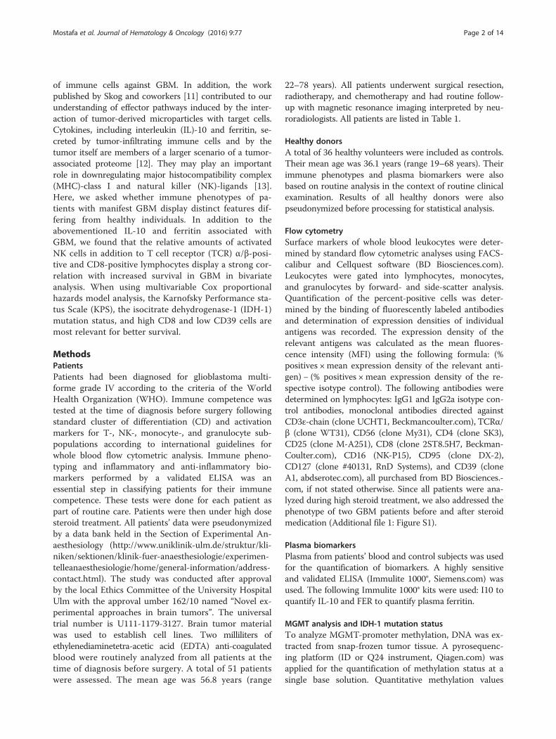
Table 1 Patients’ list showing age and sex, tumor location, total or subtotal resection, O6-methylguanine-DNA methyltransferase(MGMT), IDH-1 mutation status, and overall survival
Patient no. Age/sex Tumor location Resection MGMT IDH mutation Overall survival (months)
#1 60/m Ttemporo-occipital Subtotal p n.a. 31
#2 62/m Temporal Total n n 9
#3 41/f Fronto-parietal Total n n 18+
#4 65/m Frontal Total p n 10
#5 34/m Frontal Subtotal p n 19+
#6 34/f Occipital Total n n.a. n.a.
#7 44/f Frontal Subtotal n n 11
#8 54/m Temporal Total n n 23
#9 66/m Parietal Subtotal n n 25
#10 29/m Temporal Total p p 43+
#11 68/m Parieto-occipital Total n n 14
#12 71/m Temporo-occipital Subtotal n n 16+
#13 78/f Frontal Biopsy p n 15
#14 70/m Temporal Subtotal n n 10+
#15 45/m Parieto-occipital Subtotal p p 12+
#16 61/m Temporo-occipital Subtotal n n 11+
#17 52/m Temporal Total n n 10+
#18 63/f Frontal Total n n 7
#19 61/f Frontal Total p n.a. 53+
#20 41/m Fronto-parietal Subtotal n n 33
#21 75/m Frontal Subtotal n n 3
#22 74/f Temporal Subtotal p p 52
#23 57/m Temporal Subtotal n n 23
#24 22/m Frontal Subtotal p n 77
#25 56/m Fronto-parietal Subtotal n p 7
#26 44/m Parietal Subtotal p p 18+
#27 51/m Frontal Total p n 10
#28 67/f Frontal Total n.a. p 28+
#29 53/f Frontal Total n n 20+
#30 45/m Temporo-parietal Subtotal p p 31
#31 58/m Frontal Total n n 9+
#32 58/m Frontal Total n p 7+
#33 53/f Temporo-occipital Subtotal p n 5
#34 67/f Frontal Total p n 8+
#35 62/m Frontal + temporal Total n n 8+
#36 67/m Temporal Total n n 9+
#37 74/m Occipital Subtotal n n 9+
#38 54/m Temporal Subtotal n.a. n 7
#39 70/m Frontal Total p n 5+
#40 40/m Temporo-occipital Subtotal p n 61+
#41 68/f Temporo-occipital Total p n 6
#42 38/m Parietal Total p n 5+
#43 76/f Temporal Near total p p 25+
Mostafa et al. Journal of Hematology & Oncology (2016) 9:77 Page 3 of 14
within a sequence context were determined for individ-ual or multiple CpG sites. (http://www.varionostic.de/services/dna-methylation/)IDH-1 mutation status was tested by a monoclonal
antibody directed against the most frequent mutationsite: IDH1 R132H [14].
StatisticsDescriptive statistics, including medians, interquartileranges, and 10th and 90th percentiles were applied tocharacterize leukocyte blood parameters and surface an-tigens in healthy donors vs. controls. Between-groupcomparisons of continuous variables were performedusing the Mann-Whitney U test. The Kaplan-Meiermethod and log-rank test were applied to summarizeand compare the overall survival and time to progres-sion between trials or different conditions using Graph-PadPrism version 6.0 or the SAS procedure “LIFETEST”.In this context, median values were used as cutoffs todichotomize continuous traits for survival analysis. Forthe purpose of prediction, Cox proportional hazardsmodels were fitted using SAS PROC PHREG. Onlyparameters which showed a p value < 0.2 in univari-able analysis (Additional file 2: Table S1) were in-cluded in the multivariable Cox proportional hazardsmodel (Additional file 3: Table S2). Unbiased recur-sive partitioning analysis (RPA) was applied to identifythe risk classes. Absolute numbers of leukocytes andleukocyte subpopulations were determined by routineCoulter counting determinations. For all statisticalinvestigations, tests for significance were two-tailed,with a p value threshold of 0.05 indicating “statisticalsignificance”.
ResultsIL-10 and plasma ferritin are elevated in GBMWe determined two biomarkers in the patients’ plasmabefore surgery: IL-10 and plasma ferritin. The medianvalues of IL-10 and ferritin were significantly elevated in
the GBM patients (Fig. 1). Patients with IDH-1 muta-tions are labeled in red.
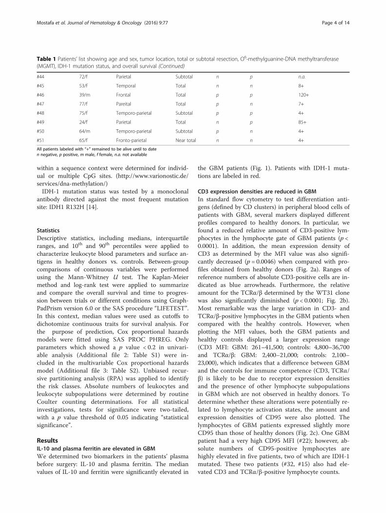
CD3 expression densities are reduced in GBMIn standard flow cytometry to test differentiation anti-gens (defined by CD clusters) in peripheral blood cells ofpatients with GBM, several markers displayed differentprofiles compared to healthy donors. In particular, wefound a reduced relative amount of CD3-positive lym-phocytes in the lymphocyte gate of GBM patients (p <0.0001). In addition, the mean expression density ofCD3 as determined by the MFI value was also signifi-cantly decreased (p = 0.0046) when compared with pro-files obtained from healthy donors (Fig. 2a). Ranges ofreference numbers of absolute CD3-positive cells are in-dicated as blue arrowheads. Furthermore, the relativeamount for the TCRα/β determined by the WT31 clonewas also significantly diminished (p < 0.0001; Fig. 2b).Most remarkable was the large variation in CD3- andTCRα/β-positive lymphocytes in the GBM patients whencompared with the healthy controls. However, whenplotting the MFI values, both the GBM patients andhealthy controls displayed a larger expression range(CD3 MFI: GBM: 261–41,500; controls: 4,800–36,700and TCRα/β: GBM: 2,400–21,000; controls: 2,100–23,000), which indicates that a difference between GBMand the controls for immune competence (CD3, TCRα/β) is likely to be due to receptor expression densitiesand the presence of other lymphocyte subpopulationsin GBM which are not observed in healthy donors. Todetermine whether these alterations were potentially re-lated to lymphocyte activation states, the amount andexpression densities of CD95 were also plotted. Thelymphocytes of GBM patients expressed slightly moreCD95 than those of healthy donors (Fig. 2c). One GBMpatient had a very high CD95 MFI (#22); however, ab-solute numbers of CD95-positive lymphocytes arehighly elevated in five patients, two of which are IDH-1mutated. These two patients (#32, #15) also had ele-vated CD3 and TCRα/β-positive lymphocyte counts.
Table 1 Patients’ list showing age and sex, tumor location, total or subtotal resection, O6-methylguanine-DNA methyltransferase(MGMT), IDH-1 mutation status, and overall survival (Continued)
#44 72/f Parietal Subtotal n p n.a.
#45 53/f Temporal Total n n 8+
#46 39/m Frontal Total p p 120+
#47 77/f Pareital Total p n 7+
#48 75/f Temporo-parietal Subtotal p p 4+
#49 24/f Parietal Total n p 85+
#50 64/m Temporo-parietal Subtotal p n 4+
#51 65/f Fronto-parietal Near total n n 4+
All patients labeled with “+” remained to be alive until to daten negative, p positive, m male, f female, n.a. not available
Mostafa et al. Journal of Hematology & Oncology (2016) 9:77 Page 4 of 14

NK cell subpopulations in GBM and healthy donorsSimilar data sets are displayed in Fig. 2; the relative andabsolute amounts of CD56+ NK-cells are presented inFig. 3. Although the percentage of CD56+ NK cells var-ied with a large range in GBM, the median values werenot significantly different from healthy donors (p =0.6857; Fig. 3a). However, when the MFI values were cal-culated, there was a trend for lower CD56 expression inGBM when compared with the lymphocyte fraction ofhealthy donors (p = 0.0972; Fig. 3a). Absolute numbersof CD56+ NK-cells again demonstrate that some patientsare unique and differ from values determined in healthydonors of advanced age [15]. Generally, these resultssuggest that GBM patients may have more CD56dim NKcells than cytokine-producing CD56bright cells. We fur-ther investigated CD56+ NK cells co-expressing the acti-vation antigen CD16. The majority of GBM samples didnot differ from the control (Fig. 3b). However, therewere unique individual GBM patients with very highrelative amounts of CD16-positive NK cells, indicatingNK-cell activation. When plotting MFI values, individ-uals with a high CD16 expression in CD56-positive NKcells could also be identified (Fig. 3b). Nevertheless, ofparticular interest were patients #12, #21, #23, and #26,who displayed elevated or very elevated CD16/CD56-positive NK cells before surgery; however, only #6 hadhigh amounts of absolute CD16/CD56-positive lympho-cytes, all within the normal range of healthy individualsat an advanced age [15]. NK cells co-expressing CD3, aT-cell receptor structure, in addition to NK markers de-fine a cell type which is particularly sensitive to cytokineactivation and have been termed cytokine-induced killer(CIK) cells; they exhibit non-MHC-restricted cytolysisactivities against tumor cells [16]. Such CD3/56 co-expressing effectors were clearly diminished in themajority of GBM patients (Fig. 3c, p = 0.0016), but twopatients (#14 and #25) were characterized by a very highproportion of this cell type. An attenuated phenotypewas also detected in a few healthy donors. The absolutenumbers of CIK were higher in healthy donors [15].
Evidence for regulatory T cell subsets in GBM patientsand healthy controlsGBM patients had a very low relative amount of CD4-positive cells (#22, #23, #26, #40) (Fig. 4a), in part corre-sponding to low absolute numbers. IDH-1-mutatedtumor patients display either increased or decreasedCD4-positive lymphocytes (Fig. 4b). One IDH-1-mutatedpatient (#32) had very high CD3-, TCRα/β-, and CD95-positive lymphocytes (Fig. 2). CD8-positive lymphocytesare also heterogeneous between GBM patients and con-trols (Fig. 4b). This accounts for the percentage of CD8-positive lymphocytes and their expression densities(MFI), as well as the absolute numbers (Fig. 4b). Twopatients with high absolute numbers of CD8-positivecells (#24, #28) also presented with high amounts ofCD16/CD56-positive cells (Fig. 3b), but not with increasedCD3- and TCRα/β-positive lymphocytes (Fig. 2b). Thedistribution of lymphocytes co-expressing CD4 andCD25 characterizes an activated lymphocyte subpopula-tion, which contains a large proportion of so-calledregulatory, that is, immune suppressive T lymphocytes.The further characterization of CD4/CD25 lymphocytesled to the recognition of the importance of FoxP3expression, a transcription factor involved in the sup-pressive function of this cell type. FoxP3 was not deter-mined in our study, but according to a recent review,active FoxP3 is mostly restricted to the CD4/CD25 highT cell subpopulation in humans [17]. According to per-ipheral blood lymphocyte analyses, neither the medianpercentage nor the MFI of CD4/CD25-positive lympho-cytes in GBM patients and control individuals was sig-nificantly different (Fig. 4c). In addition, an importantimmune suppressive function is displayed by the ex-pression of ATP-degrading enzymes, including CD39;in lymphocytes with low IL-7 receptor, CD127 medianvalues are different between patients and healthydonors (Fig. 4e). Among patients with high absolutenumbers of CD39, one IDH-1-mutated patient (#48)presented with very high relative and absolute numbersin addition to high CD39 expression densities (Fig. 4d).
Fig. 1 Increased median value of IL-10 and ferritin in plasma samples of GBM patients. Interleukin 10 (IL-10) (a), plasma ferritin (b) as relevantbiomarkers determined in plasma samples of GBM patients (P) and healthy controls (H). Median values for IL-10 is 7.18 (±1.2 pg/ml) in elderlyhealthy donors, and ferritin serum concentrations may range a lot in elderly. IDH-1 mutated patients outside of the 25–75 boxed percentile areshown as red-filled circles
Mostafa et al. Journal of Hematology & Oncology (2016) 9:77 Page 5 of 14
Results suggest that CD39 may play a unique role inimmune suppression of GBM. Exemplary analysis ofGBM tissue preparations suggests that CD39 is alsoexpressed in tumor tissue (Additional file 4: Figure S2).Regulatory T cells (Tregs) are also defined by weak ex-pression of the IL-7 receptor, CD127 (p = 0.0083).Figure 4e shows the differences of medians of CD127-positive lymphocytes in GBM and controls as comparedwith the respective expression densities of CD127.
Patients with GBM displayed a lower median of CD127-positive lymphocytes as well as a MFI of CD127. Remark-ably, the absolute numbers of CD127-positive cells werealso increased in patient #48, who was unique by highCD39 (Fig. 4e).
Immune phenotypes correlate with survival in GBMFigure 5 shows the overall survival of the GBM pa-tients and the different survival curves in association
Fig. 2 Significantly decreased CD3 and TCR α/β in peripheral blood cells but no significant change in CD95. Percent CD3 lymphocytes in EDTAwhole-blood-derived lymphocytes of GBM patients (P) and controls (H) (a, left) and the corresponding CD3 expression densities (meanfluorescence intensity (MFI)) (a, middle) and absolute numbers of CD3-positive lymphocytes in patients (a, right). Normal ranges of absolutenumbers are indicated as blue arrowheads on the y-axis (a, right). Percent TCRα/β lymphocytes in GBM patients and healthy controls (b, left) and thecorresponding MFI distribution (b, middle) and absolute numbers of TCRα/β cells/μl blood (b, right). Percent CD95 lymphocytes in GBM patients ascompared to controls (c, left) and the corresponding distribution of CD95 MFI (c, middle) and absolute CD95-expressing lymphocytes (c, right).IDH-1 mutated patients outside of the 25–75 boxed percentile are shown as red-filled circles. IDH-1 mutated patients outside of the 25–75boxed percentile are shown as red-filled circles
Mostafa et al. Journal of Hematology & Oncology (2016) 9:77 Page 6 of 14

with immune markers differentially expressed in pa-tients and controls. The median overall survival was19 months (Fig. 5a). We then analyzed the survival inthe context of immune markers. Patients were groupedaccording to their marker distribution higher or lowerof the respective median value. Patients with more than30 % CD8 expressing differed from those with < 30 %CD8-positive cells, by a better median survival (19 vs.10 months (p = 0.0419), Fig. 5b). TCRα/β expression of
>51.2 % as compared to < 51.2 % TCRα/β T cells re-sulted in significantly better survival of 43 vs. 9 months(p = 0.0498; Fig. 5c). CD95 differences were in the samerange: With a cutoff of 60.5 %, the median survival was8 and 43 months, respectively (p = 0.047, Fig. 5d). Theactivation marker CD16 on CD56+ NK cells of (> 8.35vs. <8.35 %) resulted in a median survival of 53 vs.10 months (p = 0.0031, Fig. 5e). CD127-expressinglymphocytes were analyzed in two groups with > 18.5
Fig. 3 Differences in CD56 and CD16/56 NK cells and CD3/56 cytokine-activated NK (CIK) cells in GBM patients and controls. Percent CDcluster-positive lymphocytes are shown for patients (P) and healthy donors (H) on the left; relative expression densities, given as MFI are shownin the middle column, and absolute numbers of CD cluster-positive lymphocytes for the patients are shown on the right half of the figure.CD56 NK cells (a). CD56/16 co-expressing, activated NK cells (b). CD3/56 co-expressing, cytokine-induced killer (CIK) cells (c). IDH-1mutatedpatients outside of the 25-75 boxed percentile are shown as red-filled circles
Mostafa et al. Journal of Hematology & Oncology (2016) 9:77 Page 7 of 14

vs. < 18.5 % positive cells. The median survival in thelow and high CD127 groups was 25 and 11 months, re-spectively (p = 0.0857, Fig. 5f ). The IDH-1 mutation
showed no evidence for a different survival (p = 0.6656,Fig. 5h). Despite strong arguments for lymphocyte sub-populations in GBM patients and survival, high
Fig. 4 Differences in CD8, CD4, CD4/25, CD39, and CD127-positive lymphocytes. Percent CD cluster-positive lymphocytes are shown for patients(P) and healthy donors (H) on the left; relative expression densities, given as MFI are shown in the middle column, and absolute numbers of CDcluster positive lymphocytes for the patients are shown on the right half of the figure. CD8 (a). CD4 (b). CD4+/CD25+ (c). CD39 (d). CD127 (e).IDH-1 mutated patients outside of the 25-75 boxed percentile are shown as red-filled circles
Mostafa et al. Journal of Hematology & Oncology (2016) 9:77 Page 8 of 14

absolute leukocyte counts (c.f. Additional file 5: FigureS3) are related to inferior survival when analyzed byunivariable Cox regression analysis and multivariableCox proportional hazards model (Additional file 2:Table S1, Additional file 3: Table S2, Additional file 6:Figure S4).
DiscussionIn the attempt to answer the question whether the per-ipheral immune system senses a brain tumor, we investi-gated lymphocyte subpopulations in peripheral bloodshortly before surgery. These analyses are potentiallyhampered by steroids administered to acutely ill patients
Fig. 5 Kaplan-Meier curves of overall survival and lymphocyte subpopulations, IDH-1 and MGMT mutation status. Overall survival of 51 patientswith GBM (a), distinguished by relative amounts of CD expressing lymphocytes. Patients were grouped according to the median values of markersdetermined by Mann-Whitney U statistical analysis. CD8 cells (b), TCRα/β T cells (c), CD95 expressing lymphocytes (d), CD16/56 activated NK cells(e), and CD127 expressing lymphocytes (f); IDH-1 mutation status (g) and MGMT mutation status (h)
Mostafa et al. Journal of Hematology & Oncology (2016) 9:77 Page 9 of 14

before admission. All of our patients had steroid treatmentbefore surgery. However, a major effect on lymphocytesubpopulations was not observed in two patients whichwere investigated before and 1–3 days after steroid treat-ment. By contrast, monocyte numbers were significantlyaffected (Additional file 1: Figure S1).
GBM patients show reduced amounts of CD3 and TCRα/βGBM patients presented with reduced numbers andexpression densities of CD3 and the TCRα/β. CD3 is anessential member of the TCR complex and differentchains of the CD3 complex contribute to signaling ofthe TCR. T-cell activation results in up-regulation of theCD3 complex. When the CD3-expression density is sig-nificantly diminished in patients compared to healthydonors, this indicates impaired responsiveness by TCR-mediated activation. In addition, the expression of theTCRα/β complex was also different between the GBMpatients and controls, a fact which may be related to im-mune suppression by a higher proportion of immuneregulatory elements [6] including cytokines, such as IL-10 [18, 19].
Elevated levels of IL-10 and ferritin in GBM patientsThe immune suppressive and activation pathways inGBM were addressed by quantification of two plasmabiomarkers: IL-10 and plasma ferritin. The IL-10 medianwas higher in GBM (p = 0.0006). In addition to immunesuppression, a direct effect of IL-10 on GBM prolifera-tion has been reported [20, 21]. Higher ferritin was alsodetected in GBM patients (p < 0.0001) and may be re-lated to tumor-associated changes in iron hemostasis[22] as well as macrophage activation, which has alsobeen documented by investigating macrophage infiltra-tion in GBM tissue [23].
Reduced amounts of CD56 NK cells in GBM patientsDue to low or even negative MHC expression in GBM,the activation of NK cells may contribute to immunesurveillance. A difference in NK cell numbers was foundin a subpopulation of GBM patients by using the broadNK marker CD56 (Fig. 2). The remaining GBM patientswere similar to healthy donors. NK cells may be eithercytotoxic (CD56dim) or secreting cytokines (CD56bright),and co-expressing CD16, when activated [24]. A definedgroup of our GBM patients (n = 16/51) presented withgreater numbers of CD56 NK cells (Fig. 3). Since themajority of GBM are low in MHC expression, NK cellshave been favored to target GBM tumors by immunetherapeutic approaches [25]. Whether activated NK cellsdetected in a subgroup of GBM patients (Fig. 3b) indeedrecognize GBM targets awaits further analysis. Cytokineactivation also triggers NK function in CD3-expressinglymphocytes. As shown in Fig. 3c, 5/51 patients and 3/36
healthy donors had increased numbers of such CIK[26]. This cell population can be generated from TCRα/β-positive T cells, acquires NK-receptor expressionby cytokine activation, and displays important tumor-specific cytolysis [26]. Absolute numbers of CIK arealso higher in four patients, two of which were IDH-1positive, when compared with reference values of eld-erly healthy donors [15].
Reduced amounts of CD4 and CD8 but increased CD39CIK may also express CD4 and CD8. CD8-positive lym-phocytes T cells were distinct in GBM patients both interms of their relative numbers and expression densities.CD8 is expressed as a homodimer (α/α) in suppressive Tlymphocytes presenting with a high CD8 expressiondensity because monoclonal antibodies applied in flowcytometry detect the alpha chain. In contrast, cytotoxicT lymphocytes co-express the CD8β chain and appearwith a lower CD8 MFI [27]. Accordingly, GBM patientshad higher amounts of potentially cytotoxic, i.e., CD8α/βco-expressing lymphocytes. Absolute numbers of CD8cells were high in four GBM patients but remained inthe normal range of healthy, elderly donors (Fig. 4b).Interestingly, higher absolute numbers of CD8-positivelymphocytes was an important characteristic for bettersurvival, in addition to the IDH-1 mutation status andhigh KPS according to RPA (Additional file 6: Figure S4).These results are in line with promising results oftumor vaccination attempts [28] and may eventuallysupport vaccination in IDH-1-mutated glioblastoma[14]. We also investigated CD4-positive lymphocytesubpopulations. Similar to a previous study performedby RNA expression analysis [29], we found that GBMpatients had less relative amounts of CD4-positivelymphocytes and a reduced expression density of CD4(Fig. 4a). Absolute amounts of CD4-positive lympho-cytes were also lower when compared with referencevalues of healthy individuals > 50 years of age [30]. Animportant subpopulation of CD4-positive T cells is theCD4/25 co-expressing activated T cell. Parts of thesecomprise activated T cells and Tregs. CD4/25-positiveTregs have been previously shown to play a criticalrole in glioma-related immune surveillance [31]. Thisobservation is supported in appropriate mouse modelswith experimental brain tumors [32]. In our analysis,the absolute numbers of a CD4/25 population clearlyincreased in GBM (up to 410/μl, Fig. 4c) when comparedwith the reference values of CD4/CD25 co-expressinglymphocytes (7–60 cell/μl [30]). In contrast, GBM patientspresented with a higher relative amount of another markerprotein expressed in Tregs, CD39. CD39 is a membrane-bound nucleotidase, which promotes survival in an ATP-rich environment and counteracts ATP-induced apoptosis[33]. Currently, CD39 positivity is regarded superior to
Mostafa et al. Journal of Hematology & Oncology (2016) 9:77 Page 10 of 14
FoxP3 expression in the characterization of Tregs [34, 35].The median difference of CD39 positivity determined asthe percent and expression density (MFI) was significantbetween the healthy donors and patients (p < 0.0001and p = 0.0003, respectively). These observations arein agreement with a previous study analyzing CD39-positive lymphocytes cooperating with CD73 in GBM pa-tients [36]. Preliminary investigation suggests that CD39 ex-pression is positive in GBM tumor tissue (Additional file 4:Figure S2), an observation that needs to be further ad-dressed in the future. Tregs expressing CD39 are furtherdefined by a low CD127 expression density [37, 38]. Indeed,the percent CD127-positive lymphocytes were decreased inGBM patients; results were significant for CD127 fluores-cence intensities (Fig. 4e), and some patients were uniqueby high absolute amounts of CD127 cells (including theIDH-patient #48 with inferior survival).
Overall survival of GBM patientsKaplan-Meier analysis has been performed for severalmarkers including median of overall survival whichwas 19 months (Fig. 5), which is similar to publisheddata in patients treated using standard surgery and
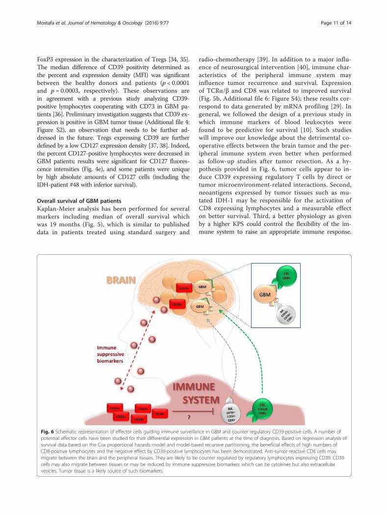
radio-chemotherapy [39]. In addition to a major influ-ence of neurosurgical intervention [40], immune char-acteristics of the peripheral immune system mayinfluence tumor recurrence and survival. Expressionof TCRα/β and CD8 was related to improved survival(Fig. 5b, Additional file 6: Figure S4); these results cor-respond to data generated by mRNA profiling [29]. Ingeneral, we followed the design of a previous study inwhich immune markers of blood leukocytes werefound to be predictive for survival [10]. Such studieswill improve our knowledge about the detrimental co-operative effects between the brain tumor and the per-ipheral immune system even better when performedas follow-up studies after tumor resection. As a hy-pothesis provided in Fig. 6, tumor cells appear to in-duce CD39 expressing regulatory T cells by direct ortumor microenvironment-related interactions. Second,neoantigens expressed by tumor tissues such as mu-tated IDH-1 may be responsible for the activation ofCD8 expressing lymphocytes and a measurable effecton better survival. Third, a better physiology as givenby a higher KPS could control the flexibility of the im-mune system to raise an appropriate immune response.
Fig. 6 Schematic representation of effector cells guiding immune surveillance in GBM and counter regulatory CD39-positive cells. A number ofpotential effector cells have been studied for their differential expression in GBM patients at the time of diagnosis. Based on regression analysis ofsurvival data based on the Cox proportional hazards model and model-based recursive partitioning, the beneficial effects of high numbers ofCD8-positive lymphocytes and the negative effect by CD39-positive lymphocytes has been demonstrated. Anti-tumor reactive CD8 cells maymigrate between the brain and the peripheral tissues. They are likely to be counter regulated by regulatory lymphocytes expressing CD39. CD39cells may also migrate between tissues or may be induced by immune suppressive biomarkers which can be cytokines but also extracellularvesicles. Tumor tissue is a likely source of such biomarkers.
Mostafa et al. Journal of Hematology & Oncology (2016) 9:77 Page 11 of 14
The immune alterations leading to tumor manifestationare possibly related to remodeling of tolerogenic lym-phocytes, a process which may be promoted in the eld-erly [15, 30, 41]. We found a better survival in patientswith high relative amounts of CD95 lymphocytes(Fig. 5d). Along these lines, Todo-Bom and colleagues[41] detected more CD95 expression in elderly donors.Activated NK cells may express CD95 and provide dir-ect immune control by recognizing malignant cells withlow or negative expression of MHC antigens [42].Moreover, glioblastoma cells express the CD95 ligand[43] and may be susceptible to CD95 ligand-initiatedcell killing and therapy [44]. Moreover, CD95-positivecytotoxic cells may trigger several pathways of caspaseactivation in glioblastoma cell lines [45]. Although themigration of activated lymphocytes between the periph-eral blood and the brain has not been proven, the over-all changes of immune phenotypes in GBM patientsstrongly suggest ongoing crosstalk between the tumorenvironment and the peripheral immune system (Fig. 6).Based on univariable regression analysis followed by mul-tivariable Cox proportional hazards model and recursivepartitioning, the absolute numbers of CD8-positive lym-phocytes appear to be relevant for improved survival(Additional file 1: Figure S2). Moreover, the relativeamounts of CD39-positive lymphocytes appear to be anegative predictor. The correlation of CD39 (relativeamounts) and CD8 (absolute cell numbers) is significant(p = 0.0033; r = −0.4332, (Additional file 7: Figure S5). Amodel implementing these variables for survival withfunctional aspects of immune cells has been provided inFig. 6. Accordingly, increased amounts of CD8 lympho-cytes may eventually invade the tumor tissue and controltumor cell proliferation. Effector cells are likely to becounter regulated by regulatory T cells expressing CD39.In addition to migrating immune cells between the brainand the peripheral immune system, extracellular vesiclesreleased from tumor cells appear to play a major role topotently activate tolerogenic T cells [46, 47], such vesiclesmay be termed “immune suppressive biomarkers” as well.Further studies and follow-up may substantiate thecurrent working hypothesis. After all, immunogenicity re-mains to be the most important factor to control malignan-cies. This hypothesis is currently supported by a likelysuccessful approach to immunize IDH-1 mutation-positivetumor patients with IDH-1-derived peptides [14] and im-proved survival of IDH-1-mutated patients [48].
ConclusionsImmune phenotypes in individual patients with GBM in-dicate sensitization as well as anergy against glioblast-oma tumor tissue. These observations may be exploitedto individualized immune-based therapeutic interventionsto eventually improve survival. Parameters can be also
used for clinical studies. Most important predictors aremutated genes such as IDH-1, a patient's KPS, the ATPnucleotidase CD39, and CD8 positive cytotoxic cells.
Additional files
Additional file 1: Figure S1. GBM patients before and after steroidadministration. (DOCX 690 kb)
Additional file 2: Table S1. Univariable Cox regression analysis.(DOCX 15 kb)
Additional file 3: Table S2. Multivariable proportional hazards model.(DOCX 25 kb)
Additional file 4: Figure S2. CD39 expression in GBM tumor lysate.(DOCX 206 kb)
Additional file 5: Figure S3. Absolute numbers of leukocytes.(DOCX 163 kb)
Additional file 6: Figure S4. Recursive Partitioning Analysis (RPA).(DOCX 94.7 kb)
Additional file 7: Figure S5 Spearman rank correlation of CD8 andCD39. (DOCX 183 kb)
AbbreviationsCD: cluster of differentiation; CIK: cytokine-induced killer(s);GBM: glioblastoma multiforme; IDH-1: isocitrate dehydrogenase-1;IL: interleukin(s); MFI: mean fluorescence intensity; MGMT: O6-Methylguanine-DNA methyltransferase; MHC: major histocompatibility complex; NK: naturalkiller; RPA: recursive partitioning analysis; TCR: T cell receptor complex;Treg: regulatory T cell(s).
Competing interestsThe authors declare that they have no competing interests.
Authors’ contributionsHM performed flow cytometry and data analysis and wrote the manuscript.AP selected the patients for study recruitment and performed surgery. JHperformed multivariable Cox regression analysis. RPA and MH performed thesurgery. ED contributed to data analysis. MAW and TB contributed to theconcept of the study. LN headed the clinical study. MG supervised theexperimental analysis. CRW performed the surgery and guided the clinicaldata analysis. EMS wrote the manuscript and supervised the analysis. Allauthors read and approved the final manuscript.
AcknowledgementsThe authors thank Julian M. Schneider for graphical support and Dr. HaraldHohmann and Sarah J. Gruber for flow cytometry and cytokinedeterminations. HM and ED are fellows of the International Graduate Schoolin Molecular Medicine Ulm (IGradU).
Author details1Sektion Experimentelle Anaesthesiologie, University Hospital Ulm, AlbertEinstein Allee 23, 89081 Ulm, Germany. 2Klinik für Anaesthesiologie,University Hospital Ulm, Albert Einstein Allee 23, 89081 Ulm, Germany.3Department of Neurosurgery, University Hospital Ulm Albert Einstein Allee23, 89081 Ulm and Bezirkskrankenhaus Günzburg, Ludwig-Heilmeyer-Str. 2,89312 Günzburg, Germany. 4Institute for Human Genetics, Albert EinsteinAllee 11, 89081 Ulm, Germany. 5Department of Pediatric Hematology andOncology, University Hospital Ulm, Prittwitzstr. 43, 89075 Ulm, Germany.
Received: 5 February 2016 Accepted: 13 April 2016
References1. Kamiya-Matsuoka C, Gilbert MR. Treating recurrent glioblastoma: an update.
CNS Oncol. 2015;4(2):91–104.2. Gagner JP, Law M, Fischer I, Newcomb EW, Zagzag D. Angiogenesis in gliomas:
imaging and experimental therapeutics. Brain Pathol. 2005;15(4):342–63.
Mostafa et al. Journal of Hematology & Oncology (2016) 9:77 Page 12 of 14

3. Wurth R, Barbieri F, Florio T. New molecules and old drugs as emergingapproaches to selectively target human glioblastoma cancer stem cells.Biomed Res Int. 2014;2014.
4. Reardon DA, Wen PY. Glioma in 2014: unravelling tumour heterogeneity-implications for therapy. Nat Rev Clin Oncol. 2015;12(2):69–70.
5. Lowther DE, Hafler DA. Regulatory T cells in the central nervous system.Immunol Rev. 2012;248(1):156–69.
6. Rolle CE, Sengupta S, Lesniak MS. Mechanisms of immune evasion bygliomas. Adv Exp Med Biol. 2012;746:53–76.
7. Urbanska K, Sokolowska J, Szmidt M, Sysa P. Glioblastoma multiforme—anoverview. Contemp Oncol (Pzn). 2014;18(5):307–12.
8. Baglietto L, Giles GG, English DR, Karahalios A, Hopper JL, Severi G. Alcoholconsumption and risk of glioblastoma; evidence from the MelbourneCollaborative Cohort Study. Int J Cancer. 2011;128(8):1929–34.
9. Tanaka S, Louis DN, Curry WT, Batchelor TT, Dietrich J. Diagnostic andtherapeutic avenues for glioblastoma: no longer a dead end? Nat Rev ClinOncol. 2013;10(1):14–26.
10. Kmiecik J, Poli A, Brons NH, Waha A, Eide GE, Enger PO, Zimmer J, ChekenyaM. Elevated CD3+ and CD8+ tumor-infiltrating immune cells correlate withprolonged survival in glioblastoma patients despite integratedimmunosuppressive mechanisms in the tumor microenvironment and atthe systemic level.J Neuroimmunol. 2013;264(1-2):71–83.
11. Skog J, Wurdinger T, van Rijn S, Meijer DH, Gainche L, Sena-Esteves M, CurryWT, Jr., Carter BS, Krichevsky AM, Breakefield XO. Glioblastoma microvesiclestransport RNA and proteins that promote tumour growth and providediagnostic biomarkers. Nat Cell Biol. 2008;10(12):1470–6.
12. Gautam P, Nair SC, Gupta MK, Sharma R, Polisetty RV, Uppin MS,Sundaram C, Puligopu AK, Ankathi P, Purohit AK, Chandak GR, HarshaHC, Sirdeshmukh R. Proteins with altered levels in plasma fromglioblastoma patients as revealed by iTRAQ-based quantitativeproteomic analysis. PLoS One. 2012;7(9):e46153.
13. Yang I, Kremen TJ, Giovannone AJ, Paik E, Odesa SK, Prins RM, Liau LM.Modulation of major histocompatibility complex class I molecules andmajor histocompatibility complex-bound immunogenic peptides inducedby interferon-alpha and interferon-gamma treatment of humanglioblastoma multiforme. J Neurosurg. 2004;100(2):310–9.
14. Bunse L, Schumacher T, Sahm F, Pusch S, Oezen I, Rauschenbach K, GonzalezM, Solecki G, Osswald M, Capper D, Wiestler B, Winkler F, Herold-Mende C, vonDeimling A, Wick W, Platten M. Proximity ligation assay evaluates IDH1R132Hpresentation in gliomas. J Clin Invest. 2015;125(2):593–606.
15. Ginaldi L, De Martinis M, D'Ostilio A, Marini L, Loreto F, Modesti M,Quaglino D. Changes in the expression of surface receptors onlymphocyte subsets in the elderly: quantitative flow cytometric analysis.Am J Hematol. 2001;67(2):63–72.
16. Jiang J, Wu C, Lu B. Cytokine-induced killer cells promote antitumorimmunity. J Transl Med. 2013;11:83.
17. Huehn J, Beyer M. Epigenetic and transcriptional control of Foxp3(+)regulatory T cells. Semin Immunol. 2015;27(1):10–18.
18. Authier A, Farrand KJ, Broadley KW, Ancelet LR, Hunn MK, Stone S, McConnellMJ, Hermans IF. Enhanced immunosuppression by therapy-exposedglioblastoma multiforme tumor cells. Int J Cancer. 2015;136(11):2566–78.
19. Mittal SK, Roche PA. Suppression of antigen presentation by IL-10. Curr OpinImmunol. 2015;34:22–27.
20. Huettner C, Czub S, Kerkau S, Roggendorf W, Tonn JC. Interleukin 10 isexpressed in human gliomas in vivo and increases glioma cell proliferationand motility in vitro. Anticancer Res. 1997;17(5A):3217–24.
21. Ni G, Wang T, Walton S, Zhu B, Chen S, Wu X, Wang Y, Wei MQ, Liu X.Manipulating IL-10 signalling blockade for better immunotherapy. CellImmunol. 2015;293(2):126–9.
22. Liu X, Madhankumar AB, Slagle-Webb B, Sheehan JM, Surguladze N, ConnorJR. Heavy chain ferritin siRNA delivered by cationic liposomes increasessensitivity of cancer cells to chemotherapeutic agents. Cancer Res. 2011;71(6):2240–9.
23. Lapa C, Linsenmann T, Luckerath K, Samnick S, Herrmann K, Stoffer C,Ernestus RI, Buck AK, Lohr M, Monoranu CM. Tumor-associatedmacrophages in glioblastoma multiforme-a suitable target forsomatostatin receptor-based imaging and therapy? PLoS One. 2015;10(3):e0122269.
24. De Maria A, Bozzano F, Cantoni C, Moretta L. Revisiting human natural killercell subset function revealed cytolytic CD56(dim)CD16+ NK cells as rapid
producers of abundant IFN-gamma on activation. Proc Natl Acad Sci U S A.2011;108(2):728–32.
25. Jung TY, Choi YD, Kim YH, Lee JJ, Kim HS, Kim JS, Kim SK, Jung S, Cho D.Immunological characterization of glioblastoma cells for immunotherapy.Anticancer Res. 2013;33(6):2525–33.
26. Franceschetti M, Pievani A, Borleri G, Vago L, Fleischhauer K, Golay J,Introna M. Cytokine-induced killer cells are terminally differentiatedactivated CD8 cytotoxic T-EMRA lymphocytes. Exp Hematol. 2009;37(5):616–28. e612.
27. Terry LA, DiSanto JP, Small TN, Flomenberg N. Differential expression andregulation of the human CD8 alpha and CD8 beta chains. Tissue Antigens.1990;35(2):82–91.
28. Wood GW, Holladay FP, Turner T, Wang YY, Chiga M. A pilot study ofautologous cancer cell vaccination and cellular immunotherapy using anti-CD3 stimulated lymphocytes in patients with recurrent grade III/IVastrocytoma. J Neurooncol. 2000;48(2):113–20.
29. Learn CA, Fecci PE, Schmittling RJ, Xie W, Karikari I, Mitchell DA, Archer GE,Wei Z, Dressman H, Sampson JH. Profiling of CD4+, CD8+, and CD4+CD25+CD45RO+FoxP3+ T cells in patients with malignant glioma revealsdifferential expression of the immunologic transcriptome compared with Tcells from healthy volunteers. Clin Cancer Res. 2006;12(24):7306–15.
30. Bisset LR, Lung TL, Kaelin M, Ludwig E, Dubs RW. Reference values forperipheral blood lymphocyte phenotypes applicable to the healthy adultpopulation in Switzerland. Eur J Haematol. 2004;72(3):203–12.
31. Fecci PE, Mitchell DA, Whitesides JF, Xie W, Friedman AH, Archer GE, HerndonJE, 2nd, Bigner DD, Dranoff G, Sampson JH. Increased regulatory T-cell fractionamidst a diminished CD4 compartment explains cellular immune defects inpatients with malignant glioma. Cancer Res. 2006;66(6):3294–302.
32. El Andaloussi A, Han Y, Lesniak MS. Prolongation of survival followingdepletion of CD4+CD25+ regulatory T cells in mice with experimental braintumors. J Neurosurg. 2006;105(3):430–7.
33. Feng L, Sun X, Csizmadia E, Han L, Bian S, Murakami T, Wang X,Robson SC, Wu Y. Vascular CD39/ENTPD1 directly promotes tumor cellgrowth by scavenging extracellular adenosine triphosphate. Neoplasia.2011;13(3):206–16.
34. Parodi A, Battaglia F, Kalli F, Ferrera F, Conteduca G, Tardito S, Stringara S,Ivaldi F, Negrini S, Borgonovo G, Simonato A, Traverso P, Carmignani G,Fenoglio D, Filaci G. CD39 is highly involved in mediating the suppressionactivity of tumor-infiltrating CD8+ T regulatory lymphocytes. CancerImmunol Immunother. 2013;62(5):851–62.
35. Mandapathil M, Lang S, Gorelik E, Whiteside TL. Isolation of functionalhuman regulatory T cells (Treg) from the peripheral blood based on theCD39 expression. J Immunol Methods. 2009;346(1-2):55–63.
36. Xu S, Shao QQ, Sun JT, Yang N, Xie Q, Wang DH, Huang QB, Huang B,Wang XY, Li XG, Qu X. Synergy between the ectoenzymes CD39 and CD73contributes to adenosinergic immunosuppression in human malignantgliomas. Neuro Oncol. 2013;15(9):1160–72.
37. Su H, Longhi MS, Wang P, Vergani D, Ma Y. Human CD4+CD25(high)CD127(low/neg) regulatory T cells. Methods Mol Biol. 2012;806:287–99.
38. Yu N, Li X, Song W, Li D, Yu D, Zeng X, Li M, Leng X, Li X. CD4(+)CD25(+)CD127 (low/-) T cells: a more specific Treg population in humanperipheral blood. Inflammation. 2012;35(6):1773–80.
39. Valeriani M, Ferretti A, Franzese P, Tombolini V. High-grade gliomas: resultsin patients treated with adjuvant radiotherapy alone and with adjuvantradio-chemotherapy. Anticancer Res. 2006;26(3B):2429–35.
40. De Bonis P, Anile C, Pompucci A, Fiorentino A, Balducci M, Chiesa S, LauriolaL, Maira G, Mangiola A. The influence of surgery on recurrence pattern ofglioblastoma. Clin Neurol Neurosurg. 2013;115(1):37–43.
41. Todo-Bom A, Mota-Pinto A, Alves V, Santos-Rosa M. Aging andasthma—changes in CD45RA, CD29 and CD95 T cells subsets. AllergolImmunopathol. 2012;40(1):14–9.
42. Wu A, Wiesner S, Xiao J, Ericson K, Chen W, Hall WA, Low WC, Ohlfest JR.Expression of MHC I and NK ligands on human CD133+ glioma cells:possible targets of immunotherapy. J Neurooncol. 2007;83(2):121–31.
43. Saggioro FP, Neder L, Stavale JN, Paixao-Becker AN, Malheiros SM, SoaresFA, Pittella JE, Matias CC, Colli BO, Carlotti CG, Jr., Franco M. Fas, FasL, andcleaved caspases 8 and 3 in glioblastomas: a tissue microarray-based study.Pathol Res Pract. 2014;210(5):267–73.
44. Eisele G, Wolpert F, Decrey G, Weller M. APO010, a synthetic hexamericCD95 ligand, induces death of human glioblastoma stem-like cells.Anticancer Res. 2013;33(9):3563–71.
Mostafa et al. Journal of Hematology & Oncology (2016) 9:77 Page 13 of 14
45. Cagigi A, Nilsson A, Levitsky V, Sabri F. Cytotoxic T-lymphocytes secretesoluble factors that induce caspase-mediated apoptosis in glioblastoma celllines. J Neuroimmunol. 2010;225(1-2):34–42.
46. Choi BD, Fecci PE, Sampson JH. Regulatory T cells move in when gliomassay "I Do". Clin Cancer Res. 2012;18(22):6086–8.
47. Chistiakov DA, Chekhonin VP. Extracellular vesicles shed by glioma cells:pathogenic role and clinical value. Tumour Biol. 2014;35(9):8425–38.
48. Chen JR, Yao Y, Xu HZ, Qin ZY. Isocitrate dehydrogenase (IDH)1/2 mutations asprognostic markers in patients with glioblastomas. Medicine. 2016;95(9):e2583.
• We accept pre-submission inquiries
• Our selector tool helps you to find the most relevant journal
• We provide round the clock customer support
• Convenient online submission
• Thorough peer review
• Inclusion in PubMed and all major indexing services
• Maximum visibility for your research
Submit your manuscript atwww.biomedcentral.com/submit
Submit your next manuscript to BioMed Central and we will help you at every step:
Mostafa et al. Journal of Hematology & Oncology (2016) 9:77 Page 14 of 14