Immortalized pathological human myoblasts: towards a ... · Immortalized pathological human...
13
Immortalized pathological human myoblasts: towards a universal tool for the study of neuromuscular disorders. Kamel Mamchaoui, Capucine Trollet, Anne Bigot, Elisa Negroni, Soraya Chaouch, Annie Wolff, Prashanth Kandalla, Solenne Marie, James Di Santo, Jean St Guily, et al. To cite this version: Kamel Mamchaoui, Capucine Trollet, Anne Bigot, Elisa Negroni, Soraya Chaouch, et al.. Im- mortalized pathological human myoblasts: towards a universal tool for the study of neuromuscu- lar disorders.. Skeletal Muscle, BioMed Central, 2011, 1 (1), pp.34. <10.1186/2044-5040-1-34>. <inserm-00651121> HAL Id: inserm-00651121 http://www.hal.inserm.fr/inserm-00651121 Submitted on 12 Dec 2011 HAL is a multi-disciplinary open access archive for the deposit and dissemination of sci- entific research documents, whether they are pub- lished or not. The documents may come from teaching and research institutions in France or abroad, or from public or private research centers. L’archive ouverte pluridisciplinaire HAL, est destin´ ee au d´ epˆ ot et ` a la diffusion de documents scientifiques de niveau recherche, publi´ es ou non, ´ emanant des ´ etablissements d’enseignement et de recherche fran¸cais ou ´ etrangers, des laboratoires publics ou priv´ es.
Transcript of Immortalized pathological human myoblasts: towards a ... · Immortalized pathological human...
Immortalized pathological human myoblasts: towards a
universal tool for the study of neuromuscular disorders.
Kamel Mamchaoui, Capucine Trollet, Anne Bigot, Elisa Negroni, Soraya
Chaouch, Annie Wolff, Prashanth Kandalla, Solenne Marie, James Di Santo,
Jean St Guily, et al.
To cite this version:
Kamel Mamchaoui, Capucine Trollet, Anne Bigot, Elisa Negroni, Soraya Chaouch, et al.. Im-mortalized pathological human myoblasts: towards a universal tool for the study of neuromuscu-lar disorders.. Skeletal Muscle, BioMed Central, 2011, 1 (1), pp.34. <10.1186/2044-5040-1-34>.<inserm-00651121>
HAL Id: inserm-00651121
http://www.hal.inserm.fr/inserm-00651121
Submitted on 12 Dec 2011
HAL is a multi-disciplinary open accessarchive for the deposit and dissemination of sci-entific research documents, whether they are pub-lished or not. The documents may come fromteaching and research institutions in France orabroad, or from public or private research centers.
L’archive ouverte pluridisciplinaire HAL, estdestinee au depot et a la diffusion de documentsscientifiques de niveau recherche, publies ou non,emanant des etablissements d’enseignement et derecherche francais ou etrangers, des laboratoirespublics ou prives.


Skeletal Muscle
Immortalized pathological human myoblasts:towards a universal tool for the study ofneuromuscular disordersMamchaoui et al.
Mamchaoui et al. Skeletal Muscle 2011, 1:34
http://www.skeletalmusclejournal.com/content/1/1/34 (1 November 2011)

RESEARCH Open Access
Immortalized pathological human myoblasts:towards a universal tool for the study ofneuromuscular disordersKamel Mamchaoui1,2,3†, Capucine Trollet1,2,3†, Anne Bigot1,2,3, Elisa Negroni1,2,3, Soraya Chaouch1,2,3, Annie Wolff1,2,3
, Prashanth K Kandalla1,2,3, Solenne Marie1,2,3, James Di Santo4, Jean Lacau St Guily1,2,3,5, Francesco Muntoni6,
Jihee Kim6, Susanne Philippi1,7, Simone Spuler7, Nicolas Levy8, Sergiu C Blumen9, Thomas Voit1,2,3,
Woodring E Wright10, Ahmed Aamiri11, Gillian Butler-Browne1,2,3 and Vincent Mouly1,2,3*
Abstract
Background: Investigations into both the pathophysiology and therapeutic targets in muscle dystrophies have
been hampered by the limited proliferative capacity of human myoblasts. Isolation of reliable and stable
immortalized cell lines from patient biopsies is a powerful tool for investigating pathological mechanisms,
including those associated with muscle aging, and for developing innovative gene-based, cell-based or
pharmacological biotherapies.
Methods: Using transduction with both telomerase-expressing and cyclin-dependent kinase 4-expressing vectors,
we were able to generate a battery of immortalized human muscle stem-cell lines from patients with various
neuromuscular disorders.
Results: The immortalized human cell lines from patients with Duchenne muscular dystrophy, facioscapulohumeral
muscular dystrophy, oculopharyngeal muscular dystrophy, congenital muscular dystrophy, and limb-girdle muscular
dystrophy type 2B had greatly increased proliferative capacity, and maintained their potential to differentiate both
in vitro and in vivo after transplantation into regenerating muscle of immunodeficient mice.
Conclusions: Dystrophic cellular models are required as a supplement to animal models to assess cellular
mechanisms, such as signaling defects, or to perform high-throughput screening for therapeutic molecules. These
investigations have been conducted for many years on cells derived from animals, and would greatly benefit from
having human cell models with prolonged proliferative capacity. Furthermore, the possibility to assess in vivo the
regenerative capacity of these cells extends their potential use. The innovative cellular tools derived from several
different neuromuscular diseases as described in this report will allow investigation of the pathophysiology of
these disorders and assessment of new therapeutic strategies.
BackgroundMuscular dystrophies constitute a heterogeneous group
of genetic muscle diseases characterized by progressive
muscle weakness, wasting and degeneration, some of
these features are common to muscle aging [1,2]. Over
the past few years, the genetics and pathophysiology of
some of these diseases has been deciphered, stimulating
the development of novel gene-based (or mRNA-based)
(for example, gene therapy, exon-skipping or codon
read-through), cell-based and pharmacological therapies
[3], which can either target the mutation directly, or tar-
get the consequences of that mutation, such as muscle
wasting, atrophy or denervation. To assess these rapidly
developing therapeutic advances, there is a crucial need
to develop standardized tools to determine the cellular
and molecular mechanisms that trigger the physiopatho-
logic modifications, and to assess these new therapeutic
* Correspondence: [email protected]
† Contributed equally1Thérapie des maladies du muscle strié, Institut de Myologie, UM76, UPMC
Université Paris 6, Paris, France
Full list of author information is available at the end of the article
Mamchaoui et al. Skeletal Muscle 2011, 1:34
http://www.skeletalmusclejournal.com/content/1/1/34Skeletal Muscle
© 2011 Mamchaoui et al; licensee BioMed Central Ltd. This is an Open Access article distributed under the terms of the CreativeCommons Attribution License (http://creativecommons.org/licenses/by/2.0), which permits unrestricted use, distribution, andreproduction in any medium, provided the original work is properly cited.

strategies in preclinical trials. Transgenic mice have
often been used to investigate the physiopathology of
muscular dystrophies [4-6]; however, the mutation
remains in a murine context, and there are often major
differences between humans and mice; for example, a
mutation in the dystrophin gene results in a mild patho-
logical phenotype in mdx mice but in a progressive and
fatal disease (Duchenne muscular dystrophy; DMD) in
humans. Furthermore, not every mutation can be cre-
ated and evaluated in murine models, and mechanisms
common to aging and dystrophies may differ between
mice and humans. Consequently, human primary myo-
blasts isolated from dystrophic patient biopsies provide
the most pertinent experimental models to assess a vari-
ety of human genetic mutations in their natural genomic
environment. Although in vitro models do not fully
recapitulate the in vivo environment, cell-culture sys-
tems allow rapid, high-throughput screening of mole-
cules or oligonucleotides, and new strategies can be
easily tested prior to validation in animal models, which
is a costly and time-consuming process. The main draw-
backs of using in vitro primary cultures of human cells
derived from muscle biopsies are their purity, their lim-
ited proliferative capacity, and the variation in pheno-
type when amplified in vitro; their phenotype will always
be confounded by modifications due to cellular senes-
cence, which will progressively occur during cell amplifi-
cation [7,8].
The two major mechanisms responsible for this
replicative cellular senescence seen in human myo-
blasts are (i) activation of the p16-mediated cellular
stress pathway, and (ii) the progressive erosion of telo-
meres at each cell division until they reach a critical
length that will trigger p53 activation and cell-cycle
exit [9,10]. Introduction of the telomerase catalytic
subunit (human telomerase reverse transcriptase;
hTERT) cDNA alone will result in an extension of the
lifespan and even immortalization in a variety of cell
types, including endothelial cells and fibroblasts
[11,12]. However, we have shown that the expression
of both hTERT and cyclin-dependent kinase (CDK)-4 is
required to successfully overcome cellular senescence
in human myoblasts [13]; while hTERT elongates the
telomere, CDK-4 blocks the p16INK4a-dependent stress
pathway.
In the present study, our goal was to create a large
collection of immortalized human myoblasts isolated
from a wide range of neuromuscular disorders (DMD,
facioscapulohumeral muscular dystrophy (FSHD), oculo-
pharyngeal muscular dystrophy (OPMD), limb-girdle
muscular dystrophy (LGMD2B or dysferlinopathy) and
congenital muscular dystrophy (CMD)), which could be
used as experimental tools to study these diseases and
to develop new therapeutic strategies.
DMD is the most common childhood muscular dys-
trophy. It is caused by mutations in the dystrophin gene
encoding an essential protein of the muscle membrane
cytoskeleton [14], leading to rapid and progressive skele-
tal-muscle weakness. FSHD is a progressive muscle dis-
ease caused by contractions in a 3.3 kb repeat region
(D4Z4) located at 4q35.2 [15], which first affects the
muscles of the face and upper limb girdle with asymme-
try, and later the lower limb girdle. OPMD is a rare,
autosomal dominant, late-onset degenerative muscle dis-
order caused by a short (GCG)n triplet expansion in the
poly(A) binding protein nuclear 1 (PABPN1) gene [16],
which affects the eyelid and pharyngeal muscles.
LGMD2B is a recessive muscle disease caused by muta-
tions in the dysferlin gene, a muscle membrane protein
known to be involved in membrane repair [17] and traf-
ficking. The disease is characterized by early and slowly
progressive weakness and atrophy of the pelvic and
shoulder girdle muscles in early adulthood. Finally,
CMD refers to a clinically and genetically heterogeneous
group of dystrophies, which result in the onset of mus-
cle weakness at birth or in childhood, and involve muta-
tions in several proteins such as collagen, laminin,
integrin, and nesprin 1 [18].
In this study, we report for the first time that for each
of these muscular dystrophies, we were able to produce
reliable and stable immortalized cell lines from human
myoblasts isolated from biopsies, resulting in robust in
vitro models that can also be implanted in vivo. This
non-exhaustive list of cellular models will provide
powerful and valuable tools for the scientific community
investigating these pathological conditions and/or their
mechanisms. as they overcome the problem of limited
proliferation usually present in myoblasts. These models
should also be useful in the development of gene or cell
therapies and pharmacological strategies for muscular
dystrophies, some of which might also be used to com-
bat muscle weakness in the elderly.
MethodsEthics approval
Muscle biopsies (Table 1) were obtained from the BTR
(Bank of Tissues for Research, a partner in the EU net-
work EuroBioBank) or from neurologists, in accordance
with European recommendations and French legislation.
Surgical procedures were performed in accordance with
the legal regulations in France and European Union
ethics guidelines for animal research.
Human myoblast cultures
Human myoblasts were isolated from biopsies and culti-
vated as described previously [19] in a growth medium
consisting of 199 medium and DMEM (Invitrogen
Carlsbad, CA) in a 1:4 ratio, supplemented with 20%
Mamchaoui et al. Skeletal Muscle 2011, 1:34
http://www.skeletalmusclejournal.com/content/1/1/34
Page 2 of 10
FCS (Invitrogen), 2.5 ng/ml hepatocyte growth factor
(Invitrogen), 0.1 μmol/l dexamethasone (Sigma-Aldrich,
St. Louis, MO, USA) and 50 μg/ml gentamycin (Invitro-
gen). The myogenic purity of the populations was moni-
tored by immunocytochemistry using desmin as marker.
Enrichment of myogenic cells was performed using an
immunomagnetic cell sorting system (MACS; Miltenyi
Biotec, Paris, France) according to the manufacturer’s
instructions. Briefly, cells were labeled with anti-CD56
(a specific marker of myoblasts) microbeads, and then
separated in a MACS column placed in a magnetic field.
Purification was checked by immunochemistry using a
desmin marker. Differentiation was induced at conflu-
ence by replacing the growth medium with DMEM sup-
plemented with 100 μg/ml transferrin, 10 μg/ml insulin
and 50 μg/ml of gentamycin (Sigma-Aldrich).
Cell transduction
hTERT and Cdk4 cDNA were cloned into different
pBABE retroviral vectors containing puromycin and
neomycin selection markers, respectively. Infection was
carried out as described previously [20]. Transduced cell
cultures were selected with puromycin (0.2 μg/ml) and/
or neomycin (0.3 mg/ml) for 8 days. The infected cells
were purified as described previously if necessary, and
were then seeded at clonal density. Selected individual
myogenic clones were isolated from each population,
using glass cylinders, and their proliferation and differ-
entiation capacities were characterized.
Telomere length analysis
Genomic DNA was extracted from each proliferating
cell line using a salting-out procedure. Telomere length
was determined by using a quantitative (q)PCR method,
as previously described [21,22]. PCR amplification was
achieved using telomere (T) and single-copy gene 36B4
(acidic ribosomal phosphoprotein P0) (S) primers. The
mean telomere length was calculated as the ratio of telo-
mere repeats to 36B4 copies, represented as the T:S
ratio. Each sample was run in triplicate, using 20 ng of
DNA per replicate, and three independent runs were
analyzed. The primer sequences and detailed PCR pro-
tocols used are available on request.
Reverse transcriptase PCR
To analyze the expression of myogenic markers in pro-
liferating primary and immortalized cell lines, 1 μg RNA
from each cell line was used for the cDNA synthesis
(Superscript III; Invitrogen) using random hexamer pri-
mers. cDNA (1 μl) was used as a template for PCR
using N-CAM, MyoD and desmin specific primers. The
primer sequences and detailed PCR protocols used are
available on request.
Induction of host muscle regeneration and implantation
of human cells
Immunodeficient Rag2-/- gC-/- C5-/- mice aged 2 to 3
months were anesthetized with an intraperitoneal injec-
tion of ketamine hydrochloride (80 mg/kg) and xylasin
(10 mg/kg) (Sigma-Aldrich). To induce severe muscle
damage and trigger regeneration, the recipient tibialis
anterior (TA) muscles were exposed to cryodamage, and
a single injection of immortalized human cells (15 μl of
cell suspension containing 2.5 × 10⁵ or 5 × 10⁵ cells in
PBS) was administered as described previously [23].
Four weeks after transplantation, the recipient TA mus-
cles were dissected, mounted in gum tragacanth, and
frozen in liquid nitrogen-cooled isopentane for later
analysis.
Immunofluorescence
In vitro and in vivo characterizations were performed by
immunolabeling as described previously [23-25]. Antibo-
dies used were directed against myosin isoforms (MF20,
mouse IgG2b, 1:20 dilution; Developmental Studies
Hybridoma Bank, DSHB, Iowa City, IA), lamin A/C
(clone JOL2, mouse IgG1, 1:300; AbCam, Cambridge,
Cambridgeshire, UK), lamin A/C (NCL-LAM A/C, clone
636, mouse IgG2b, 1:400, Novocastra, Newcastle-upon-
Tyne, Tyne and Wear, UK), spectrin (NCL-Spec1, clone
RBC2/3D5, mouse IgG2b, 1:50; Novocastra), and lami-
nin (rabbit polyclonal, Z 0097, 1:400; Dako, Trappes,
France). The secondary antibodies used were Alexa
Fluor 488-conjugated goat anti-mouse IgG2b (Molecular
Probes, Montluçon, France), Alexa Fluor 647-conjugated
goat anti-rabbit (Molecular Probes), and Cy3-conjugated
goat anti-mouse IgG1 (Jackson Immunoresearch, West
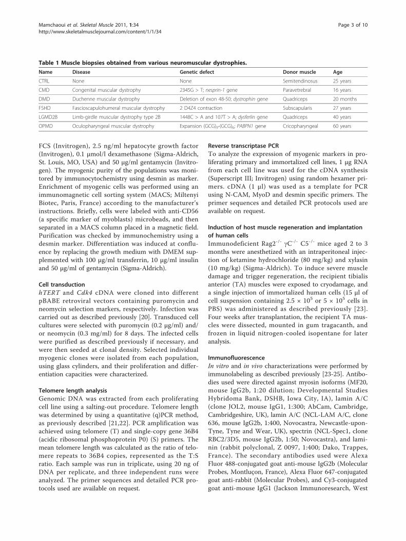
Table 1 Muscle biopsies obtained from various neuromuscular dystrophies.
Name Disease Genetic defect Donor muscle Age
CTRL None None Semitendinosus 25 years
CMD Congenital muscular dystrophy 2345G > T; nesprin-1 gene Paravetrebral 16 years
DMD Duchenne muscular dystrophy Deletion of exon 48-50; dystrophin gene Quadriceps 20 months
FSHD Fascioscapulohumeral muscular dystrophy 2 D4Z4 contraction Subscapularis 27 years
LGMD2B Limb-girdle muscular dystrophy type 2B 1448C > A and 107T > A; dysferlin gene Quadriceps 40 years
OPMD Oculopharyngeal muscular dystrophy Expansion (GCG)9-(GCG)6; PABPN1 gene Cricopharyngeal 60 years
Mamchaoui et al. Skeletal Muscle 2011, 1:34
http://www.skeletalmusclejournal.com/content/1/1/34
Page 3 of 10

Grove, PA, USA). Images were visualized using a micro-
scope (Olympus Corp., Tokyo, Japan), and digitized
using a charge-coupled device (CCD) camera (Olympus
Corp., Tokyo, Japan).
Antisense oligonucleotides transfection and reverse
transcriptase PCR
Cells were seeded in six-well plates and grown in
growth medium. Transfection of antisense oligonucleo-
tides (AONs) was performed using 1 μl of transfection
reagent (Lipofectamin 2000; Invitrogen) per μg of AONs
for 4 hours. The chemistry used for AONs was 2’-O-
methyl-phosphorothioates. All transfections were per-
formed with at least two independent duplicates. Cells
were changed to differentiation medium before transfec-
tion. Typically 24 to 48 hours after transfection, RNA
was extracted from the cells using Trizol or Qiagen col-
umn kit (Qiagen Inc., Valencia, CA, USA). 1 μg RNA
was used for the cDNA synthesis (Superscript III; Invi-
trogen) with DMD exon-specific primers. cDNA (2 μl)
was used as a template for a first PCR reaction. From
this first reaction of 25 cycles, 1 μl of the product was
removed and used as a template for a second nested
PCR of 35 cycles. PCR products were analyzed on 1.5 to
2% agarose gels. The primer sequences and detailed
PCR protocols used are available on request.
ResultsImmortalized myoblast lines generated from dystrophic
muscles
Primary cultures from distinct muscular dystrophies
(DMD, FSHD, OPMD, CMD and LGMD2B, Table 1)
were co-transduced with two retroviral vectors expres-
sing hTERT and CDK-4 cDNA. Co-transduced cells
were selected by neomycin and puromycin and then
purified using magnetic beads coupled to antibodies
directed against the myogenic marker CD56. Following
culture at clonal density, individual myogenic clones
with extended proliferative lifespans, as compared to the
untranduced cells, were isolated from each population.
In contrast to the parental populations, which stopped
proliferating at various stages of the culture, depending
on the type of dystrophy, the selected immortalized
clones were still able to proliferate after prolonged
amplification in vitro under the same culture conditions
(Figure 1). All immortalized clones were cultivated until
they had achieved at least twice as many divisions as the
parental population.
Telomere length was measured in each clone (Table
2) and ranged from 10.3 kb to 24.8 kb with no differ-
ence between the clones and control immortalized myo-
blasts (17.6 kb). The length of the telomeres in all of the
immortalized myogenic clones was always well above
the 6 to 7 kb limit usually seen in control cells that are
reaching senescence, but remained within the range of
that seen both in myoblasts and in other stem cells (12
to 20 kb).
In vitro characterization of immortalized cells
To confirm that the immortalized cell lines maintained
their myogenic signature, we compared the expression
of several markers in proliferating primary and immor-
talized cell lines from control, OPMD and DMD biop-
sies. In all of them, we confirmed the expression of the
myogenic markers desmin, neural cell adhesion mole-
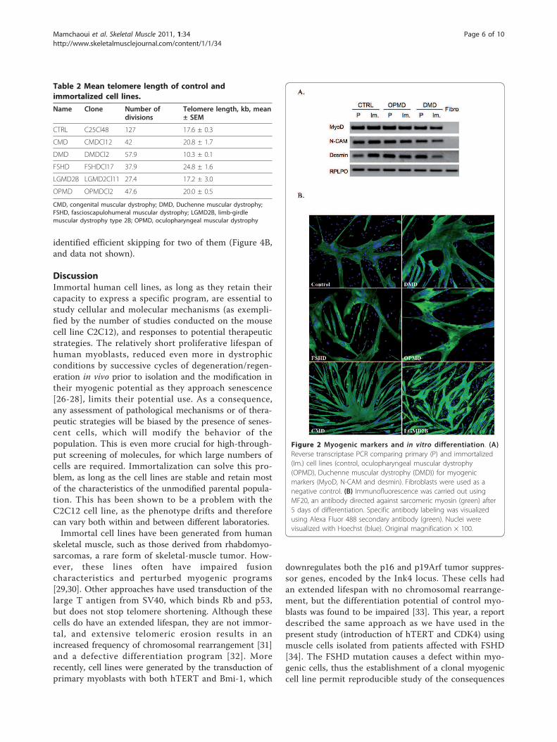
cule (N-CAM) and MyoD (Figure 2A).
In addition, we also tested their ability to differentiate
into myotubes, using immunostaining with MF20 anti-
body, which recognizes all skeletal-muscle myosin heavy
chains (MyHCs). After 5 days in differentiation condi-
tions, all of the immortalized cell lines were able to fuse
into myotubes expressing MyHCs (Figure 2B).
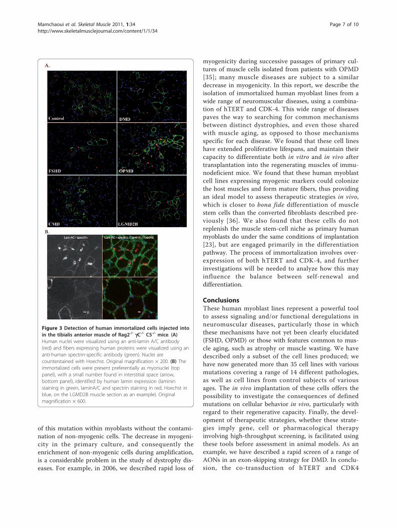
Induction of host muscle regeneration and implantation
of human cells
To investigate the in vivo behavior of these immorta-
lized cells, cells were grafted into damaged TA muscles
of Rag2-/- gC-/- C5-/- mice; injected muscles were ana-
lyzed 4 weeks after transplantation, which corresponds
to a complete fiber regeneration process, using antibo-
dies specific for human lamin A/C (expressed in all
human nuclei) and human spectrin (expressed in differ-
entiated fibers). For each injected clone (control, DMD,
FSHD, OPMD, CMD or LGMD2B), mature muscle
fibers containing human spectrin protein and human
lamin A/C+ nuclei were seen (Figure 3A). No tumors
were ever observed in these immunodeficient mice.
Using antibodies specific for the basal lamina protein
laminin, lamin A/C (human nuclei) and spectrin (speci-
fic to the human protein) to identify fiber sarcolemma,
we investigated if these cell lines could replenish the
muscle stem-cell niche (allowing self-renewal), at the
periphery of the muscle fiber and beneath the basal
lamina. Whereas the vast majority of the lamin A/C-
positive nuclei (97%) were found as myonuclei (upper
panel, Figure 3B), we observed the unexpected finding
that all the human cells outside the muscle fibers were
present in the interstitial space, separated from the
fibers by a basal lamina (lower panel, Figure 3B), and
not in the satellite-cell niche, suggesting that the
immortalized cells were engaged preferentially in the
differentiation pathway and not in the self-renewal
process.
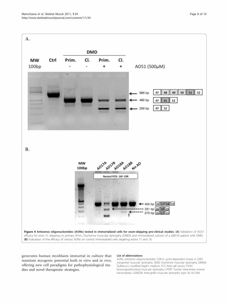
Immortalized cell lines as a useful tool for therapeutic
preclinical studies
To show that these cell lines could be powerful tools to
develop therapeutic strategies, we used them to evaluate
Mamchaoui et al. Skeletal Muscle 2011, 1:34
http://www.skeletalmusclejournal.com/content/1/1/34
Page 4 of 10

the efficiency of AONs in an exon-skipping strategy for
DMD. The first one tested was AO51, which resulted in
efficient skipping of exon 51 using both the primary and
immortalized DMD cell lines (∆48 to 50; Figure 4A)
and this AON is currently being used in a phase I/II
clinical trial. Using immortalized control cells, we were
able to screen a range of AONs targeting exons 17, 18,
21, 22, 43, 44 and 45 of the dystrophin gene. RT-PCR
Figure 1 Proliferative potential. Lifespan plots (mean population doublings; MPD) of the parental populations and the immortalized cell lines
derived from them.
Mamchaoui et al. Skeletal Muscle 2011, 1:34
http://www.skeletalmusclejournal.com/content/1/1/34
Page 5 of 10
identified efficient skipping for two of them (Figure 4B,
and data not shown).
DiscussionImmortal human cell lines, as long as they retain their
capacity to express a specific program, are essential to
study cellular and molecular mechanisms (as exempli-
fied by the number of studies conducted on the mouse
cell line C2C12), and responses to potential therapeutic
strategies. The relatively short proliferative lifespan of
human myoblasts, reduced even more in dystrophic
conditions by successive cycles of degeneration/regen-
eration in vivo prior to isolation and the modification in
their myogenic potential as they approach senescence
[26-28], limits their potential use. As a consequence,
any assessment of pathological mechanisms or of thera-
peutic strategies will be biased by the presence of senes-
cent cells, which will modify the behavior of the
population. This is even more crucial for high-through-
put screening of molecules, for which large numbers of
cells are required. Immortalization can solve this pro-
blem, as long as the cell lines are stable and retain most
of the characteristics of the unmodified parental popula-
tion. This has been shown to be a problem with the
C2C12 cell line, as the phenotype drifts and therefore
can vary both within and between different laboratories.
Immortal cell lines have been generated from human
skeletal muscle, such as those derived from rhabdomyo-
sarcomas, a rare form of skeletal-muscle tumor. How-
ever, these lines often have impaired fusion
characteristics and perturbed myogenic programs
[29,30]. Other approaches have used transduction of the
large T antigen from SV40, which binds Rb and p53,
but does not stop telomere shortening. Although these
cells do have an extended lifespan, they are not immor-
tal, and extensive telomeric erosion results in an
increased frequency of chromosomal rearrangement [31]
and a defective differentiation program [32]. More
recently, cell lines were generated by the transduction of
primary myoblasts with both hTERT and Bmi-1, which
downregulates both the p16 and p19Arf tumor suppres-
sor genes, encoded by the Ink4 locus. These cells had
an extended lifespan with no chromosomal rearrange-
ment, but the differentiation potential of control myo-
blasts was found to be impaired [33]. This year, a report
described the same approach as we have used in the
present study (introduction of hTERT and CDK4) using
muscle cells isolated from patients affected with FSHD
[34]. The FSHD mutation causes a defect within myo-
genic cells, thus the establishment of a clonal myogenic
cell line permit reproducible study of the consequences
Table 2 Mean telomere length of control and
immortalized cell lines.
Name Clone Number ofdivisions
Telomere length, kb, mean± SEM
CTRL C25Cl48 127 17.6 ± 0.3
CMD CMDCl12 42 20.8 ± 1.7
DMD DMDCl2 57.9 10.3 ± 0.1
FSHD FSHDCl17 37.9 24.8 ± 1.6
LGMD2B LGMD2Cl11 27.4 17.2 ± 3.0
OPMD OPMDCl2 47.6 20.0 ± 0.5
CMD, congenital muscular dystrophy; DMD, Duchenne muscular dystrophy;
FSHD, fascioscapulohumeral muscular dystrophy; LGMD2B, limb-girdle
muscular dystrophy type 2B; OPMD, oculopharyngeal muscular dystrophy
Figure 2 Myogenic markers and in vitro differentiation. (A)
Reverse transcriptase PCR comparing primary (P) and immortalized
(Im.) cell lines (control, oculopharyngeal muscular dystrophy
(OPMD), Duchenne muscular dystrophy (DMD)) for myogenic
markers (MyoD, N-CAM and desmin). Fibroblasts were used as a
negative control. (B) Immunofluorescence was carried out using
MF20, an antibody directed against sarcomeric myosin (green) after
5 days of differentiation. Specific antibody labeling was visualized
using Alexa Fluor 488 secondary antibody (green). Nuclei were
visualized with Hoechst (blue). Original magnification × 100.
Mamchaoui et al. Skeletal Muscle 2011, 1:34
http://www.skeletalmusclejournal.com/content/1/1/34
Page 6 of 10
of this mutation within myoblasts without the contami-
nation of non-myogenic cells. The decrease in myogeni-
city in the primary culture, and consequently the
enrichment of non-myogenic cells during amplification,
is a considerable problem in the study of dystrophy dis-
eases. For example, in 2006, we described rapid loss of
myogenicity during successive passages of primary cul-
tures of muscle cells isolated from patients with OPMD
[35]; many muscle diseases are subject to a similar
decrease in myogenicity. In this report, we describe the
isolation of immortalized human myoblast lines from a
wide range of neuromuscular diseases, using a combina-
tion of hTERT and CDK-4. This wide range of diseases
paves the way to searching for common mechanisms
between distinct dystrophies, and even those shared
with muscle aging, as opposed to those mechanisms
specific for each disease. We found that these cell lines
have extended proliferative lifespans, and maintain their
capacity to differentiate both in vitro and in vivo after
transplantation into the regenerating muscles of immu-
nodeficient mice. We found that these human myoblast
cell lines expressing myogenic markers could colonize
the host muscles and form mature fibers, thus providing
an ideal model to assess therapeutic strategies in vivo,
which is closer to bona fide differentiation of muscle
stem cells than the converted fibroblasts described pre-
viously [36]. We also found that these cells do not
replenish the muscle stem-cell niche as primary human
myoblasts do under the same conditions of implantation
[23], but are engaged primarily in the differentiation
pathway. The process of immortalization involves over-
expression of both hTERT and CDK-4, and further
investigations will be needed to analyze how this may
influence the balance between self-renewal and
differentiation.
ConclusionsThese human myoblast lines represent a powerful tool
to assess signaling and/or functional deregulations in
neuromuscular diseases, particularly those in which
these mechanisms have not yet been clearly elucidated
(FSHD, OPMD) or those with features common to mus-
cle aging, such as atrophy or muscle wasting. We have
described only a subset of the cell lines produced; we
have now generated more than 35 cell lines with various
mutations covering a range of 14 different pathologies,
as well as cell lines from control subjects of various
ages. The in vivo implantation of these cells offers the
possibility to investigate the consequences of defined
mutations on cellular behavior in vivo, particularly with
regard to their regenerative capacity. Finally, the devel-
opment of therapeutic strategies, whether these strate-
gies imply gene, cell or pharmacological therapy
involving high-throughput screening, is facilitated using
these tools before assessment in animal models. As an
example, we have described a rapid screen of a range of
AONs in an exon-skipping strategy for DMD. In conclu-
sion, the co-transduction of hTERT and CDK4
Figure 3 Detection of human immortalized cells injected into
in the tibialis anterior muscle of Rag2-/- gC-/- C5-/- mice. (A)
Human nuclei were visualized using an anti-lamin A/C antibody
(red) and fibers expressing human proteins were visualized using an
anti-human spectrin-specific antibody (green). Nuclei are
counterstained with Hoechst. Original magnification × 200. (B) The
immortalized cells were present preferentially as myonuclei (top
panel), with a small number found in interstitial space (arrow,
bottom panel), identified by human lamin expression (laminin
staining in green, laminA/C and spectrin staining in red, Hoechst in
blue, on the LGMD2B muscle section as an example). Original
magnification × 600.
Mamchaoui et al. Skeletal Muscle 2011, 1:34
http://www.skeletalmusclejournal.com/content/1/1/34
Page 7 of 10
generates human myoblasts immortal in culture that
maintain myogenic potential both in vitro and in vivo,
offering new cell paradigms for pathophysiological stu-
dies and novel therapeutic strategies.
List of abbreviations
AONs: antisense oligonucleotides; CDK-4: cyclin-dependent kinase 4; CMD:
congenital muscular dystrophy; DMD: Duchenne muscular dystrophy; DMEM:
Dulbecco’s modified Eagle’s medium; FCS: fetal calf serum; FSHD:
facioscapulohumeral muscular dystrophy; hTERT: human telomerase reverse
transcriptase; LGMD2B: limb-girdle muscular dystrophy type 2b; N-CAM:
Figure 4 Antisense oligonucleotides (AONs) tested in immortalized cells for exon-skipping pre-clinical studies. (A) Validation of AO51
efficacy for exon 51 skipping on primary (Prim.; Duchenne muscular dystrophy (DMD)) and immortalized cultures of a ∆48-50 patient with DMD.
(B) Evaluation of the efficacy of various AONs on control immortalized cells targeting exons 17 and 18.
Mamchaoui et al. Skeletal Muscle 2011, 1:34
http://www.skeletalmusclejournal.com/content/1/1/34
Page 8 of 10

neural cell adhesion molecule; OPMD: oculopharyngeal muscular dystrophy;
PBS: phosphate-buffered serum; PCR: polymerase chain reaction.
Acknowledgements
We thank all the patients who provided the biopsies to establish the
primary cultures. We also thank the MSG study group, particularly D. Furling
for fruitful discussions and L. Dollé, S. Sandal, M. Oloko, S. Vasseur and M.
Chapart for technical assistance. We thank Genosafe for help with the
transductions and Steve Wilton for providing the antisense oligonucleotides.
This work was supported by the MYORES Network of Excellence (contract
511978) and TREAT-NMD (contract LSHM-CT-2006-036825) from the
European Commission 6th FP, MYOAGE (contract HEALTH-F2-2009-223576)
from the Seventh FP, the ANR Genopath-INAFIB, the ANR MICRORNAS,
MyoGrad (GK1631, German Research Foundation), the Duchenne Parent
Project Netherlands, CNRS, INSERM, University Pierre and Marie Curie, AFM
(Association Française contre les Myopathies) (including network grant
#15123), the Jain Foundation, Parents Project of Monaco, and the European
Parent Project.
Author details1Thérapie des maladies du muscle strié, Institut de Myologie, UM76, UPMC
Université Paris 6, Paris, France. 2INSERM U974, Paris, France. 3CNRS UMR
7215, Paris, France. 4Innate Immunity Unit, INSERM U 668, Institut Pasteur,
Paris, France. 5Service d’Oto-Rhino-Laryngologie et de Chirurgie Cervico-
Faciale, Faculté de Médecine St Antoine, Université Pierre et Marie Curie,
Hôpital Tenon, Paris, France. 6The Dubowitz Neuromuscular Centre, Institute
of Child Health, University College, London, UK. 7Muscle Research Unit,
Experimental and Clinical Research Center, Charité University Hospital and
Max Delbrück Center for Molecular Medicine, Berlin, Germany. 8Faculté de
Médecine de Marseille, Université de la Méditerranée, Inserm UMRS 910
Génétique Médicale et Génomique Fonctionnelle, Marseille, France.9Department of Neurology, Hillel Yaffe Medical Center, PO Box 169, Hadera,
38100, Israel. 10UT Southwestern Medical Center, Department of Cell Biology,
Dallas, TX 75390, USA. 11Laboratoire LBCM, Departement de Biologie, Faculté
des Sciences, Agadir, Maroc.
Authors’ contributions
KM, CT, AB, EN, and SC designed and performed the in vitro and in vivo
experiments, analyzed data, and wrote the manuscript. AW, PKK, SM, JK, and
AA provided technical support. JDS provided the immunodeficient Rag2-/-
γC-/- C5-/- mice for the in vivo experiments. JLSG, FM, SP, SS, NL, SB, and TV
provided biopsies, and WEW provided DNA constructs. TV and AA discussed
the results and gave expert advice. GBB and VM provided conceptual input
and supervision. and wrote the manuscript. All authors read and approved
the final manuscript.
Competing interests
The authors declare that they have no competing interests.
Received: 20 June 2011 Accepted: 1 November 2011
Published: 1 November 2011
References
1. McNally EM, Pytel P: Muscle diseases: the muscular dystrophies. Annu Rev
Pathol 2007, 2:87-109.
2. Ruegg MA, Glass DJ: Molecular mechanisms and treatment options for
muscle wasting diseases. Annu Rev Pharmacol Toxicol 2011, 51:373-395.
3. Trollet C, Athanasopoulos T, Popplewell L, Malerba A, Dickson G: Gene
therapy for muscular dystrophy: current progress and future prospects.
Expert Opin Biol Ther 2009, 9:849-866.
4. Trollet C, Anvar SY, Venema A, Hargreaves IP, Foster K, Vignaud A, Ferry A,
Negroni E, Hourde C, Baraibar MA, et al: Molecular and phenotypic
characterization of a mouse model of oculopharyngeal muscular
dystrophy reveals severe muscular atrophy restricted to fast glycolytic
fibres. Hum Mol Genet 2010.
5. Vignaud A, Ferry A, Huguet A, Baraibar M, Trollet C, Hyzewicz J, Butler-
Browne G, Puymirat J, Gourdon G, Furling D: Progressive skeletal muscle
weakness in transgenic mice expressing CTG expansions is associated
with the activation of the ubiquitin-proteasome pathway. Neuromuscul
Disord 2010, 20:319-325.
6. Allamand V, Campbell KP: Animal models for muscular dystrophy:
valuable tools for the development of therapies. Hum Mol Genet 2000,
9:2459-2467.
7. Mouly V, Aamiri A, Perie S, Mamchaoui K, Barani A, Bigot A, Bouazza B,
Francois V, Furling D, Jacquemin V, et al: Myoblast transfer therapy: is
there any light at the end of the tunnel? Acta Myol 2005, 24:128-133.
8. Webster C, Blau HM: Accelerated age-related decline in replicative life-
span of Duchenne muscular dystrophy myoblasts: implications for cell
and gene therapy. Somat Cell Mol Genet 1990, 16:557-565.
9. Renault V, Thornell LE, Eriksson PO, Butler-Browne G, Mouly V: Regenerative
potential of human skeletal muscle during aging. Aging Cell 2002,
1:132-139.
10. Wright WE, Shay JW: Historical claims and current interpretations of
replicative aging. Nat Biotechnol 2002, 20:682-688.
11. Bodnar AG, Ouellette M, Frolkis M, Holt SE, Chiu CP, Morin GB, Harley CB,
Shay JW, Lichtsteiner S, Wright WE: Extension of life-span by introduction
of telomerase into normal human cells. Science 1998, 279:349-352.
12. Vaziri H, Benchimol S: Reconstitution of telomerase activity in normal
human cells leads to elongation of telomeres and extended replicative
life span. Curr Biol 1998, 8:279-282.
13. Zhu CH, Mouly V, Cooper RN, Mamchaoui K, Bigot A, Shay JW, Di Santo JP,
Butler-Browne GS, Wright WE: Cellular senescence in human myoblasts is
overcome by human telomerase reverse transcriptase and cyclin-
dependent kinase 4: consequences in aging muscle and therapeutic
strategies for muscular dystrophies. Aging Cell 2007, 6:515-523.
14. Nowak KJ, Davies KE: Duchenne muscular dystrophy and dystrophin:
pathogenesis and opportunities for treatment. EMBO Rep 2004, 5:872-876.
15. Tawil R: Facioscapulohumeral muscular dystrophy. Neurotherapeutics 2008,
5:601-606.
16. Brais B, Bouchard JP, Xie YG, Rochefort DL, Chretien N, Tome FM,
Lafreniere RG, Rommens JM, Uyama E, Nohira O, et al: Short GCG
expansions in the PABP2 gene cause oculopharyngeal muscular
dystrophy. Nat Genet 1998, 18:164-167.
17. Bansal D, Miyake K, Vogel SS, Groh S, Chen CC, Williamson R, McNeil PL,
Campbell KP: Defective membrane repair in dysferlin-deficient muscular
dystrophy. Nature 2003, 423:168-172.
18. Reed UC: Congenital muscular dystrophy. Part I: a review of
phenotypical and diagnostic aspects. Arq Neuropsiquiatr 2009, 67:144-168.
19. Bigot A, Klein AF, Gasnier E, Jacquemin V, Ravassard P, Butler-Browne G,
Mouly V, Furling D: Large CTG repeats trigger p16-dependent premature
senescence in myotonic dystrophy type 1 muscle precursor cells. Am J
Pathol 2009, 174:1435-1442.
20. Di Donna S, Mamchaoui K, Cooper RN, Seigneurin-Venin S, Tremblay J,
Butler-Browne GS, Mouly V: Telomerase can extend the proliferative
capacity of human myoblasts, but does not lead to their
immortalization. Mol Cancer Res 2003, 1:643-653.
21. Cawthon RM: Telomere measurement by quantitative PCR. Nucleic Acid
Research 2002, 30:e47.
22. Cawthon RM: Telomere length measurement by a novel monochrome
multiplex quantitative PCR method. Nucleic Acid Research 2009, 37:e21.
23. Negroni E, Riederer I, Chaouch S, Belicchi M, Razini P, Di Santo J, Torrente Y,
Butler-Browne GS, Mouly V: In vivo myogenic potential of human CD133+
muscle-derived stem cells: a quantitative study. Mol Ther 2009,
17:1771-1778.
24. Cooper RN, Irintchev A, Di Santo JP, Zweyer M, Morgan JE, Partridge TA,
Butler-Browne GS, Mouly V, Wernig A: A new immunodeficient mouse
model for human myoblast transplantation. Hum Gene Ther 2001,
12:823-831.
25. Cooper RN, Thiesson D, Furling D, Di Santo JP, Butler-Browne GS, Mouly V:
Extended amplification in vitro and replicative senescence: key factors
implicated in the success of human myoblast transplantation. Hum Gene
Ther 2003, 14:1169-1179.
26. Decary S, Mouly V, Hamida CB, Sautet A, Barbet JP, Butler-Browne GS:
Replicative potential and telomere length in human skeletal muscle:
implications for satellite cell-mediated gene therapy. Hum Gene Ther
1997, 8:1429-1438.
27. Renault V, Piron-Hamelin G, Forestier C, DiDonna S, Decary S, Hentati F,
Saillant G, Butler-Browne GS, Mouly V: Skeletal muscle regeneration and
the mitotic clock. Exp Gerontol 2000, 35:711-719.
Mamchaoui et al. Skeletal Muscle 2011, 1:34
http://www.skeletalmusclejournal.com/content/1/1/34
Page 9 of 10

28. Bigot A, Jacquemin V, Debacq-Chainiaux F, Butler-Browne GS, Toussaint O,
Furling D, Mouly V: Replicative aging down-regulates the myogenic
regulatory factors in human myoblasts. Biol Cell 2008, 100:189-199.
29. Rossi S, Poliani PL, Cominelli M, Bozzato A, Vescovi R, Monti E, Fanzani A:
Caveolin 1 is a marker of poor differentiation in Rhabdomyosarcoma.
Eur J Cancer 2010.
30. Wang S, Guo L, Dong L, Li S, Zhang J, Sun M: TGF-beta1 signal pathway
may contribute to rhabdomyosarcoma development by inhibiting
differentiation. Cancer Sci 2010, 101:1108-1116.
31. Stewart N, Bacchetti S: Expression of SV40 large T antigen, but not small
t antigen, is required for the induction of chromosomal aberrations in
transformed human cells. Virology 1991, 180:49-57.
32. Mouly V, Edom F, Decary S, Vicart P, Barbert JP, Butler-Browne GS: SV40
large T antigen interferes with adult myosin heavy chain expression, but
not with differentiation of human satellite cells. Exp Cell Res 1996,
225:268-276.
33. Cudre-Mauroux C, Occhiodoro T, Konig S, Salmon P, Bernheim L, Trono D:
Lentivector-mediated transfer of Bmi-1 and telomerase in muscle
satellite cells yields a duchenne myoblast cell line with long-term
genotypic and phenotypic stability. Hum Gene Ther 2003, 14:1525-1533.
34. Stadler G, Chen JC, Wagner K, Robin JD, Shay JW, Emerson CP Jr,
Wright WE: Establishment of clonal myogenic cell lines from severely
affected dystrophic muscles - CDK4 maintains the myogenic population.
Skelet Muscle 2011, 1:12.
35. Perie S, Mamchaoui K, Mouly V, Blot S, Bouazza B, Thornell LE, St Guily JL,
Butler-Browne G: Premature proliferative arrest of cricopharyngeal
myoblasts in oculo-pharyngeal muscular dystrophy: therapeutic
perspectives of autologous myoblast transplantation. Neuromuscul Disord
2006, 16:770-781.
36. Chaouch S, Mouly V, Goyenvalle A, Vulin A, Mamchaoui K, Negroni E, Di
Santo J, Butler-Browne G, Torrente Y, Garcia L, Furling D: Immortalized skin
fibroblasts expressing conditional MyoD as a renewable and reliable
source of converted human muscle cells to assess therapeutic strategies
for muscular dystrophies: validation of an exon-skipping approach to
restore dystrophin in Duchenne muscular dystrophy cells. Hum Gene
Ther 2009, 20:784-790.
doi:10.1186/2044-5040-1-34Cite this article as: Mamchaoui et al.: Immortalized pathological humanmyoblasts: towards a universal tool for the study of neuromusculardisorders. Skeletal Muscle 2011 1:34.
Submit your next manuscript to BioMed Centraland take full advantage of:
• Convenient online submission
• Thorough peer review
• No space constraints or color figure charges
• Immediate publication on acceptance
• Inclusion in PubMed, CAS, Scopus and Google Scholar
• Research which is freely available for redistribution
Submit your manuscript at www.biomedcentral.com/submit
Mamchaoui et al. Skeletal Muscle 2011, 1:34
http://www.skeletalmusclejournal.com/content/1/1/34
Page 10 of 10