Imbalance of heterologous protein folding and disulfide bond
14
RESEARCH ARTICLE Open Access Imbalance of heterologous protein folding and disulfide bond formation rates yields runaway oxidative stress Keith EJ Tyo 1,2 , Zihe Liu 1 , Dina Petranovic 1 and Jens Nielsen 1* Abstract Background: The protein secretory pathway must process a wide assortment of native proteins for eukaryotic cells to function. As well, recombinant protein secretion is used extensively to produce many biologics and industrial enzymes. Therefore, secretory pathway dysfunction can be highly detrimental to the cell and can drastically inhibit product titers in biochemical production. Because the secretory pathway is a highly-integrated, multi-organelle system, dysfunction can happen at many levels and dissecting the root cause can be challenging. In this study, we apply a systems biology approach to analyze secretory pathway dysfunctions resulting from heterologous production of a small protein (insulin precursor) or a larger protein (a-amylase). Results: HAC1-dependent and independent dysfunctions and cellular responses were apparent across multiple datasets. In particular, processes involving (a) degradation of protein/recycling amino acids, (b) overall transcription/ translation repression, and (c) oxidative stress were broadly associated with secretory stress. Conclusions: Apparent runaway oxidative stress due to radical production observed here and elsewhere can be explained by a futile cycle of disulfide formation and breaking that consumes reduced glutathione and produces reactive oxygen species. The futile cycle is dominating when protein folding rates are low relative to disulfide bond formation rates. While not strictly conclusive with the present data, this insight does provide a molecular interpretation to an, until now, largely empirical understanding of optimizing heterologous protein secretion. This molecular insight has direct implications on engineering a broad range of recombinant proteins for secretion and provides potential hypotheses for the root causes of several secretory-associated diseases. Keywords: Protein secretion, unfolded protein response, HAC1, protein production, oxidative stress Background The protein secretory pathway is an extensive process in eukaryal cells, as it is responsible for processing approxi- mately one-third of all proteins. Substantial cellular resources are therefore utilized to maintain this pathway’s functions, and stressed conditions in the secretory path- way have consequences for the whole cell [1]. Distress in secretory pathway organelles has been implicated as the molecular basis for several diseases, for example, b cell apoptosis in diabetes, cystic fibrosis, and prion-related disease, among others [2]. In biotechnology, efficient secretion of useful recombinant proteins in yeast and fungi is a key industrial objective with applications in enzyme production required for the production of bio- fuels, detergents, fabrics, food, and biologics, such as imu- noglobulins, hormones, and vaccines. Significant effort has gone into engineering yeast for increasing protein secre- tion [3]. Strategies, such as changing environmental para- meters (for example, temperature, media composition) [4] or altering genetics, can increase secretion for some pro- teins, but they rarely represent generic solutions for improving protein secretion [5,6]. The lack of a single engineering strategy that improves protein secretion across the board implies that there are several possible bottle- necks in the secretory pathway, and different proteins may be constrained in different ways. There is therefore a * Correspondence: [email protected] 1 Department of Chemical and Biological Engineering, Chalmers University of Technology, Kemivägen 10, SE-41296 Göteborg, Sweden Full list of author information is available at the end of the article Tyo et al. BMC Biology 2012, 10:16 http://www.biomedcentral.com/1741-7007/10/16 © 2012 Tyo et al; licensee BioMed Central Ltd. This is an Open Access article distributed under the terms of the Creative Commons Attribution License (http://creativecommons.org/licenses/by/2.0), which permits unrestricted use, distribution, and reproduction in any medium, provided the original work is properly cited.
Transcript of Imbalance of heterologous protein folding and disulfide bond
RESEARCH ARTICLE Open Access
Imbalance of heterologous protein folding anddisulfide bond formation rates yields runawayoxidative stressKeith EJ Tyo1,2, Zihe Liu1, Dina Petranovic1 and Jens Nielsen1*
Abstract
Background: The protein secretory pathway must process a wide assortment of native proteins for eukaryotic cellsto function. As well, recombinant protein secretion is used extensively to produce many biologics and industrialenzymes. Therefore, secretory pathway dysfunction can be highly detrimental to the cell and can drastically inhibitproduct titers in biochemical production. Because the secretory pathway is a highly-integrated, multi-organellesystem, dysfunction can happen at many levels and dissecting the root cause can be challenging. In this study, weapply a systems biology approach to analyze secretory pathway dysfunctions resulting from heterologousproduction of a small protein (insulin precursor) or a larger protein (a-amylase).
Results: HAC1-dependent and independent dysfunctions and cellular responses were apparent across multipledatasets. In particular, processes involving (a) degradation of protein/recycling amino acids, (b) overall transcription/translation repression, and (c) oxidative stress were broadly associated with secretory stress.
Conclusions: Apparent runaway oxidative stress due to radical production observed here and elsewhere can beexplained by a futile cycle of disulfide formation and breaking that consumes reduced glutathione and producesreactive oxygen species. The futile cycle is dominating when protein folding rates are low relative to disulfide bondformation rates. While not strictly conclusive with the present data, this insight does provide a molecularinterpretation to an, until now, largely empirical understanding of optimizing heterologous protein secretion. Thismolecular insight has direct implications on engineering a broad range of recombinant proteins for secretion andprovides potential hypotheses for the root causes of several secretory-associated diseases.
Keywords: Protein secretion, unfolded protein response, HAC1, protein production, oxidative stress
BackgroundThe protein secretory pathway is an extensive process ineukaryal cells, as it is responsible for processing approxi-mately one-third of all proteins. Substantial cellularresources are therefore utilized to maintain this pathway’sfunctions, and stressed conditions in the secretory path-way have consequences for the whole cell [1]. Distress insecretory pathway organelles has been implicated as themolecular basis for several diseases, for example, b cellapoptosis in diabetes, cystic fibrosis, and prion-relateddisease, among others [2]. In biotechnology, efficient
secretion of useful recombinant proteins in yeast andfungi is a key industrial objective with applications inenzyme production required for the production of bio-fuels, detergents, fabrics, food, and biologics, such as imu-noglobulins, hormones, and vaccines. Significant effort hasgone into engineering yeast for increasing protein secre-tion [3]. Strategies, such as changing environmental para-meters (for example, temperature, media composition) [4]or altering genetics, can increase secretion for some pro-teins, but they rarely represent generic solutions forimproving protein secretion [5,6]. The lack of a singleengineering strategy that improves protein secretion acrossthe board implies that there are several possible bottle-necks in the secretory pathway, and different proteins maybe constrained in different ways. There is therefore a
* Correspondence: [email protected] of Chemical and Biological Engineering, Chalmers University ofTechnology, Kemivägen 10, SE-41296 Göteborg, SwedenFull list of author information is available at the end of the article
Tyo et al. BMC Biology 2012, 10:16http://www.biomedcentral.com/1741-7007/10/16
© 2012 Tyo et al; licensee BioMed Central Ltd. This is an Open Access article distributed under the terms of the Creative CommonsAttribution License (http://creativecommons.org/licenses/by/2.0), which permits unrestricted use, distribution, and reproduction inany medium, provided the original work is properly cited.

requirement for more fundamental insight into this com-plex pathway that involves a very large number ofcomponents.In yeast, the secretory pathway is a multi-organelle sys-
tem that is responsible for trafficking proteins to theextracellular space, cell membrane, or vacuole [7]. Duringthis transit, multiple processes must be coordinated,including folding, specific proteolytic cleavage, glycosyla-tion, and disulfide bond formation, all with a layer ofquality control at key check points. The pathway requiressubstantial cellular resources to perform these tasks, suchas glycans, electron acceptors, electron donors, and ATP.In the ER, the nascent peptide is folded into its nativestructure while disulfide bonds are formed. The rate ofprotein folding is dependent upon the complexity of theprotein to be folded, the availability of chaperones toassist folding, and ATP used by the chaperones [1]. Pro-teins that are slow to fold or terminally misfolded pro-teins are removed from the ER via the ER-associateddegradation (ERAD) pathway [8]. Disulfide bond forma-tion requires the removal of electrons from cysteinethiols via protein disulfide isomerase (PDI) and Ero1p tothe final electron acceptor, typically oxygen [9,10]. Thisprocess produces reactive oxygen species (ROS) in stoi-chiometric amounts to the number of disulfide bondsformed [11]. Disulfide bond formation is random, andincorrect bond pairs must be exchanged for native bondsvia PDI-based processes [12]. In addition, reduced glu-tathione (GSH) acts as a buffer for the redox state of theER [13]. A more detailed description of oxidative proteinfolding can be found in the reviews by Sevier et al. andChakravarthi et al. [14,15].The secretory pathway must adjust the chaperone
capacity, oxidizing equivalents, ATP, glycan, and othermetabolic requirements, as well as trafficking patterns,based on the portfolio of proteins that need to beexpressed at a given time, and the resources required toprocess that set of proteins. In yeast, the unfolded proteinresponse (UPR) is one transcriptional mechanism thatadjusts secretory resources and controls to handle over-load of the folding machinery in the ER [16]. In the UPR,accumulation of unfolded proteins in the ER signals apathway that results in translation of Hac1p, a transcrip-tion factor (TF) known to activate or repress over 100genes, including many ER-associated proteins such asKar2p, Pdi1p, and Ero1p [17].In this study, we identified biological mechanisms
which alter the secretory pathway in response to secre-tion of recombinant proteins with different properties(size, number of disulfide bonds, and glycans) in aHac1p-dependent and independent manner. The secre-tory pathway was perturbed by secreting a small protein,human insulin precursor (IP), or a comparatively largerprotein, a-amylase, in wild-type (WT) and Δhac1
Saccharomyces cerevisiae. These proteins were chosenbecause the two proteins elicit different behavior in thesecretory pathway. These differences will arise becausea-amylase is a relatively larger (and likely more difficultto fold), has an odd number of cysteines (which maycomplicate disulfide isomerization) and has glycosylation,compared to insulin which is small, has even number ofcysteines, and is not glycosylated. As well, a-amylase hasone more disulfide bond than IP. To identify biologicalmechanisms, we characterized changes in physiologicalproperties (specific growth rate, carbon utilization effi-ciency, and recombinant protein secretion), TF activity(as inferred from transcriptome analysis) and metabolicdemand (as inferred by changes in metabolic flux diver-sion). Through this, we identified the following biologicalprocesses: amino acids recycling from degraded proteins,trans-Golgi network (TGN) sorting changes, overallexpression repression, and oxidative stress. Motivated bysecretory-related oxidative stress observations, we pre-sent a model for disulfide bond formation and electrontransfer in the ER which takes into account thermody-namic irreversibilities caused by differences in electronaffinity. The proposed model explains the non-stoichio-metric ROS formation that we observed that results fromdisulfide bond formation and causes oxidative stressunder folding-stress conditions. If proven by genetic andbiochemical results, the futile cycle model yields insightinto a fundamental problem in secretory stress andreveals new avenues to reduce oxidative stress andincrease productivity in industrial protein production.
ResultsProtein size and Hac1p activity affect protein secretionquantity and cell growthYeast strains were constructed that produce and secrete(a) IP or (b) a-amylase and were compared to yeaststrains containing (c) an empty vector in both wild-typeand HAC1 deletion backgrounds. IP and a-amylase werechosen because they are very different types of proteinsto secrete. IP is 51 amino acids in length, with sixcysteines forming three disulfide bonds, and no glycosyla-tion. a-amylase is 478 amino acid in length, with ninecysteines forming only four disulfide bonds and one gly-cosylation. The odd number of cysteines in a-amylasecomplicates disulfide pairing, as the random isomeriza-tion process may incorporate the cysteine that shouldnot be incorporated into a disulfide bond. Both proteinswere targeted for secretion using a YAP3 pre sequence(21 amino acids, cleaved off in the ER) and a rationallydesigned pro sequence (TA57, 42 amino acids, no glyco-sylation or disulfides) were cloned behind a TDH3 pro-moter in a high copy 2 micron plasmid [18]. a-amylasewas expressed using the same plasmid, promoter, andleader sequences. These strains are named WN (WT
Tyo et al. BMC Biology 2012, 10:16http://www.biomedcentral.com/1741-7007/10/16
Page 2 of 14

with empty vector), WI (WT secreting IP), WA (WTsecreting a-amylase), dN (Δhac1 with empty vector), dI(Δhac1 secreting IP), and dA (Δhac1 secreting a-amy-lase). Strains were characterized in batch fermentation tounderstand the effects on cell physiology.The cellular burden induced by (a) synthesizing and
secreting IP and a-amylase and (b) deleting the key TFfor the UPR, Hac1p, substantially affected the cells. Pro-tein titers in WT strain were 9 mg/L and 20 mg/L, for IPand a-amylase, respectively (Figure 1a). On a per biomassbasis, this is approximately half the insulin produced, andone-third the a-amylase reported for rich media [19,20].Rich media appears to be favorable for heterologous pro-tein production, but may present complications in down-stream separations. Comparing the small and largerproteins, a-amylase was secreted in higher levels on amass basis, but six-fold more insulin molecules weresecreted (1.52 μM IP in WI compared to 0.26 μM a-amy-lase in WA). Δhac1 strains secreted significantly less pro-tein than WT, confirming that Hac1p is important forefficient secretion (Figure 1a) [5].Reduced specific growth rates imply impairment of cel-
lular processes (Figure 1b). In WT yeast, IP productiondid not affect growth; however, a-amylase productionreduced growth by 25%. This, combined with the differ-ences in protein titers, implies that a-amylase is morechallenging to fold and secrete than IP. In the Δhac1background, recombinant protein strains dI and dA hadapproximately 20% lower growth rates compared to dN.This growth reduction occurs despite no change in speci-fic glucose uptake rate (Additional file 1, Tables S1 andS2) pointing toward higher energy requirements to main-tain homeostasis in Δhac1 while trying to secrete recom-binant proteins. Δhac1 strains had overall lower final cell
densities. Δhac1 strains produced more glycerol thanWT strains implying impaired oxidative processes in theΔhac1 strains (Additional file 2).
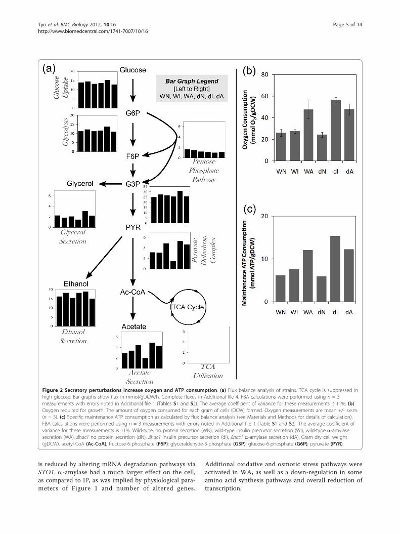
Secretory stress shifts metabolism to increase oxygen andATP requirementsThe physiological changes due to the secretory pertur-bations affect the distribution of resources through themetabolic network. The glucose uptake and range ofproducts produced were altered by the protein produc-tion conditions (Table 1). Changes in the underlyingmetabolic network were estimated by flux balance analy-sis (FBA) using a yeast central carbon metabolismmodel, constrained by measured extracellular fluxes(Additional file 1, Tables S1 and S2, Additional files 3and 4) [21]. Figure 2a shows a metabolic map of centralcarbon metabolism for each of the six conditions basedon the exchange fluxes in Table 1 and the FBA analysis.The shift in metabolic fluxes were correlated withchanges in redox requirements. As expected, the cata-bolic functions of the TCA cycle was predicted to havevery low activity due to glucose repression [22]. Figure2b shows that the oxygen uptake was twice as high inthe strains that were growth inhibited (for example,WA, dI, dA) than those that were not. This increasedoxygen uptake was not used for oxidative phosphoryla-tion, as the biomass yields on glucose were lower inWA, dI, and dA, and it may therefore be a result ofincreased oxidation in connection with formation of dis-ulfide bonds.Figure 2c shows that the maintenance ATP consump-tion is increased in WA, dI, and dA according to FBAcalculations. In WT background, WI did not consume adetectable increase in ATP, likely because IP is short
Figure 1 Secretory perturbations affect yeast physiology. (a) Final recombinant protein titer. Δhac1 strains were severely inhibited inrecombinant secretion. (b) Specific growth rate on glucose. The combination of Δhac1 and recombinant secretion had the most severe effecton growth, however even in wild-type background, a-amylase hindered growth. (c) Final cell concentration. Wild-type, no protein secretion(WN), wild-type insulin precursor secretion (WI), wild-type a-amylase secretion (WA), Δhac1 no protein secretion (dN), Δhac1 insulin precursorsecretion (dI), Δhac1 a-amylase secretion (dA). Measurements are mean +/- s.e.m. (n = 3).
Tyo et al. BMC Biology 2012, 10:16http://www.biomedcentral.com/1741-7007/10/16
Page 3 of 14
and easily folded, thereby minimally taxing the transla-tion and folding machinery. WA did increase two-foldin ATP consumption, most likely because a-amylase is10-fold larger and likely more difficult to fold and hasmore disulfide bond pairing possibilities. In the Δhac1background, folding efficiency is likely decreased due toER dysfunction. With native secretion, dN did notrequire higher ATP maintenance consumption com-pared to WT. However, even the smaller, easier to foldIP resulted in ER stress that required significant ATPconsumption compared to WT. dA, which was alreadystressed under WT, continued to show high ATP con-sumption. Despite the increased ATP consumption in dIand dA, little protein was secreted.
Transcription factors controlling oxidative stress, aminoacid salvaging, and expression repression are linked tosecretory responseGrowth phase transcriptomics measurements were carriedout to identify cellular processes that were activated underthe stresses of HAC1 deletion and recombinant proteinproduction. HAC1 deletion resulted in 339 significantlychanged genes in the no recombinant protein case (WNvs. dN). HAC1 deletions in the insulin strain and a-amy-lase strain resulted in much larger cellular responses of1628 (WI vs. dI) and 1511 (WA vs. dA) significantlyexpressed genes, respectively. KAR2 (ER chaperone)expression was significantly reduced upon HAC1 deletion(↓ three-fold dN vs WTN, P = 1 × 10⁻⁴) and the four yeastprotein disulfide isomerases (PDI1, EUG1, MPD1, MPD2)reduced an average of 2.9-fold (P < 0.05).The effects of producing IP or a-amylase within a
strain background (WT or HAC1) were not as pro-nounced as the effect of HAC1 deletion, 40 and 194genes were significantly changed in WI (compared toWN) and WA (compared to WN). Likwsise, 74 and 90genes were significantly changed for dI (compared to dN)and dA (compared to dN).To reduce the dimensionality of the data and identify
putative TFs involved in protein secretion, the ReporterTranscription Factor algorithm was used [23]. TFs werescored by the modulation in expression level of genes
that the TFs bind in the upstream region according toChIP-chip data [24]. Therefore, the score is not indica-tive of change in the TF expression level itself, but ofthe genes under its influence. Reporter TF algorithm isuseful, because although the statistical significance of anindividual gene may not meet an arbitrary threshold, ifseveral genes linked to the same TF have similar beha-vior, the likelihood of observing the group of genes islow, making TF identification very sensitive. Figure 3shows significant secretory process TFs shown to beinvolved in up- and down-regulating different cellularprocess under their control. Interestingly, different TFswere identified for the two different proteins. This islikely the combined effect of different protein size andnumber of disulfide bonds. A complete list of significanttranscription factors is provided in Additional files 5and 6.In WT (Figure 3a), several TFs were activated by pro-
tein secretion. Oxidative and osmotic stress pathway up-regulation was common to both proteins. Oxidativestress is likely caused by ROS that is formed when Ero1pshuttles electrons to oxygen in disulfide bond formation[25]. Osmotic stress response, particular hypo-osmoticstress, strengthens the cell wall to counteract internalturgor pressure by changing the cell wall composition.This change in composition requires remodeling thesecretory pathway by changing which components aretrafficked to the cell wall [26]. Surprisingly, the ReporterTF algorithm found several Hac1p-influenced genesdown-regulated. Genes that Hac1p binds from the ChIP-chip data that are significantly down-regulated are KEG1,MCD4, and ERJ5. KEG1 and MCD4 genes are involved inglycan modifications and ERJ5 is a secondary ER chaper-one [27-29]. These genes may be influenced by other TFsnot included in the ChIP-chip network. Genes known tobe regulated by Hac1p (KAR2 and ERO1) were not signif-icantly changed upon secreting recombinant protein,indicating that there is not an actual Hac1p response inthe WT.Clear differences between large and small protein
secretion emerge in WT. IP stimulated modification ofthe TGN through MCM1 and STE12. Overall expression
Table 1 Physiological parameters of recombinant protein secretion strainsa
Strainsb Μmax [h-1] YSX YSE YSG YSA YSCO2 Carbon balance
WN 0.43 +/- 0.014 0.14 +/- 0.001 0.32 +/- 0.041 0.067 +/- 0.009 0.048 +/- 0.0005 0.30 +/- 0.019 0.89
WI 0.40 +/- 0.012 0.13 +/- 0.002 0.35 +/- 0.029 0.055 +/- 0.005 0.056 +/- 0.0047 0.30 +/- 0.014 0.92
WA 0.32 +/- 0.007 0.11 +/- 0.003 0.31 +/- 0.006 0.060 +/- 0.007 0.049 +/- 0.0023 0.30 +/- 0.002 0.84
dN 0.38 +/- 0.005 0.13 +/- 0.004 0.37 +/- 0.025 0.046 +/- 0.003 0.035 +/- 0.0046 0.29 +/- 0.020 0.91
dI 0.29 +/- 0.005 0.08 +/- 0.006 0.32 +/- 0.017 0.081 +/- 0.001 0.046 +/- 0.0011 0.31 +/- 0.007 0.84
dA 0.31 +/- 0.002 0.11 +/- 0.003 0.32 +/- 0.002 0.066 +/- 0.001 0.049 +/- 0.0009 0.30 +/- 0.004 0.85aAll yields (Y) are [g/g]. Glucose (S), biomass (X), ethanol (E), glycerol (G), acetate (A), carbon dioxide (CO2).bStrain abbreviations as in Figure 1.
Tyo et al. BMC Biology 2012, 10:16http://www.biomedcentral.com/1741-7007/10/16
Page 4 of 14
is reduced by altering mRNA degradation pathways viaSTO1. a-amylase had a much larger effect on the cell,as compared to IP, as was implied by physiological para-meters of Figure 1 and number of altered genes.
Additional oxidative and osmotic stress pathways wereactivated in WA, as well as a down-regulation in someamino acid synthesis pathways and overall reduction oftranscription.
Figure 2 Secretory perturbations increase oxygen and ATP consumption. (a) Flux balance analysis of strains. TCA cycle is suppressed inhigh glucose. Bar graphs show flux in mmol/gDCW/h. Complete fluxes in Additional file 4. FBA calculations were performed using n = 3measurements with errors noted in Additional file 1 (Tables S1 and S2). The average coefficient of variance for these measurements is 11%. (b)Oxygen required for growth. The amount of oxygen consumed for each gram of cells (DCW) formed. Oxygen measurements are mean +/- s.e.m.(n = 3). (c) Specific maintenance ATP consumption as calculated by flux balance analysis (see Materials and Methods for details of calculation).FBA calculations were performed using n = 3 measurements with errors noted in Additional file 1 (Table S1 and S2). The average coefficient ofvariance for these measurements is 11%. Wild-type, no protein secretion (WN), wild-type insulin precursor secretion (WI), wild-type a-amylasesecretion (WA), Δhac1 no protein secretion (dN), Δhac1 insulin precursor secretion (dI), Δhac1 a-amylase secretion (dA). Gram dry cell weight(gDCW). acetyl-CoA (Ac-CoA); fructose-6-phosphate (F6P); glyceraldehyde-3-phosphate (G3P); glucose-6-phosphate (G6P); pyruvate (PYR).
Tyo et al. BMC Biology 2012, 10:16http://www.biomedcentral.com/1741-7007/10/16
Page 5 of 14

In the Δhac1 background (Figure 3b), many of theeffects found in WA, have become common to both IPand a-amylase producing strains. HAC1 deletion clearlymakes the cell more susceptible to recombinant secre-tion overload. Both insulin and a-amylase secretioncause considerable oxidative stress response and down-regulation of amino acid synthesis, including the generalamino acid synthesis TF, Gcn4p. In dI, translationalcapacity repression is also employed (via Fhlp/Rap1p)and adjustments in amino acid metabolism. dA shows amix of up- and down-regulation of genes that are con-trolled by Hac1p. Other TFs appear to be controllingthese genes in the absence of HAC1. Some oxidativeand osmotic stress pathways appear independent ofHAC1. Skn7p and Cin5p were similarly activated inboth WT and Δhac1. Oxidative and hypo-osmotic stress,while important for managing the secretory pathway,appears not to be directly managed through the UPR.
Thermodynamic irreversibilities in redox reactions canexplain increased oxidative stress in slow protein foldingconditionsThe increases in oxidative stress, oxygen consumption,and reduced growth observed in the study can beexplained by electron transfer in ER redox pathways.Disulfide bond formation has been established to con-sume oxygen and produce ROS (and thereby consumecellular resources to protect against the ROS) in stoi-chiometric quantities with the number of disulfidebonds formed [9]. When non-native disulfide linkagesare formed, these linkages must be rearranged to the
correct disulfide pairings for the native protein to befolded, a process called disulfide isomerization [30].Disulfide isomerization involves (a) breaking the non-
native bond by transferring electrons to the non-nativebond creating a cysteine linkage with the PDI, and (b)creating a new disulfide linkage in the nascent proteinby transferring the electrons to break the PDI-nascentprotein linkage. By random pairing, the native disulfidebonds are found.Directionality in these redox reactions is determined
by thermodynamic favorability through electron affinityof the potential disulfide bonds. Disulfide isomerizationis redox neutral, not requiring electron donors or accep-tors. However, it does require each disulfide pairing tohave a lower electron affinity than the next (non-nativedisulfide in folding protein < PDI-folding protein disul-fide < native disulfide in folding protein) to allow theelectrons to transfer. Under slow folding conditions, PDImay hold the disulfide bond (oxidized state) forextended time because a native disulfide cannot befound, resulting in PDI being reduced by other moieties,likely GSH.Given the observations in our experiments, and the
thermodynamic reasoning immediately above, we pro-pose a simple thermodynamic model of disulfide bondformation and breaking that explains increased oxidativestress, oxygen consumption, and reduced growthobserved in our experiments. This model expands uponthe mechanism by Cuozzo and Kaiser [13]. The thermo-dynamic model assumes there are PDI disulfide bondsthat have electron affinities above and below the nascent
Figure 3 Transcription factors activated by recombinant protein secretion. Transcription factors were scored based on significantlychanged genes in (a) wild-type strains, and (b) Δhac1. Venn diagram shows the number of secretory-related transcription factors activated ininsulin precursor and a-amylase compared to no protein secretion. Table lists secretory-related transcription factors in small (insulin precursor)and large (a-amylase) protein secretion. Color coding indicates common secretory mechanisms as shown in diagram: modifying trans-Golginetwork sorting (orange), oxidative stress (purple), amino acid metabolism (blue), and transcription and translation (green).
Tyo et al. BMC Biology 2012, 10:16http://www.biomedcentral.com/1741-7007/10/16
Page 6 of 14

Figure 4 Proposed thermodynamic model predicts non-stoichiometric reactive oxygen species produced with incorrect disulfide bondformation. (a) In the model, forming and breaking an incorrect disulfide bond uses two protein disulfide isomerases (PDIs), one with electronaffinity higher (PDIA) and one lower (PDIB) than the incorrect disulfide bond. In the formation phase, electrons are shuttled to molecular oxygen,resulting in ROS formation. In the breaking phase, electrons are passed from NADPH, through glutathione, to the protein. In both cases,electrons move along the electron affinity gradient. The net result is a futile cycle that is required to fix incorrect disulfide bonds, but expendsredox energy. (b) The thermodynamic model predicts at fast folding rates near stoichiometric ROS is generated per disulfide bond formed.However, when folding rates are slow, the unfolded protein may go through many futile cycles, resulting in excess ROS. Glutathione (GSH),oxidized glutathione (GSSG), disulfide bond formation (DBF), disulfide bond breaking (DBB).
Tyo et al. BMC Biology 2012, 10:16http://www.biomedcentral.com/1741-7007/10/16
Page 7 of 14

proteins disulfide bonds (Figure 4a). The disulfide isformed by the typical oxidation pathway (Figure 4a,green) catalyzed by high electron affinity PDI (calledPDIA here). Instead of isomerization, the incorrect disul-fide is reduced by an electron donor with a low electronaffinity (most likely a different PDI paralogue, calledPDIB here) (Figure 4a, blue). The difference in electronaffinity between the folding protein’s cysteines and aspecific PDI’s cysteines can only allow the electrons toflow in one direction (toward the higher electron affinitycysteines) (Figure 4a). Therefore, a different PDI isrequired to form and break the incorrect disulfide bond.This futile cycle relies on a strong electron affinity gra-dient to complete an isomerization-like process. The netresult of the futile cycle is GSH consumption and ROSproduction. This model implies that the ROS producedis not stoichiometrically linked to the number of disul-fide bonds formed, but varies by the number of futilecycles before the correct bond is formed.The metabolic and transcriptional data supports this
model. Upon HAC1 deletion, ER chaperones (KAR2) andPDIs (PDI1, FUG1, MPD1, and MPD2) expression isreduced. This downregulation of ER chaperones andPDIs results in suppressed ER folding and disulfide bondformation in the Δhac1 mutants. In the dN case, minimaloxidation stress is seen. However, when there is anincreased demand for protein folding and disulfide bondformation, as is the case for dI and dA case, we see highoxygen consumption, ATP requirements, and many oxi-dative stress pathways being activated transcriptionally.Although both folding and disulfide bond formation isdown, an imbalance toward faster disulfide bond forma-tion compared to folding will result in futile cycles.Therefore, this disulfide/folding imbalance acts as a cata-lyst for drastically increasing ROS production.Based on this thermodynamic model, the relative rates
of protein folding and disulfide bond formation for nas-cent peptides have important consequences for oxidativestress (Figure 4b). When folding is faster than disulfidebond formation, ROS is produced in near one-to-oneamounts with the disulfide bonds formed. Under theseconditions, isomerization may be more efficient to resortincorrect disulfide bonds, as native structures with lowelectron affinity disulfide pairs are favored, and isomeri-zation does not produce ROS. However, when folding isslow compared to disulfide bond formation, as is the casewhen the protein folding machinery gets overloaded, thenascent peptides cycles through the futile redox cycleproducing ROS and consuming GSH in excess to thefinal number of disulfide bonds formed. The physiologi-cal result of a high disulfide bond formation to ER foldingrate is oxidative damage to a broad range of cellular pro-teins and consumption of reducing equivalents thatcould otherwise be used for anabolism.
DiscussionIn this study, we have identified biological mechanismsrelated to protein synthesis and secretion by introducingperturbations to the cell, in the form of HAC1 deletionand different recombinant protein expression, and mea-suring the system level cellular responses, via transcrip-tomics and metabolic fluxes. These measurements,combined with data analysis algorithms, Reporter TFalgorithm and FBA, were able to identify cellular adjust-ments in (a) overall expression level, (b) post-Golgi sort-ing, (c) amino acid biosynthesis and savaging, and (d)oxidative stress. These biological effects are a result ofthe combined influence of protein synthesis and traffick-ing through the secretory pathway.Overall transcription and translation were repressed in
response to a-amylase expression (a larger protein) andin the Δhac1 strains with any recombinant proteinsecretion. Repressing overall expression is a broad spec-trum response used to adjust the rates of all other cellu-lar processes to match the reduced folding capacity inthe ER. Several mechanisms were used to alter overallexpression: repressing mRNA synthesis, increasingmRNA degradation rates, and repressing protein transla-tion rates through reducing ribosome numbers. Specifi-cally, mRNA concentrations are lowered by decreasingRNA polymerase accessibility (HIR2), inhibiting tran-scriptional elongation (THO2), and controlling RNAdegradation (STO1) [31,32]. Ribosome concentration,and thereby translation rates, can be reduced by the TFsFhl1p and Rap1p which control expression of rRNA andribosomal proteins [33]. This is seen in IP production inΔhac1 strain, both by the reporter TFs (Figure 3) andby expression of ribosomal proteins (Additional file 7).In this context, extrachromosomal plasmids offer advan-tages over chromosomal expression. HIR2, whosemechanism is to silence the chromosome, would notaffect extrachromosomal plasmids. Increased recombi-nant protein secretion would be accomplished by silen-cing native ER genes, while recombinant, plasmid-borngene would not be affected.Pronounced adjustments to the TGN were observed in
the transcriptome in all conditions. TFs involved in phero-mone responses (STE12, MCM1, ASH1), invasive/pseudo-hyphal growth (STE12, MSN1, PHD1, RIM101), andosmotic stress (CIN5, SKN7, SKO1, YAP6, MSN1) were allidentified by the Reporter TF algorithm and point to anunderlying set of activities that are required to increasethe traffic of secretory vesicles to the membrane. Invasive,pseudohyphal, and filamentous growth morphologies havea high surface to volume ratio and inherently requirehigher Golgi-to-cell membrane trafficking rates to supplycell membrane and cell wall components for growth.These altered morphologies can be activated through thefilamentous and invasive response elements (FREs) [34]
Tyo et al. BMC Biology 2012, 10:16http://www.biomedcentral.com/1741-7007/10/16
Page 8 of 14

bound by STE12 and used to regulate PHD1 [35]. HAC1deletion has been shown to cause filamentous growth [36].Osmotic stress TFs are also responsible for affecting
protein secretion, as the external cell wall must bestrengthened in response to hypo-osmotic conditions,thereby requiring an efficient secretory pathway to ferrycell wall proteins [26]. MSN1 is known to induce starchdegradation, requiring the actions necessary to secretethe appropriate enzymes through filamentous growthactivation [37]. SKN7 has a dual role in invasive growthand osmotic stress [38]. Although osmotic stress TFsare commonly associated with the hyper-osmotic gly-cerol (HOG) pathway, Ypd1p can phosphorylate Skn7p,signaling the hypo-osmotic stress pathway [39]. Becausethere were no apparent hypo-osmotic conditions in thisstudy, this indicates that these TFs are not directly con-trolled by osmotic conditions, but possibly through asecondary response to upregulation and increased secre-tion of cell wall proteins.TGN TFs and/or the genes they regulate are possible
targets for increasing Golgi-to-cell membrane trafficking.In S. cerevisiae, recombinant protein intended for secre-tion has been found mis-trafficked to the vacuole. Thishas been shown for insulin and green fluorescent pro-tein secretion in yeast [40,41]. Proteins involved in vesi-cle trafficking, namely Sly1 and Munc18 have beenfound to increase recombinant secretory rates in Chi-nese hamster ovarian (CHO) and several mammaliancell lines [42,43]. It is likely that similar proteins arepresent in yeast and could be exploited for improvingprotein secretion.Significant alterations in amino acid metabolism were
observed, particularly in the Δhac1 strains. De novoamino acid synthesis (GCN4, BAS1, MET32, ARG81,RTG3) was suppressed. On the surface, this appearscontradictory, as increased amino acid requirementsshould be observed with recombinant protein produc-tion. However, this decrease in de novo amino acidsynthesis is accompanied by observed increases inscavenging mechanisms for amino acids (SNT2, CUP9,PUT3). High scavenging rates and decrease synthesisimply high protein degradation rates where the degradedproteins result in available amino acids for scavenging;reducing the need for newly synthesized amino acids.This is consistent with either ERAD, a process whereproteins that are stalled in the ER are transported backinto the cytoplasm for degradation by the proteosome,or vacuolar-localized protein degradation. In either case,the cell is expending energy on synthesizing proteinsthat are ultimately degraded. These effects appear in thestrains that are the slowest growing with the highestATP requirements (Figures 1b and 2b). In these casesthe ER folding capacity is likely saturated, resulting inER holdup and ERAD.
Oxidative stress TFs were also found in all conditions.Several were dual oxidative/osmotic stress TFs (CIN5,SKN7, SKO1), and others were dedicated to oxidativestress only (AFT2, YAP1). TFs were found in all three ofthe major oxidative stress signaling pathways, (a) theHog1 MAPK pathway (where SKO1 is the DNA bindingagent), (b) Sln1 pathway (where SKN7 is the DNA bind-ing agent), and (c) YAP1 and CIN5, which directly senseoxidative stress and bind DNA [44]. The cell’s controlmachinery appears to have hard-wired oxidative stressresponses to increased secretory demand, as oxidative/hypo-osmotic pathways have a high degree of overlap,which is appropriate because increased secretion of cellwall proteins will result in higher oxidative stress. Inparticular, Skn7p, which has already been mentioned forits role in managing secretory pathway directly in anosmotic stress pathway, can also activate oxidative stressresponse genes [45].Oxidative stress was pronounced with all secretory
perturbations and has been identified in other studies tobe associated with secretory stress [1,17]. Futile cyclingmay be the dominant disulfide resorting pathway whenfolding is limited. In previous studies, oxidative stress,induced by tunicamycin, a N-linked glycosylation inhibi-tor, increased with ER stress, despite no increase in thenet disulfide bond formation demand [17]. The futilecycle does predict non-stoichiometric ROS formation,while isomerization does not. ROS can be formed atpotentially limitless amounts through multiple rounds ofdisulfide formation and breaking. This will occur underconditions where the rate of folding is slow, a result ofproteins that are specifically difficult to fold, or a resultof the overall ER folding capacity being saturated. Aswell, futile cycling will increase as the number of avail-able cysteine residues available for disulfide bondingincrease, as is the case for a-amylase, due to theextended amount of isomerization that may be neededto form the correct disulfide bonds.One implication of the proposed thermodynamic
model is that PDI paralogues, or cysteines within a PDI,must exist at different electron affinities that are aboveand below the electron affinity of the protein to befolded. Although in vivo redox potentials of PDI cysteinepairs were not measured, from first principles it wouldappear highly likely that these PDIs would need differentredox potentials to carry out isomerization. In Figure 4a,we assume that only PDIs interact with the folding pro-tein. This appears the case, as kinetic rates for directglutathione oxidation/reduction are too slow to be phy-siologically relevant [9]. Electron affinity (and thereforeredox potential) is broadly determined by the proximityof the two cysteines, with the proximity determined bythe current structure of the protein [46]. Cysteines thatare in the correct orientation will have a low electron
Tyo et al. BMC Biology 2012, 10:16http://www.biomedcentral.com/1741-7007/10/16
Page 9 of 14

affinity and easily form disulfide bonds, while cysteinesthat are not in the correct orientation will have a highelectron affinity and will have unstable disulfide bonds.Therefore, the electron affinity of a correctly folded/cor-rect disulfide bond would be lower than that of a mis-folded or incorrect disulfide bond. This difference inelectron affinity may allow PDIs to selectively break dis-ulfides with high electron affinity (incorrect bonds), butnot disulfide bonds with low affinity (correct bonds).The need for different PDIs to form or break disulfide
bonds may explain the need for many PDI homologuesin the ER, each with different structures, and thereforedifferent electron affinities. These PDIs can only span afinite range of electron affinities, and there may beimplications for proteins that have disulfide pairs withelectron affinities higher than the highest PDI or lowerthan the lowest PDI. If no PDI has a lower electron affi-nity than an incorrect disulfide bond, then the disulfidebond cannot be broken and the protein is terminallymisfolded. As well, a protein that has a native disulfidepairing with an electron affinity higher than any PDIcannot form a bond. This may be the case when recom-binant proteins are being processed in the ER.Futile cycling as a large potential ROS source has broad
implications on the cell. Tu and Weissman predict Ero1p-produced ROS that is one-to-one with disulfide bond for-mation could attribute approximately 25% of cellular ROSto the secretory pathway [1]. Therefore, even larger ROSproduction is likely if the futile cycle is the dominant dis-ulfide resorting pathway under folding stress. This also hasimplications on GSH and possibly NADPH availability, asit is doubly consumed (a) by the reduction of ROS and (b)directly in the futile cycle. The futile cycle limits reducingequivalents needed for anabolic processes, and mayexplain the reduced growth rates observed in foldingstressed strains (WA, dI, and dA).In all, Figure 4b highlights that the relative rates of
two processes, protein folding and disulfide bond forma-tion, must be kept in balance to avoid significant cellularstress. If disulfide bond formation is fast compared tofolding, high futile cycle use will result in high ROS for-mation, NADPH loss, and high protein degradation as aresult of ERAD. This scenario is observed in the Δhac1strains dI and dA.The engineering implications for protein secretion
become much clearer with this understanding of proteinfolding to disulfide bond formation ratio. When overex-pressing a recombinant protein, an optimal expressionmust be found, where transcription is as high as possiblewithout overloading the ER folding capacity and sendingthe cell into an oxidative stressed state. This optimalexpression level will be different for different proteins,as protein folding rates will vary according to the pro-tein size and structure. We see this in comparing IP and
a-amylase expression. The concept of an optimalexpression has been identified heuristically, in the pre-sent study we identify the competing molecular effectsthat could define these phenomena [47]. This optimalexpression ratio extends to recombinant proteins thatdo not have disulfide bonds. For recombinant proteinswithout disulfide bonds, recombinant protein folding inthe ER will consume folding resources, thus slowingdown folding rates. Although the recombinant proteinhas no disulfide bonds, many native proteins still requiredisulfide bonds. Because of this, the folding to disulfidebond formation ratio will be disturbed, resulting in simi-lar ROS stress.To maintain an optimal ratio, either protein folding
rates must increase or oxidation rates decrease. Overex-pression of chaperones that increase folding capacity hassuccessfully been used to increase protein secretion[6,48]. For particularly large or difficult to fold proteinsthis may not be adequate. A new approach may be tolimit the oxidation rate of Ero1p to slow down the firststep of the futile cycle. This would be done in concertwith repressing ERAD, as proteins would have longretention times in the ER. In this scenario, recombinantproteins would be slowly folded, albeit without high cel-lular stresses. This would result in longer overall processtimes, but may be required for difficult to fold proteins.
ConclusionIn this study, we identified post-Golgi vesicle sorting,high protein degradation rates, repressed overall expres-sion, and oxidative stress in response to +/- UPR strainssecreting different sized recombinant protein. Theseprocesses were identified through scoring TFs and esti-mating alteration to the metabolic network. Theseobservations imply our proposed futile cycling is thedominant disulfide resorting pathway in the ER andexplains non-stoichiometric ROS formation seen in ourstudy and elsewhere. The futile cycle model, producingROS and consuming GSH, has a clear thermodynamicdriving force compared to disulfide bond isomerization.If correct, futile cycling is likely the dominant mechan-ism under secretory stress. This interplay between pro-tein folding and futile cycling sheds light on a largelyempirical understanding of engineering protein secretionand implies the relative rates of protein folding and dis-ulfide bond formation are critical to maintaining cellularhomeostasis. This increased molecular understanding ofthe secretory pathway should allow for more insightfuldesign of secretory engineering strategies.
MethodsStrains and mediaAll experiments were performed in the background ofCEN.PK 113-5D (MAT a SUC2 MAL2-8c ura3-52, P.
Tyo et al. BMC Biology 2012, 10:16http://www.biomedcentral.com/1741-7007/10/16
Page 10 of 14

Kötter, Frankfurt, Germany) [49]. Genomic DNA fromY05650 (BY4741; Mat a; his3D1; leu2D0; met15D0;ura3D0; YFL031w::kanMX4, obtained from EURO-SCARF) was used as a template for the HAC1 knockoutcassette. Standard molecular biology techniques wereused [50] and all plasmids were maintained in Escheri-chia coli DH5a in Luria Bertani (LB) broth with 80 mg/Lampicillin. PCR primers are listed in Additional file 8.
CloningGenomic DNA was purified from Y05650 using FastDNA Spin Kit for Soil (MP Biomedicals Solon, OH,USA). A 2.6 kb DNA fragment containing the genomicreplacement of HAC1 with KanMX and flanking regionswas amplified by PCR using primers KT007/KT008(Additional file 8). The HAC1::kanMX4 fragment wasintegrated at the HAC1 loci of CEN.PK 113-5D by stan-dard yeast transformation [51] and selected on 200 mg/L G418 to create the Δhac1 strain. Correct integrationwas confirmed by PCR.DNA coding for an insulin precursor with a Yap3 pre-
leader sequence and the TA57 pro-leader sequence andspacers as described [18] for correct secretory processingwas synthesized with optimal codon usage for yeast anddelivered on plasmid pUC57-Yap3Insulin (GenScript Co.Piscataway, NJ, USA) (Additional file 9 for sequence). a-Amylase DNA was amplified from Saccharomyces kluyveriYKM37 [52] using LZH018 and LZH039. The pre-pro-lea-der was amplified from pUC57-Yap3Insulin using primersLZH015 and LZH016. The pre-pro-leader was connectedto the a-amylase by fusion PCR of the two segmentstogether using primers LZH015 and LZH039 [53]. Thepre-pro-insulin and pre-pro-amylase were cloned into theSpeI/SalI or SpeI/EcoRI sites of p426GPD, respectively,downstream of the constitutive GAPDH promoter [54], tocreate pYapIns and pYapAmy. Plasmids p426GPD, pYa-pIns, and pYapAmy were transformed into CEN.PK 113-5D and Δhac1 strains by standard methods [51].
Fermentor conditionsStrains were grown in SD-2xSCAA [55], containing 20 g/Lglucose, 6.7 g/L yeast nitrogen base minus amino acids(Formedium, Norfolk, UK), 2 g/L KH2PO4 (pH = 6 byNaOH), 190 mg/L Arg, 108 mg/L Met, 52 mg/L Tyr, 290mg/L Ile, 440 mg/L Lys, 200 mg/L Phe, 1260 mg/L Glu,400 mg/L Asp, 380 mg/L Val, 220 mg/L Thr, 130 mg/LGly, 400 mg/L Leu, 40 mg/L Trp, 140 mg/L His, 1 g/Lbovine serum albumin. Five hundred mL of medium wasinoculated in a 1 L bioreactor (DasGip, Jülich, Germany)at 30°C, 600 rpm agitation, 30 standard L/h air flow, pHcontrolled at 6 by KOH (2 M). Strains were inoculated toan A600 = 0.01 from late exponential phase cultures andA600 was measured throughout the cultivation. Dry cellweight (DCW) was measured by filtering 5 mL of culture
broth through a 0.45 μm nitrocellulose filter and measur-ing the increased weight of the dry filter. Glucose, ethanol,glycerol, and acetate were measured using a SummitHPLC (Dionex, Thermo Scientific, Waltham, MA, USA)with an Aminex HPX-87H column (Bio-Rad, Hercules,CA, USA). Carbon dioxide and oxygen levels were mea-sured in the off-gas and dissolved oxygen was monitored.Transcriptome samples were taken after 5+ doublings atA600 = 1.0-1.4. Triplicate fermentations were carried outfor each strain.
Protein quantificationInsulin was measured by a modification of the assay bySnell et al. [56]. One mL of cell culture was centrifugedat 4000 × g for 4 min. Eight parts supernatant wasadded to one part 0.1 N HCl and 5.5 μM sodium azideand stored at 4°C until measurement. Insulin concentra-tion was determined by HPLC using a Luna 5 μ C18(2)(250 mm × 4.6 mm) (Phenomenex, Torrance, CA, USA)column and gradient-based elution. Buffer A contained68 mM phosphoric acid, 0.2 M sodiumsulphate and 10%(w/v) acetonitrile in water, and Buffer B contained 50%acetonitrile in water. HPLC was run with 25 μL injec-tions at 1 mL/min and 50°C. Gradient protocol: 20% Bfor 10 min. Linear gradient from 20% B to 60% B over10 min. Hold at 60% B for 5 min and then to 20% B for3 min to re-equilibrate for next sample. Insulin stan-dards eluted at 22.6 min and insulin precursor at 20.0min. HPLC peaks were verified to be the correct proteinby SDS-PAGE. Human insulin was used as a standard(Sigma, St. Louis, MO, USA).a-amylase concentration was calculated from enzyme
activity. a-amylase activity was measured using the Cer-alpha kit (Megazyme K-CERA, Wicklow, Ireland) usinga-amylase from Aspergillus oryzae (Sigma, St. Louis,MO, USA) as a standard. This conversion was calculatedusing a 1.79 U/mg (weight includes salts and purifiedprotein) standard from Sigma using the Protein 80 chipon the Bioanalyzer (Agilent, Santa Clara, CA, USA). Bythis, a-amylase was found to be 0.0257 g a-amylase/gtotal.a-amylase activity was then converted to massusing 70 U/mg a-amylase protein.
Transcriptome analysisSamples for microarray were taken as described pre-viously and stored at -80°C until processing [57]. RNAwas isolated using the RNeasy Minikit (Qiagen, Valen-cia, CA, USA). Cells were lysed in RNeasy RLT bufferusing Lysing Matrix C (MP Biomedicals Solon, OH,USA) in a Fast Prep 24 (MP Biomedicals Solon, OH,USA) as follows: 20 s at speed 6, 1 min at 4°C, 20 s atspeed 6. RNA was processed to aRNA using the Gene-chip 3’ IVT Express Kit (Affymetrix, Santa Clara, CA,USA) and hybridized/scanned on the Yeast Genome 2.0
Tyo et al. BMC Biology 2012, 10:16http://www.biomedcentral.com/1741-7007/10/16
Page 11 of 14

Array (Affymetrix, Santa Clara, CA, USA) followingcommercial protocols to create CEL files.Images were analyzed using R 2.10.1 statistical soft-
ware and the ‘affy’ and ‘limma’ packages as describedpreviously [58]. Briefly, background normalization wascarried out using robust multi-array (RMA) averagemethod with perfect match (PM) probes only. Interchipnormalization used the qspline algorithm with medianpolish summary method. Statistical analysis was carriedout by comparison of triplicate bioreactor measurementsfor each strain. Emperical Bayesian statistics were usedto moderate standard errors within each gene and Ben-jamini-Hochberg’s method to adjust for multiple testing.Microarray data was submitted to the GEO databaseand have accession number GSE27062 (see http://www.ncbi.nlm.nih.gov/geo/query/acc.cgi?token=dpyzfywysoqecbk&acc=GSE27062).
Reporter transcription factor analysisTranscription factor activity was scored using theReporter Effector algorithm [23]. Transcription factor-DNA interactions were gathered from ChIP-chip with P< 0.001 [24]. Significant interactions were found for 176transcription factors regulating 3,796 genes for a total of10,849 unique interactions. Gene P values from compar-ing different strains were used to score transcription fac-tors that were known to bind to the upstream DNA.Transcription factors with P < 0.05 of being activatedbetween conditions are reported.
Flux balance analysisEstimates of intracellular reaction rates were performedusing measured exchange fluxes of glucose, ethanol,acetate, glycerol, and carbon dioxide. Model-based errorcorrection was used to close carbon and electron bal-ances [59]. Flux balance analysis was carried out using a85 reaction model of yeast central carbon metabolismand biomass yield were used [21]. Additional file 4 con-tains the complete results of the analysis which are usedto estimate ATP consumption in the different strains.
Additional material
Additional file 1: Measured exchange fluxes in strains. Measuredmetabolite exchange fluxes for strains used in this study.
Additional file 2: Final glycerol concentration of WT and Δhac1strains. Measured glycerol titers at end of fermentation for strains usedin this study.
Additional file 3: Estimated exchange fluxes. Metabolite exchangefluxes as estimated by error-correction algorithm for strains in this study.
Additional file 4: Intracellular fluxes for metabolic network. Fluxbalance analysis estimates of internal fluxes for strains in thus study.
Additional file 5: Reporter TFs for WT protein secretion. Transcriptionfactors activated by recombinant protein secretion in wild-typebackground.
Additional file 6: Reporter TFs for Δhac1 protein secretion.Transcription factors activated by recombinant protein secretion in Δhac1background.
Additional file 7: Expression profiles for ribosomal proteins. mRNAconcentrations for yeast ribosomal proteins as determined by DNAmicroarray.
Additional file 8: Oligonucleotides used in this study. PCR primersused for cloning and validation.
Additional file 9: Synthesized insulin precursor DNA sequence. DNAsequence for insulin precursor used in this study.
AbbreviationsATP: adenosine triphosphate; dA: Δhac1 secreting α-amylase; dI: Δhac1secreting IP; Dn: Δhac1 with empty vector; DNA: deoxyribonucleic acids; ER:endoplasmic reticulum; ERAD: ER-associated degradation; FBA: flux balanceanalysis; FRE: filamentous and invasive responsive elements; GSH: reducedglutatathione; HOG: hyper-osmotic glycerol; IP: insulin precursor; mRNA:messenger RNA; NADPH: nicotinamide adenine dinucleotide phosphate; PDI:protein disulfide isomerase; RNA: ribonucleic acid; ROS: reactive oxygenspecies; rRNA: ribosomal RNA; TF: transcription factor; TGN: trans-Golginetwork; UPR: unfolded protein response; WA: WT secreting α-amylase; WI:WT secreting IP; WN: WT with empty vector; WT: wild-type.
AcknowledgementsWe thank Dr Intawat Nookaew for assistance with transcriptome analysisand FBA. We thank NIH F32 Kirschstein NRSA fellowship (F32 GM083647),The Knut and Alice Wallenberg Foundation, EU Framework VII projectSYSINBIO (Grant no. 212766), European Research Council project INSYSBIO(Grant no. 247013), and the Chalmers Foundation for funding.
Author details1Department of Chemical and Biological Engineering, Chalmers University ofTechnology, Kemivägen 10, SE-41296 Göteborg, Sweden. 2Department ofChemical and Biological Engineering, Northwestern University, 2145 SheridanRd. Tech E136, Evanston, IL 60208, USA.
Authors’ contributionsKT, DP, and JN designed the experiment. KT and ZL carried out all cloning,fermentations, and analytical measurements. KT did primary calculations intranscriptomics and metabolic flux data. KT, ZL, DP, and JN analyzed dataand wrote the manuscript. DP and JN supervised the research. All authorshave read and approved of the final manuscript.
Competing interestsThe authors declare that they have no competing interests.
Received: 7 February 2012 Accepted: 1 March 2012Published: 1 March 2012
References1. Tu BP, Weissman JS: Oxidative protein folding in eukaryotes: mechanisms
and consequences. Journal of Cell Biology 2004, 164(3):341-346.2. Petranovic D, Tyo K, Vemuri GN, Nielsen J: Prospects of yeast systems
biology for human health: integrating lipid, protein and energymetabolism. FEMS Yeast Res 2010, 10(8):1046-1059.
3. Idiris A, Tohda H, Kumagai H, Takegawa K: Engineering of proteinsecretion in yeast: strategies and impact on protein production. AppliedMicrobiology and Biotechnology 2010, 86(2):403-417.
4. Wedekind A, O’Malley MA, Niebauer RT, Robinson AS: Optimization of thehuman adenosine A(2)a receptor yields in Saccharomyces cerevisiae.Biotechnology Progress 2006, 22(5):1249-1255.
5. Valkonen M, Penttila M, Saloheimo M: Effects of inactivation andconstitutive expression of the unfolded-protein response pathway onprotein production in the yeast Saccharomyces cerevisiae. Applied andEnvironmental Microbiology 2003, 69(4):2065-2072.
Tyo et al. BMC Biology 2012, 10:16http://www.biomedcentral.com/1741-7007/10/16
Page 12 of 14

6. Shusta EV, Raines RT, Pluckthun A, Wittrup KD: Increasing the secretorycapacity of Saccharomyces cerevisiae for production of single-chainantibody fragments. Nature Biotechnology 1998, 16(8):773-777.
7. Ellgaard L, Molinari M, Helenius A: Setting the standards: Quality controlin the secretory pathway. Science 1999, 286(5446):1882-1888.
8. Bhamidipati A, Denic V, Quan EM, Weissman JS: Exploration of thetopological requirements of ERAD identifies Yos9p as a lectin sensor ofmisfolded glycoproteins in the ER lumen. Molecular Cell 2005,19(6):741-751.
9. Tu BP, Ho-Schleyer SC, Travers KJ, Weissman JS: Biochemical basis ofoxidative protein folding in the endoplasmic reticulum. Science 2000,290(5496):1571-1574.
10. Frand AR, Cuozzo JW, Kaiser CA: Pathways for protein disulphide bondformation. Trends in Cell Biology 2000, 10(5):203-210.
11. Tu BP, Weissman JS: The FAD- and O-2-dependent reaction cycle of Ero1-mediated oxidative protein folding in the endoplasmic reticulum.Molecular Cell 2002, 10(5):983-994.
12. Kulp MS, Frickel EM, Ellgaard L, Weissman JS: Domain architecture ofprotein-disulfide isomerase facilitates its dual role as an oxidase and anisomerase in Ero1p-mediated disulfide formation. Journal of BiologicalChemistry 2006, 281(2):876-884.
13. Cuozzo JW, Kaiser CA: Competition between glutathione and proteinthiols for disulphide-bond formation. Nature Cell Biology 1999,1(3):130-135.
14. Sevier CS, Kaiser CA: Ero1 and redox homeostasis in the endoplasmicreticulum. Biochimica Et Biophysica Acta-Molecular Cell Research 2008,1783(4):549-556.
15. Chakravarthi S, Jessop CE, Bulleid NJ: The role of glutathione in disulphidebond formation and endoplasmic-reticulum-generated oxidative stress.EMBO Rep 2006, 7(3):271-275.
16. Patil C, Walter P: Intracellular signaling from the endoplasmic reticulumto the nucleus: the unfolded protein response in yeast and mammals.Current Opinion in Cell Biology 2001, 13(3):349-356.
17. Kimata Y, Ishiwata-Kimata Y, Yamada S, Kohno K: Yeast unfolded proteinresponse pathway regulates expression of genes for anti-oxidative stressand for cell surface proteins. Genes to Cells 2006, 11(1):59-69.
18. Kjeldsen T, Pettersson AF, Hach M: The role of leaders in intracellulartransport and secretion of the insulin precursor in the yeastSaccharomyces cerevisiae. Journal of Biotechnology 1999, 75(2-3):195-208.
19. Kjeldsen T: Yeast secretory expression of insulin precursors. Appl MicrobiolBiotechnol 2000, 54(3):277-286.
20. Nagashima T, Yamamoto Y, Gomi K, Kitamoto K, Kumagai C: A novelculture method for high level production of heterologous protein inSaccharomyces cerevisiae. Biosci Biotechnol Biochem 1994, 58(7):1292-1296.
21. Forster J, Gombert AK, Nielsen J: A functional genomics approach usingmetabolomics and in silico pathway analysis. Biotechnology andBioengineering 2002, 79(7):703-712.
22. Blank LM, Sauer U: TCA cycle activity in Saccharomyces cerevisiae is afunction of the environmentally determined specific growth andglucose uptake rates. Microbiology 2004, 150(Pt 4):1085-1093.
23. Oliveira A, Patil K, Nielsen J: Architecture of transcriptional regulatorycircuits is knitted over the topology of bio-molecular interactionnetworks. BMC Systems Biology 2008, 2(1):17.
24. Harbison CT, Gordon DB, Lee TI, Rinaldi NJ, Macisaac KD, Danford TW,Hannett NM, Tagne JB, Reynolds DB, Yoo J, et al: Transcriptional regulatorycode of a eukaryotic genome. Nature 2004, 431(7004):99-104.
25. Riemer J, Bulleid N, Herrmann JM: Disulfide Formation in the ER andMitochondria: Two Solutions to a Common Process. Science 2009,324(5932):1284-1287.
26. Levin DE: Cell wall integrity signaling in Saccharomyces cerevisiae.Microbiology and Molecular Biology Reviews 2005, 69(2):262-+.
27. Carla Fama M, Raden D, Zacchi N, Lemos DR, Robinson AS, Silberstein S:The Saccharomyces cerevisiae YFR041C/ERJ5 gene encoding a type Imembrane protein with a J domain is required to preserve the foldingcapacity of the endoplasmic reticulum. Biochimica Et Biophysica Acta-Molecular Cell Research 2007, 1773(2):232-242.
28. Nakamata K, Kurita T, Bhuiyan MSA, Sato K, Noda Y, Yoda K: KEG1/YFR042wencodes a novel Kre6-binding endoplasmic reticulum membraneprotein responsible for beta-1,6-glucan synthesis in Saccharomycescerevisiae. Journal of Biological Chemistry 2007, 282(47):34315-34324.
29. Gaynor EC, Mondesert G, Grimme SJ, Reed SI, Orlean P, Emr SD: MCD4encodes a conserved endoplasmic reticulum membrane proteinessential for glycosylphosphatidylinositol anchor synthesis in yeast.Molecular Biology of the Cell 1999, 10(3):627-648.
30. Laboissière M, Sturley S, Raines R: The essential function of protein-disulfide isomerase is to unscramble non-native disulfide bonds. Journalof Biological Chemistry 1995, 270(47):28006-28009.
31. Piruat JI, Aguilera A: A novel yeast gene, THO2, is involved in RNA pol IItranscription and provides new evidence for transcriptional elongation-associated recombination. Embo Journal 1998, 17(16):4859-4872.
32. Das B, Butler JS, Sherman F: Degradation of normal mRNA in the nucleusof Saccharomyces cerevisiae. Molecular and Cellular Biology 2003,23(16):5502-5515.
33. Rudra D, Mallick J, Zhao Y, Warner JR: Potential interface betweenribosomal protein production and pre-rRNA processing. Molecular andCellular Biology 2007, 27(13):4815-4824.
34. Madhani HD, Styles CA, Fink GR: MAP kinases with distinct inhibitoryfunctions impart signaling specificity during yeast differentiation. Cell1997, 91(5):673-684.
35. Erdman S, Lin L, Malczynski M, Snyder M: Pheromone-regulated genesrequired for yeast mating differentiation. Journal of Cell Biology 1998,140(3):461-483.
36. Schroder M, Chang JS, Kaufman RJ: The unfolded protein responserepresses nitrogen-starvation induced developmental differentiation inyeast. Genes & Development 2000, 14(23):2962-2975.
37. Lambrechts MG, Bauer FF, Marmur J, Pretorius IS: Muc1, a mucin-likeprotein that is regulated by Mss10, is critical for pseudohyphaldifferentiation in yeast. Proceedings of the National Academy of Sciences ofthe United States of America 1996, 93(16):8419-8424.
38. Palecek SP, Parikh AS, Kron SJ: Genetic analysis reveals that FLO11upregulation and cell polarization independently regulate invasivegrowth in Saccharomyces cerevisiae. Genetics 2000, 156(3):1005-1023.
39. Tao W, Deschenes RJ, Fassler JS: Intracellular glycerol levels modulate theactivity of Sln1p, a Saccharomyces cerevisiae two-component regulator.Journal of Biological Chemistry 1999, 274(1):360-367.
40. Zhang BY, Chang A, Kjeldsen TB, Arvan P: Intracellular retention of newlysynthesized insulin in yeast is caused by endoproteolytic processing inthe golgi complex. Journal of Cell Biology 2001, 153(6):1187-1197.
41. Huang DG, Gore PR, Shusta EV: Increasing Yeast Secretion ofHeterologous Proteins by Regulating Expression Rates and Post-Secretory Loss. Biotechnology and Bioengineering 2008, 101(6):1264-1275.
42. Peng RW, Guetg C, Tigges M, Fussenegger M: The vesicle-traffickingprotein munc18b increases the secretory capacity of mammalian cells.Metabolic Engineering 2010, 12(1):18-25.
43. Peng RW, Fussenegger M: Molecular Engineering of Exocytic VesicleTraffic Enhances the Productivity of Chinese Hamster Ovary Cells.Biotechnology and Bioengineering 2009, 102(4):1170-1181.
44. Ikner A, Shiozaki K: Yeast signaling pathways in the oxidative stressresponse. Mutation Research-Fundamental and Molecular Mechanisms ofMutagenesis 2005, 569(1-2):13-27.
45. Li S, Dean S, Li ZJ, Horecka J, Deschenes RJ, Fassler JS: The eukaryotic two-component histidine kinase Sln1p regulates OCH1 via the transcriptionfactor, Skn7p. Molecular Biology of the Cell 2002, 13(2):412-424.
46. Hatahet F, Ruddock LW: Protein Disulfide Isomerase: A Critical Evaluationof Its Function in Disulfide Bond Formation. Antioxidants & RedoxSignaling 2009, 11(11):2807-2850.
47. Wittrup K, Robinson A, Parekh R, Forrester K: Existence of an OptimumExpression Level for Secretion of Foreign Proteins in Yeast. Annals of theNew York Academy of Sciences 1994, 745(1):321-330.
48. Robinson AS, Bockhaus JA, Voegler AC, Wittrup KD: Reduction of BiP levelsdecreases heterologous protein secretion in Saccharomyces cerevisiae.Journal of Biological Chemistry 1996, 271(17):10017-10022.
49. van Dijken JP, Bauer J, Brambilla L, Duboc P, Francois JM, Gancedo C,Giuseppin MLF, Heijnen JJ, Hoare M, Lange HC, et al: An interlaboratorycomparison of physiological and genetic properties of fourSaccharomyces cerevisiae strains. Enzyme and Microbial Technology 2000,26(9-10):706-714.
50. Sambrook J, Fritsch EF, Maniatis T: Molecular Cloning: A LaboratoryManual. Cold Springs, NY, USA: Cold Spring Harbor Laboratory Press;, 21989.
Tyo et al. BMC Biology 2012, 10:16http://www.biomedcentral.com/1741-7007/10/16
Page 13 of 14

51. Gietz RD, Woods RA: Transformation of yeast by lithium acetate/single-stranded carrier DNA/polyethylene glycol method. Guide to Yeast Geneticsand Molecular and Cell Biology, Pt B 2002, 350:87-96.
52. Moller K, Sharif MZ, Olsson L: Production of fungal alpha-amylase bySaccharomyces kluyveri in glucose-limited cultivations. J Biotechnol 2004,111(3):311-318.
53. Pont-Kingdon G: Creation of Chimeric Juctions, Deletions, and insertionsby PCR. In PCR protocols.. 2 edition. Edited by: Bartlett JMS, Stirling D.Totowa, NJ, USA: Humana Press Inc.; 2003:511-515.
54. Mumberg D, Müller R, Funk M: Yeast vectors for the controlled expressionof heterologous proteins in different genetic backgrounds. Gene 1995,156(1):119-122.
55. Wittrup KD, Benig V: Optimization of Amino-Acid Supplements forHeterologous Protein Secretion in Saccharomyces-Cerevisiae.Biotechnology Techniques 1994, 8(3):161-166.
56. Snel L, Damgaard U: Proinsulin heterogeneity in pigs. Horm Metab Res1988, 20(8):476-480.
57. Usaite R, Patil KR, Grotkjaer T, Nielsen J, Regenberg B: Global transcriptionaland physiological responses of Saccharomyces cerevisiae to ammonium,L-alanine, or L-glutamine limitation. Applied and EnvironmentalMicrobiology 2006, 72(9):6194-6203.
58. Salazar M, Vongsangnak W, Panagiotou G, Andersen MR, Nielsen J:Uncovering transcriptional regulation of glycerol metabolism inAspergilli through genome-wide gene expression data analysis.Molecular Genetics and Genomics 2009, 282(6):571-586.
59. Stephanopoulos GN, Aristidou AA, Nielsen J: Material Balances and DataConsistency. Metabolic Engineering: Principles and Methodologies New York,New York: Academic Press; 1998.
doi:10.1186/1741-7007-10-16Cite this article as: Tyo et al.: Imbalance of heterologous protein foldingand disulfide bond formation rates yields runaway oxidative stress. BMCBiology 2012 10:16.
Submit your next manuscript to BioMed Centraland take full advantage of:
• Convenient online submission
• Thorough peer review
• No space constraints or color figure charges
• Immediate publication on acceptance
• Inclusion in PubMed, CAS, Scopus and Google Scholar
• Research which is freely available for redistribution
Submit your manuscript at www.biomedcentral.com/submit
Tyo et al. BMC Biology 2012, 10:16http://www.biomedcentral.com/1741-7007/10/16
Page 14 of 14