Imagiological Diagnosis of Gastrointestinal Diseases ... · Imagiological Diagnosis of...
8
GE Port J Gastroenterol. 2015;22(4):153---160 www.elsevier.pt/ge REVIEW ARTICLE Imagiological Diagnosis of Gastrointestinal Diseases --- Diagnostic Criteria of Hepatocellular Carcinoma Pedro Boal Carvalho a,∗ , Eduardo Pereira b a Gastroenterology Department, Centro Hospitalar do Alto Ave, Guimarães, Portugal b Gastroenterology Department, Hospital Amato Lusitano, Unidade Local de Saúde de Castelo Branco, Castelo Branco, Portugal Received 31 January 2015; accepted 12 April 2015 Available online 11 May 2015 KEYWORDS Carcinoma, Hepato- cellular/diagnosis; Carcinogenesis; Diagnostic Imaging; Magnetic Resonance Imaging; Ultrasonography Abstract Hepatocellular carcinoma (HCC) is one of the leading causes of neoplastic morbidity and mortality worldwide, and despite recent treatment advances, the prognosis remains dismal, with a 5-year mortality rate of 85%. The surveillance and timely diagnosis is therefore of crucial importance in order to improve survival rates and alleviate the health burden imposed by the HCC. Previously, HCC diagnosis warranted liver biopsy, an invasive process with limited diagnostic accuracy. In the past 15 years, HCC diagnosis based solely on imaging criteria was accepted by all the major national and international guidelines, and is now widely employed across the globe. Current European guidelines for the HCC diagnosis support the use of both dynamic contrasted computer tomography as well as magnetic resonance imaging for the non-invasive diagnosis of HCC for nodules >1cm in a cirrhotic liver. The non-invasive diagnosis of HCC depends on radiological hallmarks, such as homogeneous contrast uptake during the arterial phase and wash-out during the venous and late phases, but while such tumoral behaviour is frequent in nodules >2 cm, high-end equipment and superior expertise is often needed for the correct diagnosis of early HCC. Nevertheless, the accuracy of imaging techniques for the diagnosis of HCC is permanently improving, and supports the progressively reduced need for liver biopsy during liver nodule workout in a cirrhotic liver. © 2015 Sociedade Portuguesa de Gastrenterologia. Published by Elsevier España, S.L.U. This is an open access article under the CC BY-NC-ND license (http://creativecommons.org/ licenses/by-nc-nd/4.0/). ∗ Corresponding author. E-mail address: [email protected] (P. Boal Carvalho). http://dx.doi.org/10.1016/j.jpge.2015.04.002 2341-4545/© 2015 Sociedade Portuguesa de Gastrenterologia. Published by Elsevier España, S.L.U. This is an open access article under the CC BY-NC-ND license (http://creativecommons.org/licenses/by-nc-nd/4.0/).
-
Upload
nguyenphuc -
Category
Documents
-
view
235 -
download
0
Transcript of Imagiological Diagnosis of Gastrointestinal Diseases ... · Imagiological Diagnosis of...

GE Port J Gastroenterol. 2015;22(4):153---160
www.elsevier.pt/ge
REVIEW ARTICLE
Imagiological Diagnosis of Gastrointestinal Diseases ---Diagnostic Criteria of Hepatocellular Carcinoma
Pedro Boal Carvalhoa,∗, Eduardo Pereirab
a Gastroenterology Department, Centro Hospitalar do Alto Ave, Guimarães, Portugalb Gastroenterology Department, Hospital Amato Lusitano, Unidade Local de Saúde de Castelo Branco, Castelo Branco, Portugal
Received 31 January 2015; accepted 12 April 2015Available online 11 May 2015
KEYWORDSCarcinoma, Hepato-cellular/diagnosis;Carcinogenesis;Diagnostic Imaging;Magnetic ResonanceImaging;Ultrasonography
Abstract Hepatocellular carcinoma (HCC) is one of the leading causes of neoplastic morbidityand mortality worldwide, and despite recent treatment advances, the prognosis remains dismal,with a 5-year mortality rate of 85%.
The surveillance and timely diagnosis is therefore of crucial importance in order to improvesurvival rates and alleviate the health burden imposed by the HCC.
Previously, HCC diagnosis warranted liver biopsy, an invasive process with limited diagnosticaccuracy. In the past 15 years, HCC diagnosis based solely on imaging criteria was acceptedby all the major national and international guidelines, and is now widely employed across theglobe.
Current European guidelines for the HCC diagnosis support the use of both dynamic contrastedcomputer tomography as well as magnetic resonance imaging for the non-invasive diagnosisof HCC for nodules >1 cm in a cirrhotic liver. The non-invasive diagnosis of HCC depends onradiological hallmarks, such as homogeneous contrast uptake during the arterial phase andwash-out during the venous and late phases, but while such tumoral behaviour is frequentin nodules >2 cm, high-end equipment and superior expertise is often needed for the correctdiagnosis of early HCC.
Nevertheless, the accuracy of imaging techniques for the diagnosis of HCC is permanentlyimproving, and supports the progressively reduced need for liver biopsy during liver noduleworkout in a cirrhotic liver.© 2015 Sociedade Portuguesa de Gastrenterologia. Published by Elsevier España, S.L.U.This is an open access article under the CC BY-NC-ND license (http://creativecommons.org/licenses/by-nc-nd/4.0/).
∗ Corresponding author.E-mail address: [email protected] (P. Boal Carvalho).
http://dx.doi.org/10.1016/j.jpge.2015.04.0022341-4545/© 2015 Sociedade Portuguesa de Gastrenterologia. Published by Elsevier España, S.L.U. This is an open access article under theCC BY-NC-ND license (http://creativecommons.org/licenses/by-nc-nd/4.0/).

154 P. Boal Carvalho, E. Pereira
PALAVRAS CHAVECarcinogénese;CarcinomaHepatocelular;RessonânciaMagnética;Ultrassonografia
Diagnóstico Imagiológico de Doencas Gastrointestinais --- Critérios Diagnósticos doCarcinoma Hepatocelular
Resumo O carcinoma hepatocelular (CHC) é uma das principais causas de morbi-mortalidadea nível mundial, e apesar de avancos no tratamento, o prognóstico é sombrio, com uma mor-talidade aos 5 anos de 85%.
Assim, reveste-se de particular importância a vigilância e diagnóstico precoce do CHC, deforma a alterar substancialmente as taxas de sobrevida desta neoplasia.
Previamente, o diagnóstico do CHC exigia a realizacão de uma biópsia hepática, uma técnicainvasiva com acuidade diagnóstica limitada. Nos últimos 15 anos, o diagnóstico baseado em téc-nicas de imagem foi sendo progressivamente aceite pelas principais recomendacões nacionaise internacionais, e é agora extensamente aplicado em todo o mundo.
As recomendacões europeias mais recentes para o diagnóstico do CHC aceitam a utilizacão detomografia computorizada contrastada e ressonância magnética contrastada para o diagnósticonão invasivo de CHC em nódulos >1 cm no fígado cirrótico. Este diagnóstico depende da presencade alteracões imagiológicas típicas, como a hipercaptacão homogénea de contraste na fasearterial e o wash-out nas fases portal e tardia, características frequentes em nódulos >2 cm,mas de difícil identificacão em CHC de dimensões reduzidas.
Em conclusão, as técnicas imagiológicas para o diagnóstico do CHC apresentam uma acuidadediagnóstica progressivamente mais elevada, e permitirão reduzir significativamente a necessi-dade de biópsia hepática durante a abordagem de nódulos hepáticos num fígado cirrótico.© 2015 Sociedade Portuguesa de Gastrenterologia. Publicado por Elsevier España, S.L.U.Este é um artigo Open Access sob a licença de CC BY-NC-ND (http://creativecommons.org/licenses/by-nc-nd/4.0/).
1. Hepatocellular carcinoma epidemiology
Hepatocellular carcinoma (HCC) is the third most commontumour worldwide and the second leading cause of cancer-related deaths.1 The overall 5-year survival of patients withHCC is 15%, indicating its generally poor prognosis. However,40% of patients who are diagnosed with the disease localizedto the liver have improved 5-year survival rates of 30%.1
Cirrhosis is the most important risk factor for HCC. Morethan 80% of the cases of HCC occur in the setting of cirrhosis,and in these patients, HCC is the leading cause of death.2
Importantly, up to 20% of patients with HCC in the setting ofHBV infection develop without evidence of cirrhosis. Amongpatients with cirrhosis, alcohol, tobacco, obesity, diabetes,older age, and male gender are associated with an increasein the risk for the development of HCC.1
Moreover, the risk of HCC in cirrhotic patients dependson the aetiology of cirrhosis; 2---8% per year in hepatitis C-related liver cirrhosis, 2.5% per year in chronic hepatitis B-related cirrhosis, and <2% in primary biliary and autoimmunecirrhosis.3,4
2. HCC surveillance
National and society guidelines recommend surveillanceprogrammes for HCC5---8 on the basis of reduced mortality9,10
and cost-effectiveness.11 Current European Association forthe Study of the Liver (EASL) guidelines support HCC surveil-lance in cirrhotic patients Child---Pugh A and B, Child---PughC included in transplant lists, non-cirrhotic HBV carri-ers with active hepatitis or family history of HCC, and
non-cirrhotic patients with chronic hepatitis C and advancedliver fibrosis.5
Liver ultrasound (US) is the diagnostic procedure ofchoice across all major guidelines,12 with a pooled sensi-tivity for HCC of almost 95% in a recent meta-analysis.13 Inexperienced hands, US allows for the detection of diminu-tive nodules (Fig. 1); in a Japanese study, the average sizeof the detected malignancy was 1.6 ± 0.6 cm, and remark-ably, the tumour was larger than 3 cm in only 2% of thepatients.14 Despite being operator-dependent, with difficultidentification of a focal malignant nodule in a cirrhotic liver,US is affordable, easily accepted by patients and with noassociated risks, allowing for its progressively wider use.Therefore, in patients where surveillance is warranted, liverUS should be performed every 6 months.5
The use of serological markers, such as alpha-fetoprotein(AFP), des-gamma-carboxy prothrombin (DCP) and glyco-sylated AFP (AFP-L3), although incorporated in Japanese7
and Asian-Pacific,8 are not presently supported in the EASLguidelines for HCC surveillance.5 AFP in the surveillance set-ting has been shown to improve HCC detection comparedto US alone in just 6---8% of the patients,15 as only 20% ofearly-HCC present with elevated AFP serum levels. Addi-tionally, AFP leads as well to an increase in the numberof false positives, and consequently, in the cost for HCCdiagnosis.5,6
Dimension is of crucial importance in liver nodules, as lessthan half the nodules <1 cm in a cirrhotic liver correspondto HCC,16,17 but more than 90% of nodules >3 cm lead to thediagnosis of HCC.18 The rate of HCC in nodules between 1 and2 cm is 66% and almost 80% in nodules 2---3 cm.2,19 Therefore,the current challenge in HCC diagnosis is the detection and

Imagiological diagnosis of gastrointestinal diseases 155
Figure 1 Small hypoechoic nodule in a cirrhotic liver corre-sponding to an HCC (arrow).
characterization of nodules larger than 1 but smaller than3 cm.
In cirrhotic patients, nodules <1 cm detected by US shouldprompt a recall within 4 months, to assess either sizeor character changes,5 in order to maximize the surveil-lance effectiveness, allowing for the diagnosis of early-HCC(<2 cm). Such lesions should be evaluated every 4 monthsfor the first year, and if stable, every 6 months thereafter.5
Nodules >1 cm should be considered abnormal until oth-erwise proven, and warrant further investigation, either byguided biopsy or non-invasive diagnostic modalities.5,16
3. HCC diagnosis
Up until 2000, the diagnosis of HCC was confirmed only byhistology findings in a liver biopsy.20 However, percutaneousliver biopsy has a number of contraindications, such as asci-tis, impaired hemostasis and antithrombotic medication.21
Moreover, it is an invasive technique, with an associatedrisk of complications, the most frequent being pain andanxiety (up to 84%),2,21 while serious side effects, such asbleeding, pneumothorax, tumour seeding, perforation andsepsis, occur in up to 1% of patients22; there is mortal-ity rate for liver biopsy, although exceedingly low (<9 in10,000).21,22 Lastly, diagnostic accuracy of liver biopsy islimited by sampling error, as well as the uncertainty in thecrucial histological differentiation between advanced dys-plastic nodules and well-differentiated HCC, leading to bothfalse negative results (up to 40% in HCC ≤2 cm2,23) as wellas false positive results.2
The 2001 EASL guidelines accepted for the first timenon-invasive criteria for the diagnosis of HCC in nodules>2 cm in a cirrhotic patient,16 when coincident and sug-gestive findings of HCC were found in at least 2 imagingtechniques --- contrast-enhanced ultrasound (CEUS), dynamicCT, MRI or angiography.24---26 Non-invasive diagnosis of HCChas been subsequently validated in prospective studies,2
and is currently accepted by European,5 north-American,6
Asia-pacific8 and Japanese7 HCC diagnosis guidelines.Tumour angiogenesis is a key feature for HCC growth as
well as metastatic potential,27 and a possible target forantineoplastic therapy28 but it is also a critical malignantcharacteristic allowing for its non-invasive diagnosis.29
During the progression from dysplastic nodule to well dif-ferentiated and particularly to poorly differentiated HCC,tumour angiogenesis leads to newly formed, tortuous, exces-sively branched and short vessels, in a highly disorganizedarchitecture.27,30 This angiogenesis is driven predominantlythrough the formation of unpaired arteries, with the sup-plantation and eventual obliteration of intratumoral portaltracts.29 As such, whereas normal liver cells and pre-malignant dysplastic nodules are perfused through portalbranches,31 HCC blood supply is delivered through newlyformed arteries.29,31
Therefore, the diagnosis of HCC using imaging tech-niques requires the use of contrasting agents for identifyingneoplastic vascularization characteristics. A typical HCCvascular pattern, similar across the different imaging modal-ities, has been defined as the presence of homogeneoushyperenhancement in the arterial phase followed by washout in the venous or late phase.32
The use of dynamic techniques with contrast was vali-dated almost a decade ago,2 and current contrast-enhancedmodalities include CEUS, dynamic CT and dynamic MRI.
4. CEUS
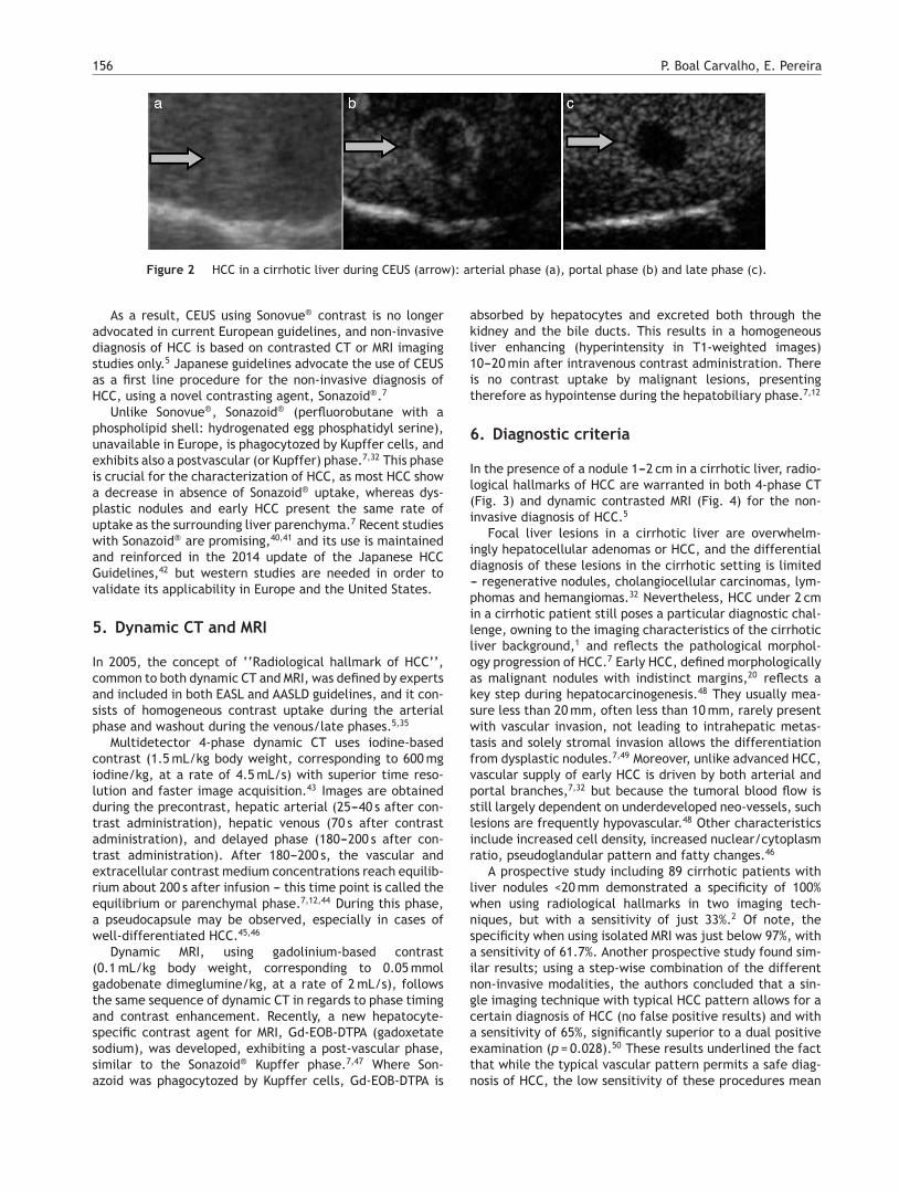
CEUS, using microbubble intravascular contrast agents suchas sulfur hexafluoride with a phospholipid shell (Sonovue®),has shown to be a useful diagnostic technique for HCC in thesetting of cirrhosis; the typical pattern of arterial hyper-enhancement and wash out in the portal and late phasescorresponds to HCC in 97% of the patients (Fig. 2).32---34 Itwas included in the 2001 EASL Guidelines16 and in the 2005AASLD guidelines35 in the diagnostic algorithm of HCC in thecirrhotic liver, as well as being incorporated in the 2010Japanese guidelines for the management of hepatocellularcarcinoma.7
In 2010, however, a study by Villana et al. in a subsetof 21 cirrhotic patients with intrahepatic cholangiocarci-noma (ICC), only 11 patients (52.4%) presented with thetypical peripheral rim arterial enhancement of ICC dur-ing CEUS, while in 10 (47.6%) of these patients, arterialhyperenhancement was homogeneous, suggestive of HCC.36
This study highlighted the likely substantial incidence ofincorrect diagnosis of HCC in patients with ICC, and con-trasted with the contrasted MRI results, where only twopatients (9.5%) presented with homogeneous hyperenhance-ment during the arterial phase. Recently, Li et al. found that68.8% (11 out of 16) of the ICC in patients with cirrhosiswill show imaging characteristics of HCC in CEUS, and suchbehaviour occurs more frequently than in patients with anon-cirrhotic liver (six out of 23; 26.1%).37 The clinical rele-vance of correct differential diagnosis between HCC and ICCrests on the increasing incidence of the latter,38 the overlap-ping risk factors38 and the vastly different approaches andmanagement of both malignancies.5,39
156 P. Boal Carvalho, E. Pereira
Figure 2 HCC in a cirrhotic liver during CEUS (arrow): arterial phase (a), portal phase (b) and late phase (c).
As a result, CEUS using Sonovue® contrast is no longeradvocated in current European guidelines, and non-invasivediagnosis of HCC is based on contrasted CT or MRI imagingstudies only.5 Japanese guidelines advocate the use of CEUSas a first line procedure for the non-invasive diagnosis ofHCC, using a novel contrasting agent, Sonazoid®.7
Unlike Sonovue®, Sonazoid® (perfluorobutane with aphospholipid shell: hydrogenated egg phosphatidyl serine),unavailable in Europe, is phagocytozed by Kupffer cells, andexhibits also a postvascular (or Kupffer) phase.7,32 This phaseis crucial for the characterization of HCC, as most HCC showa decrease in absence of Sonazoid® uptake, whereas dys-plastic nodules and early HCC present the same rate ofuptake as the surrounding liver parenchyma.7 Recent studieswith Sonazoid® are promising,40,41 and its use is maintainedand reinforced in the 2014 update of the Japanese HCCGuidelines,42 but western studies are needed in order tovalidate its applicability in Europe and the United States.
5. Dynamic CT and MRI
In 2005, the concept of ‘‘Radiological hallmark of HCC’’,common to both dynamic CT and MRI, was defined by expertsand included in both EASL and AASLD guidelines, and it con-sists of homogeneous contrast uptake during the arterialphase and washout during the venous/late phases.5,35
Multidetector 4-phase dynamic CT uses iodine-basedcontrast (1.5 mL/kg body weight, corresponding to 600 mgiodine/kg, at a rate of 4.5 mL/s) with superior time reso-lution and faster image acquisition.43 Images are obtainedduring the precontrast, hepatic arterial (25---40 s after con-trast administration), hepatic venous (70 s after contrastadministration), and delayed phase (180---200 s after con-trast administration). After 180---200 s, the vascular andextracellular contrast medium concentrations reach equilib-rium about 200 s after infusion --- this time point is called theequilibrium or parenchymal phase.7,12,44 During this phase,a pseudocapsule may be observed, especially in cases ofwell-differentiated HCC.45,46
Dynamic MRI, using gadolinium-based contrast(0.1 mL/kg body weight, corresponding to 0.05 mmolgadobenate dimeglumine/kg, at a rate of 2 mL/s), followsthe same sequence of dynamic CT in regards to phase timingand contrast enhancement. Recently, a new hepatocyte-specific contrast agent for MRI, Gd-EOB-DTPA (gadoxetatesodium), was developed, exhibiting a post-vascular phase,similar to the Sonazoid® Kupffer phase.7,47 Where Son-azoid was phagocytozed by Kupffer cells, Gd-EOB-DTPA is
absorbed by hepatocytes and excreted both through thekidney and the bile ducts. This results in a homogeneousliver enhancing (hyperintensity in T1-weighted images)10---20 min after intravenous contrast administration. Thereis no contrast uptake by malignant lesions, presentingtherefore as hypointense during the hepatobiliary phase.7,12
6. Diagnostic criteria
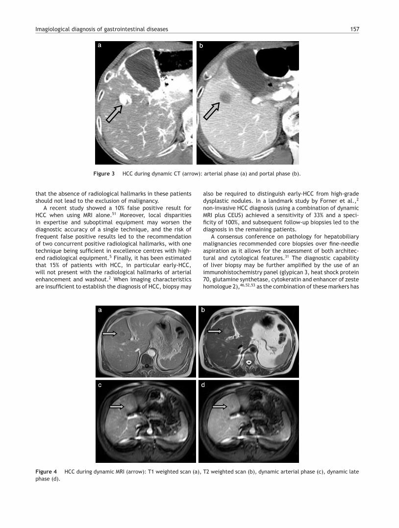
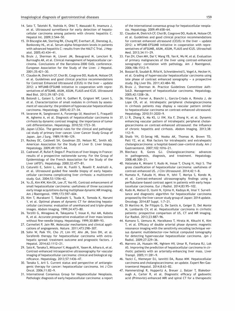
In the presence of a nodule 1---2 cm in a cirrhotic liver, radio-logical hallmarks of HCC are warranted in both 4-phase CT(Fig. 3) and dynamic contrasted MRI (Fig. 4) for the non-invasive diagnosis of HCC.5
Focal liver lesions in a cirrhotic liver are overwhelm-ingly hepatocellular adenomas or HCC, and the differentialdiagnosis of these lesions in the cirrhotic setting is limited--- regenerative nodules, cholangiocellular carcinomas, lym-phomas and hemangiomas.32 Nevertheless, HCC under 2 cmin a cirrhotic patient still poses a particular diagnostic chal-lenge, owning to the imaging characteristics of the cirrhoticliver background,1 and reflects the pathological morphol-ogy progression of HCC.7 Early HCC, defined morphologicallyas malignant nodules with indistinct margins,20 reflects akey step during hepatocarcinogenesis.48 They usually mea-sure less than 20 mm, often less than 10 mm, rarely presentwith vascular invasion, not leading to intrahepatic metas-tasis and solely stromal invasion allows the differentiationfrom dysplastic nodules.7,49 Moreover, unlike advanced HCC,vascular supply of early HCC is driven by both arterial andportal branches,7,32 but because the tumoral blood flow isstill largely dependent on underdeveloped neo-vessels, suchlesions are frequently hypovascular.48 Other characteristicsinclude increased cell density, increased nuclear/cytoplasmratio, pseudoglandular pattern and fatty changes.46
A prospective study including 89 cirrhotic patients withliver nodules <20 mm demonstrated a specificity of 100%when using radiological hallmarks in two imaging tech-niques, but with a sensitivity of just 33%.2 Of note, thespecificity when using isolated MRI was just below 97%, witha sensitivity of 61.7%. Another prospective study found sim-ilar results; using a step-wise combination of the differentnon-invasive modalities, the authors concluded that a sin-gle imaging technique with typical HCC pattern allows for acertain diagnosis of HCC (no false positive results) and witha sensitivity of 65%, significantly superior to a dual positiveexamination (p = 0.028).50 These results underlined the factthat while the typical vascular pattern permits a safe diag-nosis of HCC, the low sensitivity of these procedures mean
Imagiological diagnosis of gastrointestinal diseases 157
Figure 3 HCC during dynamic CT (arrow): arterial phase (a) and portal phase (b).
that the absence of radiological hallmarks in these patientsshould not lead to the exclusion of malignancy.
A recent study showed a 10% false positive result forHCC when using MRI alone.51 Moreover, local disparitiesin expertise and suboptimal equipment may worsen thediagnostic accuracy of a single technique, and the risk offrequent false positive results led to the recommendationof two concurrent positive radiological hallmarks, with onetechnique being sufficient in excellence centres with high-end radiological equipment.5 Finally, it has been estimatedthat 15% of patients with HCC, in particular early-HCC,will not present with the radiological hallmarks of arterialenhancement and washout.2 When imaging characteristicsare insufficient to establish the diagnosis of HCC, biopsy may
also be required to distinguish early-HCC from high-gradedysplastic nodules. In a landmark study by Forner et al.,2
non-invasive HCC diagnosis (using a combination of dynamicMRI plus CEUS) achieved a sensitivity of 33% and a speci-ficity of 100%, and subsequent follow-up biopsies led to thediagnosis in the remaining patients.
A consensus conference on pathology for hepatobiliarymalignancies recommended core biopsies over fine-needleaspiration as it allows for the assessment of both architec-tural and cytological features.31 The diagnostic capabilityof liver biopsy may be further amplified by the use of animmunohistochemistry panel (glypican 3, heat shock protein70, glutamine synthetase, cytokeratin and enhancer of zestehomologue 2),46,52,53 as the combination of these markers has
Figure 4 HCC during dynamic MRI (arrow): T1 weighted scan (a), T2 weighted scan (b), dynamic arterial phase (c), dynamic latephase (d).

158 P. Boal Carvalho, E. Pereira
shown promising results in the diagnosis of early-HCC ver-sus dysplastic nodules in prospective studies.52,53 The use ofthe coaxial biopsy technique, in which the actual needle isintroduced percutaneously into the tumour inside a sheath,can mitigate the risk of tumour seeding by insulating theneedle inside the sheath.1
Contrarily to smaller nodules, the non-invasive diagnosisof liver nodules >2 cm is very effective, as the accuracy forHCC diagnosis using either dynamic CT or MRI is over 90%.43
Therefore, radiological hallmarks in a single technique aresufficient for the diagnosis of lesions >2 cm.5
7. Dynamic CT versus dynamic MRI
When comparing dynamic MRI and dynamic CT, sev-eral studies compared either one with histology as goldstandard.54,55
Diagnostic accuracy for hypervascular HCC using dynamicCT is reported to be 61---83%,56,57 compared to 71---87% indynamic MRI.58---60 In direct comparison studies, the latterwas shown to be superior for HCC diagnosis (sensitivity 76.9%vs 53.8%),61 while the results were conflicting for the recur-rence of HCC following arterial chemoembolization.58,62,63
Most studies however are limited by selection bias,non-blindness and potential for generalization for allcentres.54,55 Therefore, the decision to use either MRI or CTshould be based on local expertise and equipment.
With the recent advent of Gd-EOB-DTPA MRI, this real-ity may change soon. In fact, in a large study including163 patients and comparing Gd-EOB-DTPA MRI with dynamicMRI, dynamic CT and US, diagnostic accuracy with the for-mer was 90%43 for lesions 1---2 cm, significantly superior(p < 0.001) to dynamic MRI (84%), dynamic CT (79%) and US(64%).43
The superiority of Gd-EOB-DTPA MRI was demonstrated byother authors, displaying diagnostic accuracy approaching90%, when compared to standard dynamic MRI (accuracy71---87%) and CT (accuracy 61---83%).58---60
However, the differential diagnosis between early-HCCand dysplastic nodule remains to be difficult, and someearly-HCC present with isointense contrast uptake dur-ing the postvascular phase, while some dysplastic nodulespresent with low-intense contrast enhancing.7
8. HCC in the non-cirrhotic liver
Current diagnostic criteria for HCC are approved only for acirrhotic liver background,5 but up to 10% of the HCC maybe diagnosed in a healthy liver64 or in the setting of non-alcoholic fatty liver disease65 or hepatitis B virus infection.1
Few studies have reported on the imaging characteristicsof HCC in a non-cirrhotic liver,64,66,67 but most report ona higher frequency of an isolated large mass when com-pared to HCC in the cirrhotic liver. The applicability ofnon-invasive diagnostic criteria to such patients has beenmet with disappointing results.68 A recent study with 32patients,67 however, has shown HCC to have the same con-trasting enhancement in both the cirrhotic and non-cirrhoticliver, and diagnostic criteria were encountered in more than90% of the patients, and the authors suggest extending suchcriteria to the non-cirrhotic liver.
Ethical disclosures
Protection of human and animal subjects. The authorsdeclare that no experiments were performed on humans oranimals for this study.
Confidentiality of data. The authors declare that no patientdata appear in this article.
Right to privacy and informed consent. The authorsdeclare that no patient data appear in this article.
Conflicts of interest
The authors declare no conflict of interests.
References
1. Marrero JA, Ahn J, Rajender Reddy K. ACG clinical guideline:the diagnosis and management of focal liver lesions. Am J Gas-troenterol. 2014;109:1328---47, quiz 48.
2. Forner A, Vilana R, Ayuso C, Bianchi L, Sole M, Ayuso JR, et al.Diagnosis of hepatic nodules 20 mm or smaller in cirrhosis:prospective validation of the noninvasive diagnostic criteria forhepatocellular carcinoma. Hepatology. 2008;47:97---104.
3. Degos F, Christidis C, Ganne-Carrie N, Farmachidi JP, DegottC, Guettier C, et al. Hepatitis C virus related cirrhosis: timeto occurrence of hepatocellular carcinoma and death. Gut.2000;47:131---6.
4. Beasley RP, Hwang LY, Lin CC, Chien CS. Hepatocellular carci-noma and hepatitis B virus. A prospective study of 22 707 menin Taiwan. Lancet. 1981;2:1129---33.
5. European Association for the Study of the L, European Orga-nisation for R, Treatment of C. EASL-EORTC clinical practiceguidelines: management of hepatocellular carcinoma. J Hepa-tol. 2012;56:908---43.
6. Bruix J, Sherman M, American Association for the Study of LiverD. Management of hepatocellular carcinoma: an update. Hepa-tology. 2011;53:1020---2.
7. Kudo M, Izumi N, Kokudo N, Matsui O, Sakamoto M, NakashimaO, et al. Management of hepatocellular carcinoma in Japan:Consensus-based Clinical Practice Guidelines proposed by theJapan Society of Hepatology (JSH) 2010 updated version. DigDis. 2011;29:339---64.
8. Omata M, Lesmana LA, Tateishi R, Chen PJ, Lin SM, Yoshida H,et al. Asian Pacific Association for the Study of the Liver consen-sus recommendations on hepatocellular carcinoma. Hepatol Int.2010;4:439---74.
9. Zhang BH, Yang BH, Tang ZY. Randomized controlled trial ofscreening for hepatocellular carcinoma. J Cancer Res ClinOncol. 2004;130:417---22.
10. Chen JG, Parkin DM, Chen QG, Lu JH, Shen QJ, Zhang BC, et al.Screening for liver cancer: results of a randomised controlledtrial in Qidong, China. J Med Screen. 2003;10:204---9.
11. Trevisani F, Cantarini MC, Labate AM, De Notariis S, RapacciniG, Farinati F, et al. Surveillance for hepatocellular carcinoma inelderly Italian patients with cirrhosis: effects on cancer stagingand patient survival. Am J Gastroenterol. 2004;99:1470---6.
12. Bota S, Piscaglia F, Marinelli S, Pecorelli A, Terzi E, Bolondi L.Comparison of international guidelines for noninvasive diagnosisof hepatocellular carcinoma. Liver Cancer. 2012;1:190---200.
13. Singal A, Volk ML, Waljee A, Salgia R, Higgins P, Rogers MA, et al.Meta-analysis: surveillance with ultrasound for early-stagehepatocellular carcinoma in patients with cirrhosis. AlimentPharmacol Ther. 2009;30:37---47.
Imagiological diagnosis of gastrointestinal diseases 159
14. Sato T, Tateishi R, Yoshida H, Ohki T, Masuzaki R, Imamura J,et al. Ultrasound surveillance for early detection of hepato-cellular carcinoma among patients with chronic hepatitis C.Hepatol Int. 2009;3:544---50.
15. Di Bisceglie AM, Sterling RK, Chung RT, Everhart JE, Dienstag JL,Bonkovsky HL, et al. Serum alpha-fetoprotein levels in patientswith advanced hepatitis C: results from the HALT-C Trial. J Hep-atol. 2005;43:434---41.
16. Bruix J, Sherman M, Llovet JM, Beaugrand M, Lencioni R,Burroughs AK, et al. Clinical management of hepatocellular car-cinoma. Conclusions of the Barcelona-2000 EASL conference.European Association for the Study of the Liver. J Hepatol.2001;35:421---30.
17. Claudon M, Dietrich CF, Choi BI, Cosgrove DO, Kudo M, Nolsoe CP,et al. Guidelines and good clinical practice recommendationsfor Contrast Enhanced Ultrasound (CEUS) in the liver --- update2012: a WFUMB-EFSUMB initiative in cooperation with repre-sentatives of AFSUMB, AIUM, ASUM, FLAUS and ICUS. UltrasoundMed Biol. 2013;39:187---210.
18. Bolondi L, Gaiani S, Celli N, Golfieri R, Grigioni WF, Leoni S,et al. Characterization of small nodules in cirrhosis by assess-ment of vascularity: the problem of hypovascular hepatocellularcarcinoma. Hepatology. 2005;42:27---34.
19. Iavarone M, Sangiovanni A, Forzenigo LV, Massironi S, FraquelliM, Aghemo A, et al. Diagnosis of hepatocellular carcinoma incirrhosis by dynamic contrast imaging: the importance of tumorcell differentiation. Hepatology. 2010;52:1723---30.
20. Japan LCSGo. The general rules for the clinical and pathologi-cal study of primary liver cancer. Liver Cancer Study Group ofJapan. Jpn J Surg. 1989;19:98---129.
21. Rockey DC, Caldwell SH, Goodman ZD, Nelson RC, Smith AD.American Association for the Study of Liver D. Liver biopsy.Hepatology. 2009;49:1017---44.
22. Cadranel JF, Rufat P, Degos F. Practices of liver biopsy in France:results of a prospective nationwide survey. For the Group ofEpidemiology of the French Association for the Study of theLiver (AFEF). Hepatology. 2000;32:477---81.
23. Caturelli E, Solmi L, Anti M, Fusilli S, Roselli P, Andriulli A,et al. Ultrasound guided fine needle biopsy of early hepato-cellular carcinoma complicating liver cirrhosis: a multicentrestudy. Gut. 2004;53:1356---62.
24. Yu JS, Kim KW, Kim EK, Lee JT, Yoo HS. Contrast enhancement ofsmall hepatocellular carcinoma: usefulness of three successiveearly image acquisitions during multiphase dynamic MR imaging.Am J Roentgenol. 1999;173:597---604.
25. Kim T, Murakami T, Takahashi S, Tsuda K, Tomoda K, NarumiY, et al. Optimal phases of dynamic CT for detecting hepato-cellular carcinoma: evaluation of unenhanced and triple-phaseimages. Abdom Imaging. 1999;24:473---80.
26. Torzilli G, Minagawa M, Takayama T, Inoue K, Hui AM, KubotaK, et al. Accurate preoperative evaluation of liver mass lesionswithout fine-needle biopsy. Hepatology. 1999;30:889---93.
27. Carmeliet P, Jain RK. Molecular mechanisms and clinical appli-cations of angiogenesis. Nature. 2011;473:298---307.
28. Sohn W, Paik YH, Cho JY, Lim HY, Ahn JM, Sinn DH, et al.Sorafenib therapy for hepatocellular carcinoma with extra-hepatic spread: treatment outcome and prognostic factors. JHepatol. 2014;62:1112---21.
29. Sato K, Tanaka S, Mitsunori Y, Mogushi K, Yasen M, Aihara A, et al.Contrast-enhanced intraoperative ultrasonography for vascularimaging of hepatocellular carcinoma: clinical and biological sig-nificance. Hepatology. 2013;57:1436---47.
30. Tanaka S, Arii S. Current status and perspective of antiangio-genic therapy for cancer: hepatocellular carcinoma. Int J ClinOncol. 2006;11:82---9.
31. International Consensus Group for Hepatocellular Neoplasia.Pathologic diagnosis of early hepatocellular carcinoma: a report
of the international consensus group for hepatocellular neopla-sia. Hepatology. 2009;49:658---64.
32. Claudon M, Dietrich CF, Choi BI, Cosgrove DO, Kudo M, Nolsoe CP,et al. Guidelines and good clinical practice recommendationsfor contrast enhanced ultrasound (CEUS) in the liver --- update2012: a WFUMB-EFSUMB initiative in cooperation with repre-sentatives of AFSUMB, AIUM, ASUM, FLAUS and ICUS. UltraschallMed. 2013;34:11---29.
33. Fan ZH, Chen MH, Dai Y, Wang YB, Yan K, Wu W, et al. Evaluationof primary malignancies of the liver using contrast-enhancedsonography: correlation with pathology. Am J Roentgenol.2006;186:1512---9.
34. Boozari B, Soudah B, Rifai K, Schneidewind S, Vogel A, Hecker H,et al. Grading of hypervascular hepatocellular carcinoma usinglate phase of contrast enhanced sonography --- a prospectivestudy. Dig Liver Dis. 2011;43:484---90.
35. Bruix J, Sherman M. Practice Guidelines Committee AAft-SoLD. Management of hepatocellular carcinoma. Hepatology.2005;42:1208---36.
36. Vilana R, Forner A, Bianchi L, Garcia-Criado A, Rimola J, deLope CR, et al. Intrahepatic peripheral cholangiocarcinomain cirrhosis patients may display a vascular pattern similarto hepatocellular carcinoma on contrast-enhanced ultrasound.Hepatology. 2010;51:2020---9.
37. Li R, Zhang X, Ma KS, Li XW, Xia F, Zhong H, et al. Dynamicenhancing vascular pattern of intrahepatic peripheral cholan-giocarcinoma on contrast-enhanced ultrasound: the influenceof chronic hepatitis and cirrhosis. Abdom Imaging. 2013;38:112---9.
38. Shaib YH, El-Serag HB, Nooka AK, Thomas M, Brown TD,Patt YZ, et al. Risk factors for intrahepatic and extrahepaticcholangiocarcinoma: a hospital-based case---control study. Am JGastroenterol. 2007;102:1016---21.
39. Blechacz B, Gores GJ. Cholangiocarcinoma: advancesin pathogenesis, diagnosis, and treatment. Hepatology.2008;48:308---21.
40. Hatanaka K, Minami Y, Kudo M, Inoue T, Chung H, Haji S. Thegross classification of hepatocellular carcinoma: usefulness ofcontrast-enhanced US. J Clin Ultrasound. 2014;42:1---8.
41. Numata K, Fukuda H, Miwa H, Ishii T, Moriya S, Kondo M,et al. Contrast-enhanced ultrasonography findings using aperflubutane-based contrast agent in patients with early hepa-tocellular carcinoma. Eur J Radiol. 2014;83:95---102.
42. Kudo M, Matsui O, Izumi N, Iijima H, Kadoya M, Imai Y. Surveil-lance and diagnostic algorithm for hepatocellular carcinomaproposed by the liver cancer study group of Japan: 2014 update.Oncology. 2014;87 Suppl. 1:7---21.
43. Di Martino M, De Filippis G, De Santis A, Geiger D, Del MonteM, Lombardo CV, et al. Hepatocellular carcinoma in cirrhoticpatients: prospective comparison of US, CT and MR imaging.Eur Radiol. 2013;23:887---96.
44. Kumano S, Uemura M, Haraikawa T, Hirata M, Kikuchi K, KimT, et al. Efficacy of double arterial phase dynamic magneticresonance imaging with the sensitivity encoding technique ver-sus dynamic multidetector-row helical computed tomographyfor detecting hypervascular hepatocellular carcinoma. Jpn JRadiol. 2009;27:229---36.
45. Marrero JA, Hussain HK, Nghiem HV, Umar R, Fontana RJ, LokAS. Improving the prediction of hepatocellular carcinoma in cir-rhotic patients with an arterially-enhancing liver mass. LiverTranspl. 2005;11:281---9.
46. Yazici C, Niemeyer DJ, Iannitti DA, Russo MW. Hepatocellularcarcinoma and cholangiocarcinoma: an update. Expert Rev Gas-troenterol Hepatol. 2014;8:63---82.
47. Hammerstingl R, Huppertz A, Breuer J, Balzer T, Blakebor-ough A, Carter R, et al. Diagnostic efficacy of gadoxeticacid (Primovist)-enhanced MRI and spiral CT for a therapeutic
160 P. Boal Carvalho, E. Pereira
strategy: comparison with intraoperative and histopathologicfindings in focal liver lesions. Eur Radiol. 2008;18:457---67.
48. Sakamoto M, Hirohashi S, Shimosato Y. Early stages of multi-step hepatocarcinogenesis: adenomatous hyperplasia and earlyhepatocellular carcinoma. Hum Pathol. 1991;22:172---8.
49. Kondo F, Kondo Y, Nagato Y, Tomizawa M, Wada K. Interstitialtumour cell invasion in small hepatocellular carcinoma. Evalua-tion in microscopic and low magnification views. J GastroenterolHepatol. 1994;9:604---12.
50. Sangiovanni A, Manini MA, Iavarone M, Romeo R, Forzenigo LV,Fraquelli M, et al. The diagnostic and economic impact of con-trast imaging techniques in the diagnosis of small hepatocellularcarcinoma in cirrhosis. Gut. 2010;59:638---44.
51. Yu NC, Chaudhari V, Raman SS, Lassman C, Tong MJ, Busut-til RW, et al. CT and MRI improve detection of hepatocellularcarcinoma, compared with ultrasound alone, in patients withcirrhosis. Clin Gastroenterol Hepatol. 2011;9:161---7.
52. Cai MY, Tong ZT, Zheng F, Liao YJ, Wang Y, Rao HL, et al.EZH2 protein: a promising immunomarker for the detectionof hepatocellular carcinomas in liver needle biopsies. Gut.2011;60:967---76.
53. Tremosini S, Forner A, Boix L, Vilana R, Bianchi L, Reig M, et al.Prospective validation of an immunohistochemical panel (glypi-can 3, heat shock protein 70 and glutamine synthetase) in liverbiopsies for diagnosis of very early hepatocellular carcinoma.Gut. 2012;61:1481---7.
54. Marrero JA, Welling T. Modern diagnosis and management ofhepatocellular carcinoma. Clin Liver Dis. 2009;13:233---47.
55. de Ledinghen V, Laharie D, Lecesne R, Le Bail B, Winnock M,Bernard PH, et al. Detection of nodules in liver cirrhosis: spi-ral computed tomography or magnetic resonance imaging? Aprospective study of 88 nodules in 34 patients. Eur J Gastroen-terol Hepatol. 2002;14:159---65.
56. Shapiro RS, Katz R, Mendelson DS, Halton KP, Schwartz ME,Miller CM. Detection of hepatocellular carcinoma in cirrhoticpatients: sensitivity of CT and ultrasonography. J UltrasoundMed. 1996;15:497---502, quiz 3-4.
57. Lim JH, Kim CK, Lee WJ, Park CK, Koh KC, Paik SW, et al.Detection of hepatocellular carcinomas and dysplastic nodulesin cirrhotic livers: accuracy of helical CT in transplant patients.Am J Roentgenol. 2000;175:693---8.
58. Kudo M. Will Gd-EOB-MRI change the diagnostic algorithm inhepatocellular carcinoma? Oncology. 2010;78 Suppl. 1:87---93.
59. Marin D, Di Martino M, Guerrisi A, De Filippis G, Rossi M, GinanniCorradini S, et al. Hepatocellular carcinoma in patients withcirrhosis: qualitative comparison of gadobenate dimeglumine-enhanced MR imaging and multiphasic 64-section CT. Radiology.2009;251:85---95.
60. Di Martino M, Marin D, Guerrisi A, Baski M, Galati F, Rossi M, et al.Intraindividual comparison of gadoxetate disodium-enhancedMR imaging and 64-section multidetector CT in the detection ofhepatocellular carcinoma in patients with cirrhosis. Radiology.2010;256:806---16.
61. Rode A, Bancel B, Douek P, Chevallier M, Vilgrain V, PicaudG, et al. Small nodule detection in cirrhotic livers: evaluationwith US, spiral CT, and MRI and correlation with patho-logic examination of explanted liver. J Comput Assist Tomogr.2001;25:327---36.
62. Yu MH, Kim JH, Yoon JH, Kim HC, Chung JW, Han JK, et al. Roleof C-arm CT for transcatheter arterial chemoembolization ofhepatocellular carcinoma: diagnostic performance and predic-tive value for therapeutic response compared with gadoxeticacid-enhanced MRI. Am J Roentgenol. 2013;201:675---83.
63. Kim BK, Kim KA, An C, Yoo EJ, Park JY, Kim DY, et al.Prognostic role of magnetic resonance imaging versus com-puted tomography for hepatocellular carcinoma undergoingchemoembolization. Liver Int. 2014 (in press).
64. Brancatelli G, Federle MP, Grazioli L, Carr BI. Hepatocellularcarcinoma in noncirrhotic liver: CT, clinical, and pathologic find-ings in 39 US residents. Radiology. 2002;222:89---94.
65. Iannaccone R, Piacentini F, Murakami T, Paradis V, BelghitiJ, Hori M, et al. Hepatocellular carcinoma in patients withnonalcoholic fatty liver disease: helical CT and MR imag-ing findings with clinical-pathologic comparison. Radiology.2007;243:422---30.
66. Winston CB, Schwartz LH, Fong Y, Blumgart LH, Panicek DM.Hepatocellular carcinoma: MR imaging findings in cirrhotic liversand noncirrhotic livers. Radiology. 1999;210:75---9.
67. Di Martino M, Saba L, Bosco S, Rossi M, Miles KA, Di Mis-cio R, et al. Hepatocellular carcinoma (HCC) in non-cirrhoticliver: clinical, radiological and pathological findings. Eur Radiol.2014;24:1446---54.
68. Bialecki ES, Ezenekwe AM, Brunt EM, Collins BT, Ponder TB,Bieneman BK, et al. Comparison of liver biopsy and noninva-sive methods for diagnosis of hepatocellular carcinoma. ClinGastroenterol Hepatol. 2006;4:361---8.


