Imaging of the Paranasal Sinuses and Nasal Cavity: Normal ...Imaging of the Paranasal Sinuses and...
15
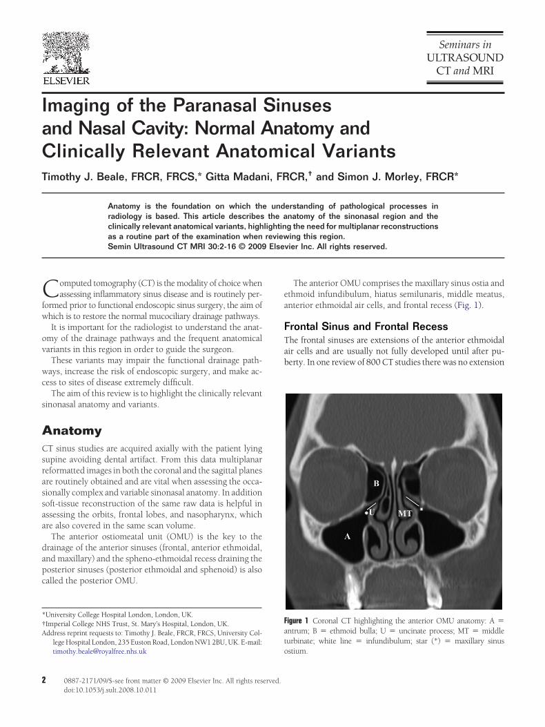
Imaging of the Paranasal Sinuses and Nasal Cavity: Normal Anatomy and Clinically Relevant Anatomical Variants Timothy J. Beale, FRCR, FRCS,* Gitta Madani, FRCR, † and Simon J. Morley, FRCR* Anatomy is the foundation on which the understanding of pathological processes in radiology is based. This article describes the anatomy of the sinonasal region and the clinically relevant anatomical variants, highlighting the need for multiplanar reconstructions as a routine part of the examination when reviewing this region. Semin Ultrasound CT MRI 30:2-16 © 2009 Elsevier Inc. All rights reserved. C omputed tomography (CT) is the modality of choice when assessing inflammatory sinus disease and is routinely per- formed prior to functional endoscopic sinus surgery, the aim of which is to restore the normal mucociliary drainage pathways. It is important for the radiologist to understand the anat- omy of the drainage pathways and the frequent anatomical variants in this region in order to guide the surgeon. These variants may impair the functional drainage path- ways, increase the risk of endoscopic surgery, and make ac- cess to sites of disease extremely difficult. The aim of this review is to highlight the clinically relevant sinonasal anatomy and variants. Anatomy CT sinus studies are acquired axially with the patient lying supine avoiding dental artifact. From this data multiplanar reformatted images in both the coronal and the sagittal planes are routinely obtained and are vital when assessing the occa- sionally complex and variable sinonasal anatomy. In addition soft-tissue reconstruction of the same raw data is helpful in assessing the orbits, frontal lobes, and nasopharynx, which are also covered in the same scan volume. The anterior ostiomeatal unit (OMU) is the key to the drainage of the anterior sinuses (frontal, anterior ethmoidal, and maxillary) and the spheno-ethmoidal recess draining the posterior sinuses (posterior ethmoidal and sphenoid) is also called the posterior OMU. The anterior OMU comprises the maxillary sinus ostia and ethmoid infundibulum, hiatus semilunaris, middle meatus, anterior ethmoidal air cells, and frontal recess (Fig. 1). Frontal Sinus and Frontal Recess The frontal sinuses are extensions of the anterior ethmoidal air cells and are usually not fully developed until after pu- berty. In one review of 800 CT studies there was no extension *University College Hospital London, London, UK. †Imperial College NHS Trust, St. Mary’s Hospital, London, UK. Address reprint requests to: Timothy J. Beale, FRCR, FRCS, University Col- lege Hospital London, 235 Euston Road, London NW1 2BU, UK. E-mail: [email protected] Figure 1 Coronal CT highlighting the anterior OMU anatomy: A antrum; B ethmoid bulla; U uncinate process; MT middle turbinate; white line infundibulum; star (*) maxillary sinus ostium. 2 0887-2171/09/$-see front matter © 2009 Elsevier Inc. All rights reserved. doi:10.1053/j.sult.2008.10.011
Transcript of Imaging of the Paranasal Sinuses and Nasal Cavity: Normal ...Imaging of the Paranasal Sinuses and...
IaCT
Cfw
ov
wc
s
ACsrassaa
dapc
*†A
2
maging of the Paranasal Sinusesnd Nasal Cavity: Normal Anatomy andlinically Relevant Anatomical Variants
imothy J. Beale, FRCR, FRCS,* Gitta Madani, FRCR,† and Simon J. Morley, FRCR*
Anatomy is the foundation on which the understanding of pathological processes inradiology is based. This article describes the anatomy of the sinonasal region and theclinically relevant anatomical variants, highlighting the need for multiplanar reconstructionsas a routine part of the examination when reviewing this region.Semin Ultrasound CT MRI 30:2-16 © 2009 Elsevier Inc. All rights reserved.
ea
FTab
Fat
omputed tomography (CT) is the modality of choice whenassessing inflammatory sinus disease and is routinely per-
ormed prior to functional endoscopic sinus surgery, the aim ofhich is to restore the normal mucociliary drainage pathways.It is important for the radiologist to understand the anat-
my of the drainage pathways and the frequent anatomicalariants in this region in order to guide the surgeon.
These variants may impair the functional drainage path-ays, increase the risk of endoscopic surgery, and make ac-
ess to sites of disease extremely difficult.The aim of this review is to highlight the clinically relevant
inonasal anatomy and variants.
natomyT sinus studies are acquired axially with the patient lying
upine avoiding dental artifact. From this data multiplanareformatted images in both the coronal and the sagittal planesre routinely obtained and are vital when assessing the occa-ionally complex and variable sinonasal anatomy. In additionoft-tissue reconstruction of the same raw data is helpful inssessing the orbits, frontal lobes, and nasopharynx, whichre also covered in the same scan volume.
The anterior ostiomeatal unit (OMU) is the key to therainage of the anterior sinuses (frontal, anterior ethmoidal,nd maxillary) and the spheno-ethmoidal recess draining theosterior sinuses (posterior ethmoidal and sphenoid) is alsoalled the posterior OMU.
University College Hospital London, London, UK.Imperial College NHS Trust, St. Mary’s Hospital, London, UK.ddress reprint requests to: Timothy J. Beale, FRCR, FRCS, University Col-
lege Hospital London, 235 Euston Road, London NW1 2BU, UK. E-mail:
[email protected] o0887-2171/09/$-see front matter © 2009 Elsevier Inc. All rights reserved.doi:10.1053/j.sult.2008.10.011
The anterior OMU comprises the maxillary sinus ostia andthmoid infundibulum, hiatus semilunaris, middle meatus,nterior ethmoidal air cells, and frontal recess (Fig. 1).
rontal Sinus and Frontal Recesshe frontal sinuses are extensions of the anterior ethmoidalir cells and are usually not fully developed until after pu-erty. In one review of 800 CT studies there was no extension
igure 1 Coronal CT highlighting the anterior OMU anatomy: A �ntrum; B � ethmoid bulla; U � uncinate process; MT � middleurbinate; white line � infundibulum; star (*) � maxillary sinus
stium.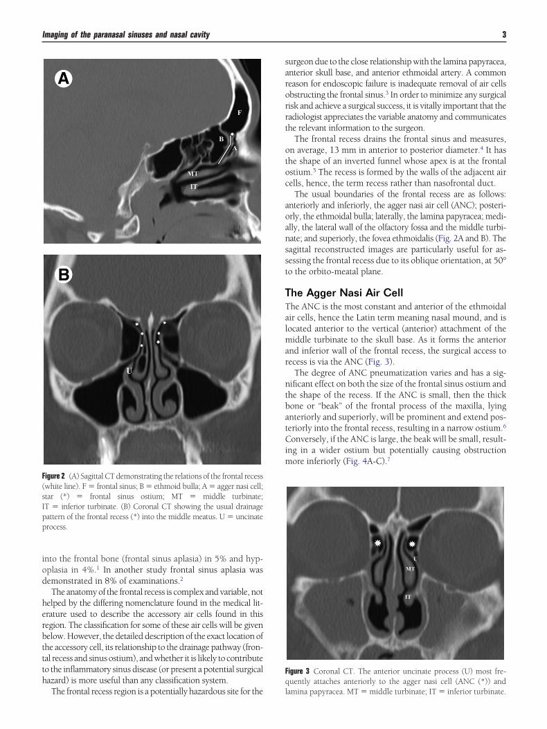
iod
herbttth
sarorrt
otoc
aoansst
TTalmar
ntbatCim
F(sIpp
Fq
Imaging of the paranasal sinuses and nasal cavity 3
nto the frontal bone (frontal sinus aplasia) in 5% and hyp-plasia in 4%.1 In another study frontal sinus aplasia wasemonstrated in 8% of examinations.2
The anatomy of the frontal recess is complex and variable, notelped by the differing nomenclature found in the medical lit-rature used to describe the accessory air cells found in thisegion. The classification for some of these air cells will be givenelow. However, the detailed description of the exact location ofhe accessory cell, its relationship to the drainage pathway (fron-al recess and sinus ostium), and whether it is likely to contributeo the inflammatory sinus disease (or present a potential surgicalazard) is more useful than any classification system.
igure 2 (A) Sagittal CT demonstrating the relations of the frontal recesswhite line). F � frontal sinus; B � ethmoid bulla; A � agger nasi cell;tar (*) � frontal sinus ostium; MT � middle turbinate;T � inferior turbinate. (B) Coronal CT showing the usual drainageattern of the frontal recess (*) into the middle meatus. U � uncinaterocess.
The frontal recess region is a potentially hazardous site for the l
urgeon due to the close relationship with the lamina papyracea,nterior skull base, and anterior ethmoidal artery. A commoneason for endoscopic failure is inadequate removal of air cellsbstructing the frontal sinus.3 In order to minimize any surgicalisk and achieve a surgical success, it is vitally important that theadiologist appreciates the variable anatomy and communicateshe relevant information to the surgeon.
The frontal recess drains the frontal sinus and measures,n average, 13 mm in anterior to posterior diameter.4 It hashe shape of an inverted funnel whose apex is at the frontalstium.5 The recess is formed by the walls of the adjacent airells, hence, the term recess rather than nasofrontal duct.
The usual boundaries of the frontal recess are as follows:nteriorly and inferiorly, the agger nasi air cell (ANC); posteri-rly, the ethmoidal bulla; laterally, the lamina papyracea; medi-lly, the lateral wall of the olfactory fossa and the middle turbi-ate; and superiorly, the fovea ethmoidalis (Fig. 2A and B). Theagittal reconstructed images are particularly useful for as-essing the frontal recess due to its oblique orientation, at 50°o the orbito-meatal plane.
he Agger Nasi Air Cellhe ANC is the most constant and anterior of the ethmoidalir cells, hence the Latin term meaning nasal mound, and isocated anterior to the vertical (anterior) attachment of the
iddle turbinate to the skull base. As it forms the anteriornd inferior wall of the frontal recess, the surgical access toecess is via the ANC (Fig. 3).
The degree of ANC pneumatization varies and has a sig-ificant effect on both the size of the frontal sinus ostium andhe shape of the recess. If the ANC is small, then the thickone or “beak” of the frontal process of the maxilla, lyingnteriorly and superiorly, will be prominent and extend pos-eriorly into the frontal recess, resulting in a narrow ostium.6
onversely, if the ANC is large, the beak will be small, result-ng in a wider ostium but potentially causing obstruction
ore inferiorly (Fig. 4A-C).7
igure 3 Coronal CT. The anterior uncinate process (U) most fre-uently attaches anteriorly to the agger nasi cell (ANC (*)) and
amina papyracea. MT � middle turbinate; IT � inferior turbinate.
tcaa
TTgt
aal
FAfMllec
Ft
4 T.J. Beale, G. Madani, and S.J. Morley
This beak of bone represents the anterior floor of the fron-al sinus and can be clearly seen on both sagittal and anteriororonal reformatted images. In the coronal plane it is seen ascontinuous bony ridge and is useful for assessing whether
n accessory air cell has extended into the frontal sinus.6
he Frontalethmoidal (Kuhn) Cellshere are various accessory air cells in the frontoethmoidal re-ion that may or may not be present. It is important to work outhe drainage pathway of the frontal sinus around these cells.
Frontalethmoidal air cells, also known as Kuhn air cells,8
re categorized into four types depending on their numbernd degree of extension into the frontal sinus.6 They are allocated superior to the ANC.
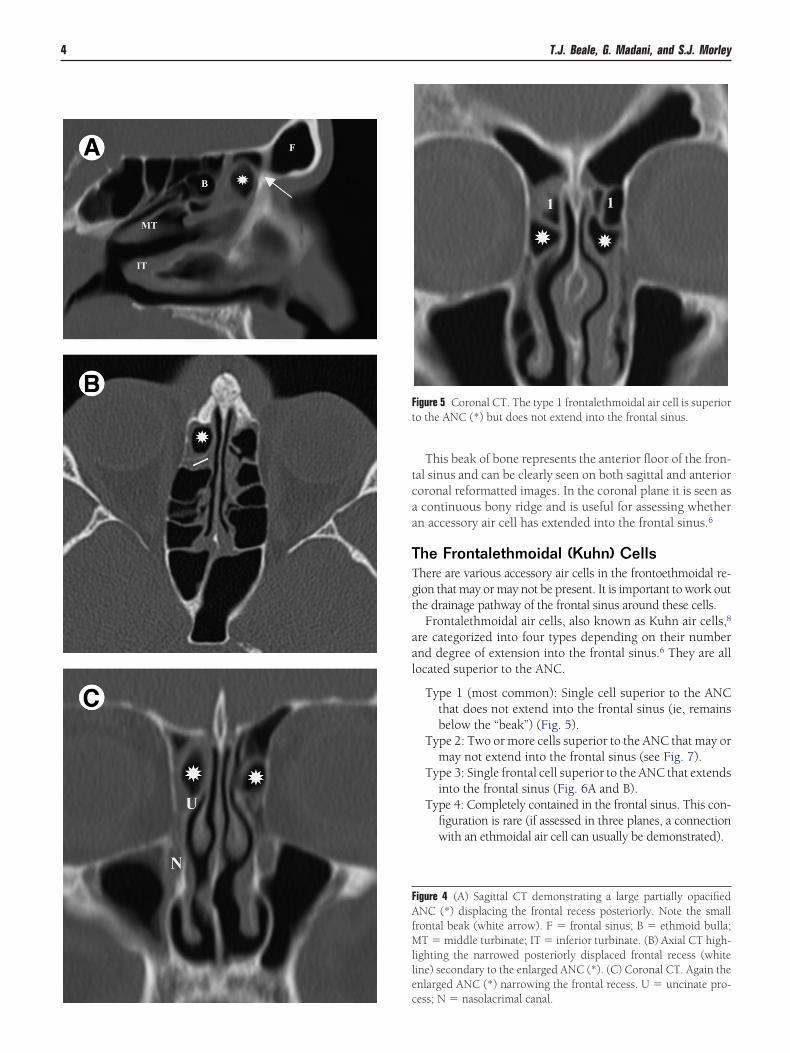
Type 1 (most common): Single cell superior to the ANCthat does not extend into the frontal sinus (ie, remainsbelow the “beak”) (Fig. 5).
Type 2: Two or more cells superior to the ANC that may ormay not extend into the frontal sinus (see Fig. 7).
Type 3: Single frontal cell superior to the ANC that extendsinto the frontal sinus (Fig. 6A and B).
Type 4: Completely contained in the frontal sinus. This con-figuration is rare (if assessed in three planes, a connectionwith an ethmoidal air cell can usually be demonstrated).
igure 4 (A) Sagittal CT demonstrating a large partially opacifiedNC (*) displacing the frontal recess posteriorly. Note the small
rontal beak (white arrow). F � frontal sinus; B � ethmoid bulla;T � middle turbinate; IT � inferior turbinate. (B) Axial CT high-
ighting the narrowed posteriorly displaced frontal recess (whiteine) secondary to the enlarged ANC (*). (C) Coronal CT. Again thenlarged ANC (*) narrowing the frontal recess. U � uncinate pro-
igure 5 Coronal CT. The type 1 frontalethmoidal air cell is superioro the ANC (*) but does not extend into the frontal sinus.
ess; N � nasolacrimal canal.
bdef
OCOas
ea(eaewtrodc
FmMss
Fafs
Fs
Imaging of the paranasal sinuses and nasal cavity 5
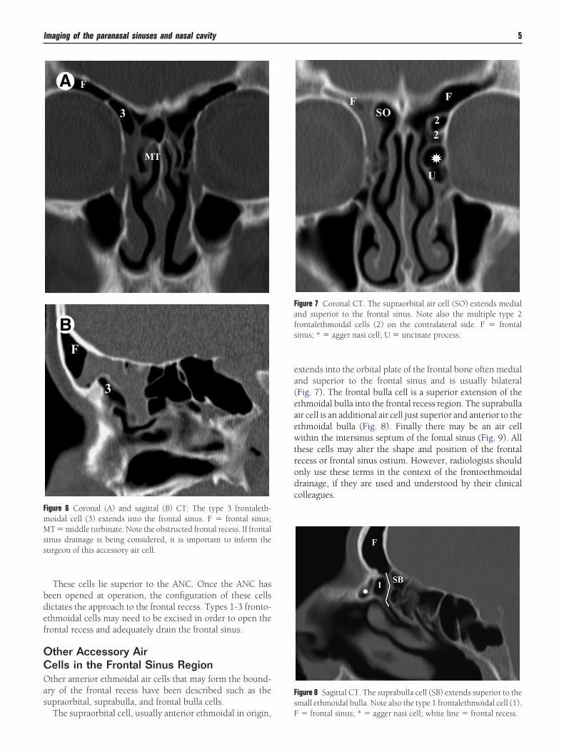
These cells lie superior to the ANC. Once the ANC haseen opened at operation, the configuration of these cellsictates the approach to the frontal recess. Types 1-3 fronto-thmoidal cells may need to be excised in order to open therontal recess and adequately drain the frontal sinus.
ther Accessory Airells in the Frontal Sinus Regionther anterior ethmoidal air cells that may form the bound-
ry of the frontal recess have been described such as theupraorbital, suprabulla, and frontal bulla cells.
igure 6 Coronal (A) and sagittal (B) CT: The type 3 frontaleth-oidal cell (3) extends into the frontal sinus. F � frontal sinus;T � middle turbinate. Note the obstructed frontal recess. If frontal
inus drainage is being considered, it is important to inform theurgeon of this accessory air cell.
The supraorbital cell, usually anterior ethmoidal in origin, F
xtends into the orbital plate of the frontal bone often medialnd superior to the frontal sinus and is usually bilateralFig. 7). The frontal bulla cell is a superior extension of thethmoidal bulla into the frontal recess region. The suprabullair cell is an additional air cell just superior and anterior to thethmoidal bulla (Fig. 8). Finally there may be an air cellithin the intersinus septum of the fontal sinus (Fig. 9). All
hese cells may alter the shape and position of the frontalecess or frontal sinus ostium. However, radiologists shouldnly use these terms in the context of the frontoethmoidalrainage, if they are used and understood by their clinicalolleagues.
igure 7 Coronal CT. The supraorbital air cell (SO) extends medialnd superior to the frontal sinus. Note also the multiple type 2rontalethmoidal cells (2) on the contralateral side. F � frontalinus; * � agger nasi cell; U � uncinate process.
igure 8 Sagittal CT. The suprabulla cell (SB) extends superior to themall ethmoidal bulla. Note also the type 1 frontalethmoidal cell (1).
� frontal sinus; * � agger nasi cell; white line � frontal recess.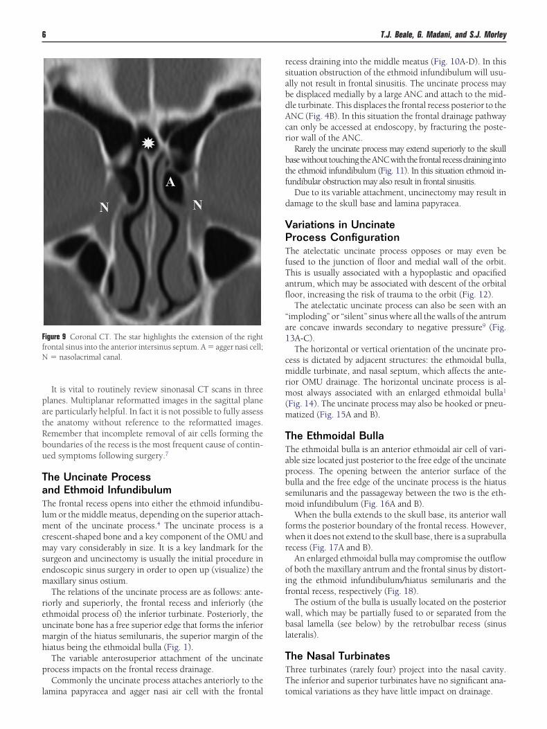
patRbu
TaTlmcmsem
reumh
p
l
rsabdAcr
btf
d
VPTfTafl
“a1
cmrm(m
TTapbsm
fwr
oif
wbl
TTT
FfN
6 T.J. Beale, G. Madani, and S.J. Morley
It is vital to routinely review sinonasal CT scans in threelanes. Multiplanar reformatted images in the sagittal planere particularly helpful. In fact it is not possible to fully assesshe anatomy without reference to the reformatted images.emember that incomplete removal of air cells forming theoundaries of the recess is the most frequent cause of contin-ed symptoms following surgery.7
he Uncinate Processnd Ethmoid Infundibulumhe frontal recess opens into either the ethmoid infundibu-
um or the middle meatus, depending on the superior attach-ent of the uncinate process.4 The uncinate process is a
rescent-shaped bone and a key component of the OMU anday vary considerably in size. It is a key landmark for the
urgeon and uncinectomy is usually the initial procedure inndoscopic sinus surgery in order to open up (visualize) theaxillary sinus ostium.The relations of the uncinate process are as follows: ante-
iorly and superiorly, the frontal recess and inferiorly (thethmoidal process of) the inferior turbinate. Posteriorly, thencinate bone has a free superior edge that forms the inferiorargin of the hiatus semilunaris, the superior margin of theiatus being the ethmoidal bulla (Fig. 1).The variable anterosuperior attachment of the uncinate
rocess impacts on the frontal recess drainage.Commonly the uncinate process attaches anteriorly to the
igure 9 Coronal CT. The star highlights the extension of the rightrontal sinus into the anterior intersinus septum. A � agger nasi cell;
� nasolacrimal canal.
amina papyracea and agger nasi air cell with the frontal t
ecess draining into the middle meatus (Fig. 10A-D). In thisituation obstruction of the ethmoid infundibulum will usu-lly not result in frontal sinusitis. The uncinate process maye displaced medially by a large ANC and attach to the mid-le turbinate. This displaces the frontal recess posterior to theNC (Fig. 4B). In this situation the frontal drainage pathwayan only be accessed at endoscopy, by fracturing the poste-ior wall of the ANC.
Rarely the uncinate process may extend superiorly to the skullasewithout touchingtheANCwith the frontal recessdraining intohe ethmoid infundibulum (Fig. 11). In this situation ethmoid in-undibular obstruction may also result in frontal sinusitis.
Due to its variable attachment, uncinectomy may result inamage to the skull base and lamina papyracea.
ariations in Uncinaterocess Configurationhe atelectatic uncinate process opposes or may even be
used to the junction of floor and medial wall of the orbit.his is usually associated with a hypoplastic and opacifiedntrum, which may be associated with descent of the orbitaloor, increasing the risk of trauma to the orbit (Fig. 12).The atelectatic uncinate process can also be seen with an
imploding” or “silent” sinus where all the walls of the antrumre concave inwards secondary to negative pressure9 (Fig.3A-C).The horizontal or vertical orientation of the uncinate pro-
ess is dictated by adjacent structures: the ethmoidal bulla,iddle turbinate, and nasal septum, which affects the ante-
ior OMU drainage. The horizontal uncinate process is al-ost always associated with an enlarged ethmoidal bulla1
Fig. 14). The uncinate process may also be hooked or pneu-atized (Fig. 15A and B).
he Ethmoidal Bullahe ethmoidal bulla is an anterior ethmoidal air cell of vari-ble size located just posterior to the free edge of the uncinaterocess. The opening between the anterior surface of theulla and the free edge of the uncinate process is the hiatusemilunaris and the passageway between the two is the eth-oid infundibulum (Fig. 16A and B).When the bulla extends to the skull base, its anterior wall
orms the posterior boundary of the frontal recess. However,hen it does not extend to the skull base, there is a suprabulla
ecess (Fig. 17A and B).An enlarged ethmoidal bulla may compromise the outflow
f both the maxillary antrum and the frontal sinus by distort-ng the ethmoid infundibulum/hiatus semilunaris and therontal recess, respectively (Fig. 18).
The ostium of the bulla is usually located on the posteriorall, which may be partially fused to or separated from theasal lamella (see below) by the retrobulbar recess (sinus
ateralis).
he Nasal Turbinateshree turbinates (rarely four) project into the nasal cavity.he inferior and superior turbinates have no significant ana-
omical variations as they have little impact on drainage.
Frd
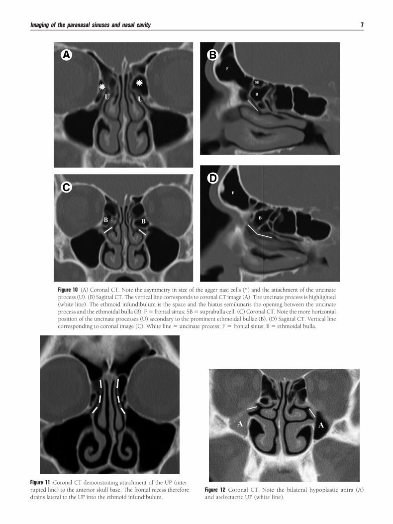
corresponding to coronal image (C). White line � uncinate process; F � frontal sinus; B � ethmoidal bulla.
F
Imaging of the paranasal sinuses and nasal cavity 7
igure 11 Coronal CT demonstrating attachment of the UP (inter-upted line) to the anterior skull base. The frontal recess therefore
Figure 10 (A) Coronal CT. Note the asymmetry in size of the agger nasi cells (*) and the attachment of the uncinateprocess (U). (B) Sagittal CT. The vertical line corresponds to coronal CT image (A). The uncinate process is highlighted(white line). The ethmoid infundibulum is the space and the hiatus semilunaris the opening between the uncinateprocess and the ethmoidal bulla (B). F � frontal sinus; SB � suprabulla cell. (C) Coronal CT. Note the more horizontalposition of the uncinate processes (U) secondary to the prominent ethmoidal bullae (B). (D) Sagittal CT. Vertical line
rains lateral to the UP into the ethmoid infundibulum. a
igure 12 Coronal CT. Note the bilateral hypoplastic antra (A) nd atelectactic UP (white line).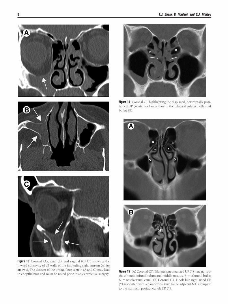
FtN(t
Fiato enopthalmos and must be noted prior to any corrective surgery.
Ftbullae (B).
8 T.J. Beale, G. Madani, and S.J. Morley
igure 15 (A) Coronal CT. Bilateral pneumatized UP (*) may narrowhe ethmoid infundibulum and middle meatus. B � ethmoid bulla;
� nasolacrimal canal. (B) Coronal CT. Hook-like right-sided UP*) associated with a paradoxical turn to the adjacent MT. Compare
igure 13 Coronal (A), axial (B), and sagittal (C) CT showing thenward concavity of all walls of the imploding right antrum (whiterrows). The descent of the orbital floor seen in (A and C) may lead
igure 14 Coronal CT highlighting the displaced, horizontally posi-ioned UP (white line) secondary to the bilateral enlarged ethmoid
o the normally positioned left UP (*).
oslbitTi
TVCPn
Fwesat(
FiAsrTpANC (*) and the ethmoid bulla (B).
Fba
Imaging of the paranasal sinuses and nasal cavity 9
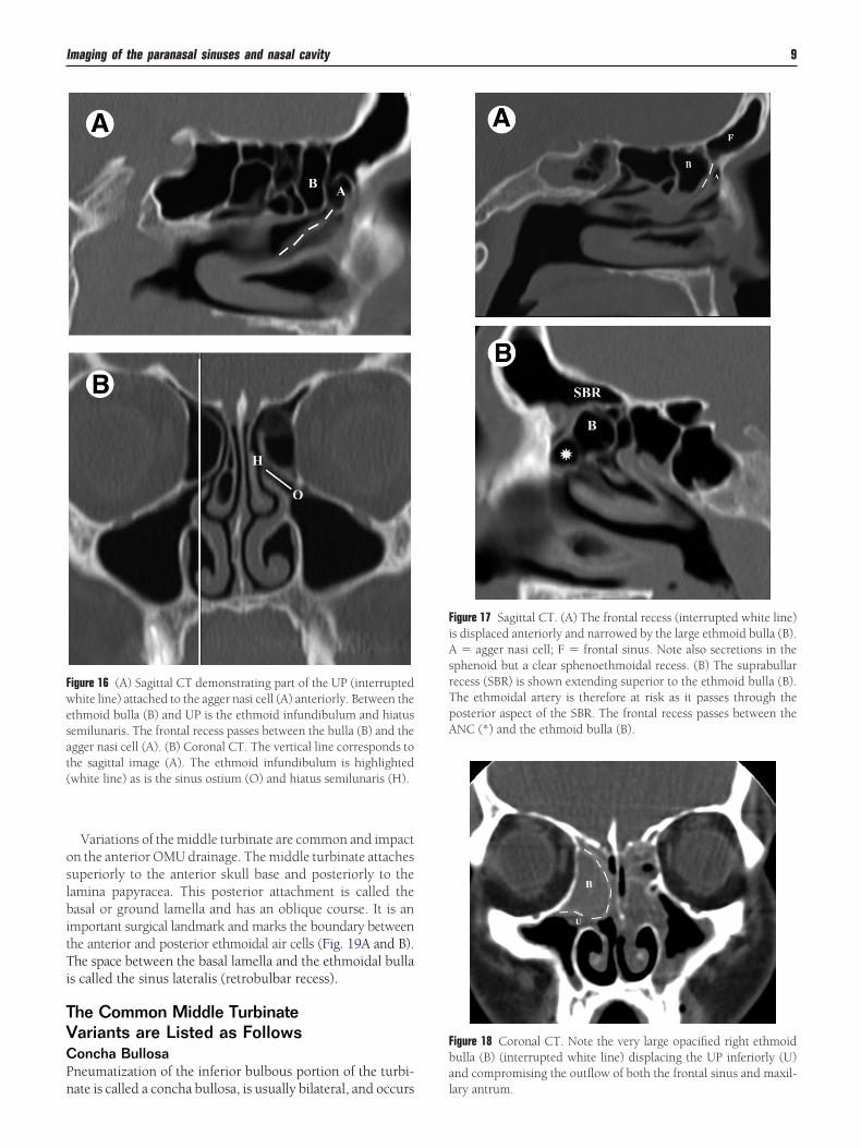
Variations of the middle turbinate are common and impactn the anterior OMU drainage. The middle turbinate attachesuperiorly to the anterior skull base and posteriorly to theamina papyracea. This posterior attachment is called theasal or ground lamella and has an oblique course. It is an
mportant surgical landmark and marks the boundary betweenhe anterior and posterior ethmoidal air cells (Fig. 19A and B).he space between the basal lamella and the ethmoidal bulla
s called the sinus lateralis (retrobulbar recess).
he Common Middle Turbinateariants are Listed as Followsoncha Bullosaneumatization of the inferior bulbous portion of the turbi-
igure 16 (A) Sagittal CT demonstrating part of the UP (interruptedhite line) attached to the agger nasi cell (A) anteriorly. Between the
thmoid bulla (B) and UP is the ethmoid infundibulum and hiatusemilunaris. The frontal recess passes between the bulla (B) and thegger nasi cell (A). (B) Coronal CT. The vertical line corresponds tohe sagittal image (A). The ethmoid infundibulum is highlightedwhite line) as is the sinus ostium (O) and hiatus semilunaris (H).
ate is called a concha bullosa, is usually bilateral, and occurs l
igure 17 Sagittal CT. (A) The frontal recess (interrupted white line)s displaced anteriorly and narrowed by the large ethmoid bulla (B).
� agger nasi cell; F � frontal sinus. Note also secretions in thephenoid but a clear sphenoethmoidal recess. (B) The suprabullarecess (SBR) is shown extending superior to the ethmoid bulla (B).he ethmoidal artery is therefore at risk as it passes through theosterior aspect of the SBR. The frontal recess passes between the
igure 18 Coronal CT. Note the very large opacified right ethmoidulla (B) (interrupted white line) displacing the UP inferiorly (U)nd compromising the outflow of both the frontal sinus and maxil-
ary antrum.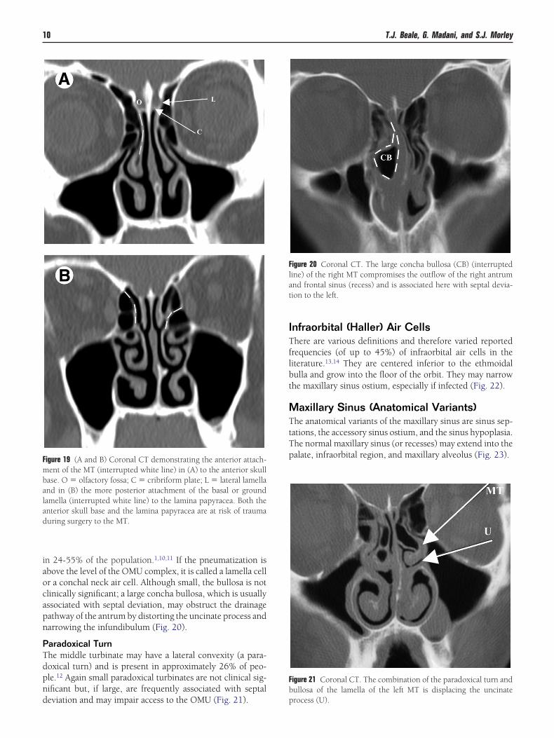
iaocapn
PTdpnd
ITflbt
MTtTpF
mbalad
Flat
Fb
10 T.J. Beale, G. Madani, and S.J. Morley
n 24-55% of the population.1,10,11 If the pneumatization isbove the level of the OMU complex, it is called a lamella cellr a conchal neck air cell. Although small, the bullosa is notlinically significant; a large concha bullosa, which is usuallyssociated with septal deviation, may obstruct the drainageathway of the antrum by distorting the uncinate process andarrowing the infundibulum (Fig. 20).
aradoxical Turnhe middle turbinate may have a lateral convexity (a para-oxical turn) and is present in approximately 26% of peo-le.12 Again small paradoxical turbinates are not clinical sig-ificant but, if large, are frequently associated with septal
igure 19 (A and B) Coronal CT demonstrating the anterior attach-ent of the MT (interrupted white line) in (A) to the anterior skull
ase. O � olfactory fossa; C � cribriform plate; L � lateral lamelland in (B) the more posterior attachment of the basal or groundamella (interrupted white line) to the lamina papyracea. Both thenterior skull base and the lamina papyracea are at risk of traumauring surgery to the MT.
eviation and may impair access to the OMU (Fig. 21). p
nfraorbital (Haller) Air Cellshere are various definitions and therefore varied reported
requencies (of up to 45%) of infraorbital air cells in theiterature.13,14 They are centered inferior to the ethmoidalulla and grow into the floor of the orbit. They may narrowhe maxillary sinus ostium, especially if infected (Fig. 22).
axillary Sinus (Anatomical Variants)he anatomical variants of the maxillary sinus are sinus sep-
ations, the accessory sinus ostium, and the sinus hypoplasia.he normal maxillary sinus (or recesses) may extend into thealate, infraorbital region, and maxillary alveolus (Fig. 23).
igure 20 Coronal CT. The large concha bullosa (CB) (interruptedine) of the right MT compromises the outflow of the right antrumnd frontal sinus (recess) and is associated here with septal devia-ion to the left.
igure 21 Coronal CT. The combination of the paradoxical turn andullosa of the lamella of the left MT is displacing the uncinate
rocess (U).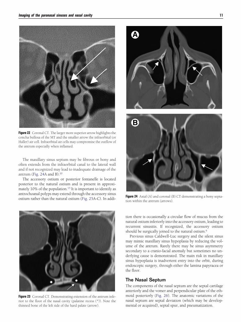
oaa
pmao
tnrs
musdset
TTamn
FcHt
Frt
Ft
Imaging of the paranasal sinuses and nasal cavity 11
The maxillary sinus septum may be fibrous or bony andften extends from the infraorbital canal to the lateral wallnd if not recognized may lead to inadequate drainage of thentrum (Fig. 24A and B).10
The accessory ostium or posterior fontanelle is locatedosterior to the natural ostium and is present in approxi-ately 10% of the population.15 It is important to identify as
ntrochoanal polyps may extend through the accessory sinusstium rather than the natural ostium (Fig. 25A-C). In addi-
igure 22 Coronal CT. The larger more superior arrow highlights theoncha bullosa of the MT and the smaller arrow the infraorbital (oraller) air cell. Infraorbital air cells may compromise the outflow of
he antrum especially when inflamed.
igure 23 Coronal CT. Demonstrating extension of the antrum infe-ior to the floor of the nasal cavity (palatine recess (*)). Note the
mhinned bone of the left side of the hard palate (arrow).
ion there is occasionally a circular flow of mucus from theatural ostium inferiorly into the accessory ostium, leading toecurrent sinusitis. If recognized, the accessory ostiumhould be surgically joined to the natural ostium.6
Previous sinus Caldwell-Luc surgery and the silent sinusay mimic maxillary sinus hypoplasia by reducing the vol-me of the antrum. Rarely there may be sinus asymmetryecondary to a cranio-facial anomaly but sometimes no un-erlying cause is demonstrated. The main risk in maxillaryinus hypoplasia is inadvertent entry into the orbit, duringndoscopic surgery, through either the lamina papyracea orhe floor.
he Nasal Septumhe components of the nasal septum are the septal cartilagenteriorly and the vomer and perpendicular plate of the eth-oid posteriorly (Fig. 26). The anatomic variations of theasal septum are septal deviation (which may be develop-
igure 24 Axial (A) and coronal (B) CT demonstrating a bony septa-ion within the antrum (arrows).
ental or acquired), septal spur, and pneumatization.
FoCoe
Fsa
Fv
12 T.J. Beale, G. Madani, and S.J. Morley
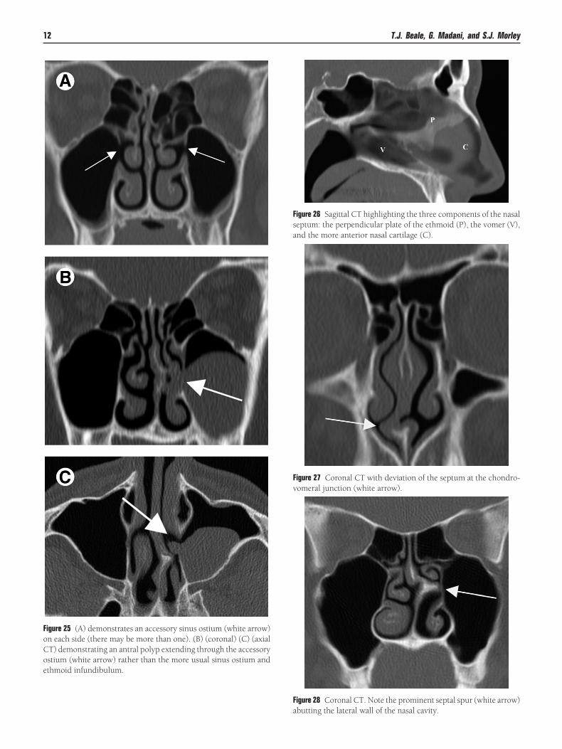
igure 25 (A) demonstrates an accessory sinus ostium (white arrow)n each side (there may be more than one). (B) (coronal) (C) (axialT) demonstrating an antral polyp extending through the accessorystium (white arrow) rather than the more usual sinus ostium and
thmoid infundibulum.Fa
igure 26 Sagittal CT highlighting the three components of the nasaleptum: the perpendicular plate of the ethmoid (P), the vomer (V),
nd the more anterior nasal cartilage (C).igure 27 Coronal CT with deviation of the septum at the chondro-
omeral junction (white arrow).igure 28 Coronal CT. Note the prominent septal spur (white arrow)
butting the lateral wall of the nasal cavity.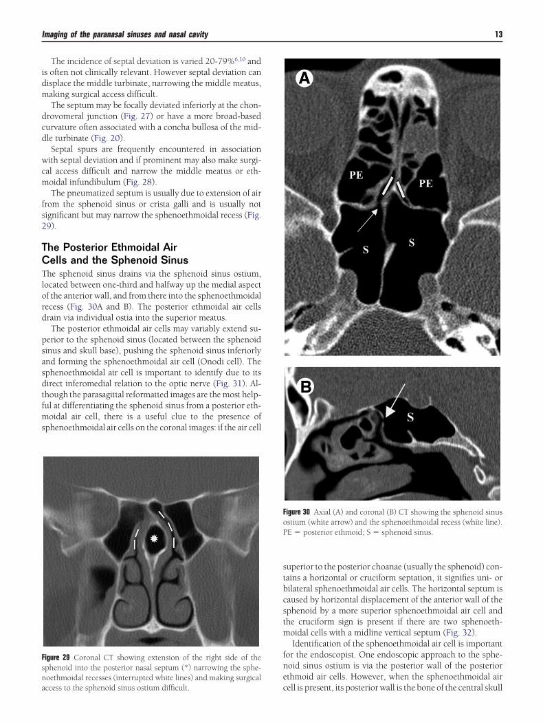
idm
dcd
wcm
fs2
TCTlord
psasdtfms
stbcstm
fne
Fsna
FoP
Imaging of the paranasal sinuses and nasal cavity 13
The incidence of septal deviation is varied 20-79%6,10 ands often not clinically relevant. However septal deviation canisplace the middle turbinate, narrowing the middle meatus,aking surgical access difficult.The septum may be focally deviated inferiorly at the chon-
rovomeral junction (Fig. 27) or have a more broad-basedurvature often associated with a concha bullosa of the mid-le turbinate (Fig. 20).Septal spurs are frequently encountered in association
ith septal deviation and if prominent may also make surgi-al access difficult and narrow the middle meatus or eth-oidal infundibulum (Fig. 28).The pneumatized septum is usually due to extension of air
rom the sphenoid sinus or crista galli and is usually notignificant but may narrow the sphenoethmoidal recess (Fig.9).
he Posterior Ethmoidal Airells and the Sphenoid Sinus
he sphenoid sinus drains via the sphenoid sinus ostium,ocated between one-third and halfway up the medial aspectf the anterior wall, and from there into the sphenoethmoidalecess (Fig. 30A and B). The posterior ethmoidal air cellsrain via individual ostia into the superior meatus.The posterior ethmoidal air cells may variably extend su-
erior to the sphenoid sinus (located between the sphenoidinus and skull base), pushing the sphenoid sinus inferiorlynd forming the sphenoethmoidal air cell (Onodi cell). Thephenoethmoidal air cell is important to identify due to itsirect inferomedial relation to the optic nerve (Fig. 31). Al-hough the parasagittal reformatted images are the most help-ul at differentiating the sphenoid sinus from a posterior eth-
oidal air cell, there is a useful clue to the presence ofphenoethmoidal air cells on the coronal images: if the air cell
igure 29 Coronal CT showing extension of the right side of thephenoid into the posterior nasal septum (*) narrowing the sphe-oethmoidal recesses (interrupted white lines) and making surgical
cccess to the sphenoid sinus ostium difficult.
uperior to the posterior choanae (usually the sphenoid) con-ains a horizontal or cruciform septation, it signifies uni- orilateral sphenoethmoidal air cells. The horizontal septum isaused by horizontal displacement of the anterior wall of thephenoid by a more superior sphenoethmoidal air cell andhe cruciform sign is present if there are two sphenoeth-oidal cells with a midline vertical septum (Fig. 32).Identification of the sphenoethmoidal air cell is important
or the endoscopist. One endoscopic approach to the sphe-oid sinus ostium is via the posterior wall of the posteriorthmoid air cells. However, when the sphenoethmoidal air
igure 30 Axial (A) and coronal (B) CT showing the sphenoid sinusstium (white arrow) and the sphenoethmoidal recess (white line).E � posterior ethmoid; S � sphenoid sinus.
ell is present, its posterior wall is the bone of the central skull
bbsistn
i
rd
shtcia
EThnihtat3
cmo
t
are
FosrM
Fss(
Fcdo
14 T.J. Beale, G. Madani, and S.J. Morley
ase. Access to the posterior ethmoidal air cells should thene made by penetrating the basal (ground) lamella. If thephenoethmoidal recess and sinus ostium need inspect-ng, it is important to dissect medially, by displacing theuperior turbinate, rather than dissecting laterally throughhe posterior ethmoidal air cells, which places the opticerve at risk.
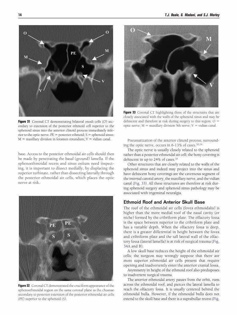
igure 31 Coronal CT demonstrating bilateral onodi cells (O) sec-ndary to extension of the posterior ethmoid cell superior to thephenoid sinus into the anterior clinoid process immediately infe-ior to the optic nerve. PE � posterior ethmoid; S � sphenoid sinus;
� maxillary division in foramen rotundum; V � vidian canal.
igure 32 Coronal CT demonstrated the cruciform appearance of thephenoethmoidal region on the same coronal plane as the choanaeecondary to posterior extension of the posterior ethmoidal air cells
ePE) superior to the sphenoid (S).
Pneumatization of the anterior clinoid process, surround-ng the optic nerve, occurs in 6-13% of cases.10,14
The optic nerve is usually closely related to the sphenoidather than a posterior ethmoidal air cell; the bony covering isehiscent in up to 24% of cases.16
Other structures that are closely related to the walls of thephenoid sinus and indeed may project into the sinus andave dehiscent bony coverings are the cavernous segment ofhe internal carotid artery, the maxillary nerve, and the vidiananal (Fig. 33). All these structures are therefore at risk dur-ng sphenoid surgery and sphenoid sinus pathology may bessociated with trigeminal neuralgia.
thmoid Roof and Anterior Skull Basehe roof of the ethmoidal air cells (fovea ethmoidalis) isigher than the more medial roof of the nasal cavity (oriche) formed by the cribriform plate. The olfactory fossa
s the space between superior to the cribriform plate andas a variable depth. When the olfactory fossa is deep,here is a greater differential in height between the foveand cribriform plate and the tall lateral wall of the olfac-ory fossa (lateral lamella) is at risk of surgical trauma (Fig.4A and B).A low skull base reduces the height of the ethmoidal air
ells; the surgeon may wrongly suppose that there areore superior ethmoidal air cells present that require
pening and inadvertently enter the anterior cranial fossa.Asymmetry in height of the ethmoid roof also predisposes
o inadvertent surgical trauma.The anterior ethmoidal artery passes from the orbit, runs
cross the ethmoidal roof, and pierces the lateral lamella toeach the olfactory fossa. It is usually centered behind thethmoidal bulla. However, if the ethmoidal bulla does not
igure 33 Coronal CT highlighting three of the structures that arelosely associated with the walls of the sphenoid sinus and may beehiscent and therefore at risk during surgery to this region. O �ptic nerve; M � maxillary division 5th nerve; V � vidian canal.
xtend to the skull base and there is a suprabullar recess (Fig.
1e
nia
CIstru
R
1
1
Fatcd
FcpW
Imaging of the paranasal sinuses and nasal cavity 15
7A and B), the artery may be exposed and travel on a mes-ntery and be at risk of injury.
The anterior ethmoidal canal should be identified on coro-al images (Fig. 35). It is important to note whether the artery
s closely applied to the skull base, has a bony covering, is onmesentery, or if the suprabullar recess is present.17
igure 34 (A and B) Coronal CT demonstrating a deep olfactory fossand therefore low cribriform plate (C). Both the cribriform plate andhe lateral lamella (L) are at risk if frontoethmoidal surgery is beingonsidered. (B) The measurement (8.4 mm) highlights the vertical
epth of the olfactory fossa in this case.onclusiont is important to have a logical approach to assessing theinonasal anatomy and understand the anatomical variantshat are relevant to the surgeon. The CT study should beeviewed in all three orthogonal planes in order to accuratelynderstand this complex region.
eferences1. Earwacker J: Anatomic variants in sinonasal CT. Radiographics 13:381-
415, 19932. Lee WT, Kuhn FA, Citardi MJ: 3D computed tomographic analysis of
frontal recess anatomy in patients without frontal sinusitis. Otolaryn-gology Head Neck Surg 131(3):164-173, 2004
3. Stammberger H, Kopp W, Dekornfeld TJ, et al: Functional endoscopicsinus surgery? Eur Arch Otorhinolaryngol 63-76, 1990
4. Kasper KA: Nasofrontal connections. A study based on one hundredconsecutive dissections. Arch Otolaryngol 23:322-343, 1936
5. Kuhn FA, Javer AR: Primary endoscopic management of the frontalsinus. Oper Tech Otolaryngol Head Neck Surg 7:222-229, 1996
6. Wormald P-J: Endoscopic Sinus Surgery. New York, NY, Thieme,2005
7. Bruner E, Jacobs JB, Lebowitz RA, et al: Role of the agger asi air cellin chronic frontal sinusitis. Ann Otol Laryngol 105:694-700, 1996
8. Owen RG, Kuhn FA: Supraorbital ethmoid cell. Otolaryngol HeadNeck Surg 116:254-261, 1997
9. Joe J, Ho S, Yanagisawa E: Documentation of variations in sinonasalanatomy by intraoperative nasal endoscopy. Laryngoscope 110:229-235, 2000
0. Sarna A, Hayman A, Laine F, et al: Coronal imaging of the ostiomeatalunit: anatomy of 24 variants. J Computer Assisted Tomogr 26(1):153-157, 2002
1. Zeinrich SJ, Mattox DE, Kennedy DW, et al: Concha bullosa: CT eval-
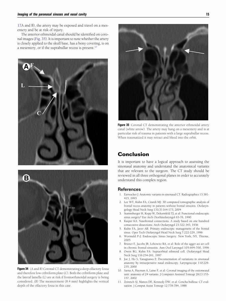
igure 35 Coronal CT demonstrating the anterior ethmoidal arteryanal (white arrow). The artery may hang on a mesentery and is atarticular risk of trauma in patients with a large suprabullar recess.hen traumatized it may retract and bleed into the orbit.
uation. J Comput Assist Tomogr 12:778-784, 1988

1
1
1
1
1
1
16 T.J. Beale, G. Madani, and S.J. Morley
2. Cannon CR: Endoscopic management of concha bullosa. OtolaryngolHead Neck Surg 110:449-454, 1994
3. Stammberger H, Wolf G: Headaches and sinus disease: the endoscopicapproach. Ann Otol Rhinol Laryngol Suppl 134:3-23, 1988
4. Bolger WE, Butzin CA, Parsons DS: Paranasal sinus bony anatomicvariations and mucosal abnormalities: CT analysis for endoscopic sinus
surgery. Laryngoscope 101:56-64, 19915. Jog M, McGarry GW: How frequent are accessory sinus ostia? J Laryn-gol Otol 117:270-272, 2003
6. Maniscalo JE, Habal MB: Microanatomy of the optic canal. J Neurosurg48:402-406, 1978
7. Bayram M, Sirikci A, Bayazit YA: Important anatomic variations of thesinonasal anatomy in light of endoscopic surgery: a pictorial review.
Eur Radiol 11:1991-1997, 2001