Imaging of Focal Lung Lesions
68
IMAGING OF FOCAL LUNG LESIONS DR SAKHER-ALKHADERI CONSULTANT RADIOLOGIST AMC
-
Upload
sakher-alkhaderi -
Category
Health & Medicine
-
view
1.805 -
download
1
Transcript of Imaging of Focal Lung Lesions

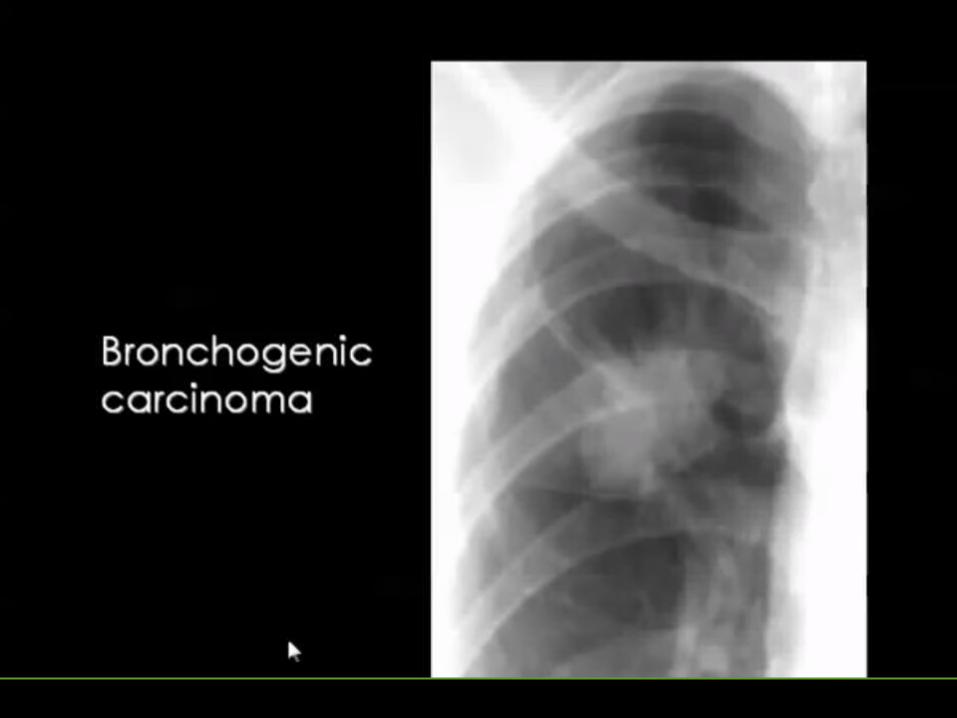
IMAGING OF FOCAL LUNG LESIONS
DR SAKHER-ALKHADERICONSULTANT RADIOLOGIST AMC


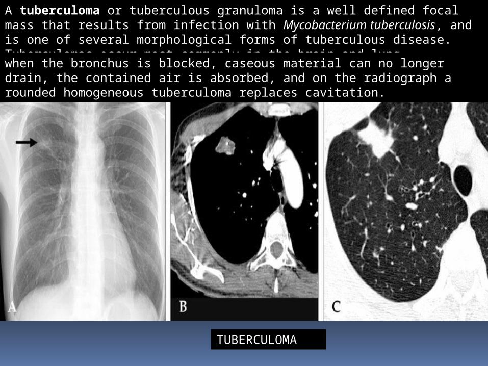
A pulmonary hamartoma is a benign neoplasm composed of cartilage, connective tissue, muscle, fat, and bone. It is one of the commonest benign tumours of the lung, and accounts for ~8% of all lung neoplasms and 6% of solitary pulmonary nodules.


PULMONARY HAMARTOMA

PULMONARY HAMARTOMA
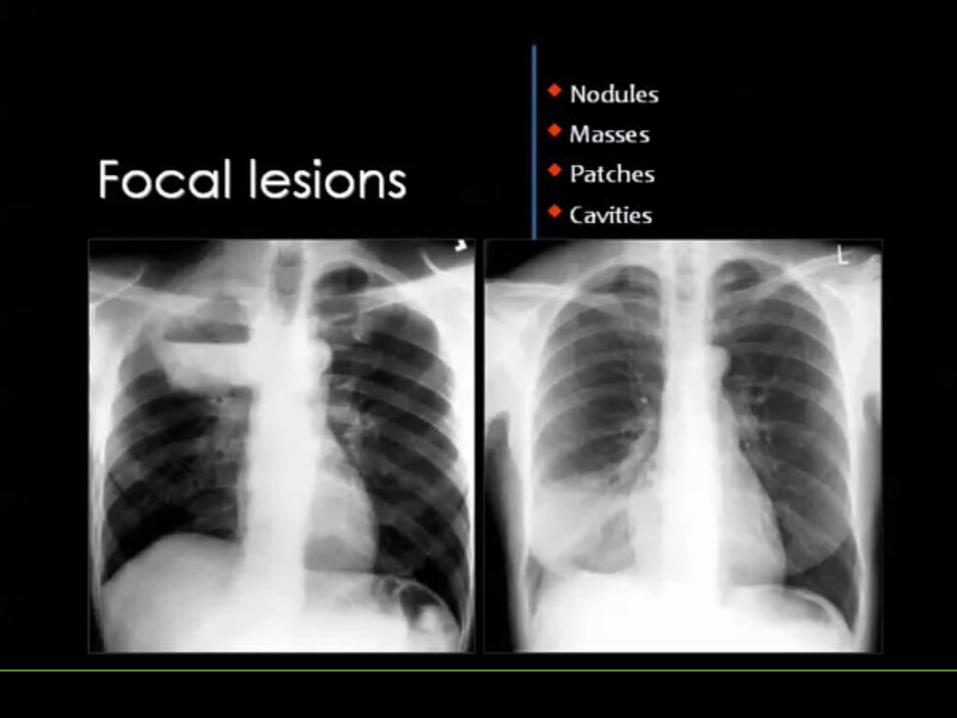
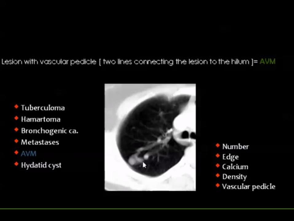
TUBERCULOMA
A tuberculoma or tuberculous granuloma is a well defined focal mass that results from infection with Mycobacterium tuberculosis, and is one of several morphological forms of tuberculous disease. Tuberculomas occur most commonly in the brain and lung.when the bronchus is blocked, caseous material can no longer drain, the contained air is absorbed, and on the radiograph a rounded homogeneous tuberculoma replaces cavitation.


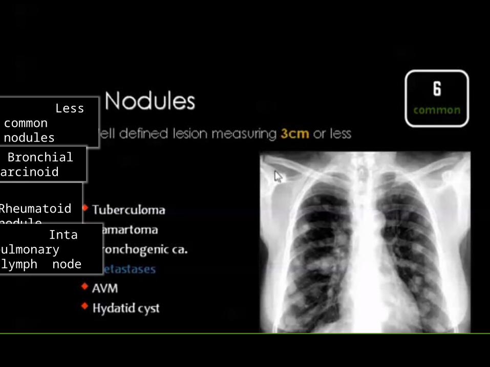
Less common nodules Bronchial
carcinoid
Rheumatoid nodule
Inta pulmonary lymph node

BRONCHIAL CARCINOID TUMOR

Clinical presentationPresentation can vary dependent on location. Central neoplasms usually give symptoms due to bronchial obstruction (such symptoms can include pneumonia, atelectasis, bronchiectasis, emphysema or even a lung abscess); if airway obstruction is partial, symptoms such as cough, wheezing and recurrent pulmonary infections can occur. Peripheral tumours on the contrary are generally asymptomatic and they are discovered occasionally. Most (~60%) tend to be central within the tracheo-bronchial tree 3.

Rheumatoid pulmonary nodules are a rare pulmonary manifestation of rheumatoid arthritis. The are thought to occur in <1% of patients with rheumatoid arthritis.Rheumatoid nodules tend to be located in peripheral zones of the upper and middle lung with tendency of cavitation .

INTRAPULMONARY LYMPHNODE


Chronic

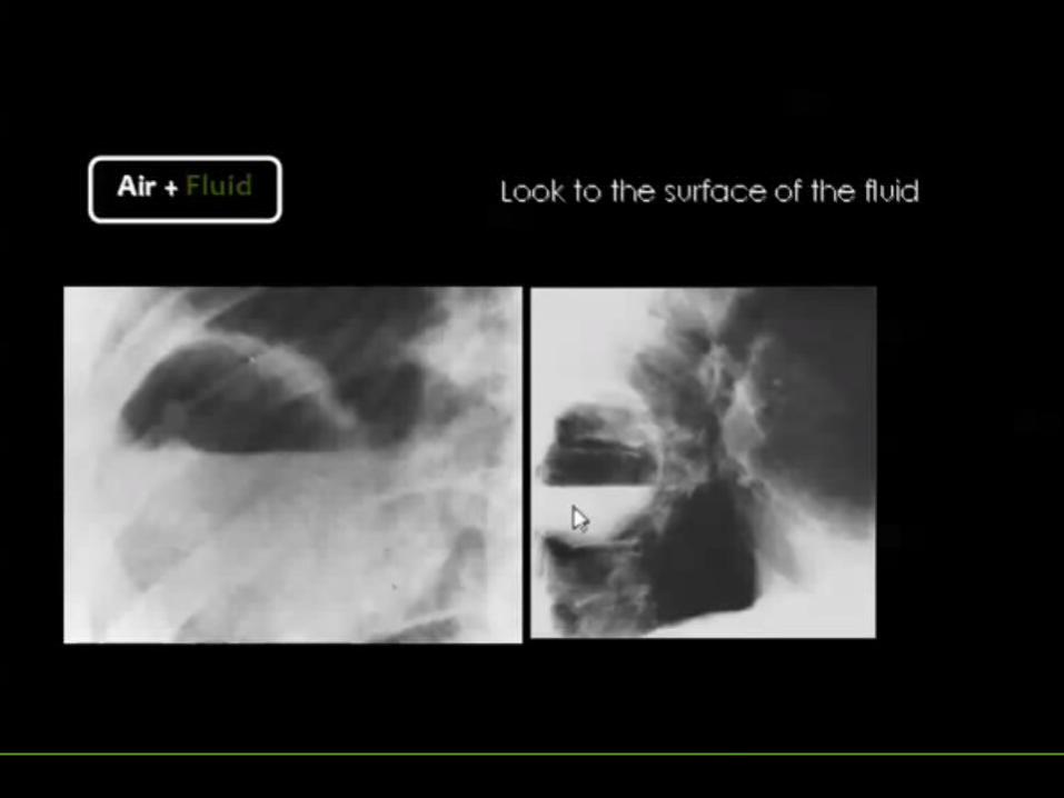
PNEUMATOCELE

Pneumatocoeles are intrapulmonary air-filled cystic spaces that can have a variety of sizes and appearances. They may contain air-fluid levels and are usually the result of ventilator-inducted lung injury in neonates or post-pneumonic. They should not be mistaken for a cavitating lung mass. The majority of pneumatocoeles occur as a result of pneumonia (post pneumotic pneumatocoele).
In addition to infection, pneumatococele are also seen in a number of other settings including:trauma - usually blunt traumapositive pressure ventilation, especially in preterm neonates 4
hydrocarbon ingestion-smooth inner margins-contain little if any fluid-wall (if visible) is thin and regular-persist despite absence of symtpoms


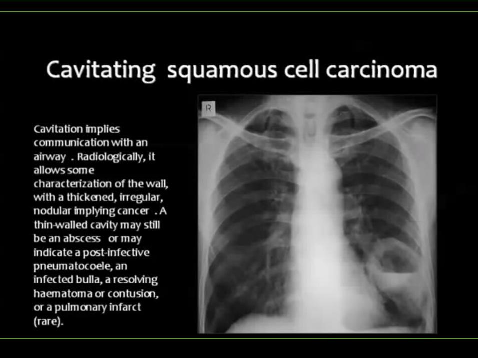

THE END