Thermal-Optical-Transmittance Analysis for Organic, Elemental ...

300 J. Opt. Soc. Am. A/Vol. 14, No. 1 /January 1997 Zhu et al.
Imaging objects in tissuelike media with opticaltagging and the diffuse
photon differential transmittance
X. D. Zhu and Sung-po Wei
Department of Physics, University of California, Davis, Davis, California 95616
Xiao Wen Guo
Department of Biological Chemistry, School of Medicine, University of California, Davis, Davis, California 95616
Received December 19, 1995; revised manuscript received July 5, 1996; accepted July 8, 1996
In a proof-of-concept experiment, we optically tagged objects embedded in an inhomogeneous and multiple-scattering medium and measured the difference between the transmitted diffuse photons at two optical wave-lengths, one at and the other off a sharp absorption peak of the exogenous contrast agent. We demonstratedthat the visibility of tagged objects was significantly enhanced in comparison with that of untagged objects.From our analysis it seems possible to use dual-wavelength differential transmittance spectrometry togetherwith monoclonal-antibody-delivered optical contrast agents to detect tumors as small as 0.1–0.3 cm in size andembedded as deeply as a few centimeters beneath a tissue surface. © 1997 Optical Society of America.[S0740-3232(97)01301-X]
1. INTRODUCTIONIn recent years the propagation and distribution of diffusephotons in highly scattering media have attracted consid-erable attention in the field of medical imaging.1–19 Thisinterest is motivated by the fact that near-infrared elec-tromagnetic waves from 730 to 900 nm are strongly scat-tered and yet only weakly absorbed in human tissuessuch as skin and breast.7 Since light is multiply scat-tered, its propagation characteristics can be described bya diffusion approximation.20,21 The latter is perturbed byinhomogeneities with differing optical properties, such astumor growths in an otherwise normal tissue. By mea-suring the transmitted or the reflected diffuse photons,one may deduce information about the locations and sizesof these inhomogeneities with sufficient certainty. Eventhough the extraction of imaging information from diffuselight is complicated and less straightforward in compari-son with the x-ray-attenuation-based imaging techniques,the research efforts of many groups have continued to befueled by the possibility that the near-infrared optical im-aging technique in some form may eventually become anoninvasive alternative to x-ray mammography for earlydetection of cancerous growths.1–19,22,23
In the past decade diffuse photon spectrometry in theform of transillumination light-scanning spectroscopy hasbeen explored and tested in the field of medicalimaging.1–6 By detecting the transmittance of diffuselight at red and near-infrared wavelengths with an eight-bit video imaging camera, one obtains either a differentialtransmittance or a relative transmittance. The identifi-cation of cancerous growth was based on empirical obser-vations that malignant tumors, accompanied by higherdensities of blood vessels, cause either an enhanced re-gional or a total absorption in one breast while not in the
0740-3232/97/010300-06$10.00 ©
other, or they cause a relative enhanced absorption of thenear-infrared light. In a series of clinical tests in the1980’s, however, the transillumination light-scanningtechnique was shown to be inferior to x-ray mammogra-phy because of its lack of specificity and its inadequatesensitivity to nonpalpable tumors of sizes less than 1 cmand to even larger tumors that are embedded a few cen-timeters beneath the skin surface.3–5 It is recognizedthat with improved understanding of light propagation inhighly scattering media and the advancement of the de-tection and biotechnology, one should be able to improvethe sensitivity, specificity, and spatial resolution of light-scan transillumination.6 At the present time, diffusephoton or transillumination imaging for cancer detectionremains experimental.Significant progress has been made recently as a
result of strong research efforts in both condensed matterphysics and medical imaging communities to better un-derstand light propagation in multiple-scatteringmedia.6,7,11–19,20 For example, it is well known now thatmodulating the amplitude of an illuminating light sourcecauses the alternating portion of the diffuse photon den-sity to behave much like a damped wave that interferesand diffracts.11–14 By exploiting the interference effect,many groups have been able to improve the spatial reso-lution of diffuse photon imaging to a fraction of amillimeter.11–14,17 The high accuracy in locating objectsin multiple-scattering media has benefited from a morequantitative understanding of diffuse photon prop-agation.15–19 It is also recognized that the use of opticalcontrast agents in transillumination imaging improvesthe sensitivity.1,12 The same concept has already beenused in magnetic resonance imaging. In the studies per-formed so far, optical contrast agents were explored
1997 Optical Society of America
Zhu et al. Vol. 14, No. 1 /January 1997 /J. Opt. Soc. Am. A 301
mainly as a means to enhance the overall absorption of anoptically tagged tissue in the near-infrared region from730 to 900 nm. On the technology side, it has recentlybecome feasible to deliver large enough amounts of con-trast agents with monoclonal antibodies to specifictumors.24 However, one crucial question remains as towhether diffuse photon imaging with the current use ofoptical contrast agents would have a sufficient specificityin detecting tumors in a human breast, where there existnormal inhomogeneities such as macro cysts. This is thesubject of this paper.We intend to show another aspect of optical contrast
agents that can be exploited in diffuse photon imaging todramatically enhance the visibility of a tumor in the pres-ence of other lumpy structures that are present even in anormal human breast. Our approach is to exploit thesharpness of the absorption peak of an optical contrastagent, not just the enhanced optical absorption. Thisform of dual-wavelength differential measurement en-ables us to distinguish with significantly improved con-trast optically tagged objects from untagged lumpy struc-tures. As far as we know, this aspect has not beenexplored by others. To prove the concept, we conduct acontinuous-wave or d.c. transillumination imaging ex-periment in which we selectively tag objects embedded ina tissue phantom with an optical contrast agent. Wethen measure the difference in the transmittance at twooptical wavelengths, one at an absorption maximum ofthe agent and the other off the maximum on the steepside of the absorption peak. We find that the visibility ofthe tagged objects is significantly enhanced in comparisonwith that of the untagged objects. Our preliminary re-sults further show that by using 16-bit rather than 8-bitCCD detectors, we can improve the sensitivity of tumordetection such that it seems feasible to detect opticallytagged tumors as small as 0.1–0.3 cm in size and embed-ded as deep as a few centimeters beneath the skin surfaceof human tissue.
2. SAMPLE PREPARATION ANDEXPERIMENTAL PROCEDURESA sketch of the experimental setup is shown in Fig. 1.We use suspended TiO2 powder (supplied by Sigma Coat-ings) in glycerin at a concentration of 0.7% by volume andan Intralipid emulsion at a concentration of 20% byweight (from Kabi Pharmacia, Clayton, N.C.) as tissuephantoms. They are contained in a 5-mm 3 50-mm3 50-mm glass cell. For objects or inhomogeneities, weuse glass capillary tubes and 0.3-mm Pentel carbon fila-ment. The capillary tubes have inner diameter (i.d.) of0.3 mm and outer diameter (o.d.) of 1.2 mm and can befilled with optical contrast agents. For the latter, we usesolutions of dichlorotriazinylaminofluorescein (DTAF) indimethyl sulfoxide (DMSO) (from Calbiochem, La Jolla,Calif., lmax 5 492 nm, emax 5 82 cm21 mM21, M.W.5 531.7). From the measurements of Blakeslee andBaines, we know that the extinction coefficient e at 488.0nm is close to 80% of the maximum, drops to 20% of themaximum at 514.5 nm, and is essentially zero at 632.8nm.25 We thus choose two of the emission lines of an
Argon-ion laser, one at 488.0 nm and the other at 514.5nm, and a He–Ne laser at 632.8 nm as illuminating lightsources. We measured the light-scattering mean freepaths in the two tissue phantoms using the same methodas described in Ref. 26. In the TiO2 suspension, the val-ues at all three wavelengths are essentially the same, lsc5 15.0 mm 6 0.3 mm. In the Intralipid emulsion, thescattering mean free paths are more dispersive, with lsc5 8.60 6 0.02 mm at 488.0 nm, lsc 5 9.77 6 0.02 mm at514.5 nm, and lsc 5 16.15 6 0.06 mm at 632.8 nm. Ourmeasurement compares well with that of van Staverenand co-workers; in particular, our result reproduces thewavelength dependence of lsc } l2.4 reported by theseauthors.27 The optical path length of the cell (L 5 5mm) thus covers 300–500 scattering mean free paths.We expand the laser beam with a combination of lenses
such that one of the 50-mm 3 50-mm cell surfaces is uni-formly illuminated. We measure the near-field distribu-tion of the transmitted light from the opposite surface.This is done by imaging an area of 46 mm 3 46 mm onthe opposite (back) surface at a 4-to-1 reduction ratio ontothe detection surface of either a CCD camera or a scan-ning photodiode. The solid angle of collection is 33 1023 steradians, so we can neglect the influence of theglass–air interface on the intensity distribution. TheCCD camera is a 16-bit TK512CB/AR (Photometrics, Tuc-son, Ariz.). The photodiode is a EG&G FND100 (EG&GPhoton Devices, Sunnyvale, Calif.). With the photodiodewe modulate the amplitude of the illuminating lightsources at a low frequency of 1000 Hz and detect the a.c.component of the photocurrent with a lock-in amplifier.With the CCD camera, we use the unmodulated illumi-nating sources and detect the total photon counts. Wetypically accumulate 5 3 105 photon counts in each pixel.In all the measurements we first record the near-field
distribution of the transmitted diffuse photons with onlythe tissue phantom; we then insert objects into the celland repeat the measurement again. We take the ratio ofthe two data sets as the normalized transmittance.When the CCD detector is used, we also take care to sub-tract the dark counts accumulated over the same expo-sure time period from the two data sets before taking theratio.
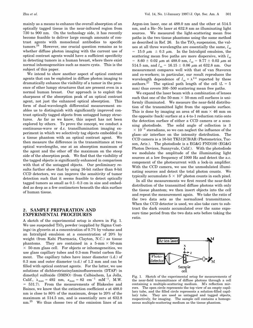
Fig. 1. Sketch of the experimental setup for measurements ofthe near-field transmittance of diffuse photons through a cellcontaining a multiple-scattering medium. M’s reflection mir-rors. The open circle represents the top view of an empty capil-lary tube, and the filled circle represents a solution-filled capil-lary tube. They are used as untagged and tagged objects,respectively, for imaging. The sample cell contains a homoge-neous multiple-scattering medium as the tissue phantom.

302 J. Opt. Soc. Am. A/Vol. 14, No. 1 /January 1997 Zhu et al.
3. EXPERIMENTAL RESULTSTo demonstrate the sharp wavelength dependence of thenear-field transmittance as a result of optical tagging ofan embedded object, we use a capillary tube filled with aDTAF solution at a concentration of 7.6 3 1022 molar(M). The tube is placed (along the y axis) in the middle ofthe cell, parallel to both the front (the illuminated) andthe back surface. In this measurement the TiO2 suspen-sion (in glycerin) is used as the tissue phantom, and thephotodiode is used to scan horizontally across the imageplane (along the x axis). Hence the object is at a position160 scattering mean paths from both surfaces of the tis-sue phantom. The measurement is performed at each ofthe three optical wavelengths; the results are displayed inFig. 2. The near-field transmittance dips at the lateralposition (the x coordinate) where the tube is placed. Thisfeature reproduces the observation made by den Ouderand co-workers.17 The most important feature here isthe sharp wavelength dependence. The magnitude of thedip decreases as the wavelength increases from 488.0 to632.8 nm. In fact, at l 5 632.8 nm, where DTAF is nolonger absorbant, the dip disappears and the near-fieldtransmittance bears a weak feature that is typical of anonabsorbing object in a highly scattering medium.17
The difference between any two of the three curves inFig. 2 may serve as an example of the enhancement of theobject visibility in a dual-wavelength differential trans-mittance. To demonstrate this aspect more directly, weshow in Fig. 3 the near-field transmittance at 514.5 and488.0 nm with three objects in the cell. One object is anempty capillary tube. The second one is a capillary tubethat contains the DTAF solution (7.6 3 1022 M). Thethird object is a 0.3-mm-diameter Pentel carbon filamentas a perfect absorber. Also shown in Fig. 3 is the differ-ence between the transmittance at 514.5 nm and the oneat 488.0 nm, which we call the differential transmittance.The visibility of the tube containing DTAF is clearly en-hanced as the visibility of the spectrally flat empty tubeand carbon filament are suppressed in the differentialtransmittance. The signal-to-noise ratios displayed inFigs. 2 and 3 are limited by the total data acquisitiontime. The latter is limited to a few minutes by the down-ward drift of TiO2 powder toward the cell bottom. Overthe few minutes’ time, we had to perform measurementsat 200 sampling positions.To show that the signal-to-noise ratio can be improved
with a stable tissue phantom and to assess properly thesensitivity of the dual-wavelength differential transmit-tance to small objects, we performed a series of measure-ments with the Intralipid emulsion as the tissuephantom.27 The latter is stable in the air for tens ofhours. Furthermore, we use a 16-bit CCD camera (Pho-tometrics TK512CB/AR) as a photodetector so that we cansimultaneously record the near-field transmittance at allsampling positions on the image plane. Assuming thatthe noise is limited by the photon statistics, this enablesus to achieve the highest signal-to-noise ratio for a fixeddata acquisition time. In the results shown below, we ac-cumulate roughly 5 3 105 photon counts in each pixel be-fore normalization. In Figs. 4(a) and 4(b) we display thenear-field transmittance over an area 3.2 3 1.3 mm on
the back surface of the cell. We have as objects an emptycapillary tube and a tube filled with a DTAF solution at3.8 3 1022 M. These objects are roughly in the middle ofthe tissue phantom. In Fig. 4(c) we display the differen-tial transmittance. We note again that the visibility ofthe tube with the DTAF solution is preferentially en-hanced. Furthermore, the signal-to-noise ratio is alsogreatly improved. In fact, the peak-to-peak noise isroughly 0.2–0.3% in the differential transmittance, whichis predominantly statistical, associated with 5 3 105 pho-ton counts per pixel. The residual signal from the emptytube is the result of the difference in the scattering meanfree paths at 514.5 and 488.0 nm.
Fig. 2. Normalized near-field transmittance of diffuse photonsat three wavelengths. The sample cell is filled with a TiO2 sus-pension (in glycerin) at a volume concentration of 0.7%. In themiddle of the cell is a 0.3-mm-i.d. capillary tube filled with aDTAF solution (in DMSO) at a concentration of 7.6 3 1022 M.
Fig. 3. Normalized near-field transmittance at 514 and 488 nm.The sample cell is filled with a TiO2 suspension (in glycerin) at avolume concentration of 0.7%. In the middle are an empty 0.3-mm-i.d. capillary tube, a 0.3-mm capillary tube filled with aDTAF solution (in DMSO) at 7.6 3 1022 M, and a 0.3-mm-diameter Pentel carbon filament. Also displayed is the differ-ence between the transmittance at 514 nm and the one at488 nm.

Zhu et al. Vol. 14, No. 1 /January 1997 /J. Opt. Soc. Am. A 303
Fig. 4. (a) Normalized near-field transmittance at 514 nm overan area 3.2 mm 3 1.3 mm on the exit surface. The sample cellis filled with a 20% Intralipid emulsion (see text). It also con-tains two capillary tubes. On the far left is the feature causedby the tube filled with a DTAF solution (in DMSO) at 3.83 1022 M; on the right is the feature caused by the emptytube. (b) Normalized near-field transmittance at 488 nm overthe same area under the same condition. (c) The difference be-tween the transmittance at 514 nm and the one at 488 nm. Thevisibility of the tube with the DTAF solution is clearly enhancedin the differential transmittance.
Fig. 5. Near-field differential transmittance between the trans-mittance at 514 nm and that at 488 nm. The sample cell is filledwith a 20% Intralipid emulsion. In the middle of the cell is a0.3-mm-i.d. capillary tube containing a 0.5-mm-long segment ofDTAF solution (in DMSO) at 3.8 3 1022 M.
To quantify the sensitivity of the differential transmit-tance to a minute amount of optical contrast agents local-ized in a tissue phantom, we use a capillary tube thatholds a small segment of DTAF solution. The dimensionof the segment is 0.5 mm in length and 0.3 mm in diam-eter. At the concentration of 3.8 3 1022 M the segmentcontains 1.3 3 1029 moles of DTAF. The tube is placedin the middle of the cell or 250 scattering mean free pathsfrom both the front and the back surfaces of the tissuephantom. In Fig. 5 we display a line scan of the differ-ential transmittance along the axis of the segment. Thepeak of the differential transmittance is 0.013, or 1.3% ofthe total normalized transmittance. For a small solutionsegment the spatial distribution of the differential trans-mittance far from the segment is independent of the seg-ment shape, so the peak change measures the totalamount of the contrast agents. The noise of 0.3% isagain limited predominantly by photon statistics.
4. DISCUSSIONFrom the experimental results presented in Section 3, itis clear that using optical contrast agents in a dual-wavelength differential transmittance measurement withthe two wavelengths near a sharp absorption peak of theagent dramatically improves the visibility of the opticallytagged objects. We emphasize here that simply taggingthe objects of interest without exploiting the sharp ab-sorption feature of the contrast agent is not sufficient foridentifying the tagged objects in the presence of other un-tagged and yet more massive inhomogeneities. Conse-quently, our approach clearly enhances the specificity oftransillumination imaging, provided that the optical con-trast agents can be delivered with specificity through, forexample, monoclonal antibodies. Such monoclonal anti-bodies in fact exist and have been used to deliver contrastagents suitable for magnetic resonance imaging of breastcancer.28
It is also clear that the use of a 16-bit CCD camera im-proves the signal-to-noise ratio and in turn the sensitivityof transillumination imaging when the noise is limited byphoton statistics (as is the case in our investigation).The improvement of the sensitivity with the use of a 16-bit CCD camera over that with an 8-bit CCD camera canbe understood as follows. For scientific CCD cameras theachievable detection limit is limited by the readout noise,which is typically one analog-to-digital conversion unit.For a 16-bit camera this corresponds to one part in65,000, or 0.0015%. For an 8-bit camera it is one part in256, or 0.4%. If we require a detection signal-to-noiseratio of 4, the minimum detectable differential trans-mittance will be 1.6% for an 8-bit camera and 0.006% fora 16-bit camera. Therefore by using 16-bit or even 18-bit cameras as photodetectors, we should be able toimprove the sensitivity of transillumination imaging bytwo to three orders of magnitude from what was achiev-able in the previous transillumination light-scanningstudies.1–5,29,30
There remain a number of practical issues that shouldbe addressed if the present improvement is to lead to aviable optical method for early detection of small cancer-ous growths in human breasts. The first is whether the

304 J. Opt. Soc. Am. A/Vol. 14, No. 1 /January 1997 Zhu et al.
absolute sensitivity of the present differential transmit-tance technique is adequate under realistic conditions oftissues and deliverable concentrations of optical contrastagents. Since the noise in our measurement is limited byphoton statistics, it can be reduced further by a factor of50 by simply increasing the photon counts. In that casewe should be able to detect 25 picomoles of DTAF (withthe same signal-to-noise ratio of 4) on a tagged object at adistance of 250 scattering mean free paths from a tissuesurface. Since the average thickness of a compressed hu-man breast is 4.2 cm and the scattering mean free path inthe near-infrared transparency window of a humanbreast is 0.06 cm,7 the middle of a compressed humanbreast is only 35 scattering mean free paths from the skinsurface. This means that we should be able to detect atumor in the middle of a human breast carrying only 4 pi-comoles of optical contrast agents such as DTAF.15,17
Moreover, the detection limit can be further reduced toless than 2 picomoles if indocyanine green (ICG) (lmax5 805 nm, emax 5 200 cm21 mM21, M.W. 5 770) isused as a contrast agent. ICG is 2.5 times as absorptivein the near-infrared as DTAF in the visible range.7 For atumor 0.1 cm in diameter, this translates to a concentra-tion of 1–2 nanomoles or 2–3 mg of indocyanine green pergram of tumor, or 2 3 1023 mM. At this level of opticaltagging, the absorption coefficient of the tagged tumor atlmax 5 805 nm is ma 5 0.4 cm21, which is ten times aslarge as the averaged absorption coefficient of a normalhuman breast. Such a concentration is far below the safelevel of 50 mg per gram of tumor approved by the U.S.Food and Drug Administration. The question then be-comes whether such a concentration of indocyanine greencan be delivered to a designated tumor. In nuclear medi-cine, monoclonal antibodies are often used to deliver ra-dioactive labeling or heavy elements to specific tissuessuch as malignant tumors for radioimmunodiagnosis orradioimmunotherapy. It is common that a concentrationof 50 picomoles or more of antibodies per gram of tumor isdelivered. Usually, 10–20 radioactive labelings can becarried by one antibody with current signal amplificationschemes.31 Consequently, it should be fairly easy to de-liver 0.5–2 nanomoles of optical contrast agent such as in-docyanine green. This is already adequate for our pro-posed goal to detect 0.1 cm tagged tumor with a photonstatistics-limited signal-to-noise ratio better than 4. Re-cently Kassis and co-workers have demonstrated that thenumber of labelings per monoclonal antibody can be en-hanced even further.24 If the targeted-signal augmenta-tion reaches 200–500, we expect to be able to deliver adosage of 10–25 nanomoles of indocyanine green pergram of tumor.32 At such a dosage level we should beable to detect tumors 0.1 cm in size and embedded a fewcentimeters beneath the skin surface with a signal-to-noise ratio much better than 4.In the present study, we use the empty glass tube and
the Pentel filament to simulate inhomogeneities that arenot tagged by optical contrast agents. In reality, the in-homogeneous structures, including blood vessels that al-ready exist in a normal human breast, do not have as flatan optical response as our phantom objects. In addition,the absorption of a normal tissue should also vary to somedegree with the optical wavelength within the near-
infrared window.7 It is possible to find a region in thenear-infrared spectral window where the absorption ofthe tissue varies little with the wavelength. The dual-wavelength transmittance is preferably carried out in thisregion. We need more spectroscopic information to fur-ther quantify and model the residual contributions fromthe normally present lumpy structures in a breast. Em-pirically, the differential transmittance from these struc-tures can be measured in the absence of optical contrastagents. Once their contributions are characterized, theycan be subtracted from the transmittance obtained afterthe optical contrast agents are delivered. The effective-ness of such an empirical approach remains to be furtherexplored and optimized.
5. CONCLUSIONWe reiterate that the dual-wavelength differential trans-mittance measurement of multiply scattered light makespossible the detection of optically tagged objects with sig-nificantly enhanced visibility. Our analysis further dem-onstrates the feasibility of detecting tumors as small as0.1 cm in diameter and embedded as deeply as a few cen-timeters beneath a tissue surface. Although many tech-nical issues remain to be worked out, such an effort seemsvery worthwhile. Considering that the early d.c. transil-lumination light-scanning technique already has had rea-sonable success in detecting breast cancers larger than 1cm in diameter, it is more than simply optimistic to be-lieve that with the currently prescribed object visibilityenhancement in combination with the use of high-resolution CCD detectors and the interference effect ofdiffuse photon density waves (a.c. transillumination), oneshould be able to detect deeply embedded tumors with di-ameters far less than 1 cm with adequate sensitivity, spa-tial resolution, and specificity.
ACKNOWLEDGMENTThe authors thank Photometrics for the loan of the CCDcamera used in this work.
Address correspondence to X. D. Zhu at the address onthe title page or tel: 916-752-4689; fax: 916-752-4717;e-mail: [email protected].
REFERENCES1. M. Cutler, ‘‘Transillumination of the breast,’’ Surg. Gynecol.
Obstet. 48, 721–727 (1929); C. J. D’Orsi, R. J. Bartrum, andM. M. Moskowitz, ‘‘Lightscanning of the breast,’’ in BreastCancer Detection: Mammography and Other Methods inBreast Imaging, Lawrence W. Bassett and Richard H. Goldeds., 2nd ed. (Grune & Stratton, Orlando, Fla., 1987), pp.169–177.
2. R. Morton and S. S. Miller, ‘‘Infrared transillumination us-ing photography and television (videodioscopy),’’ J. Audiovi-sual Media Med. 4, 86–90 (1981); D. J. Watmough, ‘‘Trans-illumination of breast tissues: factors governing optimalimaging of lesions,’’ Radiology 147, 89–92 (1982).
3. R. J. Bartrum and H. C. Crow, ‘‘Transillumination light-scanning to diagnose breast cancer: a feasibility study,’’Am. J. Roentgenology 142, 409–414 (1984); E. A. Sickles,‘‘Breast cancer detection with transillumination and mam-mography,’’ Am. J. Roentgenology 142, 841–844 (1984);

Zhu et al. Vol. 14, No. 1 /January 1997 /J. Opt. Soc. Am. A 305
G. E. Geslien, J. R. Fisher, and C. DeLaney, ‘‘Transillumi-nation in breast cancer detection: screening failure andpotential,’’ Am. J. Roentgenology 144, 619–622 (1985); J. J.Gisvold, L. R. Brown, R. G. Swee, D. J. Raygor, N. Dicker-son, and M. K. Ranfranz, ‘‘Comparison of mammographyand transillumination light scanning in the detection ofbreast cancer,’’ Am. J. Roentgenology 147, 191–194 (1986).
4. E. A. Sickles, ‘‘Imaging techniques other than mammogra-phy for the detection and diagnosis of breast cancer,’’ in Re-cent Results in Cancer Research, S. Brunner and B. Lang-feldt, eds. (Springer-Verlag, New York, 1990), Vol. 119, pp.127–135.
5. H. Key, P. C. Jackson, and P. N. T. Wells, ‘‘New approachesto transillumination imaging,’’ J. Biomed. Eng. 10, 113–118(1988).
6. S. Fantini, M. Franceschini, G. Gaida, E. Gratton, H. Jess,W. W. Mantulin, K. T. Moesta, P. Schlag, and M. Kaschke,‘‘Frequency-domain optical mammography: edge effectcorrections,’’ Med. Phys. 23, 149–157 (1996), and referencestherein.
7. A. Yodh and B. Chance, ‘‘Spectroscopy and imaging withdiffusing light,’’ Phys. Today 48, 34–40 (1995); W. F.Cheong, S. A. Prahl, and A. J. Welch, ‘‘A review of the op-tical properties of biological tissues,’’ IEEE J. QuantumElectron. 26, 2166–2185 (1990).
8. Photon Migration and Imaging in Random Media and Tis-sues, B. Chance and R. R. Alfano, eds., Proc. SPIE 1888(1993).
9. R. R. Alfano, ed., Advances in Optical Imaging and PhotonMigration, Vol. 21 of OSA Proceedings Series (Optical Soci-ety of America, Washington, D.C., 1994).
10. B. Chance and R. R. Alfano, eds., Optical Tomography,Photon Migration, and Spectroscopy of Tissue and ModelMedia: Theory, Human Studies, and Instrumentation,Proc. SPIE 2389 (1995).
11. J. M. Schmitt, A. Knuttel, and J. R. Knutson, ‘‘Interferenceof diffusive light waves,’’ J. Opt. Soc. Am. A 9, 1832–1843(1992); A. Knuttel, J. M. Schmitt, and J. R. Knutson, ‘‘Spa-tial localization of absorbing bodies by interfering diffusivephoton-density waves,’’ Appl. Opt. 32, 381–389 (1993); A.Knuttel, J. M. Schmitt, R. Barnes, and J. R. Knutson,‘‘Acoustic-optic scanning and interfering photon densitywaves for precise localization of an absorbing (or fluores-cent) body in a turbid medium,’’ Rev. Sci. Instrum. 64, 638–644 (1993).
12. B. Chance, K. Kang, L. He, J. Weng, and E. Sevick, ‘‘Highlysensitive object location in tissue models with linear in-phase and anti-phase multi-element optical arrays in oneand two dimensions,’’ Proc. Natl. Acad. Sci. USA 90, 3423–3427 (1993).
13. M. A. O’Leary, D. A. Boas, B. Chance, and A. G. Yodh, ‘‘Ex-perimental images of heterogeneous turbid media byfrequency-domain diffuse-photon tomography,’’ Opt. Lett.20, 426–428 (1995).
14. D. A. Boas, M. A. O’Leary, B. Chance, and A. G. Yodh,‘‘Scattering and wavelength transduction of diffuse photondensity waves,’’ Phys. Rev. E 47, R2999–3002 (1993).
15. D. A. Boas, M. A. O’Leary, B. Chance, and A. G. Yodh,‘‘Scattering of diffuse photon density waves by spherical in-homogeneities within turbid media: analytic solution an-dapplications,’’ Proc. Natl. Acad. Sci. USA 91, 4887–4891(1994).
16. R. Berkovits and Shechao Feng, ‘‘Theory of speckle-pattern@001a#
tomography in multiple-scattering media,’’ Phys. Rev. Lett.65, 3120–3123 (1990).
17. P. N. den Outer, Th. M. Nieuwenhuizen, and A. Lagendijk,‘‘Location of objects in multiple-scattering media,’’ J. Opt.Soc. Am. 10, 1209–1218 (1993); Th. M. Nieuwenhuizen andM. C. W. van Rossum, ‘‘Role of a single scatterer in a mul-tiple scattering medium,’’ Phys. Lett. A 177, 102–106(1993).
18. Shechao Feng, Fan-An Zeng, and B. Chance, ‘‘Photon mi-gration in the presence of a single defect—a perturbationanalysis,’’ Appl. Opt. 34, 3826–3837 (1995).
19. S. Feng, F. Zeng, and B. Chance, ‘‘Monte Carlo simulationsof photon migration path distribution in multiple scatteringmedia,’’ in Photon Migration and Imaging in Random Me-dia and Tissues, B. Chance and R. R. Alfano, eds., Proc.SPIE 1888, 78–89 (1993).
20. A. Ishimaru, Wave Propagation and Scattering in RandomMedia (Academic, New York, 1978).
21. H. C. van de Hulst, Multiple Light Scattering (Academic,New York, 1980), Vols. I and II.
22. Breast Cancer Detection: Mammography and Other Meth-ods in Breast Imaging, Lawrence W. Bassett and RichardH. Gold, eds., 2nd ed. (Grune & Stratton, Orlando, Fla.,1987).
23. R. H. Gold, Breast Cancer Detection through Mammography(Medcom, New York, 1976).
24. A. I. Kassis, P. L. Jones, K. Z. Matalka, and S. J. Adelstein,‘‘Antibody-dependent signal amplification in tumor xe-nografts after pretreatment with biotinylated monoclonalantibody and avidin or streptavidin,’’ J. Nucl. Med. 37,343–352 (1995).
25. D. Blakeslee and M. G. Baines, ‘‘Immunofluorescence usingdichlorotriazinylaminofluorescein (DTAF). I. Preparationand fractionation of labeled IgG,’’ J. Immunol. Methods 13,305–320 (1976).
26. M. B. van der Mark, M. P. van Albada, and A. Lagendijk,‘‘Light scattering in strong scattering media: multiplescattering and weak localization,’’ Phys. Rev. B 37, 3575–3592 (1988).
27. H. J. van Staveren, C. J. M. Moes, J. van Marle, S. A. Prahl,and M. J. C. van Gemert, ‘‘Light scattering in Intralipid-10% in the wavelength range of 400–1100 nm,’’ Appl. Opt.30, 4507–4514 (1991); S. T. Flock, S. L. Jacques, B. C.Wilson, W. M. Star, and M. J. van Gemert, ‘‘Optical prop-erties of Intralipid: a phantom medium for light propaga-tion studies,’’ Lasers Surg. Med. 12, 510–519 (1992).
28. K. Orang-Khadivi, B. L. Pierce, C. M. Ollom, L. Jean Floyd,R. L. Siegle, and R. F. Williams, ‘‘New magnetic resonanceimaging techniques for the detection of breast cancer,’’Breast Cancer Res. Treat. 32, 119–135 (1994).
29. J. R. Singer, F. A. Grunbaum, P. Kohn, and J. P. Zubelli,‘‘Imaging reconstruction of the interior of bodies that dif-fuse radiation,’’ Science 48, 990–993 (1990).
30. H. Jiang, Y. Qian, and K. T. Rhee, ‘‘High-speed dual-spectrainfrared imaging,’’ Opt. Eng. 32, 1281–1289 (1992).
31. Sui Shen and G. DeNardo, School of Medicine, University ofCalifornia, Davis, Davis, Calif. 95616 (personal communica-tion, August 1995).
32. The feasibility of a target-signal augmentation up to 200–1000 was communicated to the authors by A. I. Kassis, Har-vard Medical School, Boston, Mass. (personal communica-tion, October 1995).