Imaging Intrinsic Signals in the Retina Using Different Powers of Red Light as a Stimulus
17
Marilyn Zúñiga Advisor: Dr. Roorda Supervisor: Dr. Grieve Site: University of California- Berkeley Imaging Intrinsic Signals in the Retina Using Different Powers of Red Light as a Stimulus
-
Upload
forrest-lloyd -
Category
Documents
-
view
17 -
download
0
description
Imaging Intrinsic Signals in the Retina Using Different Powers of Red Light as a Stimulus. Marilyn Zúñiga Advisor: Dr. Roorda Supervisor: Dr. Grieve Site: University of California-Berkeley. Outline. Background Anatomy of Eye References Experiment - PowerPoint PPT Presentation
Transcript of Imaging Intrinsic Signals in the Retina Using Different Powers of Red Light as a Stimulus

Marilyn ZúñigaAdvisor: Dr. Roorda
Supervisor: Dr. GrieveSite: University of California-Berkeley
Imaging Intrinsic Signals in the Retina Using Different Powers of Red Light as a
Stimulus

Outline• Background
– Anatomy of Eye– References
• Experiment– Adaptive Optics Scanning Laser
Ophthalmoscope (AOSLO)– Methods: Dual Frame Imaging
• Results– Systematic Trend Observed with Higher
Energies– Limitations– Future Experiments

Background: The Retina
Fovea

Motivation
• In brain imaging, brain function is assessed by looking for changes in scattered light in response to a stimulus
• Researchers have recently observed similar reflectance changes in the retina in response to a visible light stimulus
• We want to look for these intrinsic signals in the retina using AOSLO

Previous Research
Detected IR Changes On Subjects After Stimulus Exposure
AuthorImaging Method Subject
Flash Type Flash Duration
Flash Power IR Change
Tsunoda Fundus Macaque White 1 ms41.9 cd-s/m2 Decrease
Abramoff Fundus Human Green 3 sec 10 cd/m2 Decrease
Grinvald Fundus Cat Green 1 sec N/A
Increase dim stimulus; decrease bright stimulus
Bizheva OCTRabbit in vitro White 200 ms
2300 photons/rod/flash Increase
Srinivasan OCT Rat in vivo White 1.3 s1400 cd/m2 Increase
Roorda and Grieve AOSLO Human Red 0.5-5 s Various

Horizontal Scanner
Wavefront Correction
Vertical Scanner
Eye
Laser
ConfocalPinhole
PMTWavefront
Sensing
LightDetection
LightDelivery
Raster Scanning
DeformableMirror
Experimental Methods: The AOSLO
red on
red on
1.2 degrees (~ 360 microns)

Method: Dual Frame Imaging
Frame 1: Red Laser Frame 2: Infrared Laser
red on
IR on
red on
IR on

Procedure: Intrinsic Imaging of Retina
Frame 1: Red Laser Frame 2: Infrared Laser
Dual Imaging
Sets of 5 Randomized Trials (30 seconds)
Constant infrared light
(840 nm)
Control (no red flash
stimulus)
Flashes of Red Light (658 nm;
0.5-5 s)
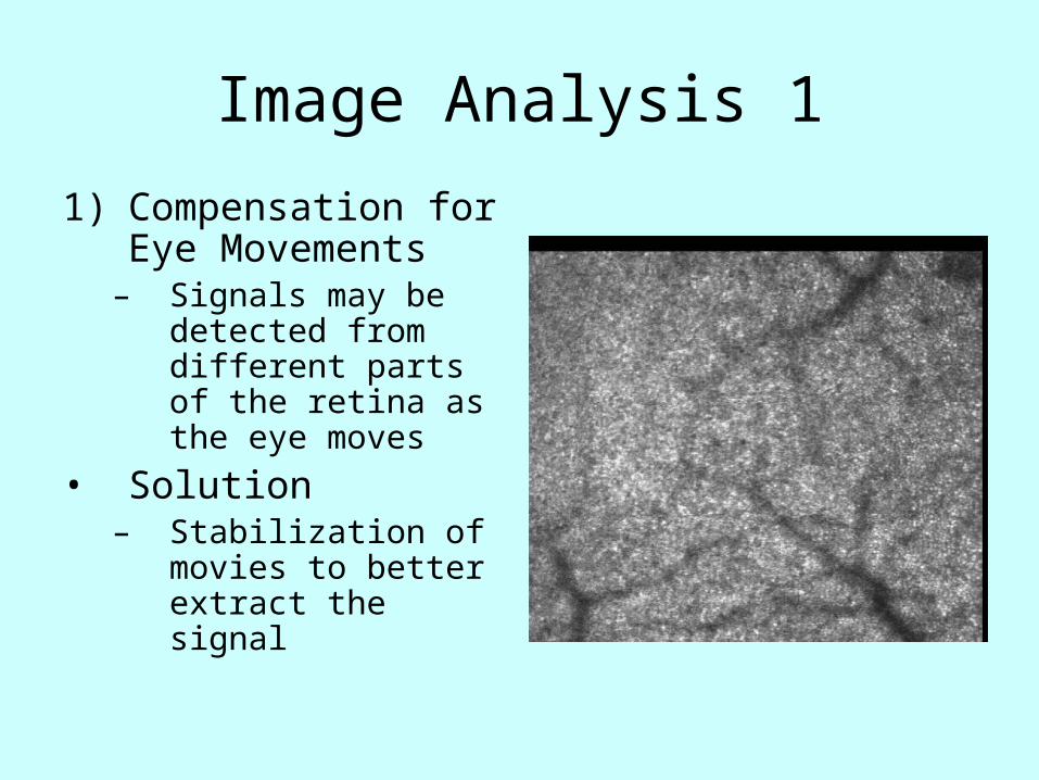
Image Analysis 1
1) Compensation for Eye Movements
– Signals may be detected from different parts of the retina as the eye moves
• Solution– Stabilization of
movies to better extract the signal

Image Analysis 22) Compensation for Eye Blinking
- Intensity changes due to blinking are larger than the desired signal
• Solution- Compare stimulated vs. unstimulated region of the
retina (ratio of upper to lower half of IR image)
Frame 1: Red Laser Frame 2: Infrared Laser
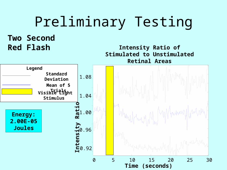
Intensity Ratio of Stimulated to Unstimulated Retinal Areas
0 5 10 15 20 25 30
Preliminary Testing
Standard Deviation
Mean of 5 Trials
Legend
Inte
nsit
y R
ati
o
Time (seconds)
0.92
0.96
1.00
1.04
1.08
Two Second Red Flash
Energy: 2.00E-05 Joules
Visible Light Stimulus

Experiment One:
Half Second Red Flash
with Designated
Powers
Time (seconds)
Energy (Joules)
1.00E-08
2.14E-08 2.04E-06
9.55E-07
4.47E-07
2.09E-07
9.77E-08
4.57E-08
Control
9.33E-06
4.37E-06
Inte
nsi
ty R
atio
0 5 10 15 20 25 30
0.92
0.96
1.00
1.04
1.08
0 5 10 15 20 25 30
Standard Deviation
Mean of 5 Trials
Legend

Energy (Joules)
1.02E-05
3.12E-05 8.40E-05
7.26E-05
5.16E-05 Control
Experiment Two:
Three Second Red Flash with Designated
Powers
Time (seconds)
Inte
nsi
ty R
atio
0.92
0.96
1.00
1.04
1.08
5 10 15 20 25 300 5 10 15 20 25 300
Standard Deviation
Mean of 5 Trials
Legend

Image of Retina Before Stimulation
Image of Retina After Stimulation

Summary/Conclusion
• Systematic change (increase in scattered lR light in response to stimulation) observed with higher flash energies and a longer flash duration
Future Plans
• More testing with different stimuli and possibly patients with retinal disease
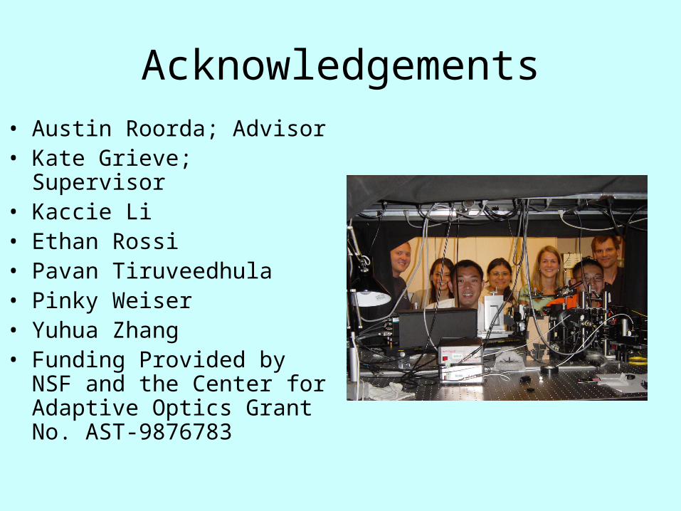
Acknowledgements• Austin Roorda; Advisor• Kate Grieve; Supervisor• Kaccie Li• Ethan Rossi• Pavan Tiruveedhula• Pinky Weiser• Yuhua Zhang• Funding Provided by NSF
and the Center for Adaptive Optics Grant No. AST-9876783

Thank you!
Questions?