Imaging in Avascular Necrosis of the Femoral Head · View Media Gallery Frogleg lateral view of the...
24
Imaging in Avascular Necrosis of the Femoral Head Updated: Sep 26, 2016 Author: Michael R Aiello, MD; Chief Editor: Felix S Chew, MD, MBA, MEd more... OVERVIEW Overview Avascular necrosis (AVN) of the femoral head is an increasingly common cause of musculoskeletal disability, and it poses a major diagnostic and therapeutic challenge. Although patients are initially asymptomatic, avascular necrosis (AVN) of the femoral head usually progresses to joint destruction, requiring total hip replacement (THR), usually before the fifth decade (see the images below). In fact, 50% of patients with avascular necrosis experience severe joint destruction as a result of deterioration and undergo a major surgical procedure for treatment within 3 years of diagnosis. Femoral head collapse usually occurs within 2 years after development of hip pain. [1, 2, 3] Axial computed tomography scan in a patient without avascular necrosis of the femoral head shows prominent and thickened but normal trabeculae (arrow) within the femoral head. Note the delicate, sclerotic, raylike branchings emanating in a radial fashion from the central dense band. This is the asterisk sign. See also the next image. View Media Gallery Page 1 of 24 Imaging in Avascular Necrosis of the Femoral Head: Overview, Radiography, Computed ... 9/26/2017 http://emedicine.medscape.com/article/386808-overview
Transcript of Imaging in Avascular Necrosis of the Femoral Head · View Media Gallery Frogleg lateral view of the...

Imaging in Avascular Necrosis of the Femoral HeadUpdated: Sep 26, 2016 Author: Michael R Aiello, MD; Chief Editor: Felix S Chew, MD, MBA, MEd more...
OVERVIEW
OverviewAvascular necrosis (AVN) of the femoral head is an increasingly common cause of musculoskeletal disability, and it poses a major diagnostic and therapeutic challenge. Although patients are initially asymptomatic, avascular necrosis (AVN) of the femoral head usually progresses to joint destruction, requiring total hip replacement (THR), usually before the fifth decade (see the images below). In fact, 50% of patients with avascular necrosis experience severe joint destruction as a result of deterioration and undergo a major surgical procedure for treatment within 3 years of diagnosis. Femoral head collapse usually occurs within 2 years after development of hip pain. [1, 2, 3]
Axial computed tomography scan in a patient without avascular necrosis of the femoral head shows prominent and thickened but normal trabeculae (arrow) within the femoral head. Note the delicate, sclerotic, raylike branchings emanating in a radial fashion from the central dense band. This is the asterisk sign. See also the next image.
View Media Gallery
Page 1 of 24Imaging in Avascular Necrosis of the Femoral Head: Overview, Radiography, Computed ...
9/26/2017http://emedicine.medscape.com/article/386808-overview
tighe
Reviewed

Avascular necrosis of the femoral head. Illustration of the normal circulation of the femoral head, viewed from the posterior approach. The posterior-superior retinacular arteries provide the major blood supply to the epiphysis. They traverse the femoral neck and are contained within the joint capsule and give rise to the lateral epiphyseal vessels at the junction of the femoral head and neck. From there, they penetrate the femur and supply the femoral epiphysis. A. = artery.
View Media Gallery
Avascular necrosis of the femoral head. Illustration demonstrating that the blood supply to the femoral head is compromised by subcapital femoral fractures or slipped capital femoral epiphysis. As the epiphysis or femoral neck separates from the femoral head, the femoral metaphysis displaces superolaterally and the femur rotates externally. This causes the distal posterior-superior retinacular arteries and proximal lateral epiphyseal vessels to kink or rotate, compromising the blood flow to the epiphysis. If this condition were to persist, the femoral head would be at high risk for developing avascular necrosis.
Page 2 of 24Imaging in Avascular Necrosis of the Femoral Head: Overview, Radiography, Computed ...
9/26/2017http://emedicine.medscape.com/article/386808-overview

View Media Gallery
Frogleg lateral view of the right hip in a patient with avascular necrosis shows the crescent sign, indicating subchondral fracture. Therapeutic interventions are less likely to halt progression of the disease once this sign appears. The frogleg lateral view is better than anteroposterior (AP) projection for demonstrating this sign, because the anterior and posterior margins of the acetabulum on the AP projection are superimposed over the superior portion of the femoral head, the usual location of the sign.
View Media Gallery
The incidence of avascular necrosis (AVN) is increasing. The causes include greater use of exogenous steroids and an increase in trauma. [4, 5, 6, 7, 8, 9, 10, 11, 12, 13, 14, 15, 16, 17, 18, 19] In 54-80% of renal transplant recipients in whom AVN is detected with plain radiographs, the disease is bilateral.
It is estimated that almost 10% of the nearly 500,000 THRs performed each year in the United States are intended to treat AVN; at a cost of more than $1 billion, THRs performed to treat avascular necrosis (AVN) of the femoral head constitute approximately 25% of the total national costs for THR. Trauma is the most common cause of avascular necrosis; however, nontraumatic avascular necrosis (AVN) is commonly bilateral and occurs in younger persons. In addition, nontraumatic bilateral AVN usually occurs at different times and progresses at different rates in different hips.
Treatment of avascular necrosis (AVN) has been facilitated by the adoption of an international classification system, by effective early diagnosis using magnetic resonance imaging (MRI), and by more aggressive surgical management; nevertheless, no universally satisfactory therapy has been developed, even for early disease.
Because measures to preserve the joint are associated with better prognoses when the diagnosis of avascular necrosis (AVN) is made early in the course of the disease and
Page 3 of 24Imaging in Avascular Necrosis of the Femoral Head: Overview, Radiography, Computed ...
9/26/2017http://emedicine.medscape.com/article/386808-overview

because the results of joint replacement therapy are poorer in younger age groups than in older patients, it is critical to diagnose this condition as early as possible to prevent or delay progression of the disease.
Avascular necrosis (AVN) is characterized by areas of dead trabecular bone and marrow extending to involve the subchondral plate. The anterolateral aspect of the femoral head, the principal weightbearing region, is typically involved, but any region of the femoral head may be involved. In the adult, the involved segment usually never fully revascularizes, and collapse of the femoral head usually occurs sometime after avascular necrosis (AVN) is detected radiographically.
The femoral head is the most vulnerable site for the development of avascular necrosis (AVN). The site of necrosis is usually immediately below the weightbearing articular surface of the bone (ie, the anterolateral aspect of the femoral head). This is the site of greatest mechanical stress.
Elderly persons are at decreased risk for developing avascular necrosis (AVN). Fat cells become smaller in elderly persons. The space between fat cells fills with a loose reticulum and mucoid fluid, which are resistant to avascular necrosis (AVN). This condition is termed gelatinous marrow. Even in the presence of increased intramedullary pressure, interstitial fluid is able to escape into the blood vessels, leaving the spaces free to absorb additional fluid.
Anatomy
To understand the changes of avascular necrosis (AVN) on radiologic studies such as computed tomography (CT) scanning and MRI, it is necessary to understand the anatomy of the hip and to understand the vascular anatomy of the hip in children.
Gross anatomy
The hip is a ball-and-socket joint. The acetabulum, which provides bony coverage of 40% of the femoral head, has a horseshoe-shaped lunate surface. The femoral head is round and smooth in all imaging planes. The fovea capitis, a small depression on the medial femoral head, is the site of attachment of the ligamentum teres (see the image below).
Page 4 of 24Imaging in Avascular Necrosis of the Femoral Head: Overview, Radiography, Computed ...
9/26/2017http://emedicine.medscape.com/article/386808-overview

Coronal computed tomography image of the pelvis and hips in a patient without avascular necrosis of the femoral head. A thin low-signal line, representing the long cortex, surrounds the femur and iliac lines. The fovea is a small indentation along the medial aspect of the femoral head and is the site of penetration of the artery of the ligamentum teres into the femur. High signal is present within the medullary space of the proximal femur, representing normal fatty marrow. The physis is a thin line of low signal extending from the lateral to the medial aspect of the femoral head.
View Media Gallery
The principal sources of blood flow to the femoral head are the lateral epiphyseal vessels (LEVs). LEVs are branches of the posterior superior retinacular vessels (PSVs), which are themselves branches of the medial femoral circumflex artery; the medial femoral circumflex artery is a branch of the profunda femoris artery (see the following 2 images). The PSVs run along the posterior-superior aspect of the femoral neck under the synovial membrane. They are extraosseous in location and give rise to the LEV (see the following 2 images). [20]
The LEV enters the femoral head within a 1-cm-wide zone between the cartilage of the femoral head and the cortical bone of the femoral neck. They supply the lateral and central thirds of the femoral head (see the following 2 images). When patent, the artery of the ligamentum teres (ALT) supplies the medial third of the femoral head.
Avascular necrosis of the femoral head. Illustration of the normal circulation of the femoral head, viewed from the posterior approach. The posterior-superior retinacular arteries provide the major blood supply to the epiphysis. They traverse the femoral neck and are contained within the joint capsule and give rise to the lateral epiphyseal vessels at the junction of the femoral head and neck. From there, they penetrate the femur and supply the femoral epiphysis. A. = artery.
View Media Gallery
Page 5 of 24Imaging in Avascular Necrosis of the Femoral Head: Overview, Radiography, Computed ...
9/26/2017http://emedicine.medscape.com/article/386808-overview

Avascular necrosis of the femoral head. Illustration demonstrating that the blood supply to the femoral head is compromised by subcapital femoral fractures or slipped capital femoral epiphysis. As the epiphysis or femoral neck separates from the femoral head, the femoral metaphysis displaces superolaterally and the femur rotates externally. This causes the distal posterior-superior retinacular arteries and proximal lateral epiphyseal vessels to kink or rotate, compromising the blood flow to the epiphysis. If this condition were to persist, the femoral head would be at high risk for developing avascular necrosis.
View Media Gallery
Branches of the LEVs and the ALT anastomose in the junction of the central and medial third of the femoral head. The thickest part of the articular cartilage of the femoral head is located along the posterior-superior aspect and measures 3 mm in diameter. It thins to 0.5 mm along the peripheral and inferior margins.
Blood supply in children
In children 4-7 years of age, the vascular anatomy of the proximal femur is in a transitional stage of development. The ALT does not penetrate the epiphysis of the femoral head until age 9 or 10 years. The medial circumflex artery, a branch of the profunda femoris artery, penetrates into the femoral proximal metaphysis but is prevented from passing into the femoral epiphysis by the growth plate. The blood supply to the femoral head is especially vulnerable during this time.
Anatomy on CT scans
Physiologic thickening of bone trabeculae in the center of the femoral head is present and appears similar to a star, which is termed the asterisk sign (see the image below). The configuration is related to the stress of weightbearing.
Sclerotic raylike branches of the star usually extend to the upper surface of the femoral head (see the image below). A dense line, extending from the lateral to the medial portion of the mid femoral head, represents the fused epiphysis.
Page 6 of 24Imaging in Avascular Necrosis of the Femoral Head: Overview, Radiography, Computed ...
9/26/2017http://emedicine.medscape.com/article/386808-overview

Axial computed tomography scan in a patient without avascular necrosis of the femoral head shows prominent and thickened but normal trabeculae (arrow) within the femoral head. Note the delicate, sclerotic, raylike branchings emanating in a radial fashion from the central dense band. This is the asterisk sign. See also the next image.
View Media Gallery
Anatomy on MRI
Fatty marrow is present in the femoral capital epiphysis and the greater trochanter of all individuals older than 2 years. Fatty marrow has high signal intensity on T1-weighted images (T1WIs) and T2-weighted images (T2WIs) (see the images below). Hematopoietic marrow, when present, is found in the femoral neck, the intertrochanteric region, and the acetabulum. It has low signal intensity on T1WIs and high signal intensity on T2WIs (see the images below).
Avascular necrosis of the femoral head. Coronal T1-weighted magnetic resonance image in a patient showing hypointense signal within the proximal femoral neck and intertrochanteric regions (arrows) representing hematopoietic marrow. Increased signal is present within the greater trochanters and femoral capital epiphysis representing normal fatty marrow. See also the next image.
View Media Gallery
Page 7 of 24Imaging in Avascular Necrosis of the Femoral Head: Overview, Radiography, Computed ...
9/26/2017http://emedicine.medscape.com/article/386808-overview
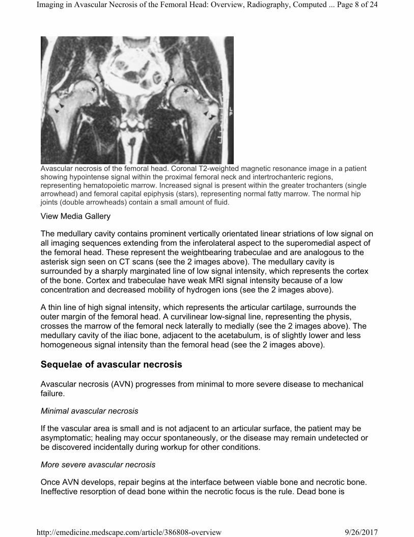
Avascular necrosis of the femoral head. Coronal T2-weighted magnetic resonance image in a patient showing hypointense signal within the proximal femoral neck and intertrochanteric regions, representing hematopoietic marrow. Increased signal is present within the greater trochanters (single arrowhead) and femoral capital epiphysis (stars), representing normal fatty marrow. The normal hip joints (double arrowheads) contain a small amount of fluid.
View Media Gallery
The medullary cavity contains prominent vertically orientated linear striations of low signal on all imaging sequences extending from the inferolateral aspect to the superomedial aspect of the femoral head. These represent the weightbearing trabeculae and are analogous to the asterisk sign seen on CT scans (see the 2 images above). The medullary cavity is surrounded by a sharply marginated line of low signal intensity, which represents the cortex of the bone. Cortex and trabeculae have weak MRI signal intensity because of a low concentration and decreased mobility of hydrogen ions (see the 2 images above).
A thin line of high signal intensity, which represents the articular cartilage, surrounds the outer margin of the femoral head. A curvilinear low-signal line, representing the physis, crosses the marrow of the femoral neck laterally to medially (see the 2 images above). The medullary cavity of the iliac bone, adjacent to the acetabulum, is of slightly lower and less homogeneous signal intensity than the femoral head (see the 2 images above).
Sequelae of avascular necrosis
Avascular necrosis (AVN) progresses from minimal to more severe disease to mechanical failure.
Minimal avascular necrosis
If the vascular area is small and is not adjacent to an articular surface, the patient may be asymptomatic; healing may occur spontaneously, or the disease may remain undetected or be discovered incidentally during workup for other conditions.
More severe avascular necrosis
Once AVN develops, repair begins at the interface between viable bone and necrotic bone. Ineffective resorption of dead bone within the necrotic focus is the rule. Dead bone is
Page 8 of 24Imaging in Avascular Necrosis of the Femoral Head: Overview, Radiography, Computed ...
9/26/2017http://emedicine.medscape.com/article/386808-overview
reabsorbed only partially. Reactive and reparative bone is laid down on dead trabeculae, resulting in a sclerotic margin of thickened trabeculae within an advancing front of hyperemia, inflammation, bone resorption, and fibrosis. The incomplete resorption of dead bone has a mixed sclerotic and cystic appearance on radiographs. Necrosis and repair are ongoing in various stages of evolution within a single lesion.
Mechanical failure
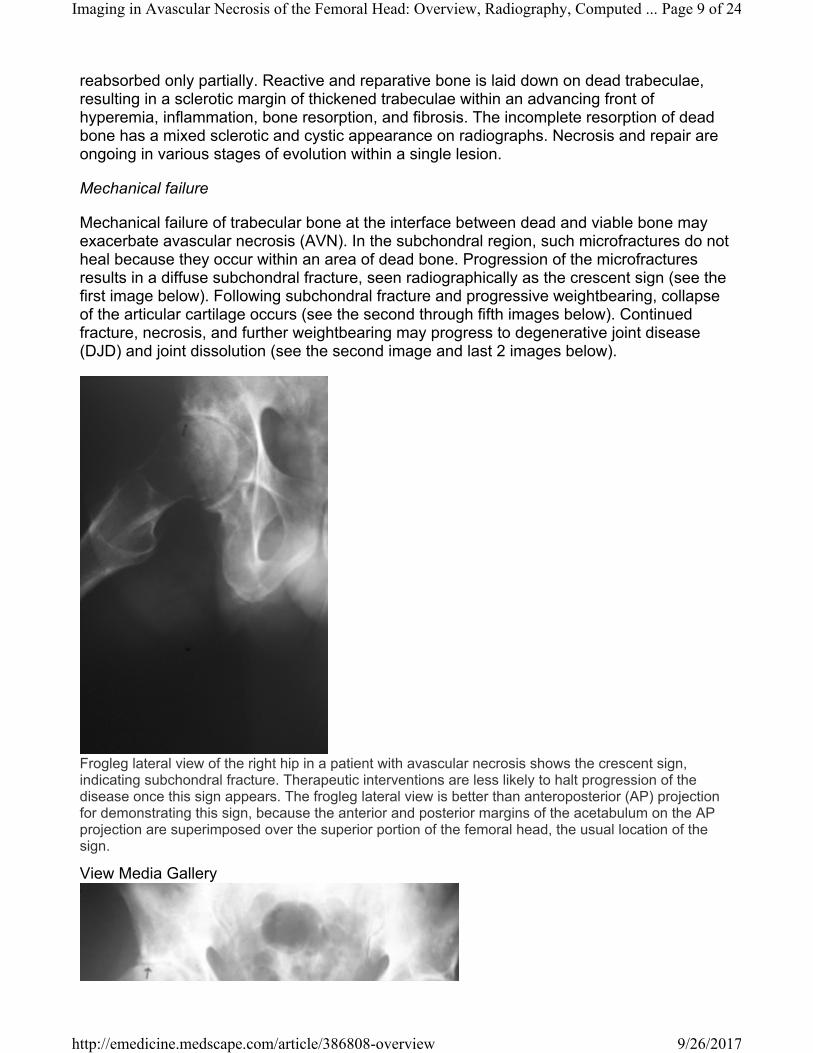
Mechanical failure of trabecular bone at the interface between dead and viable bone may exacerbate avascular necrosis (AVN). In the subchondral region, such microfractures do not heal because they occur within an area of dead bone. Progression of the microfractures results in a diffuse subchondral fracture, seen radiographically as the crescent sign (see the first image below). Following subchondral fracture and progressive weightbearing, collapse of the articular cartilage occurs (see the second through fifth images below). Continued fracture, necrosis, and further weightbearing may progress to degenerative joint disease (DJD) and joint dissolution (see the second image and last 2 images below).
Frogleg lateral view of the right hip in a patient with avascular necrosis shows the crescent sign, indicating subchondral fracture. Therapeutic interventions are less likely to halt progression of the disease once this sign appears. The frogleg lateral view is better than anteroposterior (AP) projection for demonstrating this sign, because the anterior and posterior margins of the acetabulum on the AP projection are superimposed over the superior portion of the femoral head, the usual location of the sign.
View Media Gallery
Page 9 of 24Imaging in Avascular Necrosis of the Femoral Head: Overview, Radiography, Computed ...
9/26/2017http://emedicine.medscape.com/article/386808-overview

Avascular necrosis of the femoral head. Anteroposterior view of the pelvis shows flattening of the outer portion of the right femoral head from avascular necrosis (arrow), with adjacent joint-space narrowing, juxta-articular sclerosis, and osteophytes representing degenerative joint disease. See also the next image.
View Media Gallery
Anteroposterior view of the left hip in a patient with avascular necrosis obtained 6 months after presentation shows that the patient has undergone core decompression but has developed mild flattening of the femoral head, indicating progression of disease despite treatment.
View Media Gallery
Page 10 of 24Imaging in Avascular Necrosis of the Femoral Head: Overview, Radiography, Comput...
9/26/2017http://emedicine.medscape.com/article/386808-overview
Plain film finding in a patient with bilateral avascular necrosis of the femoral head 6 months after presentation demonstrates that subtle flattening of the left femoral head (open arrow) has occurred. This indicates progression of disease from stage 2 to stage 3 despite conservative treatment. The patient underwent bilateral core decompression and bone grafting. The defects within the proximal femurs represent removal of the dead bone, and the tubular densities within each femur represent bone grafts in an attempt to revascularize the region of avascular necrosis.
View Media Gallery
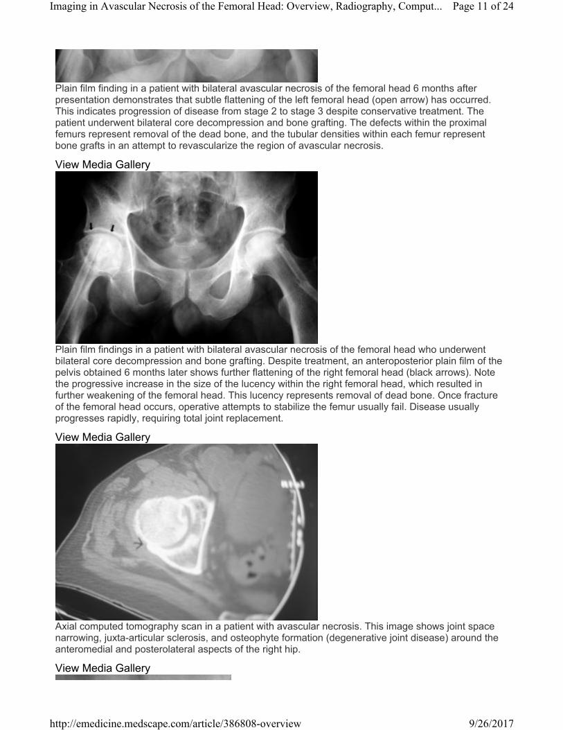
Plain film findings in a patient with bilateral avascular necrosis of the femoral head who underwent bilateral core decompression and bone grafting. Despite treatment, an anteroposterior plain film of the pelvis obtained 6 months later shows further flattening of the right femoral head (black arrows). Note the progressive increase in the size of the lucency within the right femoral head, which resulted in further weakening of the femoral head. This lucency represents removal of dead bone. Once fracture of the femoral head occurs, operative attempts to stabilize the femur usually fail. Disease usually progresses rapidly, requiring total joint replacement.
View Media Gallery
Axial computed tomography scan in a patient with avascular necrosis. This image shows joint space narrowing, juxta-articular sclerosis, and osteophyte formation (degenerative joint disease) around the anteromedial and posterolateral aspects of the right hip.
View Media Gallery
Page 11 of 24Imaging in Avascular Necrosis of the Femoral Head: Overview, Radiography, Comput...
9/26/2017http://emedicine.medscape.com/article/386808-overview

Anteroposterior plain film of the pelvis in a patient with avascular necrosis of the femoral head shows asymmetric joint-space narrowing (arrow), juxta-articular sclerosis, and subchondral cyst formation around the right hip secondary to degenerative joint disease. This may be confused with avascular necrosis, but the intimate localization of all of the bindings and the absence of femoral head collapse makes a diagnosis of avascular necrosis highly unlikely.
View Media Gallery
Preferred examination
This section will discuss various imaging modalities. For more detailed information, please see their respective sections in this article.
MRI
MRI is the most sensitive means of diagnosing avascular necrosis (AVN). This imaging modality provides the criterion standard of noninvasive diagnostic evaluation and is more sensitive than CT scanning or planar scintigraphy. In addition, MRI is much more sensitive than plain film radiography for detecting avascular necrosis (AVN). However, low-field magnets (0.1 Tesla [T]) are not as sensitive for diagnosing avascular necrosis (AVN).
7-T hip MRI showed comparable results in hip joint imaging compared with 3 T, with slight advantages in contrast detail (cartilage defects) and fluid detection at 7 T when accepting image degradation medially. Image homogeneity of 7 T compared with 3 T (3.9-4.0 for all sequences) was degraded, especially in TSE sequences at 7 T through signal variations (7 T: 2.1-2.9). [21]
MRI is indispensable for the accurate staging of avascular necrosis (AVN), because images clearly depict the size of the lesion, and gross estimates of the stage of disease can be made (see Radiograph for the radiographic and radiologic staging systems for AVN). MRI
Page 12 of 24Imaging in Avascular Necrosis of the Femoral Head: Overview, Radiography, Comput...
9/26/2017http://emedicine.medscape.com/article/386808-overview

allows sequential evaluation of asymptomatic lesions that are undetectable on plain radiographs. [1] MRI facilitates better response to treatment because, with the use of MRI, avascular necrosis (AVN) is diagnosed at an earlier stage, and therapeutic measures are more successful the earlier they are begun.
MRI does not employ ionizing radiation—a factor that is especially important in the growing skeleton—and it is accepted widely and is easy to perform. MRI is also capable of imaging in multiple planes (ie, axial, sagittal, coronal, or any variation thereof), demonstrates superior soft-tissue resolution, and has high spatial and contrast resolution, allowing evaluation of morphologic features.
MRI may help guide interventional procedures such as core decompression, may demonstrate response of the femoral head to treatment, and may detect the joint effusions and bone edema that often accompany avascular necrosis (AVN). MRI is a noninvasive means of evaluating articular cartilage congruity, and it allows sequential evaluation of asymptomatic lesions that are undetectable on plain radiographs. [22, 23, 24, 25, 26]
Early MRI detection and closed bone graft epiphysiodesis (CBGE) may mitigate the effects of AVN after slipped capital femoral epiphysis (SCFE). Seventeen patients (17 hips) had a scheduled MRI between 1 and 6 months from initial surgery. Six hips diagnosed by MRI received surgical intervention (4 CBGE, 1 free vascularized fibula graft, and 1 repinning due to screw cutout) at a mean of 4.1 months (range, 1.3 to 7.2 mo) postoperatively. None of the 4 patients treated with CBGE within 2 months postoperatively progressed to stage IVC AVN.[27]
Similar sensitivity and specificity were found between low-field MRI and planar radionuclide bone scanning. At high magnetic field strength, MRI has a higher sensitivity than radionuclide scanning. Using a 1.5-T magnet, Beltran et al reported 88% sensitivity, 100% specificity, and 94% accuracy with MRI and 78% sensitivity, 75% specificity, and 76% accuracy with bone scintigraphy. [28] The high sensitivity of MRI has been confirmed by others.
In another study, MRI performed at 0.6 T and single-photon emission CT (SPECT) bone imaging using technetium-99m (99m Tc) methylene diphosphonate (MDP) were similarly effective in diagnosing avascular necrosis (AVN). [26] MRI had a sensitivity of 87% and a specificity of 83%; whereas SPECT scanning had a sensitivity of 91% and a specificity of 78%. Both were more effective than planar bone scintigraphy, which had a sensitivity of 83% and a specificity of 83%.
MRI was also more effective than SPECT for diagnosing cases of avascular necrosis (AVN) of the hip in which pain was absent. MRI detected avascular necrosis (AVN) in 10 of 15 patients, but SPECT scanning detected AVN in only 5 of 15 patients.
Using receiver operating characteristic (ROC) curves, MRI was better than CT scanning by more than 2 standard errors and better than radionuclide scanning by more than 3 standard errors in helping diagnose early avascular necrosis (AVN).
Magnetic resonance perfusion imaging has been able to identify significant differences between avascular necrosis, bone marrow edema, and subchondral insufficiency fractures
Page 13 of 24Imaging in Avascular Necrosis of the Femoral Head: Overview, Radiography, Comput...
9/26/2017http://emedicine.medscape.com/article/386808-overview

of the proximal femur, particularly regarding maximum enhancement values (Emax), slope (Eslope) and time to peak (TTP). Diffusion weighted imaging of bone marrow of the proximal femur did not show significant differences in the same study. [29]
Gadolinium-enhanced perfusion MRI (pMRI) after closed reduction/spica casting for developmental dysplasia of the hip (DDH) has been suggested as a potential means to identify and avoid avascular necrosis (AVN) by helping the surgeon evaluate femoral head vascularity. [30]
Single-photon emission CT scanning
SPECT scanning provides images of the radioactivity within the target organ in 3 dimensions. With this modality, overlying and underlying areas of radioactivity may be separated into sequential tomographic planes, thus providing increased image contrast and improved lesion detection and localization, as compared with planar scintigraphy. SPECT scanning eliminates radioactivity resulting from hyperemia about the hip joint and from the underlying acetabulum and adjacent bladder. SPECT scanning is used as an alternative to MRI when MRI cannot be performed or when the results of MRI are indeterminate. [31, 32, 33]
Initially, SPECT images reflect vascular integrity; early in the disease, SPECT scans may demonstrate an avascular focus; such findings are missed with MRI unless contrast is used. Collier et al found a sensitivity of 85% for SPECT scanning. [34] With triple-head high-resolution SPECT scanning, Lee et al reported a sensitivity of 97%. [35]
Nuclear imaging
Planar radionuclide imaging, bone scintigraphy using pinhole collimation, and planar scintigraphic imaging using quantitative bone scanning are briefly discussed below.
Collier reported a sensitivity of 55% with planar radionuclide imaging for avascular necrosis (AVN). [34] However, bone scintigraphy equipped with a pinhole collimator has greater sensitivity for diagnosing avascular necrosis (AVN) than bone scintigraphy using a high-resolution parallel-hole collimator.
The pinhole collimator is a conical collimator with a small circular aperture (3-5 mm) that produces an inverted image of the object in a manner analogous to photographic cameras. The image obtained is magnified, allowing better visualization of small structures and improving detection of scintigraphic abnormalities. The pinhole collimator optimizes resolution in the evaluation of circumscribed areas, and the acquisition time is only 15 minutes, compared to up to 45 minutes for SPECT scanning. The technique is an alternative to MRI when MRI cannot be performed or when MRI results are not clear-cut.
Planar scintigraphic imaging using quantitative bone scanning provides physiologic data that cannot be obtained with other modalities, including MRI; for example, this technique allows quantification of uptake in the perfusion and static phases. However, correct computer programming is required. [36, 37, 38, 39]
In one study, F-18 fluoride PET/CT showed good agreement with MRI in the initial diagnosis of AVN and was better than MRI in detecting early disease. MRI was 96.5% sensitive, 100%
Page 14 of 24Imaging in Avascular Necrosis of the Femoral Head: Overview, Radiography, Comput...
9/26/2017http://emedicine.medscape.com/article/386808-overview

specific, and 98.03% accurate, while PET/CT was 100% sensitive, 100% specific, and 100% accurate in diagnosing AVN. [40]
In a comparative study of technetium-99m-methylene diphosphonate (99mTc-MDP) SPECT/CT versus planar bone scintigraphy (BS) for diagnosis of AVN, SPECT/CT was found to be superior to planar BS and SPECT alone. The diagnostic accuracy of planar BS, SPECT, and SPECT/CT was 67%, 78%, and 95%, respectively. Planar BS was found to have the lowest sensitivity (75%) and specificity (40%), whereas SPECT/CT had the highest sensitivity (98%) and specificity (87%). [41]
CT scanning
The high spatial resolution and contrast resolution of CT scanning allow analysis of morphologic features (see the images below), and . The sensitivity of CT scanning in detecting early avascular necrosis (AVN) is 55%, which is similar to the sensitivity of planar nuclear medicine imaging. Thus, CT scanning is more appropriate in evaluating the extent of involvement, such as subchondral lucencies and sclerosis during the reparative stage, before the onset of femoral head collapse and superimposed degenerative disease.
Axial computed tomography scan in a patient without avascular necrosis of the femoral head shows prominent and thickened but normal trabeculae (arrow) within the femoral head. Note the delicate, sclerotic, raylike branchings emanating in a radial fashion from the central dense band. This is the asterisk sign. See also the next image.
View Media Gallery
Page 15 of 24Imaging in Avascular Necrosis of the Femoral Head: Overview, Radiography, Comput...
9/26/2017http://emedicine.medscape.com/article/386808-overview

Coronal multiplanar reconstructed computed tomography image in a patient without avascular necrosis of the femoral head. Thickened trabeculae are seen at the medial aspect of the junction of the femoral head and neck and extend to the subchondral region. The physis (arrowhead) is a transverse white line located between the lateral and medial aspect of the femoral head.
View Media Gallery
Axial computed tomographh scan of a patient with avascular necrosis of the femoral head shows clumping and distortion of the central trabeculae representing the asterisk sign (arrowhead) and an adjacent low-density region (arrow) representing the reparative zone. See also the next image.
View Media Gallery
Axial computed tomography (CT) scan of a patient with avascular necrosis of the femoral head shows a fracture to the anterior aspect of the femoral head. This finding was demonstrated on an axial fat-saturated T2-weighted magnetic resonance image, but it is delineated more clearly using CT scanning because of the superior resolution of CT scanning. See also the next image.
View Media Gallery
Page 16 of 24Imaging in Avascular Necrosis of the Femoral Head: Overview, Radiography, Comput...
9/26/2017http://emedicine.medscape.com/article/386808-overview

Axial computed tomography scan in a patient with avascular necrosis. This image shows joint space narrowing, juxta-articular sclerosis, and osteophyte formation (degenerative joint disease) around the anteromedial and posterolateral aspects of the right hip.
View Media Gallery
CT scanning is better able to help define the extent of disease at stages II and higher than MRI and plain film radiography. CT scanning enables detection of subchondral or cancellous fractures and collapse, especially when multiplanar reconstruction is used. This information is essential for planning treatment (see the third and fourth images above).
Plain film radiography
Although unable to detect disease of stage 0 or 1, plain film radiography may be helpful in assessing flattening of the femoral head and associated degenerative changes (see the images below). See Radiograph.
Avascular necrosis of the femoral head. Anteroposterior view of the pelvis shows flattening of the outer portion of the right femoral head from avascular necrosis (arrow), with adjacent joint-space narrowing, juxta-articular sclerosis, and osteophytes representing degenerative joint disease. See also the next image.
View Media Gallery
Page 17 of 24Imaging in Avascular Necrosis of the Femoral Head: Overview, Radiography, Comput...
9/26/2017http://emedicine.medscape.com/article/386808-overview

Frogleg lateral view of the right hip in a patient with avascular necrosis shows the crescent sign, indicating subchondral fracture. Therapeutic interventions are less likely to halt progression of the disease once this sign appears. The frogleg lateral view is better than anteroposterior (AP) projection for demonstrating this sign, because the anterior and posterior margins of the acetabulum on the AP projection are superimposed over the superior portion of the femoral head, the usual location of the sign.
View Media Gallery
Anteroposterior view of the left hip in a patient with avascular necrosis obtained 6 months after presentation shows that the patient has undergone core decompression but has developed mild flattening of the femoral head, indicating progression of disease despite treatment.
View Media Gallery
Page 18 of 24Imaging in Avascular Necrosis of the Femoral Head: Overview, Radiography, Comput...
9/26/2017http://emedicine.medscape.com/article/386808-overview

Anteroposterior view of the pelvis in a patient with bilateral avascular necrosis of the femoral head. Mild flattening to the superior aspect of the right femoral head (open arrow) indicates stage 3 disease. The left femoral head has a normal contour, indicating stage 2 disease. The black arrows indicate the margins of the reparative zone, representing new bone formation on dead trabeculae. When avascular necrosis is bilateral, it usually occurs in each hip at different times, and the staging of disease in each hip can be, and often is, at different stages. See also the next image.
View Media Gallery
Plain film finding in a patient with bilateral avascular necrosis of the femoral head 6 months after presentation demonstrates that subtle flattening of the left femoral head (open arrow) has occurred. This indicates progression of disease from stage 2 to stage 3 despite conservative treatment. The patient underwent bilateral core decompression and bone grafting. The defects within the proximal femurs represent removal of the dead bone, and the tubular densities within each femur represent bone grafts in an attempt to revascularize the region of avascular necrosis.
View Media Gallery
Page 19 of 24Imaging in Avascular Necrosis of the Femoral Head: Overview, Radiography, Comput...
9/26/2017http://emedicine.medscape.com/article/386808-overview

Plain film findings in a patient with bilateral avascular necrosis of the femoral head who underwent bilateral core decompression and bone grafting. Despite treatment, an anteroposterior plain film of the pelvis obtained 6 months later shows further flattening of the right femoral head (black arrows). Note the progressive increase in the size of the lucency within the right femoral head, which resulted in further weakening of the femoral head. This lucency represents removal of dead bone. Once fracture of the femoral head occurs, operative attempts to stabilize the femur usually fail. Disease usually progresses rapidly, requiring total joint replacement.
View Media Gallery
Limitations of techniques
The limitations of the imaging techniques discussed above are briefly reviewed.
MRI
Bone biopsy analysis has been rarely reported to be positive when MRI findings are normal. MRI cannot be performed in patients who have cardiac pacemakers or when intracranial clips are present, nor can MRI be performed in patients who have claustrophobia. In addition, problems related to malpositioning may lead to misrepresentation. In children, slight pelvic obliquity may cause the normal dark-appearing growth plate to appear in the same axial cut as the contralateral bright-appearing epiphysis; in such cases, the normal growth plate may appear to be abnormal on MRI.
Children may require sedation as a result of the long imaging times that MRI requires. It may be difficult to detect avascular necrosis (AVN) after surgery to repair a hip fracture because of the presence of orthopedic hardware, which creates significant image distortion. Marrow cells are more resistant to ischemia than hematopoietic cells or osteocytes. Because MRI images reflect changes within marrow fat signal intensity, MRI findings of avascular necrosis (AVN) may not be seen for up to 5 days after the ischemic event, until the marrow fat cells have died. In this situation, contrast-enhanced MRI is needed.
SPECT scanning
SPECT scanning demonstrates poor spatial resolution. Artifacts from the bladder are frequently encountered; these artifacts may obscure the photon-deficient region of the femoral head. A number of techniques, such as the use of multihead cameras with shorter acquisition times that improve resolution and increase sensitivity, have been advocated, but none has gained universal acceptance.
SPECT imaging requires a cooperative patient who must remain immobile for up to 45 minutes of acquisition time, thus this modality is difficult to use in children because of the necessity to remain motionless for long periods of time. Children may require sedation. In addition, diagnosing Legg-Calve-Perthes (LCP) in small children may be difficult because of the small size of the femoral epiphysis and associated bladder artifacts.
Planar scintigraphy
Planar scintigraphy demonstrates poor spatial resolution (see the following images). The ring of increased activity reflecting hyperemia in the early stages and bone healing later obscures the photon-deficient necrotic center within the femoral head, which is indicative of
Page 20 of 24Imaging in Avascular Necrosis of the Femoral Head: Overview, Radiography, Comput...
9/26/2017http://emedicine.medscape.com/article/386808-overview

avascular necrosis (AVN). The site may show a uniform high level of activity, making it impossible to distinguish avascular necrosis (AVN) from other causes of increased activity, such as osteoarthritis, fracture, and inflammatory arthritis. A cold spot in the femoral head is highly specific but not sensitive for diagnosing avascular necrosis (AVN).
Bone scan of a patient with avascular necrosis of the femoral head shows increased uptake in the superolateral aspect of the right femoral head, indicative of avascular necrosis but providing little information concerning the structural integrity of the hip.
View Media Gallery
Planar bone scan of the pelvis in a patient with bilateral avascular necrosis of the femoral head shows marked increased uptake of radiopharmaceutical agent in both hips.
View Media Gallery
Artifacts from radioactivity in the bladder are frequently encountered, obscuring the photon-deficient region. Entities causing increased uptake about the hip joint, such as arthritis and
Page 21 of 24Imaging in Avascular Necrosis of the Femoral Head: Overview, Radiography, Comput...
9/26/2017http://emedicine.medscape.com/article/386808-overview

inflammatory disease, may obscure the photopenic necrotic focus within the femoral head. Results can be judged only by comparison with the other hip and may be of little use in the presence of bilateral involvement.
CT scanning
Although CT scanning may delineate subtle alterations of bone density when plain radiograph findings are normal, MRI and SPECT scintigraphy are much more sensitive for evaluating early manifestations of the disease, such as bone marrow edema. CT scans are insensitive for detecting stage 0 and 1 avascular necrosis (AVN), but they are excellent for detecting femoral head collapse, early degenerative joint disease (DJD), and the presence of loose bodies.
CT scanning may improve the accuracy of radiographic staging using thin-slice thicknesses of 1 mm or less and by incorporating multiplanar reconstruction. In one study, 30% of hips with stage 2 (precollapse) avascular necrosis (AVN), evaluated with plain film radiography, had stage 3 disease when evaluated with CT scans.
Plain film radiography
Using plain film radiography, the sensitivity for detecting early stages of the disease is as low as 41%. Plain film does not detect stage 0 and 1 avascular necrosis (AVN). A delay of 1-5 years may occur between the onset of symptoms and the appearance of radiographic abnormalities. Normal radiographic findings do not necessarily mean that disease is not present.
Demineralization of the femur may be detected, and the disease may be suggested only after bone resorption has occurred. If early diagnosis is needed for the prompt initiation of therapy, more sensitive imaging methods (ie, MRI) must be used, especially in patients who are at increased risk for AVN.
Differential diagnosis and other problems to be considered
Bone metastases should be considered in the differential diagnosis as well as transient osteoporosis. Other problems to be considered when viewing findings on plain film radiographs, bone scintigraphs, CT scans, and MRIs are as follows:
Plain film radiograph
• Malignancy • Osteomyelitis • Transient osteoporosis of the hip • Bone sarcoma • Advanced DJD (see the following image)
Page 22 of 24Imaging in Avascular Necrosis of the Femoral Head: Overview, Radiography, Comput...
9/26/2017http://emedicine.medscape.com/article/386808-overview

Frogleg lateral view of the right hip in a patient with avascular necrosis shows the crescent sign, indicating subchondral fracture. Therapeutic interventions are less likely to halt progression of the disease once this sign appears. The frogleg lateral view is better than anteroposterior (AP) projection for demonstrating this sign, because the anterior and posterior margins of the acetabulum on the AP projection are superimposed over the superior portion of the femoral head, the usual location of the sign.
View Media Gallery• Insufficiency fractures • Epiphyseal dysplasia • Bone metastases
Bone scintigram
• Infection • Plasma cell myeloma • Skeletal metastasis • Hemangioma • Radiation therapy • Arthritis • Sympathetic dystrophy • Bone marrow edema syndrome • Bone metastases
CT scan
• Degenerative disease • Insufficiency fracture • Malignancy • Infection • Plasma cell myeloma • Bone metastases
MRI
• Transient osteoporosis of the hip • Transient bone marrow syndrome
Page 23 of 24Imaging in Avascular Necrosis of the Femoral Head: Overview, Radiography, Comput...
9/26/2017http://emedicine.medscape.com/article/386808-overview

• Bone bruise • Epiphyseal stress fracture • Infection • Infiltrative neoplasm • Insufficiency fracture • Bone metastases
Special concerns
Failure to diagnose such a potentially devastating condition as avascular necrosis (AVN) in a young age group has the potential for serious medical-legal repercussions. Malpractice settlements reflect compensation for a lifetime of a potentially compromised lifestyle with much morbidity. Such settlements also reflect the cost of potential joint replacement and prosthesis failure.
Diagnosing AVN as early as possible is imperative for a greater chance of success of conservative treatment. Patients who are at high risk must be screened using MRI. Normal radiograph findings do not mean a normal hip. Failure to pursue this condition with more aggressive imaging in a high-risk population can potentially lead to medical malpractice.
MRI has replaced bone marrow pressure, venography, and bone biopsy. These are invasive procedures that were highly sensitive and specific for diagnosing avascular necrosis (AVN). They should be performed only when a high index of suspicion is present and all tests are equivocal.
Page 24 of 24Imaging in Avascular Necrosis of the Femoral Head: Overview, Radiography, Comput...
9/26/2017http://emedicine.medscape.com/article/386808-overview