Imaging Complex Protein Metabolism in Live … Complex Protein Metabolism in Live Organisms by...
8
Imaging Complex Protein Metabolism in Live Organisms by Stimulated Raman Scattering Microscopy with Isotope Labeling Lu Wei, † Yihui Shen, † Fang Xu, † Fanghao Hu, † Jamie K. Harrington, ‡ Kimara L. Targoff, ‡ and Wei Min* ,†,§ † Department of Chemistry, § Kavli Institute for Brain Science, Columbia University, New York, New York 10027, United States ‡ Department of Pediatrics, Columbia University, New York, New York 10032, United States * S Supporting Information ABSTRACT: Protein metabolism, consisting of both synthesis and degradation, is highly complex, playing an indispensable regulatory role throughout physiological and pathological processes. Over recent decades, extensive efforts, using approaches such as autoradiography, mass spectrometry, and fluorescence microscopy, have been devoted to the study of protein metabolism. However, noninvasive and global visualization of protein metabolism has proven to be highly challenging, especially in live systems. Recently, stimulated Raman scattering (SRS) microscopy coupled with metabolic labeling of deuterated amino acids (D-AAs) was demonstrated for use in imaging newly synthesized proteins in cultured cell lines. Herein, we significantly generalize this notion to develop a comprehensive labeling and imaging platform for live visualization of complex protein metabolism, including synthesis, degradation, and pulse−chase analysis of two temporally defined populations. First, the deuterium labeling efficiency was optimized, allowing time-lapse imaging of protein synthesis dynamics within individual live cells with high spatial−temporal resolution. Second, by tracking the methyl group (CH 3 ) distribution attributed to pre-existing proteins, this platform also enables us to map protein degradation inside live cells. Third, using two subsets of structurally and spectroscopically distinct D-AAs, we achieved two-color pulse−chase imaging, as demonstrated by observing aggregate formation of mutant hungtingtin proteins. Finally, going beyond simple cell lines, we demonstrated the imaging ability of protein synthesis in brain tissues, zebrafish, and mice in vivo. Hence, the presented labeling and imaging platform would be a valuable tool to study complex protein metabolism with high sensitivity, resolution, and biocompatibility for a broad spectrum of systems ranging from cells to model animals and possibly to humans. P roteins are dynamic entities in cells, acting coordinately through both synthesis and degradation to maintain cellular functions. Hence, the ability to image protein metabolism at a global level with subcellular resolution is extremely useful in revealing the metabolic status of a cell. Such a technique would enable functional identification of either subcellular compartments or cell locations within complex tissues during physiological and pathological processes. For example, long-term memory formation involves activity- dependent local protein synthesis in neurons, 1,2 whereas Huntington’s disease often disrupts protein degradation pathways of the affected cells. 3,4 To some extent, it is not the identities of the proteins that are important, but the complex spatial distribution and temporal dynamics. Current methods, including isotope-based analysis and bioorthogonal chemistry-based fluorescence detection, have been extensively applied to visualize complex metabolic dynamics at the proteome level. Traditional autoradiography using radioactive amino acids provides vigorous analysis for either protein synthesis or degradation. 5,6 However, samples must be fixed before exposure to films. Stable isotope labeling by amino acids in cell culture (SILAC) combined with mass spectrometry offers a quantitative approach for proteomics. 7,8 However, it lacks spatial information. Recently developed multi-isotope imaging mass spectrometry (MIMS) provides the imaging ability, but it is highly invasive and thereby not compatible with live systems. 9,10 A powerful fluorescence-based technique named bioorthogonal noncanonical amino acid tagging (BONCAT) was developed by metabolic incorporation of unnatural amino acids containing reactive groups, which are subsequently conjugated to fluorescent tags via click chem- istry. 11−13 A related labeling strategy was demonstrated with an alkyne analogue of puromycin. 14 Unfortunately, these methods generally require nonphysiological fixation of cells. 15−17 We have recently reported a live imaging technique to visualize nascent proteins by coupling stimulated Raman scattering (SRS) microscopy with metabolic labeling of deuterated amino acids (D-AAs) by a cell’s native translational Received: September 30, 2014 Accepted: January 5, 2015 Published: January 5, 2015 Articles pubs.acs.org/acschemicalbiology © 2015 American Chemical Society 901 DOI: 10.1021/cb500787b ACS Chem. Biol. 2015, 10, 901−908
Transcript of Imaging Complex Protein Metabolism in Live … Complex Protein Metabolism in Live Organisms by...
Imaging Complex Protein Metabolism in Live Organisms byStimulated Raman Scattering Microscopy with Isotope LabelingLu Wei,† Yihui Shen,† Fang Xu,† Fanghao Hu,† Jamie K. Harrington,‡ Kimara L. Targoff,‡
and Wei Min*,†,§
†Department of Chemistry, §Kavli Institute for Brain Science, Columbia University, New York, New York 10027, United States‡Department of Pediatrics, Columbia University, New York, New York 10032, United States
*S Supporting Information
ABSTRACT: Protein metabolism, consisting of both synthesis anddegradation, is highly complex, playing an indispensable regulatoryrole throughout physiological and pathological processes. Over recentdecades, extensive efforts, using approaches such as autoradiography,mass spectrometry, and fluorescence microscopy, have been devotedto the study of protein metabolism. However, noninvasive and globalvisualization of protein metabolism has proven to be highlychallenging, especially in live systems. Recently, stimulated Ramanscattering (SRS) microscopy coupled with metabolic labeling ofdeuterated amino acids (D-AAs) was demonstrated for use in imagingnewly synthesized proteins in cultured cell lines. Herein, wesignificantly generalize this notion to develop a comprehensivelabeling and imaging platform for live visualization of complex proteinmetabolism, including synthesis, degradation, and pulse−chase analysis of two temporally defined populations. First, thedeuterium labeling efficiency was optimized, allowing time-lapse imaging of protein synthesis dynamics within individual live cellswith high spatial−temporal resolution. Second, by tracking the methyl group (CH3) distribution attributed to pre-existingproteins, this platform also enables us to map protein degradation inside live cells. Third, using two subsets of structurally andspectroscopically distinct D-AAs, we achieved two-color pulse−chase imaging, as demonstrated by observing aggregate formationof mutant hungtingtin proteins. Finally, going beyond simple cell lines, we demonstrated the imaging ability of protein synthesisin brain tissues, zebrafish, and mice in vivo. Hence, the presented labeling and imaging platform would be a valuable tool to studycomplex protein metabolism with high sensitivity, resolution, and biocompatibility for a broad spectrum of systems ranging fromcells to model animals and possibly to humans.
Proteins are dynamic entities in cells, acting coordinatelythrough both synthesis and degradation to maintain
cellular functions. Hence, the ability to image proteinmetabolism at a global level with subcellular resolution isextremely useful in revealing the metabolic status of a cell. Sucha technique would enable functional identification of eithersubcellular compartments or cell locations within complextissues during physiological and pathological processes. Forexample, long-term memory formation involves activity-dependent local protein synthesis in neurons,1,2 whereasHuntington’s disease often disrupts protein degradationpathways of the affected cells.3,4 To some extent, it is not theidentities of the proteins that are important, but the complexspatial distribution and temporal dynamics.Current methods, including isotope-based analysis and
bioorthogonal chemistry-based fluorescence detection, havebeen extensively applied to visualize complex metabolicdynamics at the proteome level. Traditional autoradiographyusing radioactive amino acids provides vigorous analysis foreither protein synthesis or degradation.5,6 However, samplesmust be fixed before exposure to films. Stable isotope labeling
by amino acids in cell culture (SILAC) combined with massspectrometry offers a quantitative approach for proteomics.7,8
However, it lacks spatial information. Recently developedmulti-isotope imaging mass spectrometry (MIMS) provides theimaging ability, but it is highly invasive and thereby notcompatible with live systems.9,10 A powerful fluorescence-basedtechnique named bioorthogonal noncanonical amino acidtagging (BONCAT) was developed by metabolic incorporationof unnatural amino acids containing reactive groups, which aresubsequently conjugated to fluorescent tags via click chem-istry.11−13 A related labeling strategy was demonstrated with analkyne analogue of puromycin.14 Unfortunately, these methodsgenerally require nonphysiological fixation of cells.15−17
We have recently reported a live imaging technique tovisualize nascent proteins by coupling stimulated Ramanscattering (SRS) microscopy with metabolic labeling ofdeuterated amino acids (D-AAs) by a cell’s native translational
Received: September 30, 2014Accepted: January 5, 2015Published: January 5, 2015
Articles
pubs.acs.org/acschemicalbiology
© 2015 American Chemical Society 901 DOI: 10.1021/cb500787bACS Chem. Biol. 2015, 10, 901−908
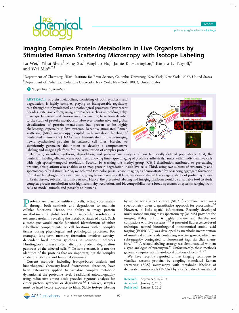
machineries.18 The newly synthesized proteins are specificallydetected by SRS through the vibrational signature fromcarbon−deuterium bonds (C−D) in the cell-silent spectralregion. This concept is particularly attractive for imaging denovo protein synthesis at the global level in live systems. On thelabeling side, cells and animals can tolerate a large amount ofdeuterium on D-AAs, which introduces minimum perturbationto protein functions. In fact, experiments using deuteratedwater or deuterated drugs have already been carried out onhumans.19−21 On the imaging side, SRS microscopy is asensitive and specific optical technique for imaging chemicalbonds. When the energy difference between incident photonsfrom two lasers (Pump beam and Stokes beam at 867.2 and1064 nm, respectively) matches the 2133 cm−1 mode of C−Dvibrations, the joint action of Pump and Stokes photons willefficiently excite a vibrational transition of C−D bonds.Whenever a molecule is transferred into the vibrational excitedstate, the Stokes pulse gains a photon, whereas the Pump pulseloses one, dictated by energy conservation (Figure 1a). Bydetecting the resulting stimulated Raman loss (or gain) of thePump beam (or the Stokes beam) in one pixel and then rasterscanning the laser spot across the sample, one can produce a3D concentration map of the targeted C−D bonds in living celland tissues (Figure 1b). Technically, SRS microscopy providesbackground-free chemical contrast with linear concentrationdependence, subcellular resolution determined by the opticaldiffraction limit (xy resolution of ∼300 nm; z resolution ordepth of field of ∼1000 nm), and intrinsic 3D sectioning that issuitable for tissue imaging, and the use of near-infraredwavelength and picosecond excitation pulses minimizes photonscattering inside turbid samples and potential phototoxic-ity.22−25
Despite the conceptual novelty, there are several notableshortcomings in the above proof-of-principle demonstration.First, only the synthesis aspect of protein metabolism wasprobed. Second, neither the D-AA labeling efficiency nor theSRS imaging instrument was optimized. Third, only culturedcell lines were demonstrated due to the limited sensitivity.18
In this article, we report a comprehensive labeling andimaging platform to probe complex protein metabolic dynamicsby fully exploiting the notion of coupling SRS with metaboliclabeling of D-AAs. Three major technical advances are beingimplemented together with a series of biological applications oncomplex tissues and model animals in vivo (Figure 1). First, weoptimized the chemical composition of the deuterated culturemedium to achieve a much higher deuterium labeling efficiencyand improved imaging sensitivity and speed of our SRSinstrumentation. These optimizations allow us to demonstratetime-lapse imaging of protein synthesis dynamics within singlelive cells. Second, we successfully imaged protein degradation inlive HeLa cells by targeting the Raman peak of the methylgroup (CH3) for pre-existing protein pools and employing arecently developed linear combination algorithm on measuredSRS images at 2940 and 2845 cm−1 channels. Third, inspired bythe classic pulse−chase analysis of complex protein dynamics,two-color pulse−chase imaging was accomplished by rationallydividing D-AAs into two structurally different subsets thatexhibit resolvable vibrational modes, as demonstrated bytracking aggregate formation of mutant huntingtin (mHtt)proteins. Finally, going beyond the cellular level to visualizingmore complex tissues and animals in vivo, we imaged the spatialdistribution of newly synthesized proteins inside live braintissue slices and in both developmental embryonic zebrafish
and mice (Figure 1). Taken together, these technical advancesand biological applications demonstrate that SRS microscopycoupled with metabolic labeling of D-AAs is a comprehensiveand generally applicable imaging platform to evaluate complexprotein metabolism with high sensitivity, resolution, andbiocompatibility in a broad spectrum of live cells, tissues, andanimals.
■ RESULTS AND DISCUSSIONSensitivity Optimization and Time-Lapse Imaging of
de Novo Proteome Synthesis Dynamics. The cell culturemedium reported previously was prepared by supplying auniformly deuterium-labeled whole set of amino acids tocommercially available medium that is deficient in leucine,lysine, and arginine.18 Due to the presence of other regularamino acids already in the commercial medium, the resultingpartially deuterated medium has only about a 60% deuteration
Figure 1. Imaging complex protein metabolism by stimulated Ramanscattering (SRS) microscopy in live cells, tissues, and animals. (a)Energy diagram of the SRS process. (b) Cartoon for SRS imagingfollowing metabolic labeling of deuterated amino acids (D-AAs) in liveorganisms (e.g., mice), which are first administered with D-AAs for acertain period of time and then imaged by SRS to probe proteinmetabolism. (c) Spontaneous Raman spectra from HeLa cellsincubated with medium containing either regular amino acids (gray,dashed) or D-AAs (black, solid) illustrate three distinct ways to probecomplex protein metabolism: imaging newly synthesized proteins bytargeting 2133 cm−1 from carbon−deuterium bonds (C−D), imagingdegradation of pre-existing proteins by targeting the pure methylgroup (CH3) distribution, and two-color pulse−chase protein imagingby labeling with two subgroups of D-AAs (i.e., groups I and II).
ACS Chemical Biology Articles
DOI: 10.1021/cb500787bACS Chem. Biol. 2015, 10, 901−908
902
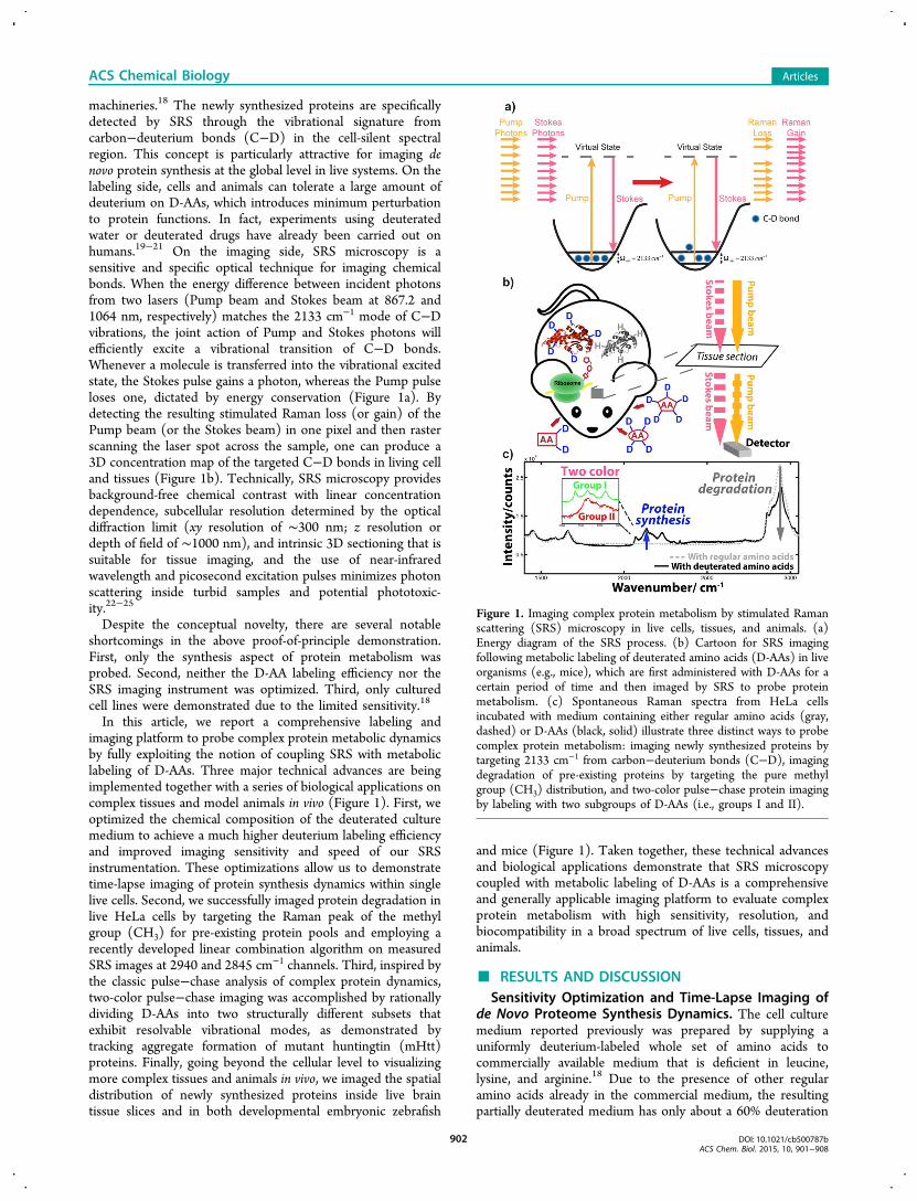
efficiency. In the present article, we custom-prepared newmedia that replace nearly all of the regular amino acids by theD-AA counterparts (details are given in the SupportingInformation). As shown in the spontaneous Raman spectra(Figure 2a), the optimized medium (red spectrum) displays a50% signal increase compared with that of the partiallydeuterated medium (blue spectrum). Indeed, SRS imagestargeting the C−D vibrational peak at 2133 cm−1 confirms a50% average intensity boost in live HeLa cells (Figure 2b). Theuse of optimized D-AA medium now leads to an about 8 timeshigher signal than that when using a single leucine-d10 (Figure2a, red vs black spectrum). In addition to improving thelabeling strategy, nontrivial instrumentation optimizations werealso carried out to further improve SRS detection sensitivityand acquisition speed, including increasing the laser output andmicroscope system’s throughput for near-IR wavelengths,replacing the acousto-optic modulator (AOM) with anelectro-optic modulator (EOM) for a 30% higher modulationdepth, and employing a high-speed lock-in amplifier for fasterimage acquisition.With much-improved sensitivity, protein synthesis can now
be imaged with superb spatial and temporal resolution.Spatially, we visualized newly synthezied proteins from finestructures (likely dendritic spines, indicated by arrow heads) oflive neurons (Figure 2c). Temporally, we could readily imagenewly synthesized proteins in live HeLa cells in less than a 1 hincubation with the optimized deuteration medium (Figure2d). A control image of cells in the presence of proteinsynthesis inhibitors displays only vague and homogeneous celloutlines, which, presumably, come from the free D-AA pool(submillimolar concentration, much more dilute than themetabolically enriched pool in the protein-bound form).18,26
Moreover, using a fast lock-in amplifier (details are given inMethods), our current imaging speed can be as fast as 3 s perframe (512 × 512 pixels), nearly 10 times faster than before,which enables time-lapse imaging in live cells with minimumphototoxicity to cell viabilities. Figure 2e presents time-lapseSRS imaging of the same set of live HeLa cells graduallysynthesizing new proteins over time from a 10 min to 5 hincubation in optimized D-AA medium. The obviousobservation of cell migration and division prove the viabilityof the cells, supporting the high biocompatibility of ourtechnique. To our knowledge, this is the first time that long-
term time-lapse imaging of proteome synthesis dynamics hasbeen demonstrated on single live mamamian cells.
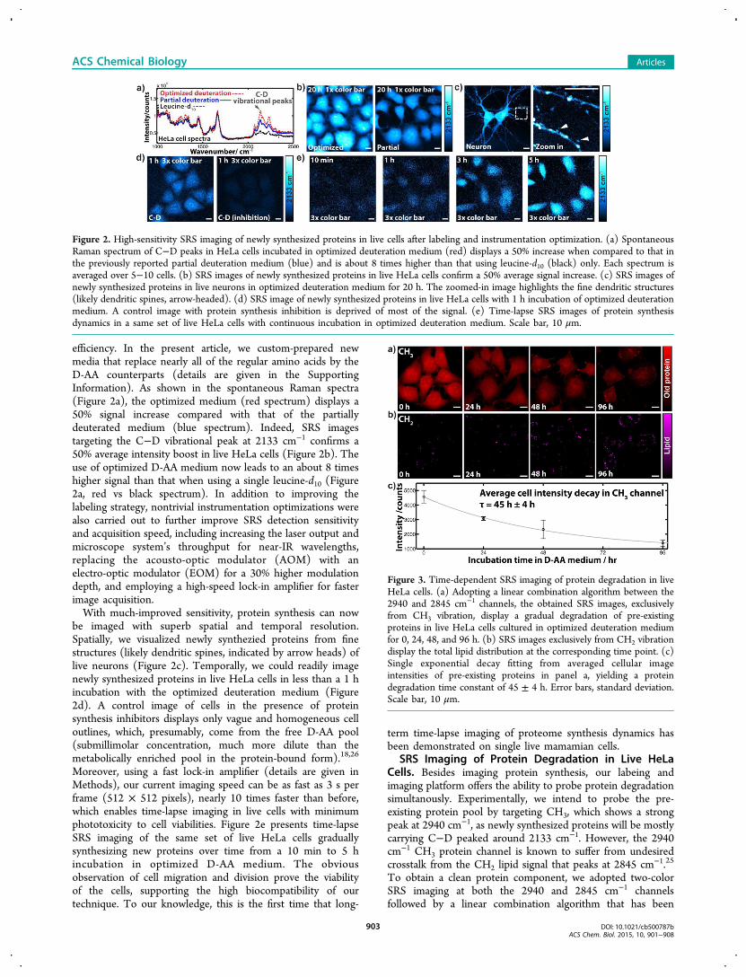
SRS Imaging of Protein Degradation in Live HeLaCells. Besides imaging protein synthesis, our labeing andimaging platform offers the ability to probe protein degradationsimultanously. Experimentally, we intend to probe the pre-existing protein pool by targeting CH3, which shows a strongpeak at 2940 cm−1, as newly synthesized proteins will be mostlycarrying C−D peaked around 2133 cm−1. However, the 2940cm−1 CH3 protein channel is known to suffer from undesiredcrosstalk from the CH2 lipid signal that peaks at 2845 cm−1.25
To obtain a clean protein component, we adopted two-colorSRS imaging at both the 2940 and 2845 cm−1 channelsfollowed by a linear combination algorithm that has been
Figure 2. High-sensitivity SRS imaging of newly synthesized proteins in live cells after labeling and instrumentation optimization. (a) SpontaneousRaman spectrum of C−D peaks in HeLa cells incubated in optimized deuteration medium (red) displays a 50% increase when compared to that inthe previously reported partial deuteration medium (blue) and is about 8 times higher than that using leucine-d10 (black) only. Each spectrum isaveraged over 5−10 cells. (b) SRS images of newly synthesized proteins in live HeLa cells confirm a 50% average signal increase. (c) SRS images ofnewly synthesized proteins in live neurons in optimized deuteration medium for 20 h. The zoomed-in image highlights the fine dendritic structures(likely dendritic spines, arrow-headed). (d) SRS image of newly synthesized proteins in live HeLa cells with 1 h incubation of optimized deuterationmedium. A control image with protein synthesis inhibition is deprived of most of the signal. (e) Time-lapse SRS images of protein synthesisdynamics in a same set of live HeLa cells with continuous incubation in optimized deuteration medium. Scale bar, 10 μm.
Figure 3. Time-dependent SRS imaging of protein degradation in liveHeLa cells. (a) Adopting a linear combination algorithm between the2940 and 2845 cm−1 channels, the obtained SRS images, exclusivelyfrom CH3 vibration, display a gradual degradation of pre-existingproteins in live HeLa cells cultured in optimized deuteration mediumfor 0, 24, 48, and 96 h. (b) SRS images exclusively from CH2 vibrationdisplay the total lipid distribution at the corresponding time point. (c)Single exponential decay fitting from averaged cellular imageintensities of pre-existing proteins in panel a, yielding a proteindegradation time constant of 45 ± 4 h. Error bars, standard deviation.Scale bar, 10 μm.
ACS Chemical Biology Articles
DOI: 10.1021/cb500787bACS Chem. Biol. 2015, 10, 901−908
903
effectively applied in cells, tissues, and animals.27−29 Thesubsequently obtained images show the pure distribution of oldprotein pools (exclusively from CH3) and the distribution oflipids (exclusively from CH2), respectively. Hence, proteindegradation could be tracked by imaging the old proteindistributions over time when cells are growing in D-AAmedium.Figure 3a shows time-dependent SRS images of old protein
distributions (CH3) in live HeLa cells when incubated with D-AAs from 0 to 96 h. Clearly, the old protein pool is degrading,as shown by the decay of its average intensity. In contrast, thetotal lipid images display no obvious intensity change (Figure3b). In addition, the spatial patterns of old proteins (Figure 3a)reveal a faster decay in the nucleoli than that in the cytoplasm.This observation is consistent with the fact that neucleoli haveactive protein turnover30 and also with our previous report thatC−D labeled newly synthesized proteins are more prominentin nucleoli.18 Single exponential decay fitting of the averageintensities in Figure 3a yields a decay time constant of 45 ± 4 h(Figure 3c), corresponding to a proteome half-life of 31 ± 3 h,which is very close to that reported by mass spectrometry (35h).31 Therefore, our imaging platform is capable of observingboth protein synthesis and degradation by imaging the C−Dchannel and CH3 channel, respectively, thus capturingproteomic metabolism dynamics in full-scope.
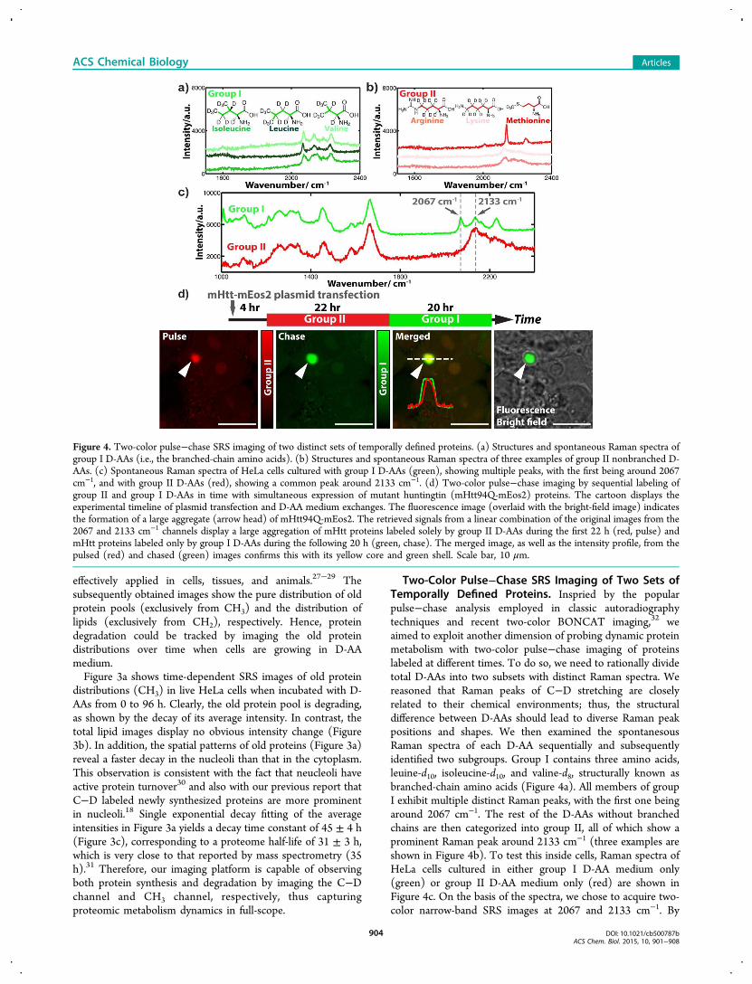
Two-Color Pulse−Chase SRS Imaging of Two Sets ofTemporally Defined Proteins. Inspried by the popularpulse−chase analysis employed in classic autoradiographytechniques and recent two-color BONCAT imaging,32 weaimed to exploit another dimension of probing dynamic proteinmetabolism with two-color pulse−chase imaging of proteinslabeled at different times. To do so, we need to rationally dividetotal D-AAs into two subsets with distinct Raman spectra. Wereasoned that Raman peaks of C−D stretching are closelyrelated to their chemical environments; thus, the structuraldifference between D-AAs should lead to diverse Raman peakpositions and shapes. We then examined the spontanesousRaman spectra of each D-AA sequentially and subsequentlyidentified two subgroups. Group I contains three amino acids,leuine-d10, isoleucine-d10, and valine-d8, structurally known asbranched-chain amino acids (Figure 4a). All members of groupI exhibit multiple distinct Raman peaks, with the first one beingaround 2067 cm−1. The rest of the D-AAs without branchedchains are then categorized into group II, all of which show aprominent Raman peak around 2133 cm−1 (three examples areshown in Figure 4b). To test this inside cells, Raman spectra ofHeLa cells cultured in either group I D-AA medium only(green) or group II D-AA medium only (red) are shown inFigure 4c. On the basis of the spectra, we chose to acquire two-color narrow-band SRS images at 2067 and 2133 cm−1. By
Figure 4. Two-color pulse−chase SRS imaging of two distinct sets of temporally defined proteins. (a) Structures and spontaneous Raman spectra ofgroup I D-AAs (i.e., the branched-chain amino acids). (b) Structures and spontaneous Raman spectra of three examples of group II nonbranched D-AAs. (c) Spontaneous Raman spectra of HeLa cells cultured with group I D-AAs (green), showing multiple peaks, with the first being around 2067cm−1, and with group II D-AAs (red), showing a common peak around 2133 cm−1. (d) Two-color pulse−chase imaging by sequential labeling ofgroup II and group I D-AAs in time with simultaneous expression of mutant huntingtin (mHtt94Q-mEos2) proteins. The cartoon displays theexperimental timeline of plasmid transfection and D-AA medium exchanges. The fluorescence image (overlaid with the bright-field image) indicatesthe formation of a large aggregate (arrow head) of mHtt94Q-mEos2. The retrieved signals from a linear combination of the original images from the2067 and 2133 cm−1 channels display a large aggregation of mHtt proteins labeled solely by group II D-AAs during the first 22 h (red, pulse) andmHtt proteins labeled only by group I D-AAs during the following 20 h (green, chase). The merged image, as well as the intensity profile, from thepulsed (red) and chased (green) images confirms this with its yellow core and green shell. Scale bar, 10 μm.
ACS Chemical Biology Articles
DOI: 10.1021/cb500787bACS Chem. Biol. 2015, 10, 901−908
904
constructing and utilizing a linear combination algorithm(Supplementary Figure 1), similar to the one used for CH3
and CH2 above, pure signals of proteins labeled by group I D-AAs and by group II D-AAs can be successfully separated andquantitatively visualized. Note that hyperspectral imagingapproaches using broadband femtosecond lasers might alsowork here.33−35
We now chose the mutant huntingtin (mHtt) protein inHuntington’s disease as our model system for the pulse−chaseimaging demonstration. It is believed that Huntington’s diseaseis caused by a mutation from a normal huntingtin gene to amHtt gene expressing aggregation-prone mHtt proteins withpolyglutamine (polyQ) expansion.3 For easy visualization byfluorescence, we tagged mHtt (with 94Q) with a fluorescentprotein marker, mEos2. As illustrated by the cartoon in Figure4d, HeLa cells were first transfected with mHtt94Q-mEos2plasmid in regular medium for 4 h, which was then replacedwith group II D-AA medium for 22 h before changing to groupI D-AA medium for another 20 h. SRS images are acquired inthe 2067 and 2133 cm−1 channels, respectively, andsubsequently processed with linear combination.A fluorescence image overlaid with a bright-field image
demonstrates the formation of a large aggregate triggered byaggregation-prone polyQ expansion in mHtt94Q-mEos2(Figure 4d, fluorescence). Interestingly, proteins labeled withgroup II D-AAs during the initial pulse period concentratemainly within the core of the aggregate (Figure 4d, red),whereas proteins labeled with group I D-AAs during thesubsequent chase period occupy the entire volume of theaggregate (Figure 4d, green). The merged image between
group I and group II images, as well as the intensity profilesacross the aggregate, further confirm the observation of ayellow core inside and a green shell outside (Figure 4d,merged). This two-color pulse−chase result suggests that thecore is aggregated earlier in time and that the later producedmHtt proteins are then recruited to and percolate through theaggregate to increases its overall size, in agreement withrecently reported results by fluorescence.36 The demonstrationhere thus illustrates that our imaging platform using the twosubgroups of D-AAs is readily applicable for performing pulse−chase imaging to probe the complex and dynamic aspects ofproteome metabolism.
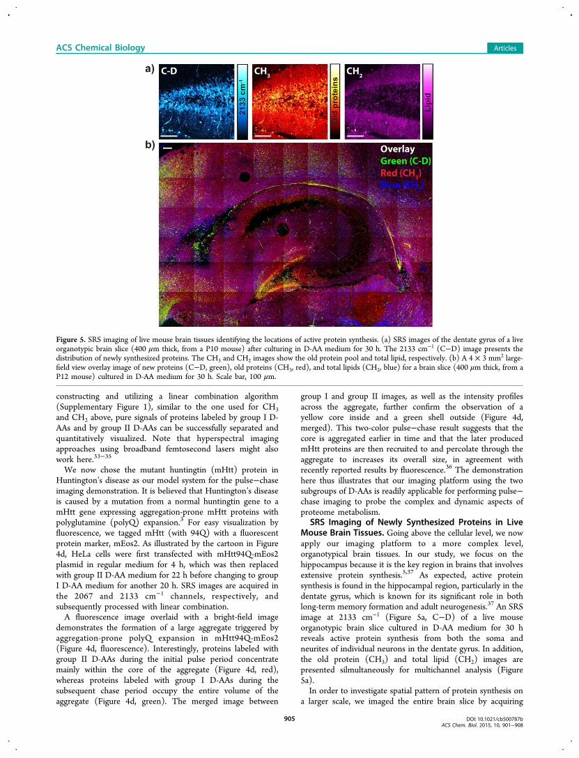
SRS Imaging of Newly Synthesized Proteins in LiveMouse Brain Tissues. Going above the cellular level, we nowapply our imaging platform to a more complex level,organotypical brain tissues. In our study, we focus on thehippocampus because it is the key region in brains that involvesextensive protein synthesis.5,37 As expected, active proteinsynthesis is found in the hippocampal region, particularly in thedentate gyrus, which is known for its significant role in bothlong-term memory formation and adult neurogenesis.37 An SRSimage at 2133 cm−1 (Figure 5a, C−D) of a live mouseorganotypic brain slice cultured in D-AA medium for 30 hreveals active protein synthesis from both the soma andneurites of individual neurons in the dentate gyrus. In addition,the old protein (CH3) and total lipid (CH2) images arepresented silmultaneously for multichannel analysis (Figure5a).In order to investigate spatial pattern of protein synthesis on
a larger scale, we imaged the entire brain slice by acquiring
Figure 5. SRS imaging of live mouse brain tissues identifying the locations of active protein synthesis. (a) SRS images of the dentate gyrus of a liveorganotypic brain slice (400 μm thick, from a P10 mouse) after culturing in D-AA medium for 30 h. The 2133 cm−1 (C−D) image presents thedistribution of newly synthesized proteins. The CH3 and CH2 images show the old protein pool and total lipid, respectively. (b) A 4 × 3 mm2 large-field view overlay image of new proteins (C−D, green), old proteins (CH3, red), and total lipids (CH2, blue) for a brain slice (400 μm thick, from aP12 mouse) cultured in D-AA medium for 30 h. Scale bar, 100 μm.
ACS Chemical Biology Articles
DOI: 10.1021/cb500787bACS Chem. Biol. 2015, 10, 901−908
905
large-area image mosaics. A 4 × 3 mm2 image (Figure 5b) ofanother organotypic slice displays an overlaid pattern from newproteins (2133 cm−1, green), old proteins (CH3, red), andlipids (CH2, blue). Intriguing spatial variation is observed: whilethe distribution of old proteins is relatively homogeneousacross the field of view, newly synthesized proteins are eitherconcentrated in the dentate gyrus or scattered within individualneurons throughout the cortex, suggesting high activity in thesetwo regions. Thus, we have demonstrated the ability to directlyimage protein synthesis dynamics on living brain tissues withsubcellular resolution and multichannel analysis, which wasdifficult to achieve with other existing methods.38 The intricaterelationship between protein synthesis and neuronal plasticity39
is currently under investigation on this platform.SRS Imaging of Newly Synthesized Proteins in Vivo.
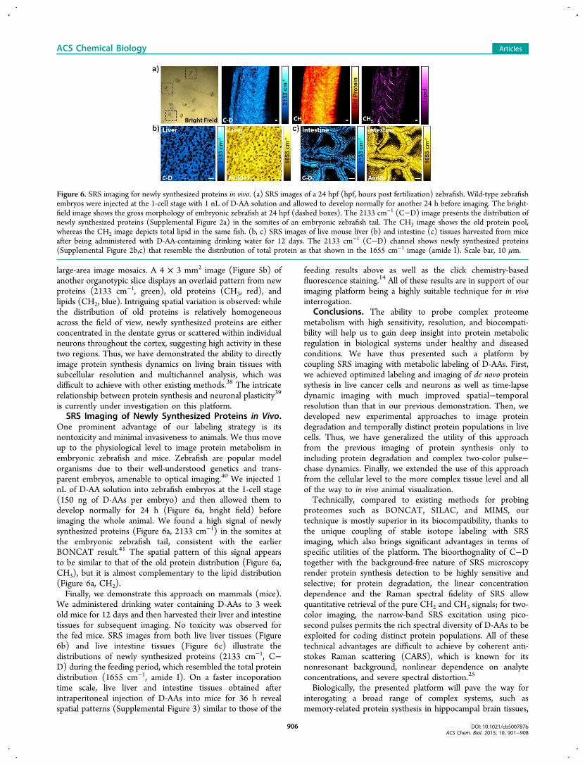
One prominent advantage of our labeling strategy is itsnontoxicity and minimal invasiveness to animals. We thus moveup to the physiological level to image protein metabolism inembryonic zebrafish and mice. Zebrafish are popular modelorganisms due to their well-understood genetics and trans-parent embryos, amenable to optical imaging.40 We injected 1nL of D-AA solution into zebrafish embryos at the 1-cell stage(150 ng of D-AAs per embryo) and then allowed them todevelop normally for 24 h (Figure 6a, bright field) beforeimaging the whole animal. We found a high signal of newlysynthesized proteins (Figure 6a, 2133 cm−1) in the somites atthe embryonic zebrafish tail, consistent with the earlierBONCAT result.41 The spatial pattern of this signal appearsto be similar to that of the old protein distribution (Figure 6a,CH3), but it is almost complementary to the lipid distribution(Figure 6a, CH2).Finally, we demonstrate this approach on mammals (mice).
We administered drinking water containing D-AAs to 3 weekold mice for 12 days and then harvested their liver and intestinetissues for subsequent imaging. No toxicity was observed forthe fed mice. SRS images from both live liver tissues (Figure6b) and live intestine tissues (Figure 6c) illustrate thedistributions of newly synthesized proteins (2133 cm−1, C−D) during the feeding period, which resembled the total proteindistribution (1655 cm−1, amide I). On a faster incoporationtime scale, live liver and intestine tissues obtained afterintraperitoneal injection of D-AAs into mice for 36 h revealspatial patterns (Supplemental Figure 3) similar to those of the
feeding results above as well as the click chemistry-basedfluorescence staining.14 All of these results are in support of ourimaging platform being a highly suitable technique for in vivointerrogation.
Conclusions. The ability to probe complex proteomemetabolism with high sensitivity, resolution, and biocompati-bility will help us to gain deep insight into protein metabolicregulation in biological systems under healthy and diseasedconditions. We have thus presented such a platform bycoupling SRS imaging with metabolic labeling of D-AAs. First,we achieved optimized labeling and imaging of de novo proteinsythesis in live cancer cells and neurons as well as time-lapsedynamic imaging with much improved spatial−temporalresolution than that in our previous demonstration. Then, wedeveloped new experimental approaches to image proteindegradation and temporally distinct protein populations in livecells. Thus, we have generalized the utility of this approachfrom the previous imaging of protein synthesis only toincluding protein degradation and complex two-color pulse−chase dynamics. Finally, we extended the use of this approachfrom the cellular level to the more complex tissue level and allof the way to in vivo animal visualization.Technically, compared to existing methods for probing
proteomes such as BONCAT, SILAC, and MIMS, ourtechnique is mostly superior in its biocompatibility, thanks tothe unique coupling of stable isotope labeling with SRSimaging, which also brings significant advantages in terms ofspecific utilities of the platform. The bioorthognality of C−Dtogether with the background-free nature of SRS microscopyrender protein synthesis detection to be highly sensitive andselective; for protein degradation, the linear concentrationdependence and the Raman spectral fidelity of SRS allowquantitative retrieval of the pure CH2 and CH3 signals; for two-color imaging, the narrow-band SRS excitation using pico-second pulses permits the rich spectral diversity of D-AAs to beexploited for coding distinct protein populations. All of thesetechnical advantages are difficult to achieve by coherent anti-stokes Raman scattering (CARS), which is known for itsnonresonant background, nonlinear dependence on analyteconcentrations, and severe spectral distortion.25
Biologically, the presented platform will pave the way forinterogating a broad range of complex systems, such asmemory-related protein systhesis in hippocampal brain tissues,
Figure 6. SRS imaging for newly synthesized proteins in vivo. (a) SRS images of a 24 hpf (hpf, hours post fertilization) zebrafish. Wild-type zebrafishembryos were injected at the 1-cell stage with 1 nL of D-AA solution and allowed to develop normally for another 24 h before imaging. The bright-field image shows the gross morphology of embryonic zebrafish at 24 hpf (dashed boxes). The 2133 cm−1 (C−D) image presents the distribution ofnewly synthesized proteins (Supplemental Figure 2a) in the somites of an embryonic zebrafish tail. The CH3 image shows the old protein pool,whereas the CH2 image depicts total lipid in the same fish. (b, c) SRS images of live mouse liver (b) and intestine (c) tissues harvested from miceafter being administered with D-AA-containing drinking water for 12 days. The 2133 cm−1 (C−D) channel shows newly synthesized proteins(Supplemental Figure 2b,c) that resemble the distribution of total protein as that shown in the 1655 cm−1 image (amide I). Scale bar, 10 μm.
ACS Chemical Biology Articles
DOI: 10.1021/cb500787bACS Chem. Biol. 2015, 10, 901−908
906

protein aggregation and degradation in neurodegenerativediseases, and protein metabolism in animal disease models.Furthermore, considering that stable isotope labeling and SRSimaging are both compatible with live humans,42 we envisionthat the prospects are bright for applying this platform toperforming diagnostic and theraputic imaging in humans.
■ METHODSStimulated Raman Scattering Microscopy. Spatially and
temporally overlapped pulsed Pump (tunable from 720 to 990 nm,5−6 ps, 80 MHz repetition rate) and Stokes (1064 nm, 6 ps, 80 MHzrepetition rate, modulated at 8 MHz) beams, which are provided by acustom-modified picoEMERALD system from Applied Physics &Electronics, Inc., are coupled into an inverted laser-scanningmicroscope (FV1200 MPE, Olympus) optimized for near-IRthroughput. A 60× water objective (UPlanAPO/IR, 1.2 N.A.,Olympus) is used for all cell imaging, and a 25× water objective(XLPlan N, 1.05 N.A., MP, Olympus) with both a high near-IRtransmission and a large field of view is used for brain tissue and in vivoimaging. After passing through the sample, the forward-going Pumpand Stokes beams are collected in transmission by a high N.A. oilcondenser. A high O.D. bandpass filter (890/220, Chroma) is used toblock the Stokes beam completely and to transmit the Pump beamonly onto a large area Si photodiode for the detection of thestimulated Raman loss signal. The output current from the photodiodeis terminated, filtered, and demodulated by a lock-in amplifier at 8MHz to ensure shot-noise-limited detection sensitivity. (Details aregiven in the Supporting Information.)Metabolic Incorporation of Deuterated Amino Acids. For
HeLa cells, cells were seeded on a coverslip in a Petri dish with 2 mLof regular medium for 20 h, which was then replaced with D-AAmedium (or group I and group II D-AA media) for the designatedamount of time. The coverslip was taken out to make an imagingchamber filled with PBS for SRS imaging. For hippocampal neurons,the dissociated neurons from newborn mice were seeded for 10 days inregular Neurobasal A medium, which was then replaced with thecorresponding D-AA medium for the designated amount of timebefore imaging. For organotypic brain slices, 400 μm thick, P10 mousebrain slices were cultured on Millicell-CM inserts (PICM03050,Millipore) in 1 mL of CD-MEM culture medium for 2 h, which wasthen changed to 1 mL of CD-neurobasal A culture medium foranother 28 h before imaging. For a detailed recipe of D-AA media andthe in vivo labeling procedure in zebrafish and mice, see the SupportingInformation. The experimental protocols for in vivo mice experiments(AC-AAAG2702) and zebrafish experiments (AC-AAAD6300) wereapproved by the Institutional Animal Care and Use Committee atColumbia University.Spontaneous Raman spectroscopy. The spontaneous Raman
spectra were acquired using a laser Raman spectrometer (inVia Ramanmicroscope, Ranishaw) at room temperature. A 27 mW (afterobjective), 532 nm diode laser was used to excite the sample througha 50×, N.A. 0.75 objective (NPLAN EPI, Leica). The total dataacquisition was performed during 60 s using WiRE software. All of thespontaneous Raman spectra subtracted the PBS solution as back-ground.Image Progressing. Images were acquired with FluoView
scanning software and assigned color or overlaid by ImageJ. Linearcombination was processed with MATLAB. Graphs were assembledwith Adobe Illustrator.
■ ASSOCIATED CONTENT*S Supporting InformationAdditional materials and methods. Figure S1: SRS images at2067 and 2133 cm−1 channels of proteins labeled with group ID-AA only and group II D-AA only. Figure S2: Raw C-D on-resonance (2133 cm−1) and off-resonance (2000 cm−1) SRSimages of newly synthesized proteins in vivo. Figure S3: SRSimaging for newly synthesized proteins in vivo with intra-
peritoneal injection of mice with D-AA solutions. This materialis available free of charge via the Internet at http://pubs.acs.org.
■ AUTHOR INFORMATIONCorresponding Author*E-mail: [email protected] ContributionsL.W., Y.S., F.X., F.H., J.K.H., and K.L.T. performed experimentsand analyzed data. L.W., Y.S., and W.M. designed theexperiments. L.W. and W.M. conceived the concept andwrote the article.NotesThe authors declare the following competing financialinterest(s): Columbia University has filed a patent applicationbased on this work.
■ ACKNOWLEDGMENTSWe thank J. Jackson and C. Dupre for assistance with the brainslices and J. C. Tapia, M. C. Wang, Z. Chen, D. Peterka, and R.Yuste for helpful discussions. We are grateful to Y. Shin fortechnical assistance with the in vivo mice experiments. W.M.acknowledges support from Columbia University, an NationalInstitutes of Health Director’s New Innovator Award, the U.S.Army Research Office (W911NF-12-1-0594), the BrainResearch Foundation, and an Alfred P. Sloan ResearchFellowship.
■ REFERENCES(1) Mayford, M., Siegelbaum, S.-A., and Kandel, E.-R. (2012)Synapses and memory storage. Cold Spring Harbor Perspect. Biol. 4,a005751.(2) Sutton, M.-A., and Schuman, E.-M. (2006) Dendritic proteinsynthesis, synaptic plasticity, and memory. Cell 127, 49−58.(3) Walker, F.-O. (2007) Huntington’s disease. Lancet. 369, 218−228.(4) Bennett, E.-J., Shaler, T.-A., Woodman, B., Ryu, K.-Y., Zaitseva,T.-S., Becker, C.-H., Bates, G.-P., Schulman, H., and Kopito, R. R.(2007) Global changes to the ubiquitin system in Huntington’sdisease. Nature 448, 704−708.(5) Lipton, P., and Raley-Susman, K.-M. (1999) Autoradiographicmeasurements of protein synthesis in hippocampal slices from rats andguinea pigs. Methods 18, 127−143.(6) Bachmair, A., Finley, D., and Varshavsky, A. (1986) In vivo half-life of a protein is a function of its amino-terminal residue. Science 234,179−186.(7) Ong, S.-E., Blagoev, B., Kratchmarova, I., Kristensen, D.-B., Steen,H., Pandey, A., and Mann, M. (2002) Stable isotope labeling by aminoacids in cell culture, SILAC, as a simple and accurate approach toexpression proteomics. Mol. Cell. Proteomics 1, 376−386.(8) Mann, M. (2006) Functional and quantitative proteomics usingSILAC. Nat. Rev. Mol. Cell Biol. 7, 952−958.(9) Lechene, C., Hillion, F., McMahon, G., Benson, D., Kleinfeld, A.-M., Kampf, J.-P., Distel, D., Luyten, Y., Bonventre, J., Hentschel, D.,Park, K.-M., Ito, S., Schwartz, M., Benichou, G., and Slodzian, G.(2006) High-resolution quantitative imaging of mammalian andbacterial cells using stable isotope mass spectrometry. J. Biol. 5, 20.(10) Zhang, D.-S., Piazza, V., Perrin, B.-J., Rzadzinska, A.-K.,Poczatek, J.-C., Wang, M., Prosser, H.-M., Ervasti, J.-M., Corey, D.-P., and Lechene, C.-P. (2012) Multi-isotope imaging massspectrometry reveals slow protein turnover in hair-cell stereocilia.Nature 481, 520−524.(11) Beatty, K.-E., Liu, J.-C., Xie, F., Dieterich, D.-C., Schuman, E.-M., Wang, Q., and Tirrell, D.-A. (2006) Fluorescence visualization ofnewly synthesized proteins in mammalian cells. Angew. Chem., Int. Ed.45, 7364−7367.
ACS Chemical Biology Articles
DOI: 10.1021/cb500787bACS Chem. Biol. 2015, 10, 901−908
907
(12) Ngo, J.-T., and Tirrell, D.-A. (2011) Noncanonical amino acidsin the interrogation of cellular protein synthesis. Acc. Chem. Res. 44,677−685.(13) Yuet, K.-P., and Tirrell, D.-A. (2014) Chemical tools fortemporally and spatially resolved mass spectrometry-based proteomics.Ann. Biomed. Eng. 42, 299−311.(14) Liu, J., Xu, Y., Stoleru, D., and Salic, A. (2012) Imaging proteinsynthesis in cells and tissues with an alkyne analog of puromycin. Proc.Natl. Acad. Sci. U.S.A. 109, 413−418.(15) Prescher, J.-A., and Bertozzi, C.-R. (2005) Chemistry in livingsystems. Nat. Chem. Biol. 1, 13−21.(16) Bertozzi, C.-R. (2011) A decade of bioorthogonal chemistry.Acc. Chem. Res. 44, 651−653.(17) Grammel, M., and Hang, H.-C. (2013) Chemical reporters forbiological discovery. Nat. Chem. Biol. 9, 475−484.(18) Wei, L., Yu, Y., Shen, Y., Wang, M.-C., and Min, W. (2013)Vibrational imaging of newly synthesized proteins in live cells bystimulated Raman scattering microscopy. Proc. Natl. Acad. Sci. U.S.A.110, 11226−11231.(19) Moore, F.-D. (1946) Determination of total body water andsolids with isotopes. Science 104, 157−160.(20) Pinkus, J.-L., Charles, D., and Chattoraj, S.-C. (1971)Deuterium-labeled steroids for study in humans: I. estrogenproduction rates in normal pregnancy. J. Biol. Chem. 246, 633−636.(21) Tang, G., Qin, J., and Dolnikowski, G.-G. (1998) Deuteriumenrichment of retinol in humans determined by gas chromatographyelectron capture negative chemical ionization mass spectrometry. J.Nutr. Biochem. 9, 408−414.(22) Freudiger, C.-W., Min, W., Saar, B.-G., Lu, S., Holtom, G.-R.,He, C., Tsai, J.-C., Kang, J.-X., and Xie, X.-S. (2008) Label-freebiomedical imaging with high sensitivity by stimulated Ramanscattering microscopy. Science 322, 1857−1861.(23) Min, W. (2011) Label-free optical imaging of nonfluorescentmolecules by stimulated radiation. Curr. Opin. Chem. Biol. 15, 831−837.(24) Min, W., Freudiger, C.-W., Lu, S., and Xie, X.-S. (2011)Coherent nonlinear optical imaging: beyond fluorescence microscopy.Annu. Rev. Phys. Chem. 62, 507−530.(25) (2012) Coherent Raman Scattering Microscopy (Cheng, J.-X., andXie, X.-S., Eds.) CRC Press, Boca Raton, FL.(26) Piez, K.-A., and Eagle, H. (1958) The free amino acid pool ofcultured human cells. J. Biol. Chem. 231, 533−545.(27) Lu, F.-K., Ji, M., Fu, D., Ni, X., Freudiger, C.-W., Holtom, G.,and Xie, X.-S. (2012) Multicolor stimulated Raman scattering (SRS)microscopy. Mol. Phys. 110, 1927−1932.(28) Yu, Z., Chen, T., Zhang, X., Fu, D., Liao, X., Shen, J., Liu, X.,Zhang, B., Xie, X.-S., Su, X.-D., Chen, J., and Huang, Y. (2012) Label-free chemical imaging in vivo: three-dimensional non-invasivemicroscopic observation of amphioxus notochord through stimulatedRaman scattering (SRS). Chem. Sci. 3, 2646−2654.(29) Ji, M., Orringer, D.-A., Freudiger, C.-W., Ramkissoon, S., Liu, X.,Lau, D., Golby, A.-J., Norton, I., Hayashi, M., Agar, N.-Y., Young, G.-S.,Spino, C., Santagata, S., Camelo-Piragua, S., Ligon, K.-L., Sagher, O.,and Xie, X.-S. (2013) Rapid, label-free detection of brain tumors withstimulated Raman scattering microscopy. Sci. Transl. Med. 5, 201ra119.(30) Andersen, J.-S., Lam, Y.-W., Leung, A.-K., Ong, S.-E., Lyon, C.-E., Lamond, A.-I., and Mann, M. (2005) Nucleolar proteomedynamics. Nature 433, 77−83.(31) Cambridge, S.-B., Gnad, F., Nguyen, C., Bermejo, J.-L., Kruger,M., and Mann, M. (2011) Systems-wide proteomic analysis inmammalian cells reveals conserved, functional protein turnover. J.Proteome Res. 10, 5275−5284.(32) Beatty, K.-E., and Tirrell, D.-A. (2008) Two-color labeling oftemporally defined protein populations in mammalian cells. Bioorg.Med. Chem. Lett. 18, 5995−5999.(33) Zhang, D., Wang, P., Slipchenko, M.-N., Ben-Amotz, D.,Weiner, A.-M., and Cheng, J.-X. (2013) Quantitative vibrationalimaging by hyperspectral stimulated Raman scattering microscopy andmultivariate curve resolution analysis. Anal. Chem. 85, 98−106.
(34) Zhang, D., Slipchenko, M.-N., and Cheng, J.-X. (2011) Highlysensitive vibrational imaging by femtosecond pulse stimulated Ramanloss. J. Phys. Chem. Lett. 2, 1248−1253.(35) Fu, D., Holtom, G., Freudiger, C.-W., Zhang, X., and Xie, X.-S.(2013) Hyperspectral imaging with stimulated Raman scattering bychirped femtosecond lasers. J. Phys. Chem. B 117, 4634−4640.(36) Schipper-Krom, S., Juenemann, K., Jansen, A.-H., Wiemhoefer,A., van den Nieuwendijk, R., Smith, D.-L., Hink, M.-A., Bates, G.-P.,Overkleeft, H., Ovaa, H., and Reits, E. (2014) Dynamic recruitment ofactive proteasomes into polyglutamine initiated inclusion bodies. FEBSLett. 588, 151−159.(37) Deng, W., Aimone, J.-B., and Gage, F.-H. (2010) New neuronsand new memories: how does adult hippocampal neurogenesis affectlearning and memory? Nat. Rev. Neurosci. 11, 339−350.(38) Dieterich, D.-C., Hodas, J.-J., Gouzer, G., Shadrin, I.-Y., Ngo, J.-T., Triller, A., Tirrell, D.-A., and Schuman, E.-M. (2010) In situvisualization and dynamics of newly synthesized proteins in rathippocampal neurons. Nat. Neurosci. 13, 897−905.(39) Hinz, F.-I., Dieterich, D.-C., and Schuman, E.-M. (2013)Teaching old NCATs new tricks: using non-canonical amino acidtagging to study neuronal plasticity. Curr. Opin. Chem. Biol. 17, 738−746.(40) Kari, G., Rodeck, U., and Dicker, A.-P. (2007) Zebrafish: anemerging model system for human disease and drug discovery. Clin.Pharmacol. Ther. 82, 70−80.(41) Hinz, F.-I., Dieterich, D.-C., Tirrell, D.-A., and Schuman, E.-M.(2012) Non-canonical amino acid labeling in vivo to visualize andaffinity purify newly synthesized proteins in larval zebrafish. ACSChem. Neurosci. 3, 40−49.(42) Saar, B.-G., Freudiger, C.-W., Reichman, J., Stanley, C.-M.,Holtom, G.-R., and Xie, X.-S. (2010) Video-rate molecular imaging invivo with stimulated Raman scattering. Science 330, 1368−1370.
ACS Chemical Biology Articles
DOI: 10.1021/cb500787bACS Chem. Biol. 2015, 10, 901−908
908