Imaging breast mammogram
72
IMAGING BREAST MAMMOGRAM By : DR REKHA KHARE MD RADIOLOGY
-
Upload
rekhakhare -
Category
Health & Medicine
-
view
489 -
download
0
Transcript of Imaging breast mammogram
IMAGING BREAST MAMMOGRAM
By : DR REKHA KHARE MD RADIOLOGY
What is a mammogram ?
A mammogram is a x-ray exam of the
breast to detect and evaluate any
change in the breast

History of mammography
• X-rays were first used to examine breast
tissue by the German surgeon
ALBERT SALOMON about a century ago
• Modern mammography came in to existence since the late 1960
• Technology has advanced and today’s machine is different even from those of
the 1980s and 1990s
Mammography machine
• Today’s mammography machine expose
the breast to much less radiation
• X-rays do not go through tissue as easily as those used for routine chest x-ray or x-rays of extremity
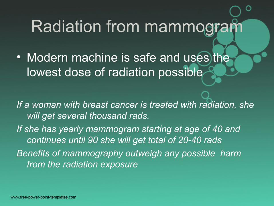
Radiation from mammogram
• Modern machine is safe and uses the lowest dose of radiation possible
If a woman with breast cancer is treated with radiation, she will get several thousand rads.
If she has yearly mammogram starting at age of 40 and continues until 90 she will get total of 20-40 rads
Benefits of mammography outweigh any possible harm from the radiation exposure
Types of mammogram
• Screening mammogram
• Diagnostic mammogram
Screening mammogram
• Mammogram of the breast for the women
who have no sign or symptom of breast
cancer, usually with two x-ray views
• Finding breast cancer early greatly improves a woman’s chance for successful treatment
Diagnostic mammogram
• X-ray of the breast for a woman with breast problem like lump or nipple discharge or an abnormal area found in screening by taking spot view or magnification view
What diagnostic mammogram does? One of three ways----• It may reveal that an area that looked abnormal
on screening is actually NORMAL—routine checkup
• It could reveal that an area of abnormal tissue probably is NOT CANCER but radiologist may not ready to say it normal based on these
x-rays--- re-check up in 4-6 months• The results could also suggest that a biopsy is
needed to find out if the abnormal area is CANCER
Types of mammogram machine
• Screen- film units
• Full- field digital mammography units (better in woman younger than 50/or
with dense breast tissue)
How is mammogram done?
• Breast is briefly compressed between 2 plates attached to the mammogram machine– an adjustable plastic plate on top and a fixed plate on bottom which holds the x-ray film or the digital detector that makes the image
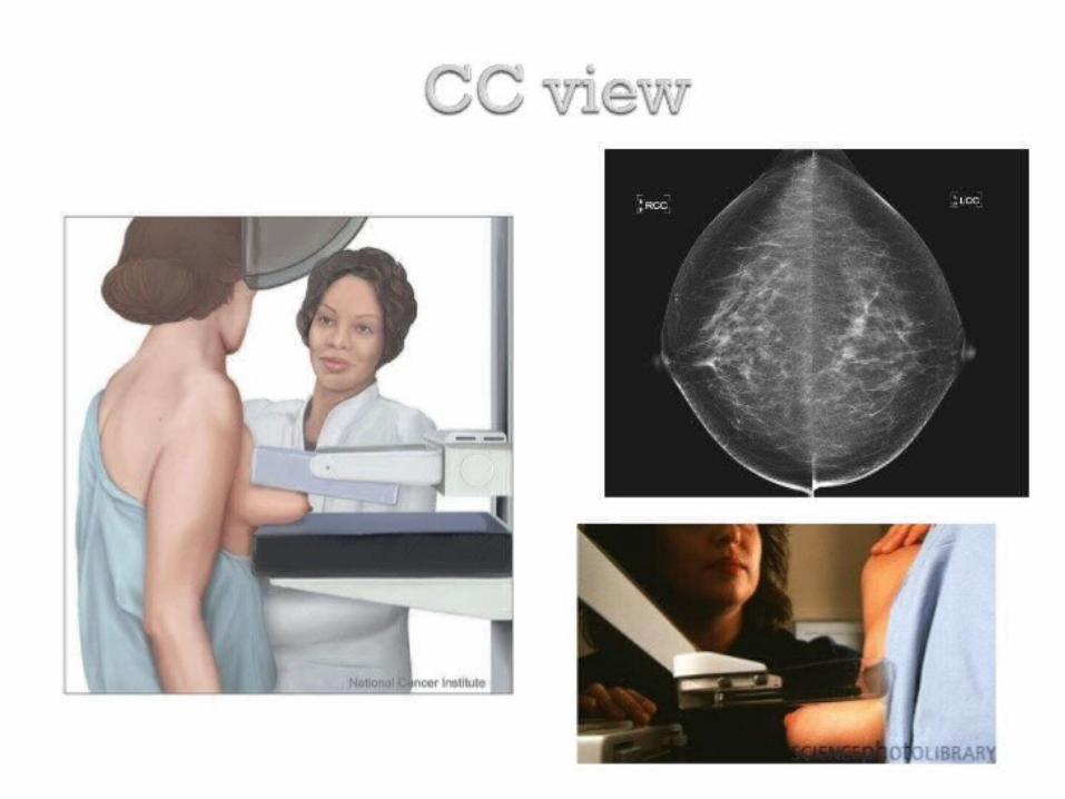
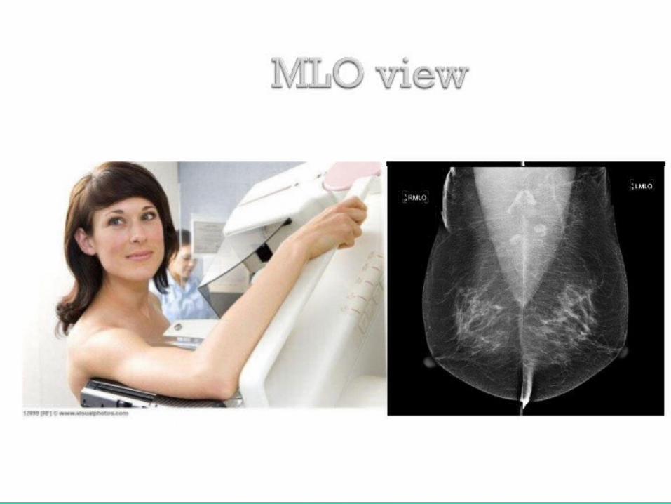
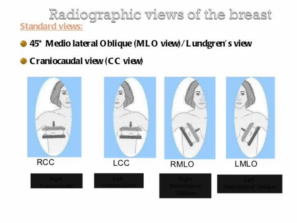
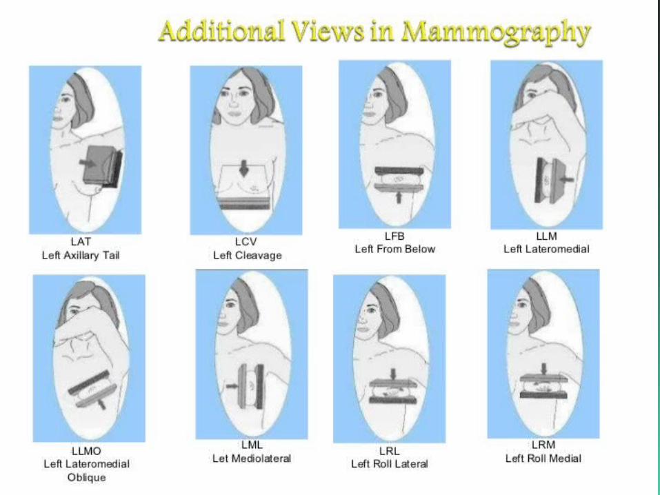
Typical views
• For screening: Cranio- caudal view(CC)
Medio-lateral oblique(MLO)
• For diagnostic: CC
MLO
- lateromedial(from side towards center of chest)
- mediolateral(from the center of the chest out)
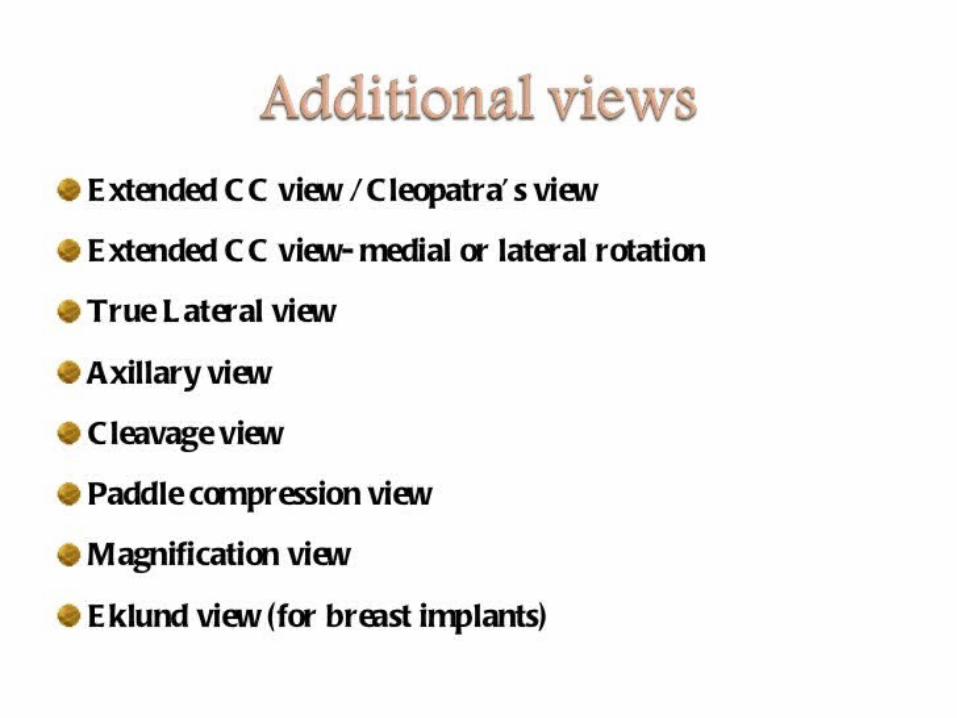
- Spot compression view
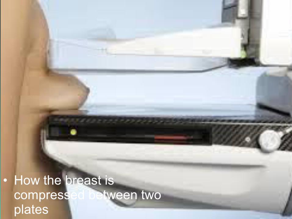
Position
• How the breast is compressed between two plates
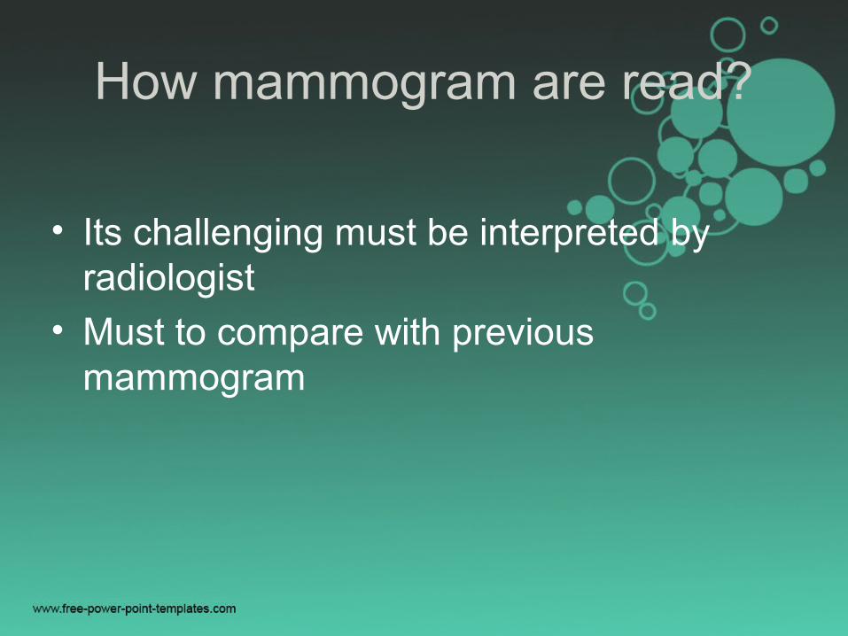
How mammogram are read?
• Its challenging must be interpreted by radiologist
• Must to compare with previous mammogram
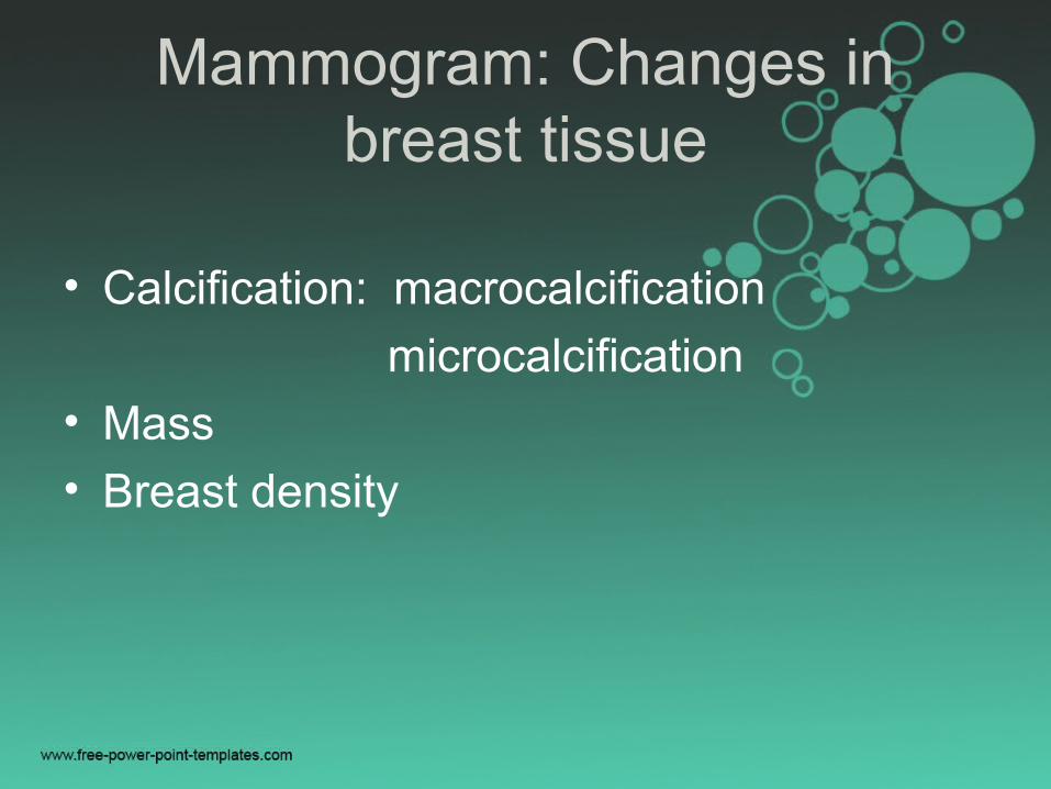
Mammogram: Changes in breast tissue
• Calcification: macrocalcification
microcalcification
• Mass
• Breast density
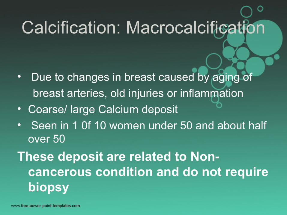
Calcification: Macrocalcification
• Due to changes in breast caused by aging of
breast arteries, old injuries or inflammation• Coarse/ large Calcium deposit• Seen in 1 0f 10 women under 50 and about half
over 50
These deposit are related to Non-cancerous condition and do not require biopsy
Calcification: Microcalcifiation
• Tiny specks of calcium
• If seen it’s a matter of concern though not necessarily it is cancer or does not mean biopsy
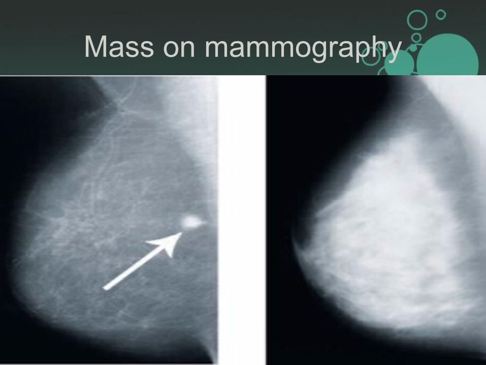
Mass
• Mass with or without calcification
• Noncancerous mass:
Cystic– fluid filled sacs/ simple cyst
Solid – Fibro adenoma
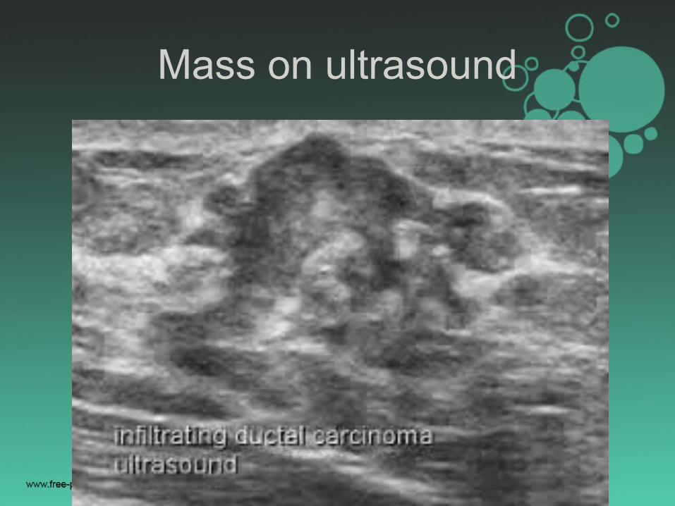
• Complex or mixed mass: suspect cancer
needs FNAC or biopsy
Breast ultrasound is complementary
Breast density
• Density is based on
: how much fibrous and glandular
tissue
: how is the distribution within breast tissue
: how is breast made up of fatty tissue
Dense breasts are not abnormal but they are linked to higher risk of breast cancer
Findings on mammogram
• Primary signs of breast cancer may include spiculated masses or clustered pleomorphic microcalcification
• Secondary signs of breast cancer may include asymmetrical tissue density, skin thickening or retraction or focal distortion of tissue
Impression
• Overall assessment of the radiological findings often includes a classification of the mammogram using the BI-RADS system developed by the American College Of Radiology(ACR)
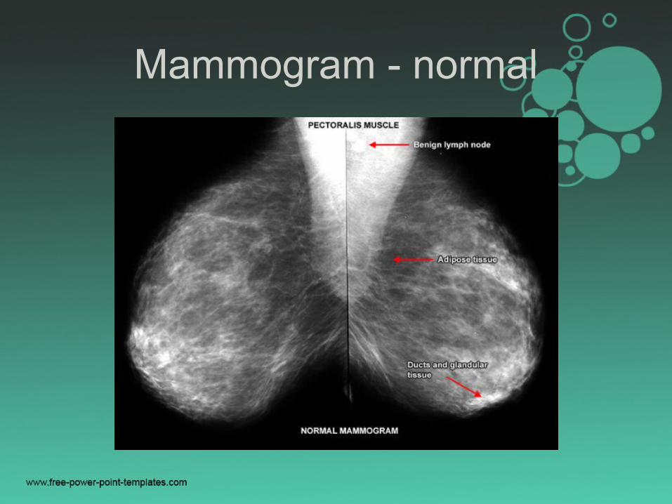
Mammogram - normal
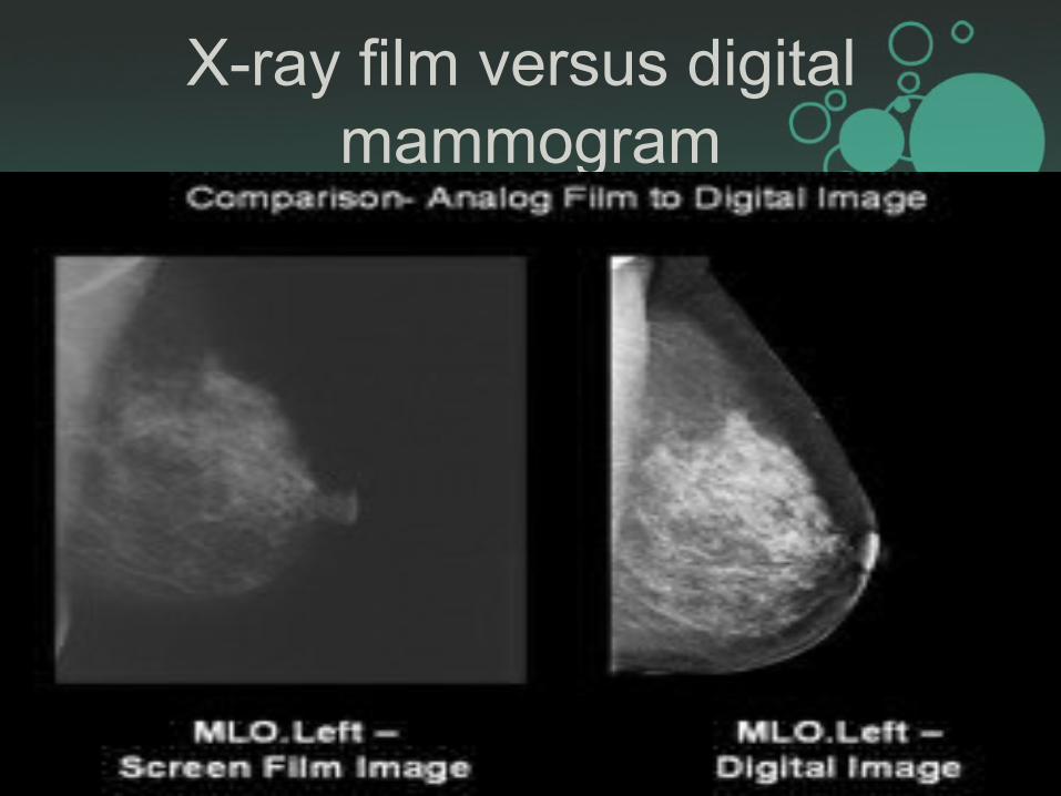
X-ray film versus digital mammogram
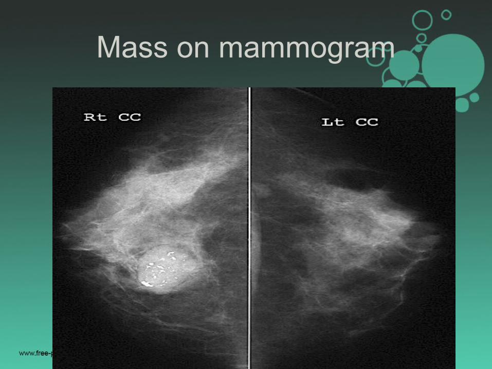
Mass on mammogram
Mass on mammography
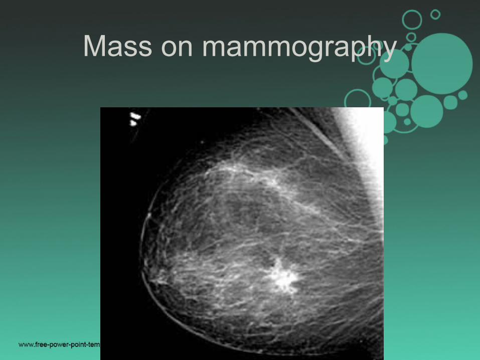
Mass on mammography
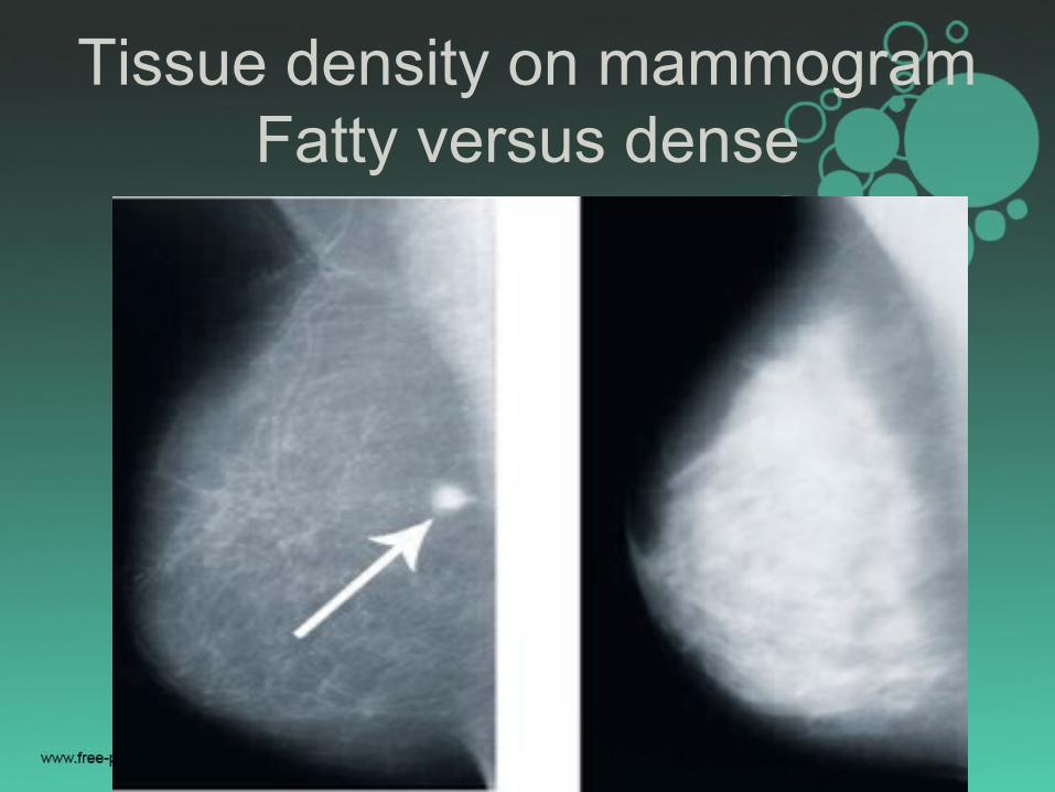
Tissue density on mammogramFatty versus dense
Recommendation(optional)
• No action necessary
• A six month follow up mammogram
• Spot views
• Breast ultrasound
• Biopsy etc….
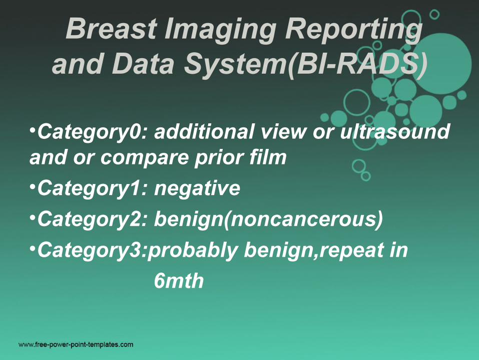
Breast Imaging Reporting and Data System(BI-RADS)
•Category0: additional view or ultrasound and or compare prior film
•Category1: negative
•Category2: benign(noncancerous)
•Category3:probably benign,repeat in
6mth
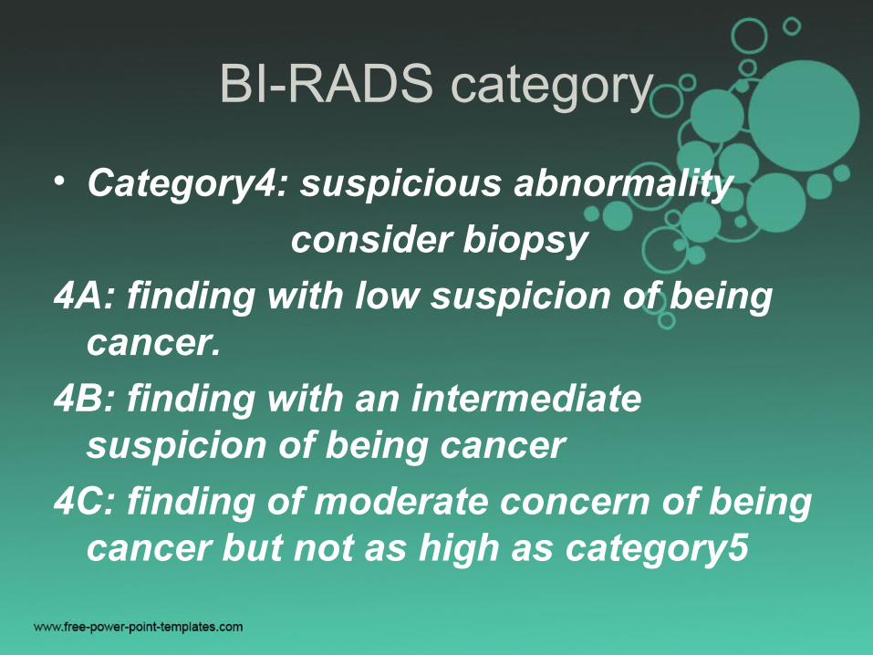
BI-RADS category
• Category4: suspicious abnormality
consider biopsy
4A: finding with low suspicion of being cancer.
4B: finding with an intermediate suspicion of being cancer
4C: finding of moderate concern of being cancer but not as high as category5

BI-RADS category
• Category5: highly suggestive of malignancy, biopsy is recommended
• Category6: known biopsy proven malignancy, appropriate action should be taken. It is only to see how well the cancer is responding to treatment
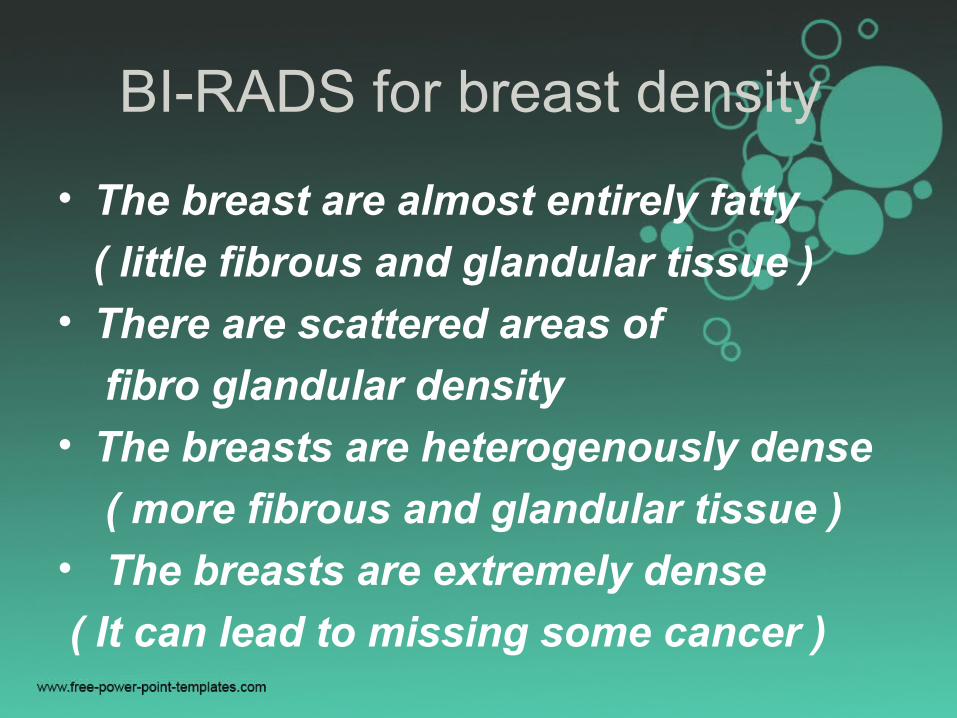
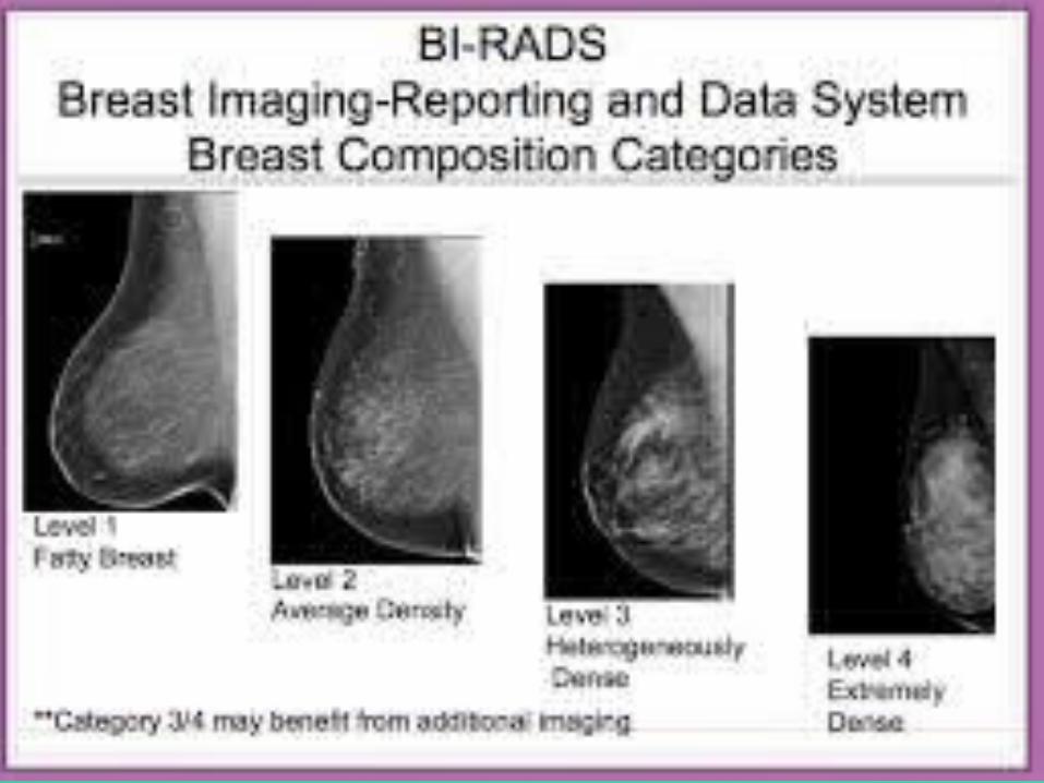
BI-RADS for breast density
• The breast are almost entirely fatty
( little fibrous and glandular tissue )
• There are scattered areas of
fibro glandular density
• The breasts are heterogenously dense
( more fibrous and glandular tissue )
• The breasts are extremely dense
( It can lead to missing some cancer )
Limitations of mammogram
• Breast cancer screening is the best way to find cancer early but finding cancer early does not always reduce a woman’s chance of dying from breast cancer
• Detecting breast cancer early may not help prolong the life of a woman who has other kind of serious or life threatening health problem like CCF, ESRD,COPD…..
False-negative results
• A false-negative mammogram appears normal even though with breast cancer
• It occur more often among younger women usually had dense breast
• False-negative results can delay treatment and promote a false sense of security for the woman
Overall, screening mammogram miss about 1 in 5 breast cancers
False- positive results
• A false- positive mammogram looks abnormal but no cancer is actually present
• It requires diagnostic mammogram, ultrasound, MRI or even biopsy
• It is common in younger woman, have dense breast, have had breast biopsy or cancer in family or are taking oesterogen
Over diagnosis andover treatment
• While mammogram can find invasive breast cancer and DCIS that need to be treated, it is also possible that some invasive cancer or DCIS detected on mammography will not keep growing so not so life threatening and never would have been detected if a woman had not gotten a mammogram. Doctors often cannot be sure which cancers & cases of DICS will become life-threatening, they are all treated
Mammogram – in younger women
• Difficult to read because breast tissue is dense and it can hide the tumour
• In some younger women who are at high risk yearly MRI or mammograms are recommended at age of 30years
Risk factors: gene mutation,strong family history or other factors
Mammogram plan after BCT
• Radiation and surgery both cause changes in the skin and breast tissue
• These changes on mammogram, making it harder to read
• The changes usually peak 6mth after RT
• Mammogram done at this time serves as a new baseline for the affected breast
Mammogram after mastectomywithout reconstruction
• Women who had total or modified radical or radical mastectomy for breast cancer need no further routine screening mammogram of the affected side
• Mammograms are usually continued on the UNAFFECTED breast each year
• Follow up mammogram does require only in the cases who had gone for Subcutaneous mastectomy

Mammogram after mastectomywith reconstruction
• Women who have had a breast fully removed and reconstructed (rebuilt) with silicone gel or saline implant do not need routine mammogram
Mammogram after BCT
• BCT: Breast conserving treatment
• Partial Mastectomy (sometimes called Lumpectomy) is another name for BCT
• It always followed by radiotherapy
• Woman after BCT will need to continue having regular mammogram of both breast
Mammogram with implants
• Its special challenge to do mammography
of breast with implant
• In order to see as much breast tissue as possible it needs 4 extra and 4 standard picture (2 on each breast)
• Extra picture is implant displacement(ID) view pushing back implant against chest wall and the breast is pulled forward over it
• MRI IS BEST WAY TO CHECK IMPLANT
49
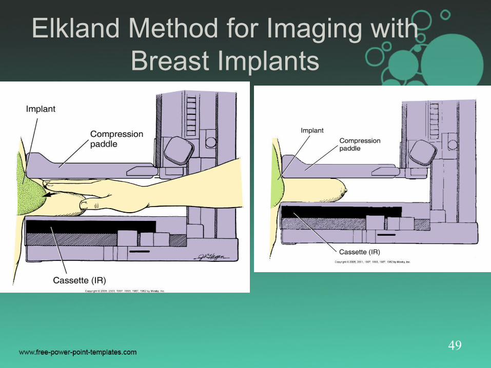
Elkland Method for Imaging with Breast Implants
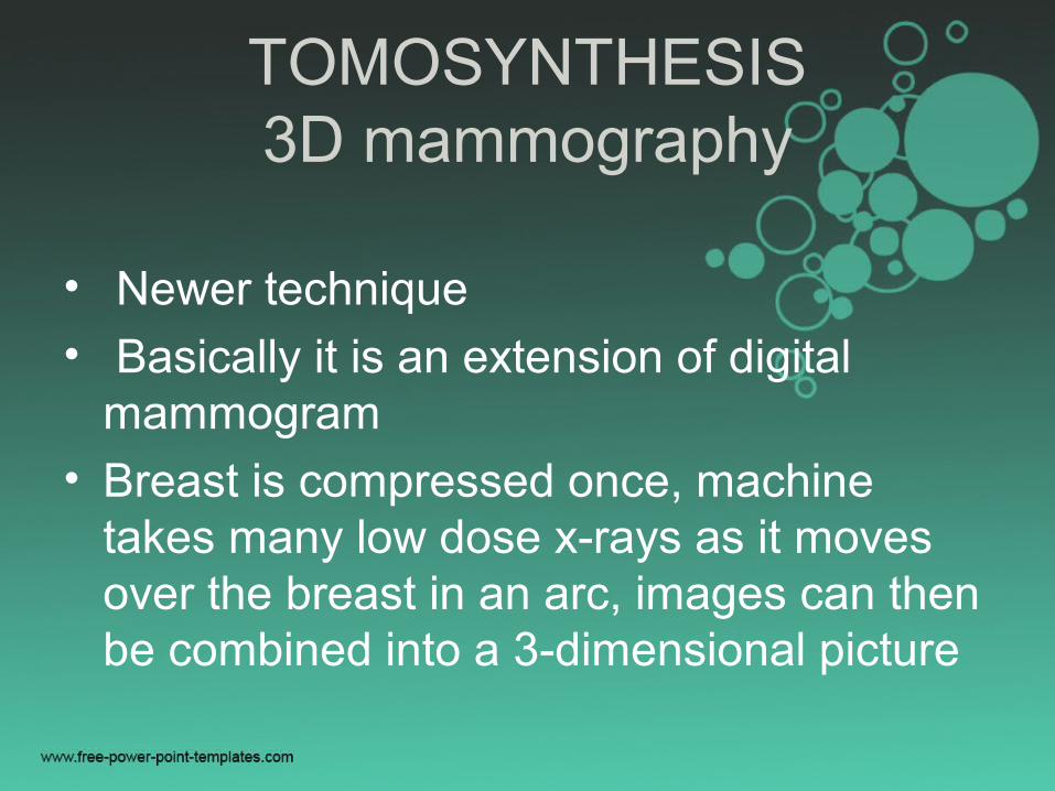
TOMOSYNTHESIS3D mammography
• Newer technique
• Basically it is an extension of digital mammogram
• Breast is compressed once, machine takes many low dose x-rays as it moves over the breast in an arc, images can then be combined into a 3-dimensional picture
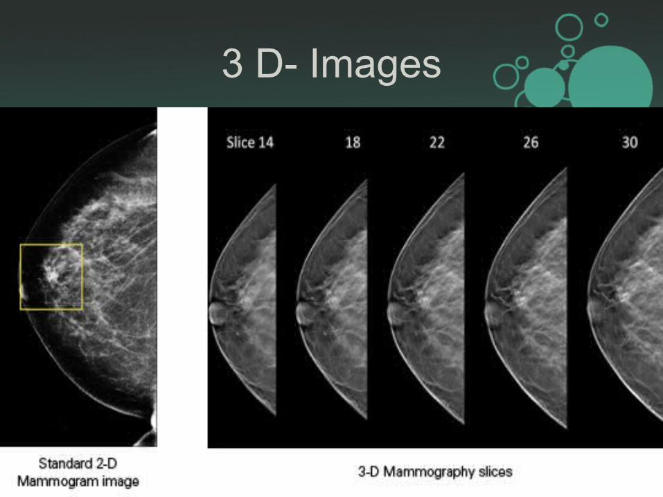
3 D- Images
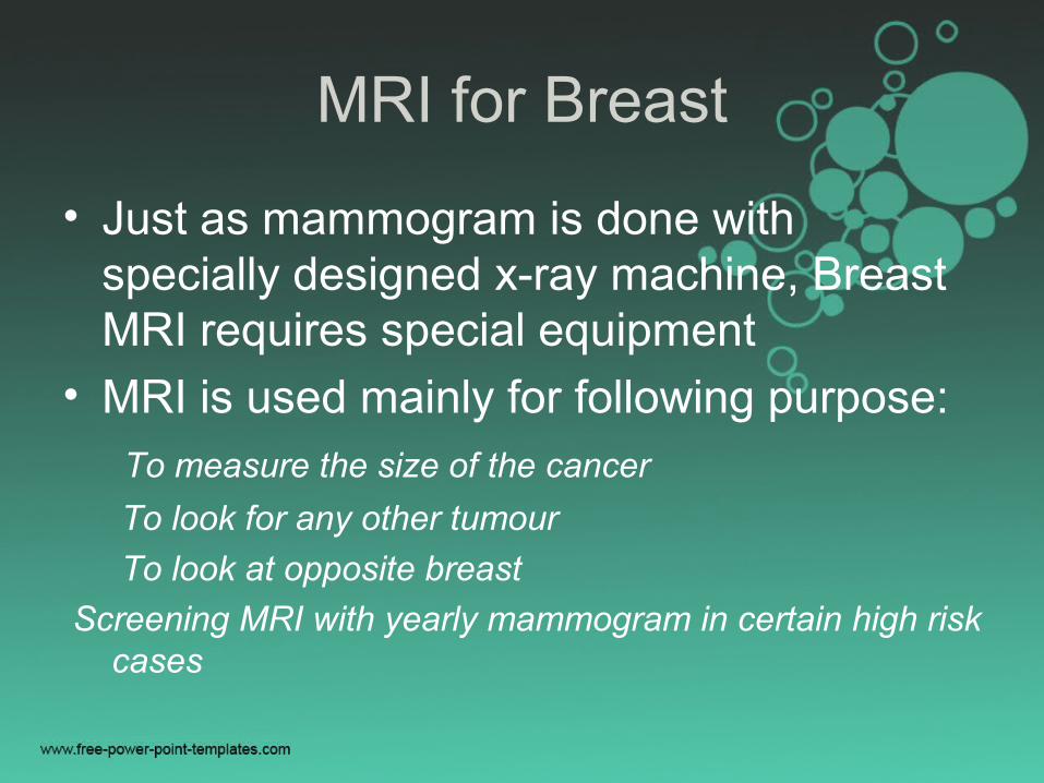
MRI for Breast
• Just as mammogram is done with specially designed x-ray machine, Breast MRI requires special equipment
• MRI is used mainly for following purpose: To measure the size of the cancer
To look for any other tumour
To look at opposite breast
Screening MRI with yearly mammogram in certain high risk cases
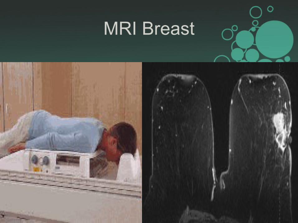
MRI Breast
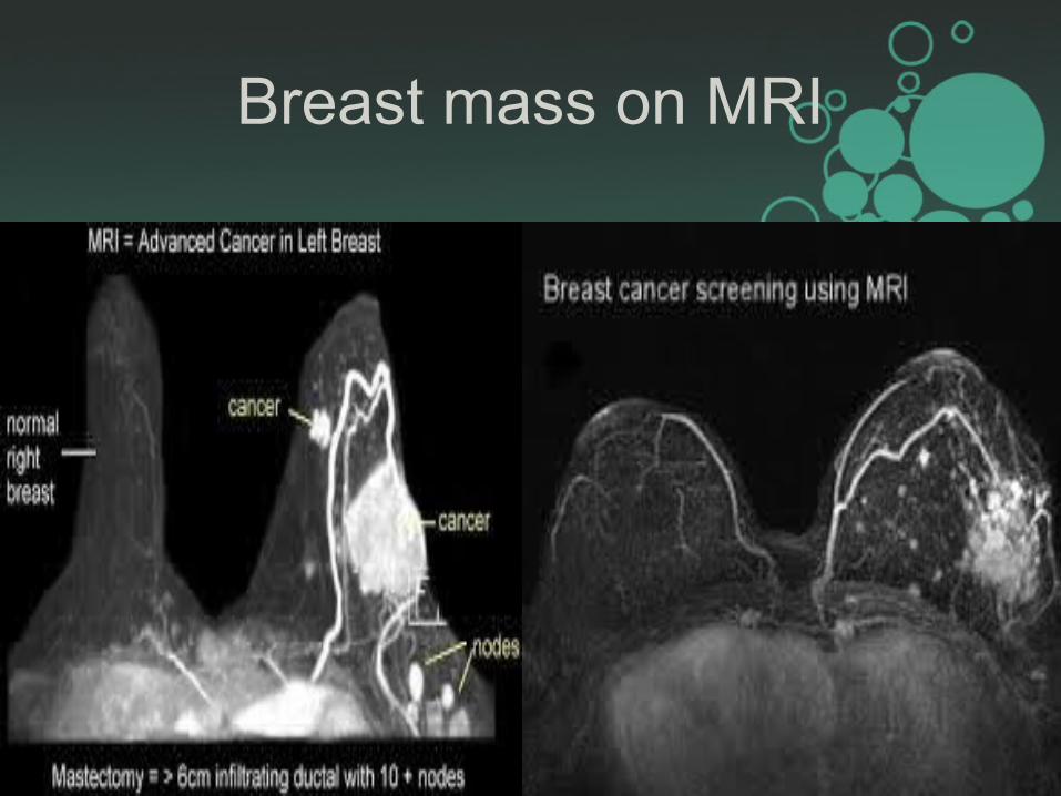
Breast mass on MRI
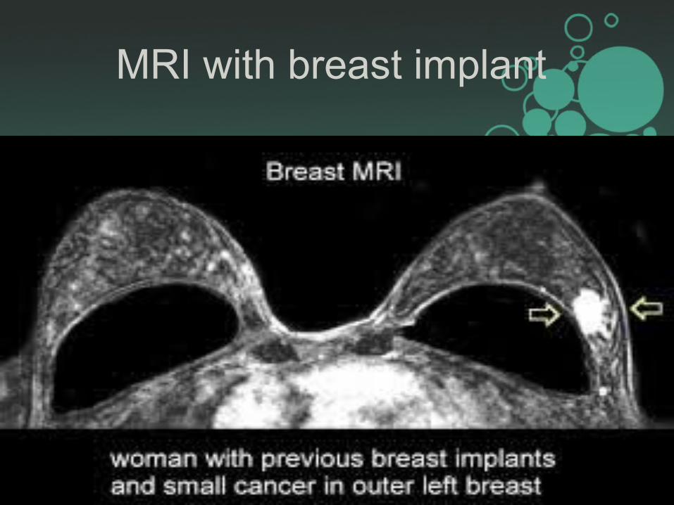
MRI with breast implant
Ultrasound for Breast
• It has become a valuable tool to use along with mammogram because it’s widely available, noninvasive and cost effective
• It is good to have a closer look at some breast masses, if required U/S guided FNAC is possible
• It is also used to look at axillary lymph nodes
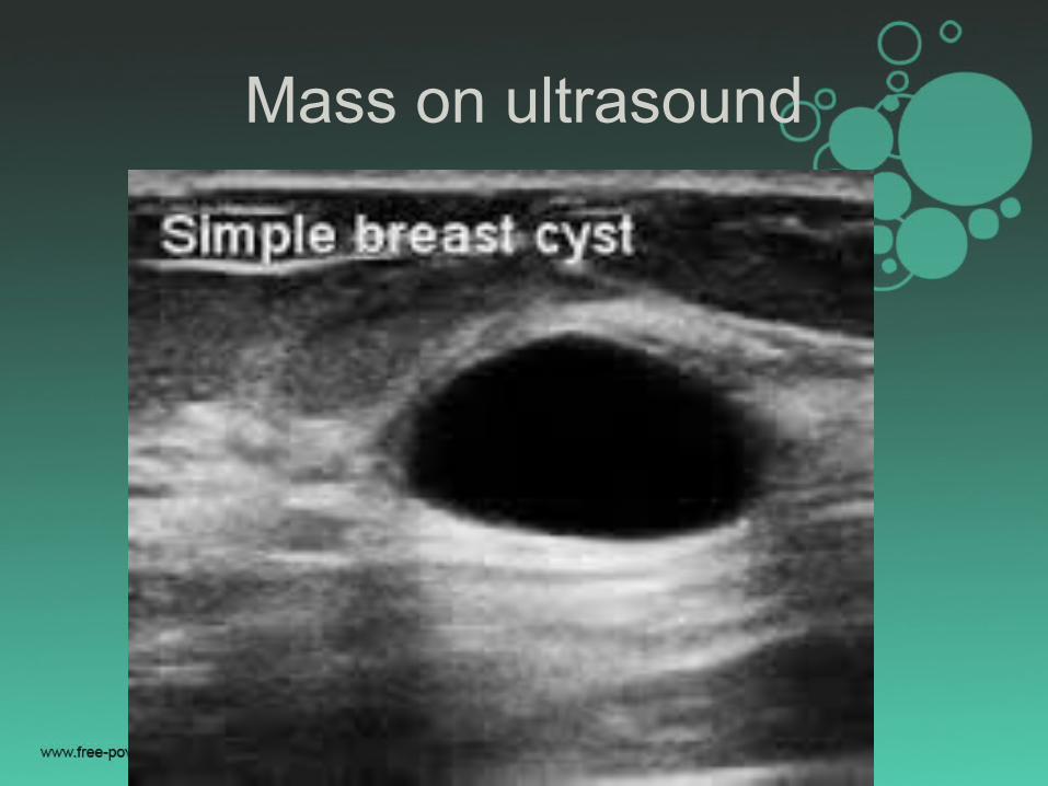
Mass on ultrasound
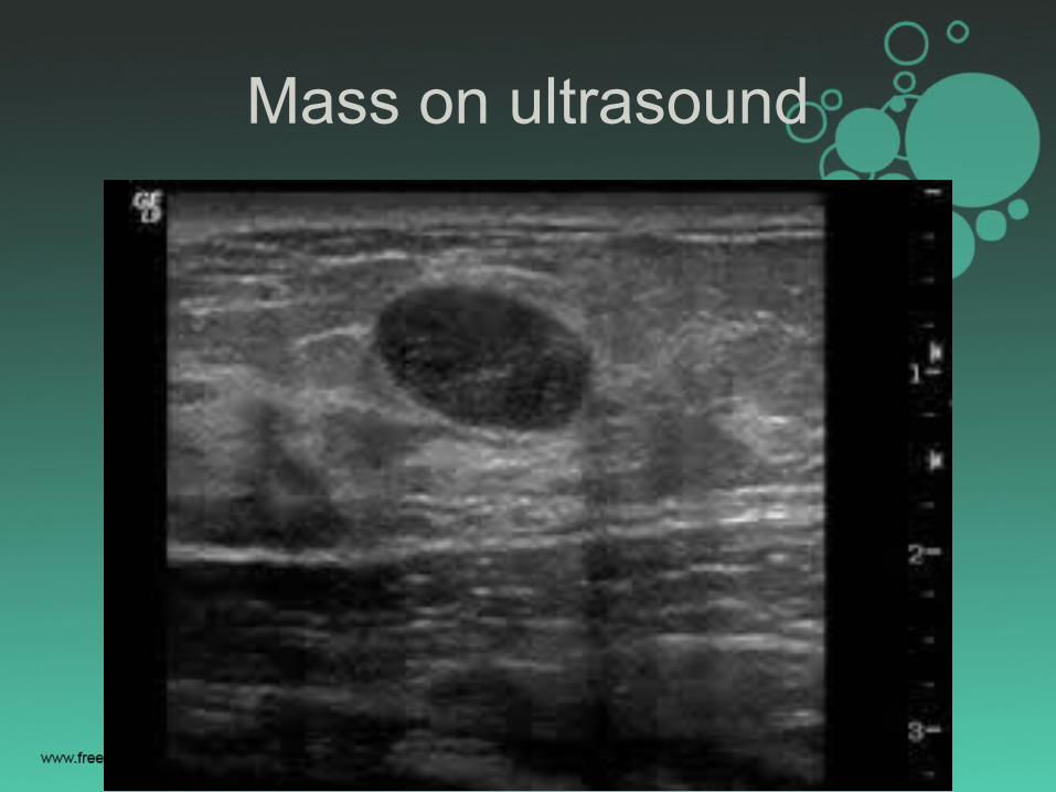
Mass on ultrasound
Mass on ultrasound
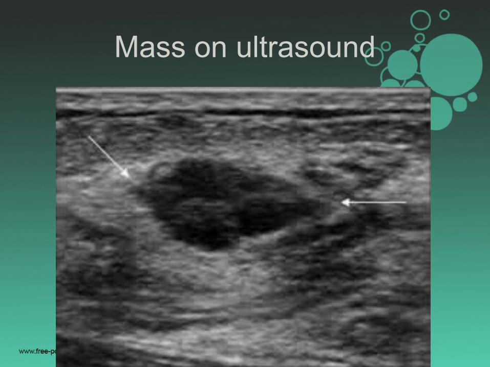
Mass on ultrasound
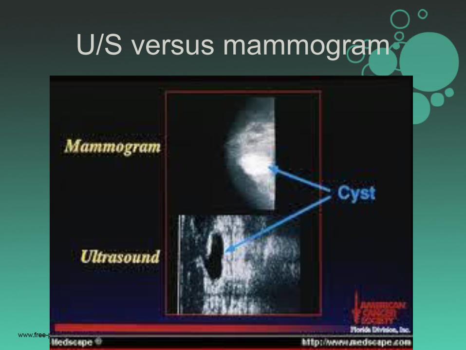
U/S versus mammogram
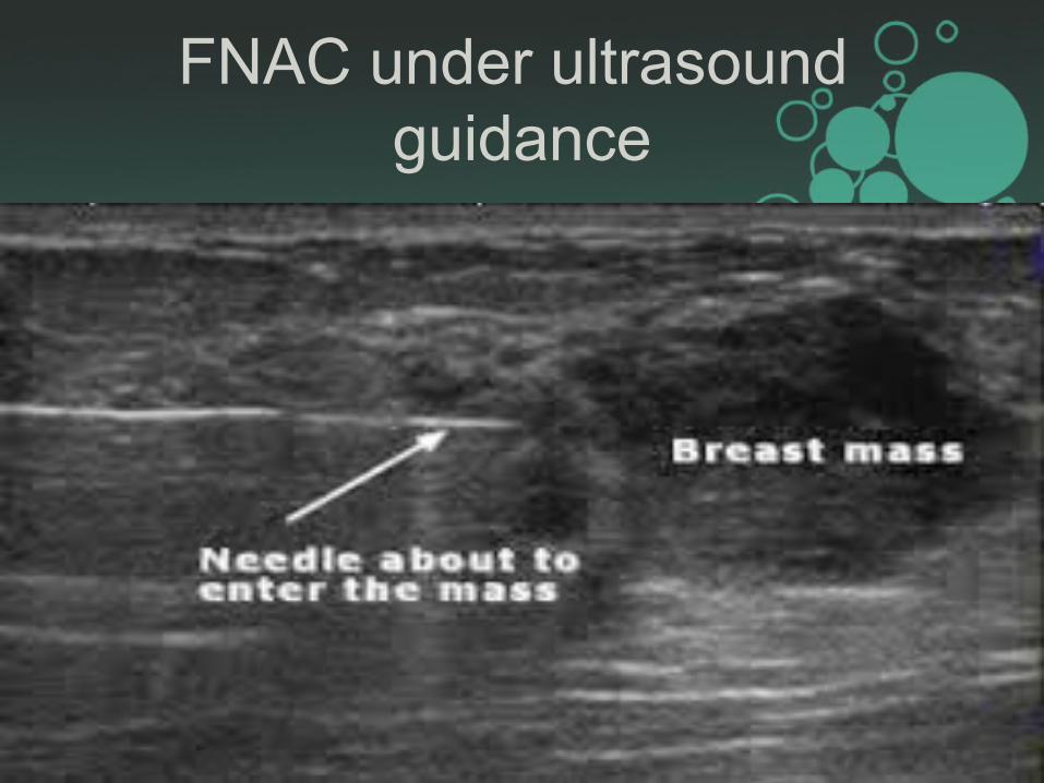
FNAC under ultrasound guidance

Ductogram Galactogram
• Sometimes it is done to find the cause of any worrisome nipple discharge
• Through a very thin plastic tube contrast is put in which outlines the duct on x-ray and can show whether there is a mass inside the duct

Experimental imaging methods
• Scintimammography/ MBI
Radioactive tracer technetium sestamibi
is given IV, it attaches to breast cells and
detected by Gamma camera • Electrical Impedance imaging
based on idea that breast cancer cells
conduct electricity in a different way than
normal cells
Other experimental imaging
• Optical imaging tests pass light into the breast and then measure the light that returns or passes through the tissue
• Positron emission mammography(PEM) uses sugar attached to a radioactive particle to detect cancer cells.
• Thermal imaging is a way to measure and map on the surface of the breast using heat sensing camera
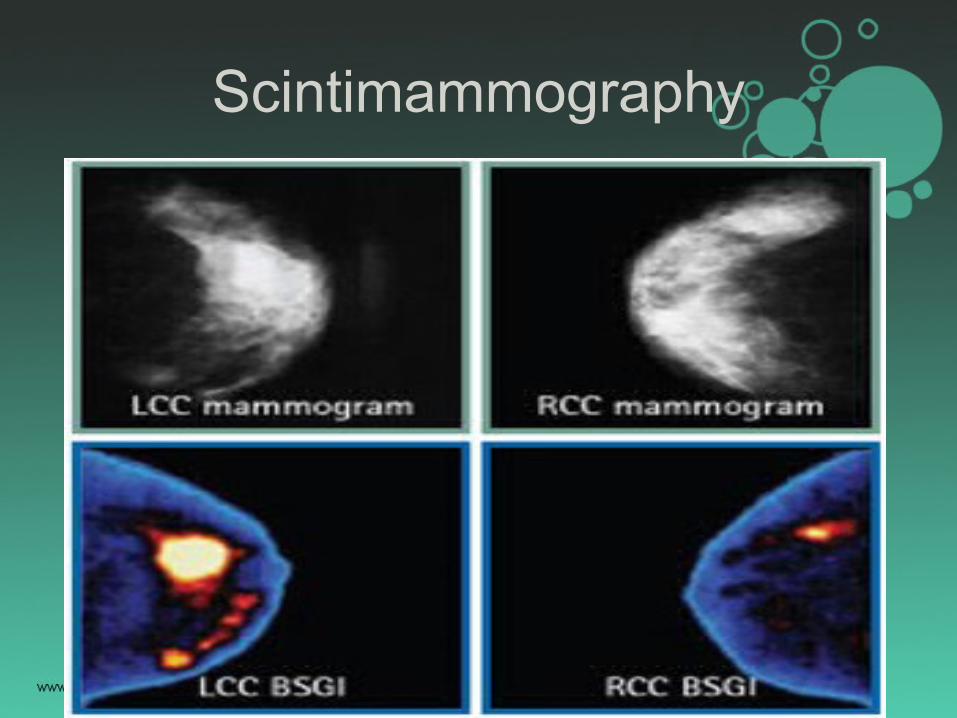
Scintimammography
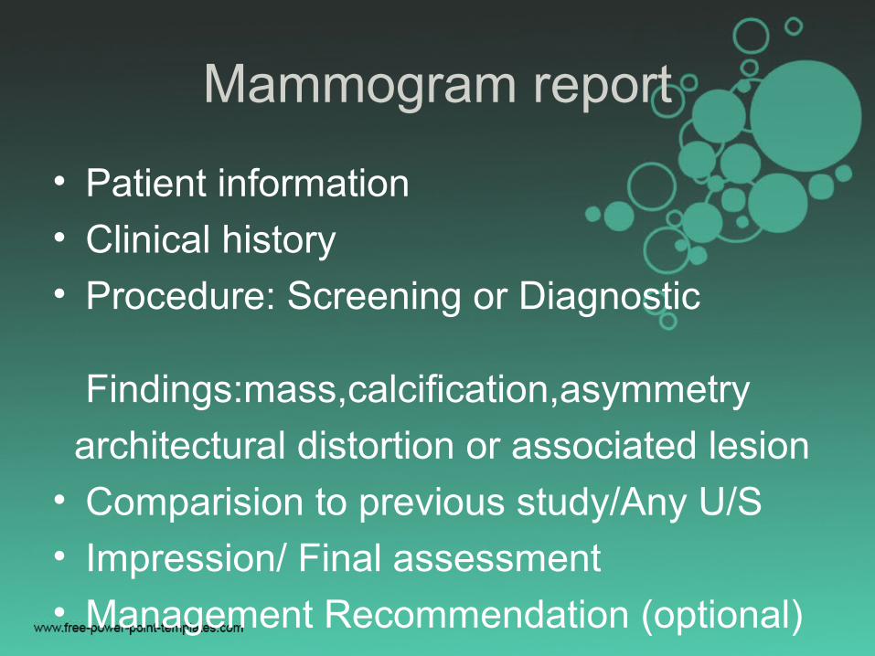
Mammogram report
• Patient information
• Clinical history
• Procedure: Screening or Diagnostic Findings:mass,calcification,asymmetry
architectural distortion or associated lesion
• Comparision to previous study/Any U/S
• Impression/ Final assessment
• Management Recommendation (optional)
Expert quote::
• Before you go to get a mammogram, make sure you know whether you're there for a screening mammogram or a diagnostic mammogram. If you're there for your annual screening mammogram, you may not meet with the radiologist or get your results the same day. Sometimes, there's an advantage to this. Getting your results later often means having two doctors look at your mammogram. A lump, pain, nipple discharge, breast implants, or breast surgery automatically make your mammogram diagnostic. If you have a lump, or other symptoms, tell the mammography center so they know what they're dealing with."•-- Susan Greenstein Orel, M.D.
69
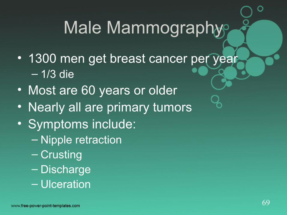
Male Mammography
• 1300 men get breast cancer per year– 1/3 die
• Most are 60 years or older• Nearly all are primary tumors• Symptoms include:
– Nipple retraction– Crusting– Discharge– Ulceration
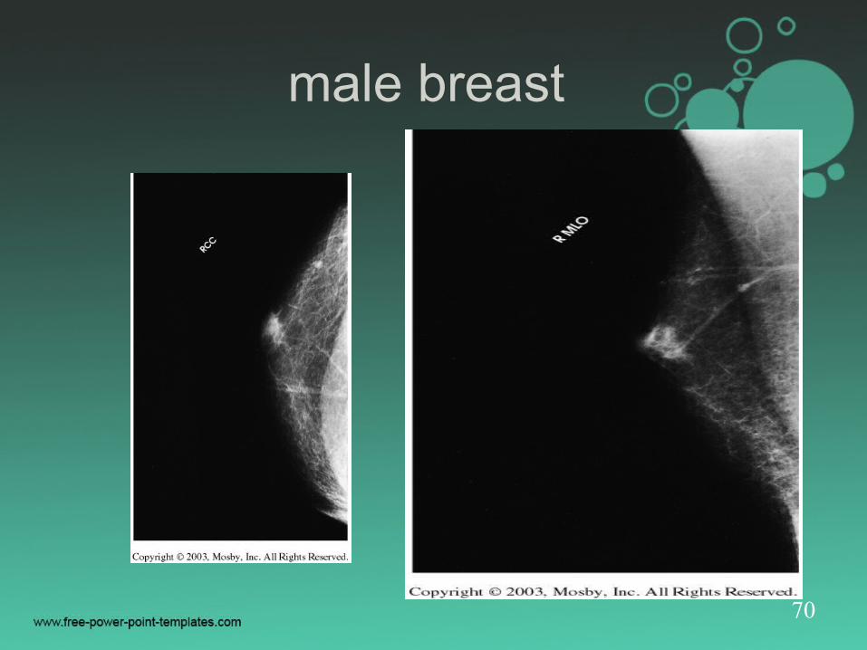
70
male breast
71
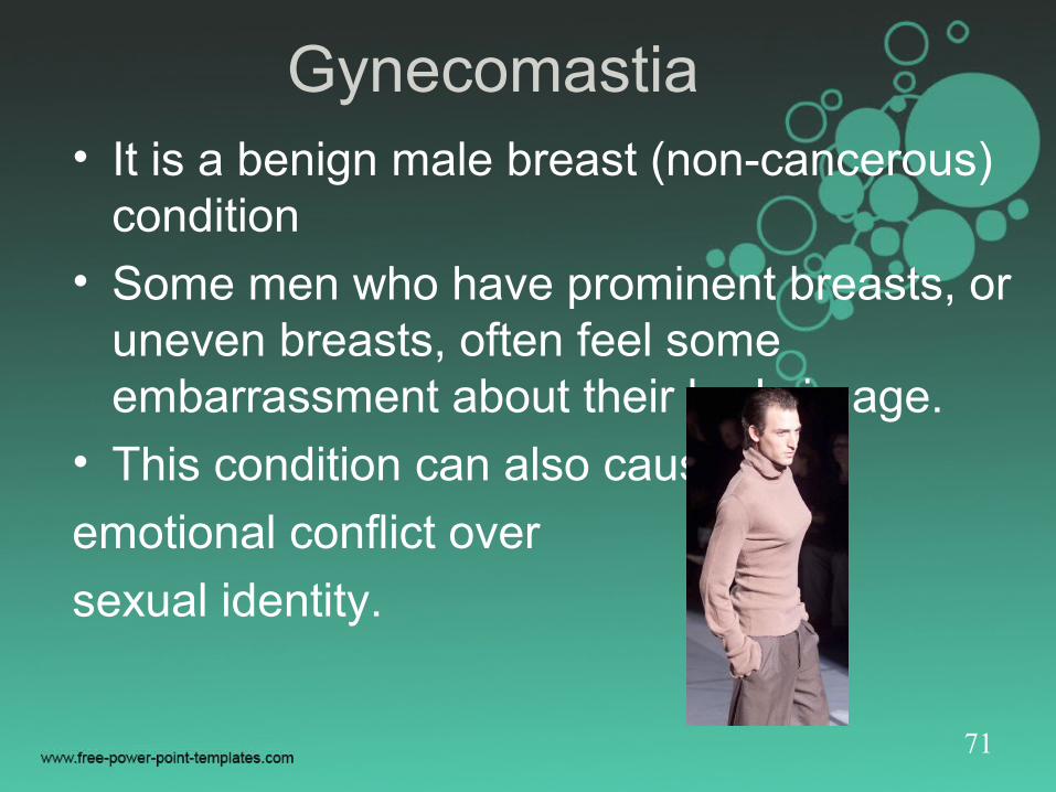
Gynecomastia• It is a benign male breast (non-cancerous)
condition
• Some men who have prominent breasts, or uneven breasts, often feel some embarrassment about their body image.
• This condition can also cause
emotional conflict over
sexual identity.

Thank you
Have a nice day