Imaginal discs1
62
GENE EXPRESSION FOLLOWING INDUCTION OF REGENERATION IN DROSOPHILA WING IMAGINAL DISCS AND EXPRESSION PROFILE OF REGENERATING WING DISCS.
-
Upload
prerna-jain -
Category
Education
-
view
294 -
download
0
Transcript of Imaginal discs1

GENE EXPRESSION FOLLOWING INDUCTION OF REGENERATION IN DROSOPHILA WING IMAGINAL DISCS AND EXPRESSION PROFILE OF REGENERATING WING DISCS.

REGENERATION
Allows organisms to recreate the original shape, size and function of body parts that have been lost or damaged.
First established by T.H. Morgan. Varies between species. Drosophila imaginal discs(the larval
primordia of adult cuticular structures. Limited cell plasticity and identity is
maintained.

In some intial fate lost…..called transdetermination.
Involves wound healing and blastema formation.
Epithelial and cytoskeletal changes occur during the first 24 hours.
Local proliferation increases and peaks around 2-3 days after fragmentation.
Helps in studying molecular mechanism in signal transduction pathway which helps in transcriptional regulation of target genes that will elicit the ultimate response.

JNK signal transduction cascade The c-Jun NH(2)-terminal kinase (JNK) is
a member of an evolutionarily conserved sub-family of mitogen-activated protein (MAP) kinases.
Activated by exposure of cells to cytokines or environmental stress.
Regulates cell proliferation, apoptosis, inflammatory responses, tissue morphogenesis, and polarity.
Several downstream target genes of this signalling pathway are involved in dorsal closure and thorax formation.

Required during imaginal disc regeneration and is activated near the wound as well as in cell death-induced regeneration.
dpp is induced by the JNK pathway in the leading edge cells during dorsal closure.

Wnt and Notch signalling cascade Wingless-type MMTV integration site family
(Wnt). Wnt signalling plays a key role in most aspects
of embryonic development. Induction of ectopic expression of wingless
(wg), a member of the Wnt family promotes cell-fate plasticity such as leg-to-wing transdetermination .
Notch signalling pathway is essential to determine cell fate and regulate pattern formation during embryonic and adult life .
Helps in transdetermination of imaginal discs.

Unclear whether regeneration requires the reactivation of earlier developmental genes or signalling pathways, or if it involves the activation of novel genes specific to
the regeneration process????? THUS, determined the expression
profile of regenerating wing imaginal discs at different times after fragmentation and culture.

METHODS AND METHODS AND MATERIALSMATERIALS
METHODS AND METHODS AND MATERIALSMATERIALS

Drosophila strains and experimental conditions
• All Drosophila strains and crosses were kept on standard media at 25°C.
• The following strains were used: CS; ash2I1/TM6C NI1N-ts2rb'; FRT82gro+: Df(3R)grob32.2/TM6B (a complete deficiency of all E(spl) complex genes, cbtEP(2)2237E1/CyO-twi:GFP en-Gal4;Gal80ts/SM6a-TM6B; UAS-hepCA.
• Wing discs were removed from third instar larvae and a 90° sector was dissected out from the posterior compartment, leaving a 3/4 anterior fragment.
• Regenerating fragments were recovered at 24 and 48 hours after implantation, fixed with 4% paraformaldehyd and immunostained with anti-PH3 ), FITC-conjugated goat anti-rabbit secondary antibody and TOPRO3 for nuclei staining.

In situ hybridization• cbt sense and antisense RNA probes were
synthesized using a complete cDNA with DIG RNA labeling Mix and hydrolyzed prior to hybridization.
• Discs were analyzed with a Leica DMLB fluorescent microscope and a Leica SP2 confocal microscope.
Quantitative RT-PCR analysis• Reverse transcription reactions with 500 ng of
RNA isolated from regenerating discs were used to synthesize cDNA with M-MLV reverse transcriptase

Microarray analysis
• Total RNA was extracted as described above from wing discs recovered after 0, 24 and 72 hours for cut discs, and at 0 and 24 hours for uncut discs.
• Four microarrays were hybridized for each experiment C0→C24, C24→C72 and NC0→NC24.
• Obtained a list of genes that displayed at least two-fold differential expression.
• We employed the program GSEA to perform gene-set-enrichment analyses in the full transcriptome of each microarray. The Enrichment Score (ES) was calculated by walking down the ranked list, increasing the cumulative sum when a gene is present in a given GO category and decreasing it if a gene is not.

Microarray production process: DNA fragments amplified by PCR technique
are spotted on a microscopic glass slide coated with polylysine prior to spotting process. Prior to hybridisation, DNA is denatured to obtained a single strand DNA on the microarray.
Target preparation: RNA are extracted from wings-cut and noncut.
Messengers RNA are then transformed in cDNA by reverse transcription. On this stage, DNA from the first culture with a green dye, whereas DNA from the second culture is labelled with a red dye.

Hybridisation: Green labelled cDNA and red labelled ones are mixed
together (call the target) and put on the matrix of spotted single strand DNA (call the probe). The chip is then incubated one night at 60 degrees. At this temperature, a DNA strand that encounter the complementary strand and match together to create a double strand DNA. The fluorescent DNA will then hybridise on the spotted ones.
Slide scanning: A laser excites each spot and the fluorescent
emission gather through a photo-multiplicator (PMT) coupled to a confocal microscope.

Data analysis: measure the signal amount in the green dye emission
wavelength and the red dye emission wavelength. the amount of fluorescent DNA fixed is proportional to the
mRNA amount present in each cell at the beginning and we calculate the red/green fluorescence ratio. If this ratio is greater than 1 (red on the image), the gene expression is greater in the second experimental condition, if this ration is smaller than 1 (green on the image), the gene expression is greater in the first condition.
Expression profile clustering: to gather genes that share the same expression profile on
several experiments. displayed as a matrix where each column represent one
experiment and each row a gene. Ratios are displayed thanks to a colour scale going from green (repressed genes) to red (induced genes).


Results and Results and DiscussionDiscussion
Results and Results and DiscussionDiscussion

Whole genome Whole genome expression analysis expression analysis
of intact and of intact and regenerating wing regenerating wing
discsdiscs

12 microarrays containing 12,254 genes annotated in RefSeq from D. melanogaster
Four microarrays (non cut, NC0→NC24) were used to assess the effect of the implantation procedure in intact wing discs.
The remaining eight were used to measure changes in gene expression in the first 24 hours after disc dissection and implantation (cut, C0→C24) and during the period between 24 hours and 72 hours after the cut (C24→C72).

Total number of up and downregulated genes in NC0→NC24, C0→C24 and C24→C72
microarrays.
NC0→NC24
C0→C24 C24→C72
GENES (UP)
407 607 116
GENES (DOWN)
356 576 165
TOTAL 763 1183 281

More genes were reported in C0→C24. 44% of 1,183 differentially expressed
genes in C0→C24 were not found in NC0→NC24.
Conversely, 87% of 763 genes in NC0→NC24 were in C0→C24.
The number of genes in C0→C24 was also higher than in C24→C72 confirming the strong initial response during the first 24 hours.

Significant enrichment in genes associated with apoptosis, response to stress, cytoskeletal activity, and JNK pathway regulation (functional annotation of microarrays).
The cellular machinery required for RNA processing and protein synthesis seems to be blocked during the first 24 hours after implantation.
Expression changes only in cut and implanted discs (200 upregulated and 220 downregulated genes in C0→C24).

A. gene ontology terms of upregulated and downregulated genes.
B-enrichment plots. The enrichment score has absolute peak in the left sideindicating overrepresentation in C0→C24.
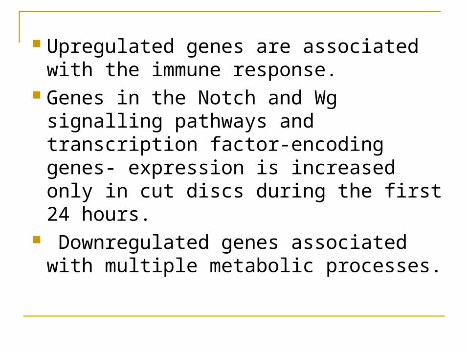
Upregulated genes are associated with the immune response.
Genes in the Notch and Wg signalling pathways and transcription factor-encoding genes- expression is increased only in cut discs during the first 24 hours.
Downregulated genes associated with multiple metabolic processes.

Gene Set Enrichment Analysis
Computational method that determines whether a defined set of genes (e.g. GO categories) shows statistically significant differences between two biological conditions (e.g. cut versus intact discs)
Detected a significant enrichment in C0→C24 of genes involved in Notch and Wg signalling pathways, several transcription factors and the immune response.
No particular categories were found to be specific only in intact discs.

For instance, genes involved in apoptosis
and regulation of the JNK cascade, which have been reported to be essential for imaginal disc wound healing and dorsal closure were identified as upregulated in microarrays for intact discs.
Implantation probably results in sufficient
stress to trigger the JNK pathway and these genes cannot be eliminated as relevant.

IDENTIFICATION OF IDENTIFICATION OF GENES WITH PUTATIVE GENES WITH PUTATIVE
ROLES IN REGENERATIONROLES IN REGENERATION

Examined the set of 281 genes showing expression changes in the C24→C72 microarrays.
Results suggest that the normal activity of imaginal discs, interrupted in response to dissection and implantation, is resumed during the 24-72 hours of regeneration.
As, a significant increase in genes involved in the regulation of RNA metabolism and gene expression in the set of upregulated genes, whereas genes involved in apoptotic processes, structural activities and dorsal closure were augmented in the set of downregulated genes.

Functional annotation of differentially expressed genes in wing imaginal discs at 24 and 72 hours of regeneration.
o The no. of genes in each category of microarray is shown within bars.
o The length of bars indicate the fold change.

GSEA analysis of C0→C24 and C24→C72 microarrays shows the functional classes overrepresented in early regeneration.
Moreover, in addition to RNA processing and protein folding activities, GSEA analysis of C24→C72 identified an enrichment in genes associated with cell proliferation and chromatin remodeling processes during late regeneration of wing discs.

Classes of gene based on expression levels. Class I genes showing differential
expression only in C0→C24. Class II genes with differential expression
only in C24→C72. Class III genes displaying changing
expression levels between the two periods. Class IV genes that steadily increase or
decrease their expression levels.

Classification of differentially expressed genes involved in
wing imaginal disc regeneration.

Class I genes A significant change, either increasing or
decreasing expression between 0 hours and 24 hours after the cut, but remain constant during the second period of time.
82% of upregulated genes and 93% of downregulated genes.
Upregulated genes associated with dorsal closure, the JNK cascade, MAP kinase activity, and the Notch and Wg signalling pathways, imaginal disc development, immune response, and apoptotic processes.

Downregulated genes associated with growth regulation or involved in chromatin remodeling and wing disc development, representation of the additive response of the implantation effect and the process of regeneration.

Class II genes Display increased or decreased expression
between 24 and 72 hours but remain constant in C0→C24.
44% of upregulated genes and 49% of downregulated genes.
Increased expression of chromatin regulator.
Enrichment of upregulated transcription factors:

a. Sox box protein 15 (Sox15 or SoxF) -codes for a transcription factor involved in the Wg signalling pathway that has been linked to control of proliferation in Drosophila .
b. Enhancer of split (E(spl)) -is an essential Notch signalling pathway mediator
c. Medea (Med) -a component of the dpp pathway .
Brahma associated protein 60 kD (Bap60) and Dalao are components of the Brahma complex involved in chromatin remodeling and Nucleosome assembly protein 1 (Nap1).

Activation of these genes together with the presence in this class of splicing and translation initiation factors indicates that the normal RNA processing machinery resumes its activity.
In contrast, genes involved in the wound healing response and cytoskeletal organization processes were downregulated, indicating that cell shape changes and cytoskeletal reorganization described in early healing have been accomplished.

Class III genes Expression changed dramatically, from significant
upregulation to downregulation or vice versa. Enriched in genes associated with the stress
response, defense response, and structural activities, as well as several downstream targets of the JNK regulatory cascade.
For example, Krüppel-like transcription factor cabut (cbt), the Collagen type IV (Cg25C) gene related to dorsal closure etc.
Account for the cellular responses to injury, which would then be switched off once wound healing is completed.

Class IV genes Expression remains significantly increased or
decreased throughout the whole process, indicating their relevance during the 72 hours after the cut.
A large fraction of upregulated and downregulated genes in C24→C72 had the same expression pattern observed in C0→C24.
Genes whose expression pattern was upregulated throughout the experiment;
a. headcase (hdc) and regucalcin, cryptocephal (crc) gene,
b. different chromatin remodeling factors such as absent, small, or homeotic discs 2 (ash2)

a. three basic helix-loop-helix transcription factors (bigmax, HLHm3 and HLHm7), indicating again that transcriptional regulation plays a critical role in regeneration.
The set of genes that remained downregulated throughout the 72-hour period comprised a group of actin and heat shock proteins that were probably activated just after the injury, and the endopeptidases tolloid (tld) and tolkin (tok), involved in imaginal disc morphogenesis.

Transcriptional Transcriptional regulators acting in early regulators acting in early
and late regenerationand late regeneration

Among the plethora of genes, we draw attention those associated with transcriptional regulation.
searched for binding sites of these transcription factors in promoter sequences of misregulated genes, using the genomes of 12 Drosophilas

JNK signalling pathway-
a necessary step in regeneration

Activator Protein 1 (AP1) AP1 transcription factor, a dimer of jun and
fos is activated by JNK signalling pathway. In C0→C24, promoter of 24 upregulated genes
were found to be the binding sites for AP1(11 out of these 24 genes were reported only in cut discs).
15 AP1 binding sites in 12 downregulated genes in C24→C72 microarray.
The genes were both from class 1 and class III (AP1 sites in six Class III genes in both C0→C24 and C24→C72 microarrays).

The phosphatase puckered (puc) has been used as a molecular readout of the activated JNK pathway and its expression seems directly controlled by AP1.
In imaginal disc fragmentation experiments, the expression of puc is activated in several rows of cells near the wound edges at 5 hours after the fragmentation, peaking at 12 hours and decreasing from 24 hours onwards, as the wound is healed.

These results suggest that the JNK pathway regulates the expression of Class I and Class III genes through AP1 during the first few hours of wing disc healing and that its activity decreases during later stages of regeneration.

Also, the suppression of Polycomb group (PcG) proteins by JNK induces transdetermination in Drosophila imaginal discs and that this downregulation is directly controlled by the JNK signalling pathway.

Enhancer of split (E(spl))
Increase in their expression levels during wound healing and regeneration stages.
E(spl) is a bHLH transcription factor that binds regulatory sequences containing the E-box palindromic motif CACGTG.
Six evolutionarily conserved E-boxes in the promoters of downregulated genes were found.

Genes belong to classIII (mojority) and classII.
The genes identified are potential downstream targets of the Notch pathway. .

Chromatin regulators encode proteins that may play a general role as transcriptional activators.
Examles - Ash2, is required for histone H3 trimethylation, BAP60 and Dalao .
The transcriptional activation of this small number of cofactors may lead to the enzymatic activation of several proteins involved in chromatin activity.
Global transcription slows at the beginning of regeneration but resumes concurrent to wound repair.

Requirement of Requirement of transcription factors and transcription factors and chromatin remodelers in chromatin remodelers in
regenerationregeneration

To study the involvement of various genes, we validated the change in expression levels of selected genes by quantitative PCR.
in N I1N-ts2 mutant discs – healing and proliferation were impaired.
with a deficiency of all E(spl) complex genes, proliferation decreased but no change in wound closure.
cbt mutant discs - discs healed, but lower proliferation.
the ash2 i1 allele were smaller and showed wound healing defects at 24 hours.

(A) Regenerating imaginal discs from wild-type (wt); NI1N-ts2; Df(E(spl))/+; cbtEP(2)2237E1/+; and ash2I1/+. Staining of mitosis (green) and nuclei (red).
Involvement of the Notch pathway, cbt and ash2 in wing imaginal disc regeneration.

The transcription factor The transcription factor Cbt as an example of Cbt as an example of
Class III genesClass III genes

Class III display a tight regulation associated with the requirement of the proteins encoded by these genes in a particular window of time.
cbt was upregulated during the first 24 hours after disc fragmentation, decreasing dramatically in the following 48-hour period.
cbt is ubiquitously expressed in the wing disc. Increase in the level of expression of cbt after
activating the JNK pathway in the posterior compartment.
During cell death-induced regeneration, the JNK pathway is activated at the leading edges of healing tissue.

And is required in the living cells for the regulation of healing and regenerative growth.
Our results point to the transcription factor Cbt as a crucial downstream mediator gene of JNK signalling during microsurgery or cell death-induced regeneration.
E(spl) binds to the E-boxes identified in the promoters of cbt and other members of Class III genes contributing to their downregulation in the 24-72 hours period.

JNK and NOTCH signalling pathway. JNK is required in the living cells for the
regulation of healing and regenerative growth. Notch pathway participate in the regulation of
growth in the wings. A sensitive balance between JNK and Notch
signalling events regulates stress responses, stem cell proliferation, and cell differentiation.
Both JNK and Notch pathways cooperate by regulating the transcriptional activity of the same set of genes during wound healing and regeneration of wing imaginal discs.

CONCLUSIONSCONCLUSIONSCONCLUSIONSCONCLUSIONS

1. Able to identify early and late genes involved.
2. The onset of wound healing is the first necessary step for regeneration and the role of the JNK pathway in this type of processes has been widely documented.
3. A significant enrichment of AP1 sites in the promoters of several genes with differential expression only in cut discs, suggesting that they could be direct targets of the JNK pathway.
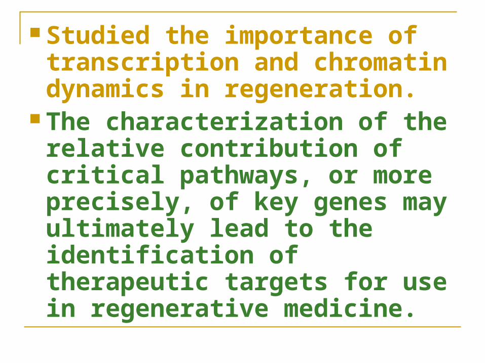
Studied the importance of transcription and chromatin dynamics in regeneration.
The characterization of the relative contribution of critical pathways, or more precisely, of key genes may ultimately lead to the identification of therapeutic targets for use in regenerative medicine.

HHREFRENCES• By Enrique Blanco, Deptt de Genetica.
• Marina Ruiz Romero, Institut de Biomedicina de la Universitat de Barcelona.
• Received:28 June 2010
• Accepted:2 September 2010
• Published:2 September 2010

Thank you