Imagerie segmentation ENPC - Sophia - Inria · Principales Modalités d’Imagerie 5 IRM...
38
1 Imagerie Médicale Olivier Clatz Projet Asclepios
Transcript of Imagerie segmentation ENPC - Sophia - Inria · Principales Modalités d’Imagerie 5 IRM...
1
Imagerie Médicale
Olivier Clatz
Projet Asclepios
2
Plan• Introduction aux images médicales :• Introduction à la segmentation d’images
– Méthodes de seuillage et classification– Modèles déformable
• Introduction au recalage d’images• Modélisation
• Simulation de chirurgie• Modélisation Cardiaque• Croissance de tumeurs

3Imagerie Médicale
Roentgen, 1895

4Caracteristiques des images médicales
Les niveaux de gris sont liées aux caractéristiques physiques des tissus qui peut également être relié à un phénomène physiologique
Physique
Anatomie
Physiologie

5
Principales Modalités d’Imagerie IRM
UltrasonScintigraphie
CT-Scanner
Densitéd’absorptionaux rayons X
Variations d’impédance Acoustique
Densitéd’isotopesinjectées
Densité et structure des
Protons

6
Images Médicales 3-D• Représentation discrète d’une partie du corps qui est
décrite par une matrice à 3 dimensions (voxels)
• I(x,y,z) mesure certaines propriétés physiques ou chimiques du corps humain dans un élément de volume
M(i,j,k) = I (x,y,z)
7
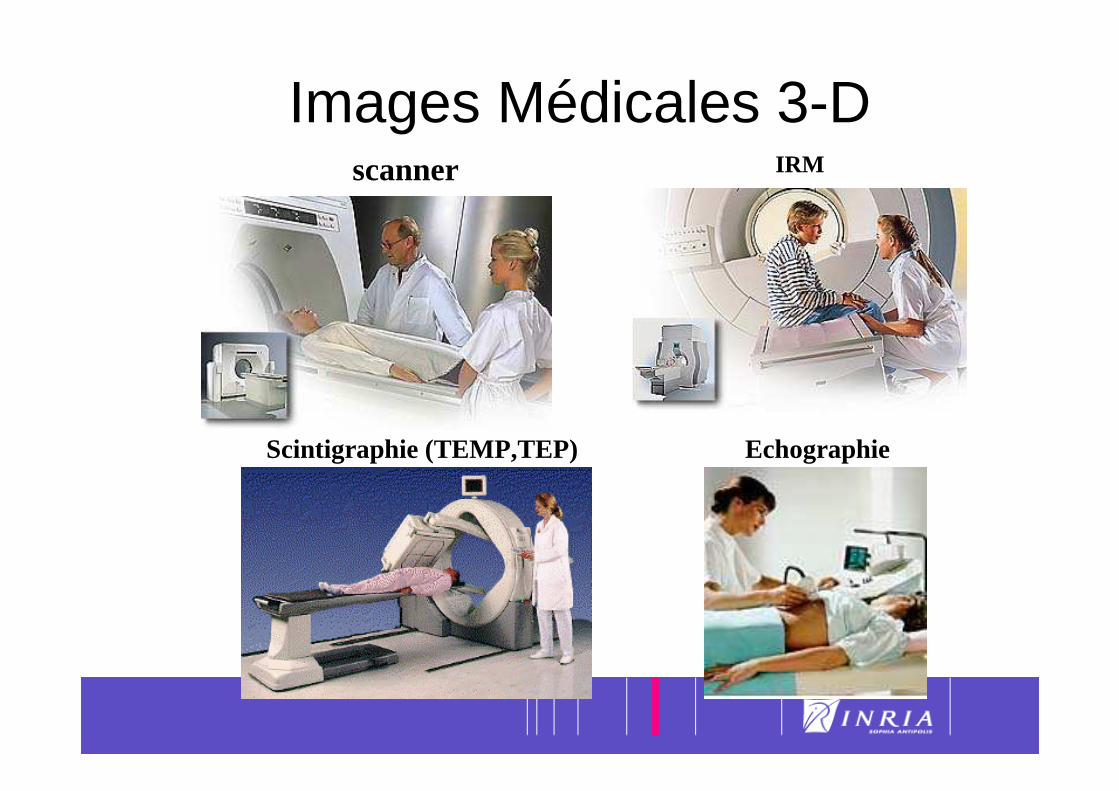
Images Médicales 3-D scanner IRM
Scintigraphie (TEMP,TEP) Echographie

8
Tomodensitomètre X (Scanner)
Taille: 512 x 512 x 128Résolution: 0.5 x 0.5 x 1 mm

9Imagerie par Résonance Magnétique
Résolution millimétrique. 16 millions de points
Coupe Sagittale Coronale ou Frontale Axiale ou Transverse
I(x,y,z) mesure une fonction de la densité et structure des protons

10Scintigraphies (médecine nucléaire)
TEMP: Tomographie par Emission MonoPhotonique
TEP: Tomographie par Emission de Positons
11
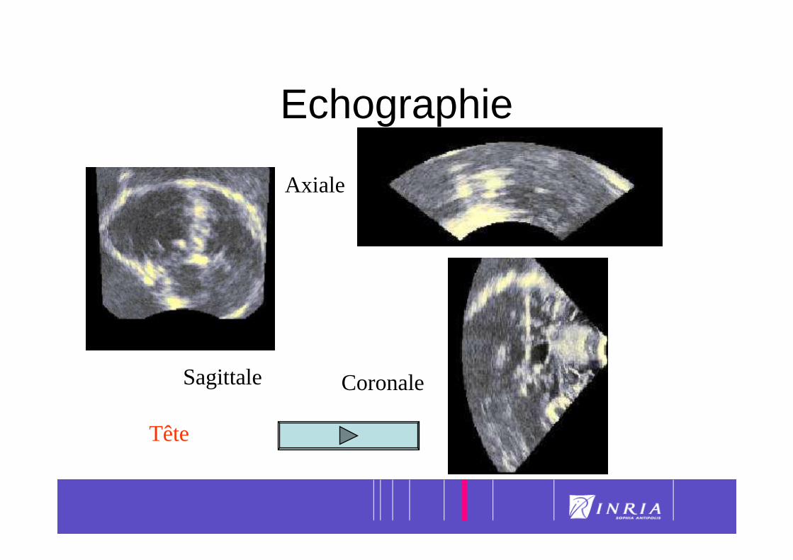
Echographie
Sagittale
Axiale
Coronale
Tête
12
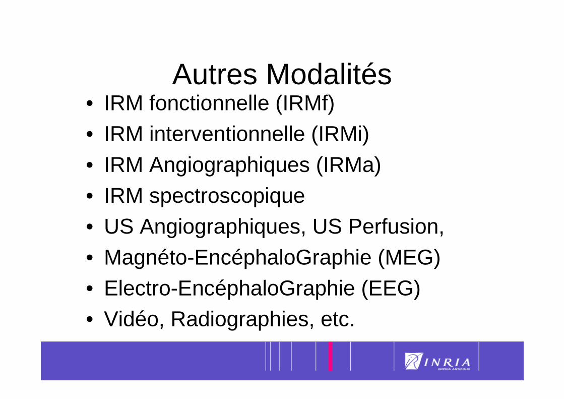
Autres Modalités• IRM fonctionnelle (IRMf)• IRM interventionnelle (IRMi)• IRM Angiographiques (IRMa)• IRM spectroscopique• US Angiographiques, US Perfusion,• Magnéto-EncéphaloGraphie (MEG)• Electro-EncéphaloGraphie (EEG) • Vidéo, Radiographies, etc.

13
Imagerie Médicale• Imagerie Médicale est utilisée à toutes
les étapes de la pratique médicale: – Diagnostic
– Planification de la thérapie– Contrôle de la thérapie
14
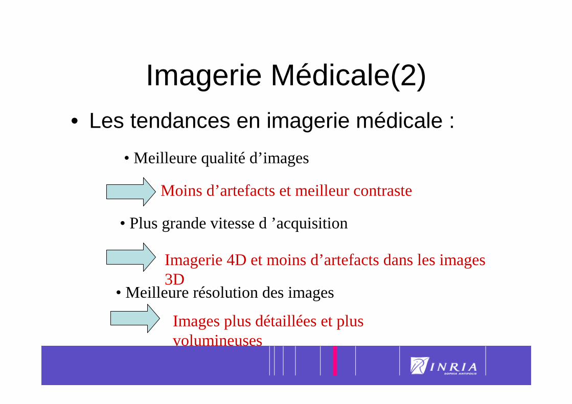
Imagerie Médicale(2)• Les tendances en imagerie médicale :
• Meilleure qualité d’images
Moins d’artefacts et meilleur contraste
• Plus grande vitesse d ’acquisition
Imagerie 4D et moins d’artefacts dans les images 3D
• Meilleure résolution des images
Images plus détaillées et plus volumineuses
15
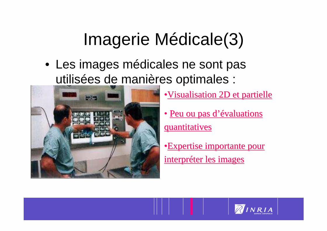
Imagerie Médicale(3)• Les images médicales ne sont pas
utilisées de manières optimales :••Visualisation 2D et partielleVisualisation 2D et partielle
• Peu ou pas dPeu ou pas d’é’évaluations valuations
quantitativesquantitatives
••Expertise importante pour Expertise importante pour
interprinterprééter les imagester les images
16
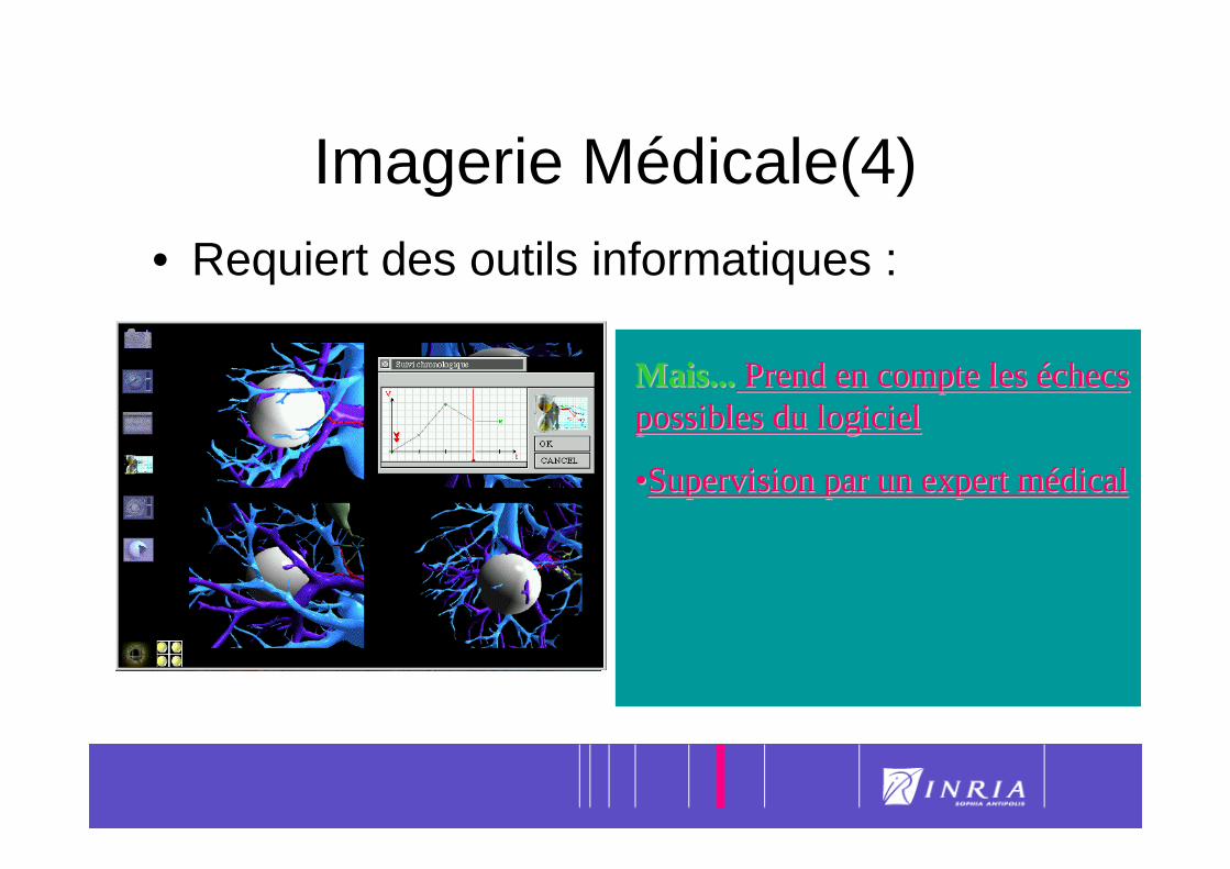
Imagerie Médicale(4)• Requiert des outils informatiques :
••Peux prendre en compte des Peux prendre en compte des images volumiques de grande images volumiques de grande tailletaille
••permet un analyse quantitative permet un analyse quantitative et reproductibleet reproductible
••Peux être intPeux être intéégrgréé dans le dans le systsystèème dme d’’ information de information de ll ’’ hôpital hôpital
Mais...Mais... Prend en compte les Prend en compte les ééchecs checs possibles du logicielpossibles du logiciel
••Supervision par un expert mSupervision par un expert méédical dical
17
Segmentation
1. Introduction

18Segmentation Task
• Large number of available algorithms
• Possible classifications :– Bottom-up vs Top-down approaches
– Boundary vs Region approaches– Explicit vs Implicit A priori knowledge
• Validation
19
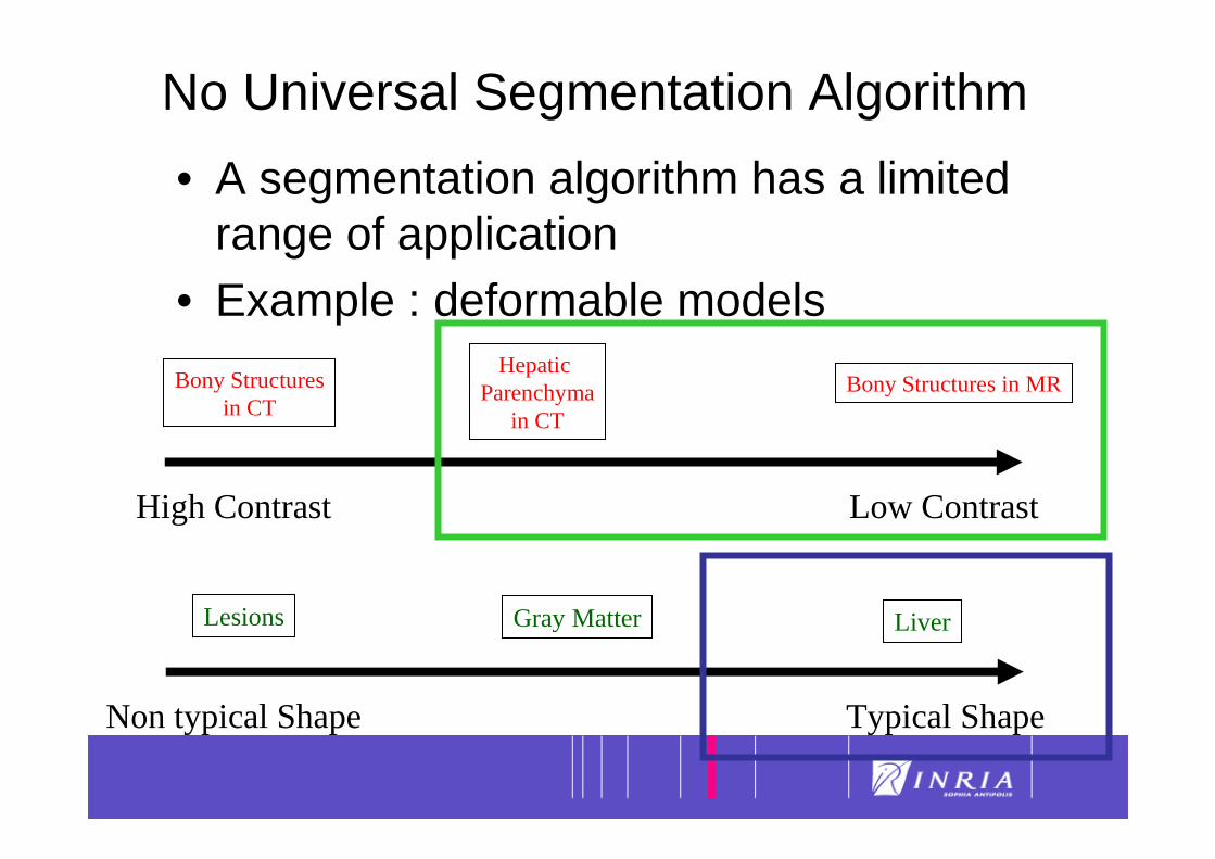
No Universal Segmentation Algorithm
• A segmentation algorithm has a limited range of application
• Example : deformable models
High Contrast Low Contrast
Bony Structuresin CT
HepaticParenchyma
in CT
Bony Structures in MR
Non typical Shape Typical Shape
Lesions Gray Matter Liver
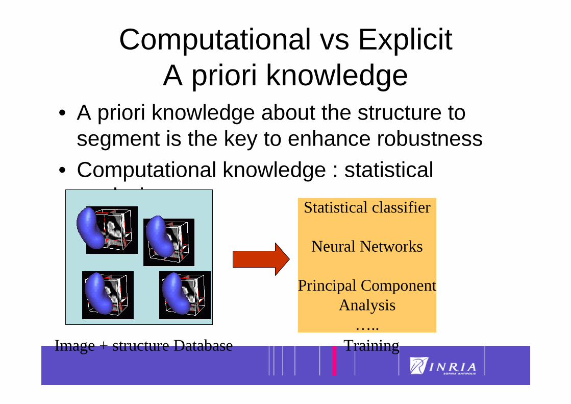
20Computational vs Explicit A priori knowledge
• A priori knowledge about the structure to segment is the key to enhance robustness
• Computational knowledge : statistical analysis
Image + structure Database
Statistical classifier
Neural Networks
Principal ComponentAnalysis
…..Training
21
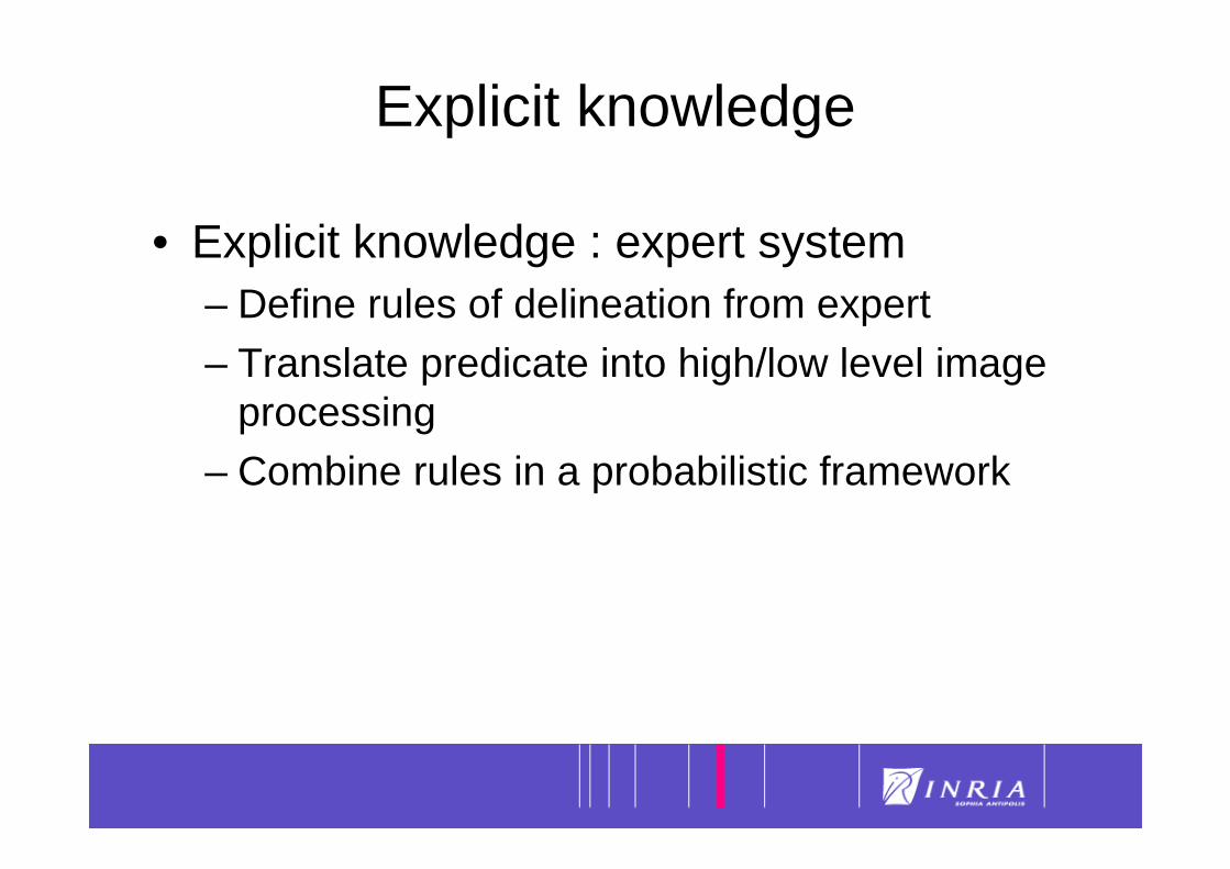
Explicit knowledge
• Explicit knowledge : expert system– Define rules of delineation from expert
– Translate predicate into high/low level image processing
– Combine rules in a probabilistic framework
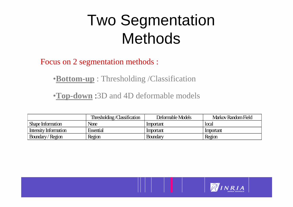
22Two Segmentation Methods
Focus on 2 segmentation methods :
•Bottom-up : Thresholding /Classification
•Top-down :3D and 4D deformable models
Thresholding /Classification Deformable Models Markov Random FieldShape Information None Important localIntensity Information Essential Important ImportantBoundary / Region Region Boundary Region

23
Segmentation
Seuillage/ Classification

24
• Idée principale : une structure est uniquement caractérisée
par ses niveaux de gris dans l’image
• Algorithme de seuillage élémentaire : – Seuillage entre deux niveaux de gris
(fenêtrage)
– Opérations de morphologie mathématique• Erosion et Dilation• Fermeture et Ouverture• Extraction de composantes connexes
Seuillage et Classification
Valide pour les structures fortement contrastées
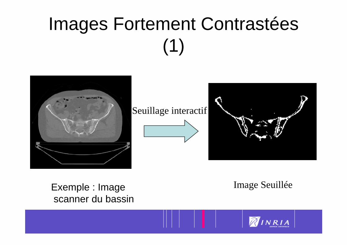
25Images Fortement Contrastées (1)
Exemple : Image scanner du bassin
Seuillage interactif
Image Seuillée
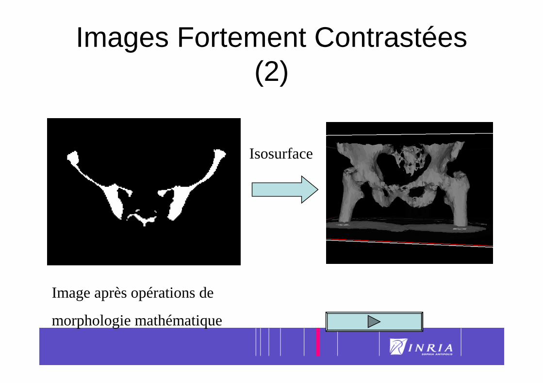
26Images Fortement Contrastées (2)
Image après opérations de
morphologie mathématique
Isosurface

27
Thresholding + mathematical morphology + connected components
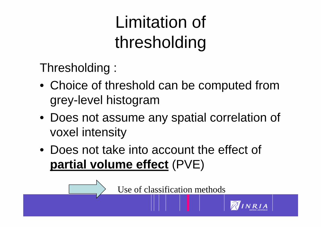
28Limitation of thresholding
Thresholding :• Choice of threshold can be computed from
grey-level histogram• Does not assume any spatial correlation of
voxel intensity• Does not take into account the effect of
partial volume effect (PVE)
Use of classification methods
29
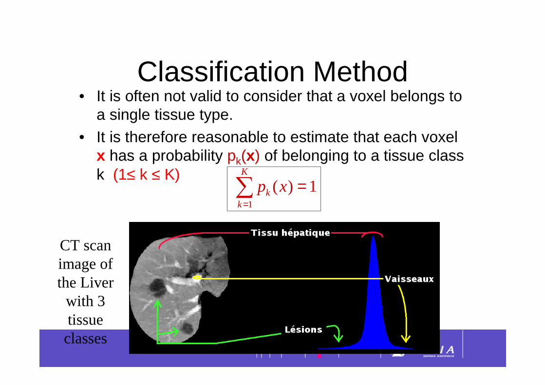
Classification Method• It is often not valid to consider that a voxel belongs to
a single tissue type.• It is therefore reasonable to estimate that each voxel
x has a probability pk(x) of belonging to a tissue class k (1≤ k ≤ K)
1)(1
=∑=
K
kk xp
CT scan image of the Liverwith 3 tissue classes
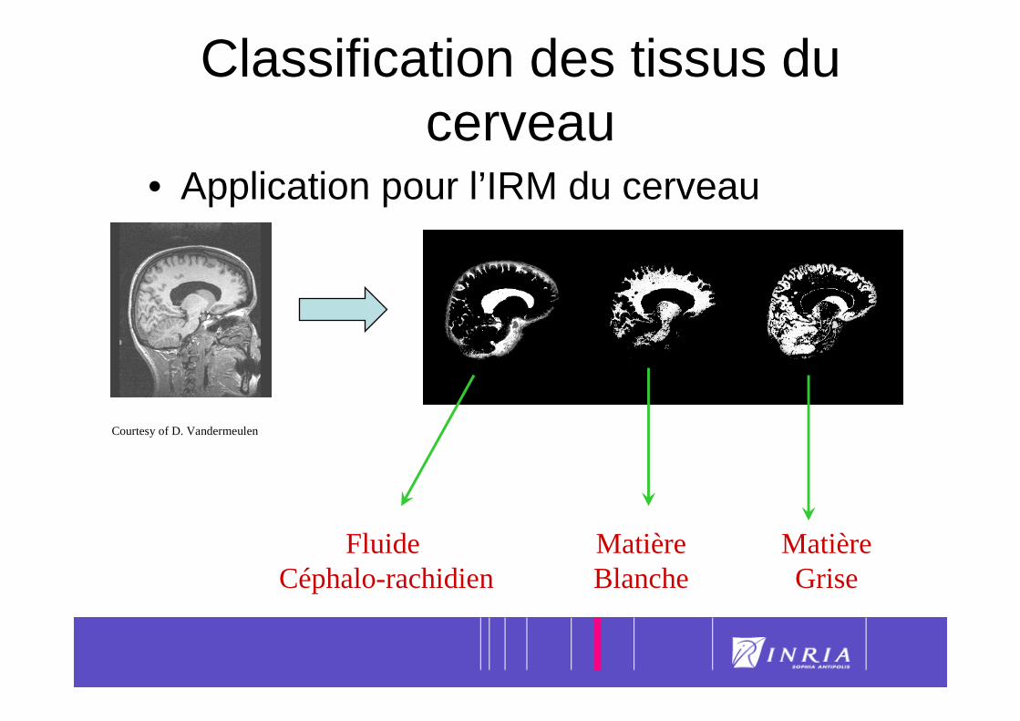
30Classification des tissus du cerveau
• Application pour l’IRM du cerveau
Fluide Céphalo-rachidien
Matière Blanche
Matière Grise
Courtesy of D. Vandermeulen
31
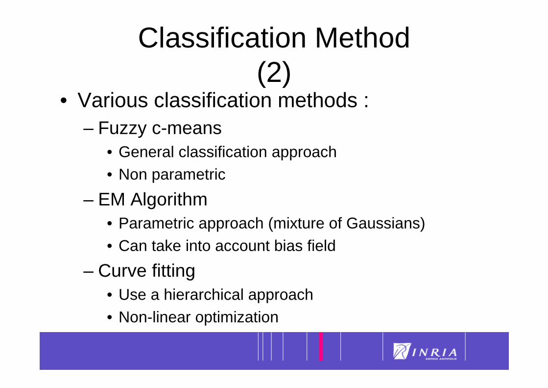
• Various classification methods :– Fuzzy c-means
• General classification approach• Non parametric
– EM Algorithm• Parametric approach (mixture of Gaussians)• Can take into account bias field
– Curve fitting• Use a hierarchical approach• Non-linear optimization
Classification Method (2)
32
Segmentation
Modèles déformables

33Segmentation d’images à l’aide de modèles déformables
• Un modèle déformable est un récipient pour stocker de l’information a priori sur la géométrie et l'apparence de structures anatomiques
• Deux niveaux de connaissance a priori:
Forme
Apparence
Faible Connaissance a priori
Contrainte de continuité C1 ou C2
Initialisation avec formes génériques (sphère, …)
Utilise information de gradientet/ou intensité
Contrainte de Forme
Initialisation avec forme moyenne
Utilise profils d’intensité ou appariement de blocs
Grande Connaissance a priori
34
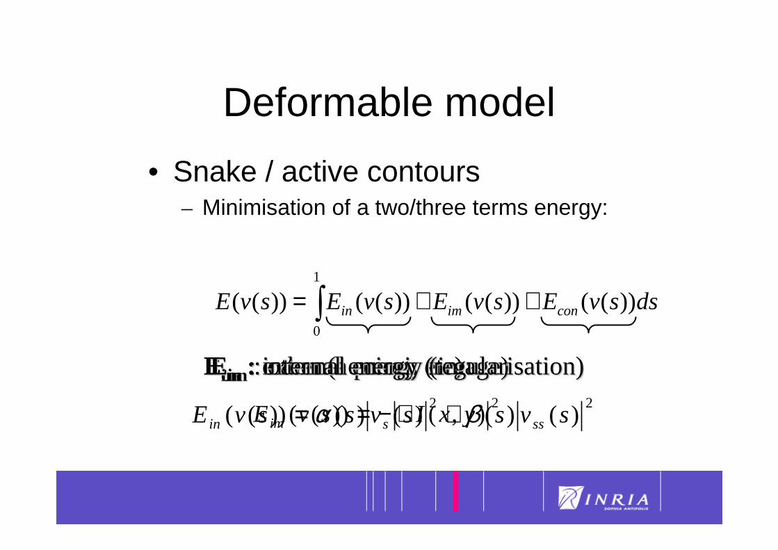
Deformable model
• Snake / active contours – Minimisation of a two/three terms energy:
22)()()()())(( svssvssvE sssin βα +=
∫ ++=1
0
))(())(())(())(( dssvEsvEsvEsvE conimin
EEconcon : : otherother(a priori, (a priori, etcetc))EEimim : : externalexternalenergyenergy(image)(image)EEinin : : internalinternalenergyenergy((regularisationregularisation))2
),())(( yxIsvE im ∇−=

35
Segmentation: endocraniumCT scan image, Bony structures
Time of convergence : 13,8 s 1169 3cmmodel:mold: 1150 3cm
36
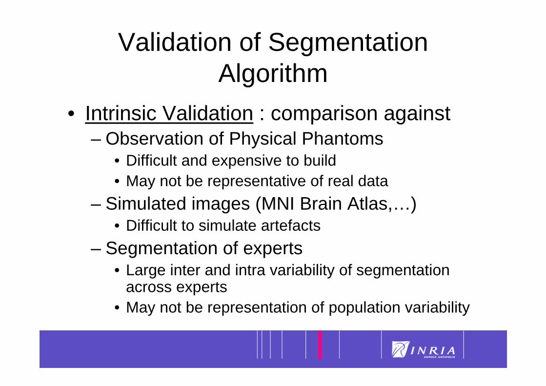
Validation of Segmentation Algorithm
• Intrinsic Validation : comparison against– Observation of Physical Phantoms
• Difficult and expensive to build• May not be representative of real data
– Simulated images (MNI Brain Atlas,…)• Difficult to simulate artefacts
– Segmentation of experts • Large inter and intra variability of segmentation
across experts• May not be representation of population variability
37
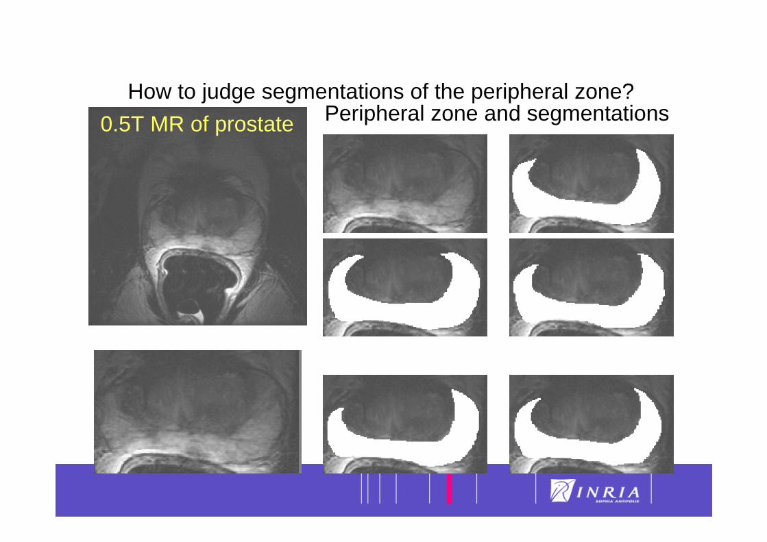
How to judge segmentations of the peripheral zone?
0.5T MR of prostate Peripheral zone and segmentations
38
Validation of Segmentation Algorithm (2)
• Extrinsic Validation : comparison againstother segmentation algorithms– Only possibility when no ground truth exists
(Inter-patient registration of images) or when itnot available
– Estimate consistency, repeatability and size of convergence basin


![VENTILATOR-INDUCED LUNG INJURY file• Jv = Kfc [(Pcap - Pint) - s (Ppl -Pint)] Pression Pression de filtration d’absorption. Albert JCI 1979 20 Inactivation du surfactant et pression](https://static.fdocuments.in/doc/165x107/5d4d1b6788c993c16c8bc982/ventilator-induced-lung-jv-kfc-pcap-pint-s-ppl-pint-pression-pression.jpg)