Innate lymphoid cells regulate CD4+ T-cell responses to intestinal ...
Upload
trinhtuongCategory
view
215download
2
IL-25 and type 2 innate lymphoid cells inducepulmonary fibrosisEmily Hamsa,b, Michelle E. Armstronga,b, Jillian L. Barlowc, Sean P. Saundersa,b, Christian Schwartzd, Gordon Cookee,Ruairi J. Fahyf, Thomas B. Crottyg, Nikhil Hiranih, Robin J. Flynnc, David Voehringerd, Andrew N. J. McKenziec,Seamas C. Donnellye,i, and Padraic G. Fallona,b,j,1
aTrinity Biomedical Sciences Institute, School of Medicine, Trinity College Dublin, Dublin 2, Ireland; bNational Children’s Research Centre, Our Lady’s Children’sHospital, Dublin 8, Ireland; cMedical Research Council Laboratory of Molecular Biology, Cambridge CB2 0QH, United Kingdom; dDepartment of InfectionBiology, Universitätsklinikum Erlangen, 91054 Erlangen, Germany; eSchool of Medicine and Medical Science, University College Dublin, Dublin 4, Ireland;fSt. James’s Hospital, Dublin 8, Ireland; gDepartment of Pathology, St. Vincent’s University Hospital, Dublin 4, Ireland; hMedical Research Council Centre forInflammation Research, University of Edinburgh, Edinburgh EH16 4SB, United Kingdom; iNational Pulmonary Fibrosis Referral Centre, St. Vincent’s UniversityHospital, Dublin 4, Ireland; and jInstitute of Molecular Medicine, School of Medicine, Trinity College Dublin, St. James’s Hospital, Dublin 8, Ireland
Edited by Shigeo Koyasu, RIKEN Center for Integrative Medical Sciences, Yokohama, Japan, and accepted by the Editorial Board November 15, 2013 (receivedfor review August 26, 2013)
Disease conditions associated with pulmonary fibrosis are pro-gressive and have a poor long-term prognosis with irreversiblechanges in airway architecture leading to marked morbidity andmortalities. Using murine models we demonstrate a role for in-terleukin (IL)-25 in the generation of pulmonary fibrosis. Mechanis-tically, we identify IL-13 release from type 2 innate lymphoid cells(ILC2) as sufficient to drive collagen deposition in the lungs ofchallenged mice and suggest this as a potential mechanism throughwhich IL-25 is acting. Additionally, we demonstrate that in humanidiopathic pulmonary fibrosis there is increased pulmonary expres-sion of IL-25 and also observe a population ILC2 in the lungs ofidiopathic pulmonary fibrosis patients. Collectively, we present aninnate mechanism for the generation of pulmonary fibrosis, via IL-25and ILC2, that occurs independently of T-cell–mediated antigen-spe-cific immune responses. These results suggest the potential of ther-apeutically targeting IL-25 and ILC2 for the treatment of humanfibrotic diseases.
innate response | cytokine | therapy | inflammation
Disease conditions associated with pulmonary fibrosis areoften progressive and have a poor long-term prognosis (1).
In the context of developing new treatments for pulmonary fi-brosis, the cytokines associated with the pathogenic milieu thatcan lead to aberrant extracellular matrix deposition are keytargets, in particular interleukin (IL)-13, TGF-β, and, more re-cently, IL-17A (2). However, to develop more effective thera-peutics for fibrotic lung diseases a greater understanding of thepathogenesis and the underlying mechanisms that lead to pul-monary fibrosis is needed (3, 4).The cytokine IL-13 was first implicated in fibrosis using
profibrotic eggs from the type 2 cytokine-inducing pathogenSchistosoma mansoni, in the presence of a soluble IL-13Rα2-Fcfusion protein (5) and in Il13−/− mice (6). IL-13 is now widelylinked to a range of fibrotic conditions (7) including asthma,where IL-13 is being targeted as a therapy (8). In the context ofthe cellular source of IL-13 in the generation of fibrosis, CD4+ Thelper (h) 2 cells are implicated (9). However, more recentlyinnate lymphoid cells (ILC) are emerging as an important sourceof IL-13 (10, 11). In this context, the type 2 cytokine IL-25 isimplicated in the generation of the recently identified IL-13–expressing ILC, termed ILC2 (11–14).Recent studies have implicated IL-25 and ILC2 in the path-
ogenesis of pulmonary conditions in both murine models and hu-man conditions such as allergic asthma (12, 13, 15, 16). In murinestudies intranasal administration of IL-25 results in evidence ofpulmonary tissue remodeling including development of perivascularfibrosis, and intratracheal administration results in increased pul-monary Th2 cytokines and airways hyper-reactivity (AHR) (17, 18),whereas blocking IL-25 reduces AHR severity (19). Herein wedescribe a potential role for IL-25 in the generation of pulmonary
fibrosis in experimental mouse models, via the activation of IL-13–producing ILC2. We also observe increases in both IL-25 and ILC2in the lung of patients with idiopathic pulmonary fibrosis (IPF).These data suggest unique mechanisms for the generation of pul-monary fibrosis and identify an interesting area for further researchon the role of IL-25 and ILC2 in the treatment of pulmonaryfibrosis.
ResultsMice Unable to Signal via IL-25 Show Decreased S. mansoni Egg-Associated Pulmonary Fibrosis. To address the mechanistic roleof IL-25 in pulmonary fibrosis we used the S. mansoni egg-induced pulmonary granuloma model. In mice deficient in IL-25(Il25−/−) and its receptor IL-17BR (Il17br−/−) there was smalleregg granuloma formation relative to WT mice (Fig. 1 A and C)with significantly (P < 0.05) reduced pulmonary collagen de-position and, more specifically, less fibrosis within the egg gran-uloma of these mice (Fig. 1 B and C). In addition, in the absenceof functional IL-25 there was reduced relative pulmonary ex-pression of type 2 cytokines IL-4 and IL-13 (Fig. 1D), and re-duced expression of TGFβ (Fig. 1D). Whereas pulmonary levelsof IL-17A were up-regulated in response to S. mansoni eggs inWT mice, as previously demonstrated (2), there were compara-ble IL-17A levels in all groups (Fig. 1D). Regarding the re-duction in IL-4 expression, a cytokine shown to be important inthe generation of egg-associated granuloma formation (20), in
Significance
Abnormal damage and scarring of tissue (fibrosis) in the lungscan lead to pulmonary fibrosis. Patients that develop the var-ious forms of pulmonary fibrosis are difficult to treat and havea high level of mortality. In this study we have used mousemodels to address the role of the cytokine interleukin (IL)-25and type 2 innate lymphoid cells (ILC2) in pulmonary fibrosis. Inanimal models we show a role for IL-25 and ILC2 in the gen-eration of pulmonary fibrosis. Furthermore, we have identifiedelevated levels of IL-25 and ILC2s in the lungs of patients withidiopathic pulmonary fibrosis. This study provides insights onthe factors and cells that may initiate pulmonary fibrosis inhumans and have therapeutic potential.
Author contributions: E.H. and P.G.F. designed research; E.H., M.E.A., J.L.B., S.P.S., C.S.,G.C., and R.J. Flynn performed research; G.C., R.J. Fahy, T.B.C., N.H., R.J. Flynn, D.V., A.N.J.M.,and S.C.D. contributed new reagents/analytic tools; E.H., M.E.A., J.L.B., S.P.S., R.J. Flynn,and A.N.J.M. analyzed data; and E.H. and P.G.F. wrote the paper.
The authors declare no conflict of interest.
This article is a PNAS Direct Submission. S.K. is a guest editor invited by the EditorialBoard.1To whom correspondence should be addressed. E-mail: [email protected].
This article contains supporting information online at www.pnas.org/lookup/suppl/doi:10.1073/pnas.1315854111/-/DCSupplemental.
www.pnas.org/cgi/doi/10.1073/pnas.1315854111 PNAS | January 7, 2014 | vol. 111 | no. 1 | 367–372
IMMUNOLO
GY
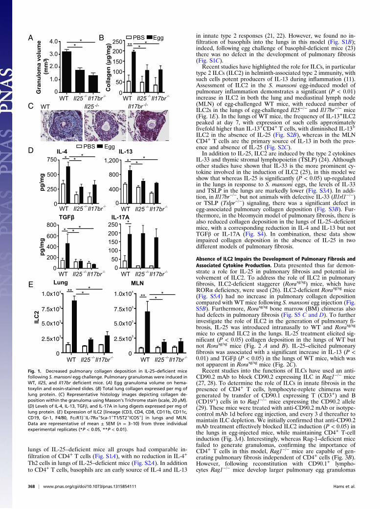
lungs of IL-25–deficient mice all groups had comparable in-filtration of CD4+ T cells (Fig. S1A), with no reduction in IL-4+
Th2 cells in lungs of IL-25–deficient mice (Fig. S2A). In additionto CD4+ T cells, basophils are an early source of IL-4 and IL-13
in innate type 2 responses (21, 22). However, we found no in-filtration of basophils into the lungs in this model (Fig. S1B);indeed, following egg challenge of basophil-deficient mice (23)there was no defect in the development of pulmonary fibrosis(Fig. S1C).Recent studies have highlighted the role for ILCs, in particular
type 2 ILCs (ILC2) in helminth-associated type 2 immunity, withsuch cells potent producers of IL-13 during inflammation (11).Assessment of ILC2 in the S. mansoni egg-induced model ofpulmonary inflammation demonstrates a significant (P < 0.01)increase in ILC2 in both the lung and mediastinal lymph node(MLN) of egg-challenged WT mice, with reduced number ofILC2s in the lungs of egg-challenged Il25−/− and Il17br−/− mice(Fig. 1E). In the lungs of WT mice, the frequency of IL-13+ILC2peaked at day 7, with expression of such cells approximatelyfivefold higher than IL-13+CD4+ T cells, with diminished IL-13+ILC2 in the absence of IL-25 (Fig. S2B), whereas in the MLNCD4+ T cells are the primary source of IL-13 in both the pres-ence and absence of IL-25 (Fig. S2C).In addition to IL-25, ILC2 are induced by the type 2 cytokines
IL-33 and thymic stromal lymphopoietin (TSLP) (24). Althoughother studies have shown that IL-33 is the more prominent cy-tokine involved in the induction of ILC2 (25), in this model weshow that whereas IL-25 is significantly (P < 0.05) up-regulatedin the lungs in response to S. mansoni eggs, the levels of IL-33and TSLP in the lungs are markedly lower (Fig. S3A). In addi-tion, in Il17br−/−, but not animals with defective IL-33 (Il1rl1−/−)or TSLP (Tslpr−/−) signaling, there was a significant defect inegg-associated pulmonary collagen deposition (Fig. S3B). Fur-thermore, in the bleomycin model of pulmonary fibrosis, there isalso reduced collagen deposition in the lungs of IL-25–deficientmice, with a corresponding reduction in IL-4 and IL-13 but notTGFβ or IL-17A (Fig. S4). In combination, these data showimpaired collagen deposition in the absence of IL-25 in twodifferent models of pulmonary fibrosis.
Absence of ILC2 Impairs the Development of Pulmonary Fibrosis andAssociated Cytokine Production. Data presented thus far demon-strate a role for IL-25 in pulmonary fibrosis and potential in-volvement of ILC2. To address the role of ILC2 in pulmonaryfibrosis, ILC2-deficient staggerer (Rorasg/sg) mice, which haveRORα deficiency, were used (26). ILC2-deficient Rorasg/sg mice(Fig. S5A) had no increase in pulmonary collagen depositioncompared with WT mice following S. mansoni egg injection (Fig.S5B). Furthermore, Rorasg/sg bone marrow (BM) chimeras alsohad defects in pulmonary fibrosis (Fig. S5 C and D). To furtherinvestigate the role of ILC2 in the generation of pulmonary fi-brosis, IL-25 was introduced intranasally to WT and Rorasg/sg
mice to expand ILC2 in the lungs. IL-25 treatment elicited sig-nificant (P < 0.05) collagen deposition in the lungs of WT butnot Rorasg/sg mice (Fig. 2 A and B). IL-25–elicited pulmonaryfibrosis was associated with a significant increase in IL-13 (P <0.01) and TGFβ (P < 0.05) in the lungs of WT mice, which wasnot apparent in Rorasg/sg mice (Fig. 2C).Recent studies into the functions of ILCs have used an anti-
CD90.2 mAb to block CD90.2 expressing ILC in Rag1−/− mice(27, 28). To determine the role of ILCs in innate fibrosis in thepresence of CD4+ T cells, lymphocyte-replete chimeras weregenerated by transfer of CD90.1 expressing T (CD3+) and B(CD19+) cells in to Rag1−/− mice expressing the CD90.2 allele(29). These mice were treated with anti-CD90.2 mAb or isotype-control mAb 1d before egg injection, and every 3 d thereafter tomaintain ILC depletion. We initially confirmed that anti-CD90.2mAb treatment effectively blocked ILC2 induction (P < 0.05) inthe lungs in egg-injected mice, while maintaining CD4+ T-cellinduction (Fig. 3A). Interestingly, whereas Rag-1–deficient micefailed to generate granulomas, confirming the importance ofCD4+ T cells in this model, Rag1−/− mice are capable of gen-erating pulmonary fibrosis independent of CD4+ cells (Fig. 3B).However, following reconstitution with CD90.1+ lympho-cytes Rag1−/− mice develop larger pulmonary egg granulomas
WT Il25 Il17br-/- -/-C
olla
ge
n (μ
g/m
g)
* ** *
* ** *
pg
/m
gp
g/m
g
* **
C
D
A BG
ra
nu
lo
ma
v
olu
me
(m
m )
34.0
3.0
2.0
1.0
0
*
WT Il25 Il17br-/- -/-
*200
150
100
50
0
PBS Egg
*750
500
250
0
1,200
800
400
0
*
*800
600
400
200
0 0
WT Il25 Il17br-/- -/-
WT Il25 Il17br-/- -/- WT Il25 Il17br
-/- -/-
WT Il25 Il17br-/- -/- WT Il25 Il17br
-/- -/-
PBS Egg
250
E
IL
C2
Lung MLN
1.0x10
7.5x10
5.0x10
2.5x10
4
3
3
3
0WT Il25 Il17br
-/- -/-
*** *
*** *
WT Il25 Il17br-/- -/-
1.0x10
7.5x10
5.0x10
2.5x10
4
3
3
3
0
IL-4 IL-13
TGFβ
50100150200250
IL-17A
Fig. 1. Decreased pulmonary collagen deposition in IL-25–deficient micefollowing S. mansoni egg challenge. Pulmonary granulomas were induced inWT, Il25, and Il17br deficient mice. (A) Egg granuloma volume on hema-toxylin and eosin-stained slides. (B) Total lung collagen expressed per mg oflung protein. (C) Representative histology images depicting collagen de-position within the granuloma using Masson’s Trichrome stain (scale, 20 μM).(D) Levels of IL-4, IL-13, TGFβ, and IL-17A in lung digests expressed per mg oflung protein. (E) Expression of ILC2 [lineage (CD3, CD4, CD8, CD11b, CD11c,CD19, Gr-1, F4/80, FceR1)−IL-7Rα+Sca-1+T1/ST2+ICOS+] in lungs and MLN.Data are representative of mean ± SEM (n = 3–10) from three individualexperimental replicates (*P < 0.05, **P < 0.01).
368 | www.pnas.org/cgi/doi/10.1073/pnas.1315854111 Hams et al.

with significant fibrosis (Fig. 3B). In anti-CD90.2 mAb-treatedCD90-disparate Rag-1−/− chimeric mice, the deficiency in ILC2was accompanied by significantly reduced granuloma and im-paired generation of pulmonary fibrosis relative to mAb-isotypecontrol-treated mice (Fig. 3 B and C). The impaired collagendeposition in the absence of ILC2 was also associated witha significant decrease in pulmonary expression of IL-13 (P <0.05) but not IL-4, TGFβ, or IL-17A (Fig. 3D). These datasuggest that specific depletion of ILCs can abrogate S. mansoniegg-induced pulmonary fibrosis.
IL-13 Expression in ILC2 Induces Pulmonary Fibrosis in S. mansoni Egg-Challenged Mice. To directly confirm that ILC2 can elicit pul-monary fibrosis via their production of IL-13, we transferredILC2 from Il13−/− or WT mice into Il13−/− during egg challenge;Il13−/− were used as these mice do not develop lung fibrosis inresponse to S. mansoni eggs (30, 31). Transfer of WT ILC2 in-duced collagen deposition in unchallenged mice, with markedfibrosis in egg-challenged mice receiving ILC2 (Fig. 4). In con-trast, transfer of IL-13–deficent ILC2 did not induce collagendeposition in either challenged or unchallenged mice (Fig. 4).Therefore, using cell transfer studies we demonstrate that ILC2can induce pulmonary fibrosis in an IL-13-dependent manner.
IL-25 and ILC2 Are Present in the Bronchalveolar Lavage and LungTissue of Patients with IPF. Data presented herein indicate a rolefor IL-25 in the generation of pulmonary fibrosis in murinemodels. To investigate the potential for IL-25 to play a role inthe generation of clinical pulmonary fibrosis, we assessed IL-25expression in the bronchalveolar lavage (BAL) fluid of a cohortof patients with IPF and control BAL samples from early-stageerythema nodosum patients, with the lungs clear of fibroticlesions. The levels of IL-25 were significantly (P < 0.01) in-creased in the BAL fluid of IPF patients relative to controlpatients at first diagnosis (Fig. 5A). Furthermore, IL-25 levelswere further enhanced in the same patients 1 y after diagnosis(Fig. 5A). Whereas other cytokines analyzed (IL-1β, IL-2, IL-4,IL-5, IL-8, IL-12p70, IL-13, IL-17A, IFNγ, and TNFα) were not
WT Rorasg/sg
PBS
IL-25
A
PBS IL-250
40
80
120
Co
llag
en
(μ
g/m
g)
Rorasg/sgWTB
PBS IL-250
50
25
75
pg
/m
g
PBS IL-250
400
800
1,200
PBS IL-250
200
100
300
400C IL-13 IL-17A TGFβ
Rorasg/sgWT
ND ND
** ** ns
**
* *
*
*
Fig. 2. IL-25 induces pulmonary collagen deposition in WT but not Rorasg/sg
mice. WT and Rorasg/sg were treated with recombinant mouse IL-25 i.n. ondays 0, 3, 7, 10, and 13. (A) Representative histology images depicting col-lagen deposition within the lung using Masson’s Trichrome stain (scale, 100μM). (B) Total collagen in lung digests expressed per mg of lung protein. (C)Levels of IL-13, IL-17A, and TGFβ in lung digests expressed per mg of lungprotein. Data are representative of mean ± SEM (n = 3–5) from two in-dividual experimental replicates (ns, not significant; *P < 0.05, **P < 0.01).
200
150
100
50
0Co
llag
en (
µg
/mg
)
A
B
PBS Egg
**
Rag1-/-
IgG
α-CD90.2
0
0.5
1.0
1.5
Gra
nu
lom
a vo
lum
e (
mm
)3
IgG α-CD90.2Rag1-/-
PBS Egg PBS Egg
PBS Egg PBS Egg
400
300
200
100
0
800
600
400
200
0
1,000
40
30
20
10
0
120
80
40
0
pg
/mg
pg
/mg
IL-4 IL-13
IL-17A TGF
D
**
*
PBS Egg
Rag1 -/- IgG α-CD90.2
PBS Egg
2.0x10
1.5x10
1.0x10
0.5x10
4
4
4
4
0
3x10
2x10
1x10
3
3
3
0
CD
4
ILC
2
**
***
Rag1 -/- IgG α-CD90.2
Rag1 -/- IgG α-CD90.2
C
ns
nsns
*
Fig. 3. ILC2 regulate pulmonary collagen deposition. Disparate CD90 chi-meras were generated by transferring CD90.1 T (CD3+) and B (CD19+) cells toRag-1−/− mice expressing the CD90.2 allele, which were subsequently treatedwith anti-CD90.2 or an isotype control mAb. (A) Lung frequency of CD4 andILC2 in Rag-1−/−, disparate CD90 chimeras treated with anti-IgG and dispa-rate CD90 chimeras treated with anti-CD90.2. (B) Granuloma volume andrepresentative histology images stained with Masson’s Trichrome (scale, 100μM). Lung collagen (C) and cytokines (D) expressed per mg of lung protein.Data are representative of mean ± SEM (n = 3–5) from two individual ex-perimental replicates (ns, not significant; *P < 0.05, **P < 0.01).
Hams et al. PNAS | January 7, 2014 | vol. 111 | no. 1 | 369
IMMUNOLO
GY

significantly enhanced at initial diagnosis, these cytokines weresignificantly (P < 0.01–0.05) elevated in the BAL 1-y post-diagnosis (Fig. 5A and Fig. S6 A and B). Interestingly, levels ofIL-25 showed a moderate positive correlation (P < 0.05; R =0.3748) with periostin levels, an extracellular matrix protein as-sociated with IPF and lung remodeling (32), in BAL from IPFpatients (Fig. 5B). As the mouse data showed infiltration of ILC2in lungs of mice with pulmonary fibrosis (Fig. 1E), we in-vestigated if ILC2 cells were in patients with IPF. A populationof lineage marker (CD3, CD4, CD8, CD11b, CD11c, CD14,CD19, CD56, CD123, FceR1) negative cells that expressedCRTH2, T1/ST2, CD45, ICOS, IL-7Rα, and IL-17BR was sig-nificantly (P < 0.05) enhanced in the BAL of IPF patientscompared with control patients (Fig. 5C). The ILC2 populationin IPF patients is similar to those previously seen in human lungtissue and BAL (16, 27), but this is an observation of these cellsin association with pulmonary fibrosis.
DiscussionPulmonary fibrotic conditions are common and often are asso-ciated with poor prognosis. The pathogenesis of pulmonary fi-brosis is not fully understood, with theories based on eithera recurrent inflammatory response and subsequent tissue de-struction and wound healing (33), or a single inflammatory phaserapidly followed by aberrant wound healing (34). Studies into thepathogenesis of pulmonary fibrosis have primarily focused on theclassical profibrotic cytokines TGFβ, and more recently, IL-13.Herein we demonstrate a role for the type 2 cytokine IL-25 andIL-25–elicited ILC2 in pulmonary fibrosis in murine models.Furthermore, in a small cohort of IPF patients there was increasedIL-25 in the presence of lung fibrosis and an up-regulation of ILC2
in the BAL fluid, inferring a potential role for IL-25 and ILC2 inhuman disease.Murine data have already provided evidence on both the role
of type 2 cytokines and innate lymphoid cells on the progressionof inflammation and fibrosis at several mucosal sites (11, 12, 27,28). Data presented herein demonstrate the ability of IL-25 todrive fibrosis, confirmed by a decrease in collagen deposition inthe absence of functional IL-25 signaling in two independentmodels of pulmonary fibrosis. One mechanism potentially un-derlying these observations involves ILC2-derived IL-13 expres-sion, confirmed by the ability of transferred ILC2 to inducepulmonary fibrosis in an IL-13–dependent manner. Indeed, todemonstrate the role of ILC2 in the progression of pulmonaryfibrosis we adopted several independent methods, using micedeficient in RORα, RORα chimeras as well as blocking CD90.2-expressing ILCs. In these mouse models a role for ILC2 inpulmonary fibrosis was shown. As there are currently no effectivemodels for specifically blocking ILC2 in vivo, without alsoinhibiting other CD90-expressing cell types such as other ILCpopulations or innate cells such as basophils, these results couldbe further interrogated once an effective specific blocking pro-tocol is developed.We show that in the absence of IL-25 there is a decreased
expression of the Th2 cytokine IL-4 and the profibrotic cytokineTGFβ, in addition to IL-13, despite comparable IL-4+Th2 cellexpression between groups. Analysis of the cell populations as-sociated with the egg-induced pulmonary granuloma suggestsILC2 are a major source of IL-13, which is absent in IL-25–deficient mice, whereas the frequency of IL-4+CD4+ was com-parable in WT- and IL-25–deficient mice. Further work is re-quired to address the reduced TGFβ in lungs of IL-25–deficientmice in the S. mansoni egg-induced model of pulmonary fibrosis,which is not apparent in the bleomycin model. Interestingly, IL-25 treatment was shown to directly elicit TGFβ in the lungs ofWT mice. Studies on human fibroblasts, known as a prominentsource of TGFβ in wound healing and fibrosis (35), have shownthat fibroblasts respond to IL-25, they express IL-17BR, andproduce TGFβ (36). These observations taken in combinationwith the apparent role for IL-25 in promoting fibrosis suggestthis could be an interesting avenue for further mechanisticresearch in mice. Interestingly, in the models we have used,whereas we observe increased IL-17A in response to egg chal-lenge in WT mice, as reported previously (2), pulmonary levelsof IL-17A are not altered in the absence of IL-25 or ILC2. Inaddition, despite noting a general increase in the frequency ofILCs in the lungs of challenged mice; we do not observe an in-crease in IL-17–producing ILC3, with the source of IL-17detected in the lungs being Th17 cells.Using a small cohort of IPF patients we observed that the
increased expression of IL-25 in BAL has a moderate correlationwith the levels of periostin, a marker recently shown to promotefibrosis in IPF patients (32). In accordance with previous studies,we also show increased expression of other cytokines, such as IL-8, IL-4, IL-1β, and IL-17A in the BAL of IPF patients (2). Inaddition, we show increased expression of ILC2 in the BAL ofIPF patients compared with BAL from patients without pulmo-nary fibrosis. ILC2 have previously been identified both inhealthy lung tissue and tissue from asthma, rhinosinitis, and in-fluenza patients (16), implying a role for these cells in pulmonaryinflammation. Whereas the human data presented herein pro-vide a tantalizing insight into the potential role of IL-25 andILC2 in fibrosis, confirmation of the observations we have seenwould be required in a larger cohort of patients before any firmerconclusions are made. This study highlights a unique role for IL-25 and ILC2 in pulmonary conditions and identifies therapeutictargets for fibrosis in the human lung as well as other chronicfibrotic disorders.
Materials and MethodsAnimals. Bagg albino (BALB/c) and C57BL/6 mice were purchased fromHarlan. IL-25– (10), IL-17BR– (11), T1/ST2- (37), TSLPR- (38), IL-13– (39), and
A
B
Co
llag
en
(μ
g/m
g)
0.6
0.4
0.2
0
200
250
150
100
50
0
**
*
PBS Egg
PBS
- Il13-/-
ILC2 transfer
Gran
ulo
ma vo
lu
me (m
m )
3
C no cellsWT ILC2
Il13 ILC2-/-
no cells Il13 ILC2-/- WT ILC2
WT
Egg
*
Fig. 4. Transferring IL-13–expressing ILC2 induces pulmonary collagen de-position in IL-13–deficient mice. ILC2 isolated from WT and Il13−/− weretransferred into unchallenged and egg-challenged IL-13–deficient mice. (A)Representative histology images depicting Masson’s Trichrome staining fromPBS and egg-challenged animals (scale, 100 μM) and (B) egg-associatedgranuloma volume. (C) Lung collagen expressed per mg of lung protein.Data are representative of mean ± SEM (n = 2–4) from two individual ex-perimental replicates (*P < 0.05).
370 | www.pnas.org/cgi/doi/10.1073/pnas.1315854111 Hams et al.

staggerer-Rorasg/sg–deficient mice (Jackson Laboratories) were bred in-houseon a BALB/c background. Rag-1–deficient mice and CD90.1+ mice and CD45.1+
on a C57BL/6 background were obtained from Jackson Laboratories andbred in-house. Mcpt8Cre mice (23) experiments were performed in theDepartment of Infection Biology, Universitätsklinikum Erlangen, Germany.In all studies, female mice of 8–12 wk of age were used, with the exceptionof Rorasg/sg, which were used at 4–5 wk of age. All animal experiments wereperformed in compliance with Irish Department of Health and Childrenregulations and approved by the Trinity College Dublin’s BioResourcesethical review board, or the UK Home Office.
S. mansoni Egg-Induced Pulmonary Granuloma Formation and AntibodyTreatment. S. mansoni eggs were isolated from the livers of mice infectedwith S. mansoni, and pulmonary granulomas were induced by i.v. injectionof 5,000 S. mansoni eggs (40). For neutralizing experiments anti-CD90.2 mAb(30H12; BioXCell), or rat IgG2b isotype control, was administered i.p. every3 d at a dose of 250 μg per mouse starting 1 d before i.v. egg administration.
Bleomycin- and IL-25–Induced Pulmonary Fibrosis. Pulmonary fibrosis was in-duced by intratracheal injection of 0.15 U bleomycin (Sigma), as previouslydescribed (2). Mice were given 0.5 μg recombinant mouse IL-25 intranasal (i.n.)on days 0, 3, 7, 10, and 13 and lung tissue and MLN harvested on day 14.
IPF Study Subjects. All IPF subjects (n = 14) were Caucasian and providedwritten informed consent. Ethical approval was obtained from the LothianResearch Ethics Committee. IPF was diagnosed according to the AmericanThoracic Society/European Respiratory Society bases on multidisciplinaryconsensus classification. Surgical lung biopsy and/or BAL was performed asdescribed previously (41). Baseline characteristics of IPF patients, in additionto characteristics at 1-y follow-up, are presented in Table S1.
Erythema Nodosum Patients. Irish patients were diagnosed at initial hospitalpresentation by the same physician (S.C.D.). All patients were classified at pre-sentation using chest radiograph (staged 0–IV, i.e., normal to fibrosis), in accor-dance with the Siltzbach staging system as per international guidelines. BALsamples from patients that were subsequently diagnosed with erythema nodo-sum (n = 3) served as a control group in this study. Ethical approval for the studywas obtained from the St. Vincent’s University Hospital Medical Ethics Commit-tee. Informed written consent was obtained from all participants.
Lung Tissue Processing and Histology. Lung tissue from mice or biopsy samplestaken from IPF patients was perfused and fixed in 10% formalin saline, thenparaffin-embedded. Healthy control slides of human lung were from Imgenex.Paraffin-embedded sections were cut to 4 μM and the slides stained withhematoxylin and eosin or Masson’s trichrome to assess pulmonary fibrosis.Histology was imaged using an Olympus BX43 with an Olympus DP25 Camera.
Lung tissue from mice was collected for cytokine and collagen analysis.Tissue was homogenized and prepared as previously described (42). Lungcollagen was analyzed using the Sircol collagen assay (Biocolor Life ScienceAssays) as described (43).
Flow Cytometry. Surface marker expression was assessed by flow cytometrywith data collection on a CyAn (Beckman Coulter). Data were analyzed usingFlowJo software (Tree Star). Murine cells were stained with BD BiosciencesmAbs; CD8-APC (Ly-2), B220-APC (RA3-6B2), F4/80-APC (BM8), ICOS-PE(7E.17G9), Siglec-F-APC (E50-2440), Sca-1-PE-Cy7 (D7), c-kit-PE (2B8), eBio-sciences mAbs; CD4-APC (RM4-5), CD11b-APC (M1/70), Gr-1-APC (RB6-8CS),FceR1-APC (MAR-1), IL-7Rα-APC-Cy7 (A7R34), BioLegend mAb; LAP (TGF-β1)-PE (TW7-16B4), and MD biosciences mAb; T1/ST2-FITC (DJ8). For intracellularcytokine, staining cells were cultured with 50 ng/mL phorbol 12-myristate13-acetate and 500 ng/mL ionomycin in the presence of 10 μg/mL Brefeldin Afor 4 h. Permeabolized cells were stained with eBiosciences mAb; IL-4-PerCP-efluor 710 (11B11), IL-5-PE (TRFK5), IL-13-efluor 450 (eBio13A), and BD Bio-sciences mAb; IL-17A-PE (TC11-18H10). Human cells were stained with BDBiosciences mAbs; CD3-PerCP (SK7), CD19-PerCP (SJ25C1), CD14-PerCP(MΦP9), CD45-AlexaFluor 700 (HI30) CD56-PECy7 (B159), eBiosciences mAbs;CD11c-PerCP (3.9), CD123-PerCP (6H6), CD127-APC (eBioRDR5), BiolegendmAbs; CD8a-PerCP (RPA-T8), FceR1a-PerCP (AER-37(CRA-a)), CRTH2-PE (BM16),
AF
old
in
crease *
**
B
10
20
30
40
pg
/m
l
P=0.0187R=0.3748
0 2 4 6
Periostin (ng/ml)
00
60
EN IPF IPF+ 1 year
EN IPF IPF+ 1 year
40
20
0
5
10
15
*
*
IL-25
TGFβ
0 2 4 6
50
100
150
200
0
P=0.0007R=0.6645
IL-25
TGFβ
EN IPF IPF+ 1 year
0
5
10
15
20
ns
**
IL-13
EN IPF IPF +1 year
IL-17A
0
0.5
1.0
1.5
2.0
2.5
ns
ns
Fo
ld
in
crease
Fo
ld
in
crease
Fo
ld
in
crease
0
0.2
0.4
0.6
0 2 4 6
IL-13
P=0.5319R=-0.1408
pg
/m
l
IL-17A
0
2
4
6
0 2 4 6
P=0.3369R=0.2149
pg
/m
lp
g/m
l
NormalBAL
IPFBAL
CR
TH
2C
RT
H2
NormalBAL
IPF BAL
T1/ST2
0 101 102 1030
20
40
60
80
100
0 101 102 1030
20
40
60
80
100
0 101 102 1030
20
40
60
80
100
0 101 102 1030
20
40
60
80
100
CD45 IL-7Rα ICOS IL-17BR
% o
f M
ax.
Isotype Antibody
3x10
2x10
1x10
3
3
3
0
IL
C2
C
0.01
0.16
*
Fig. 5. IPF patients have increased expression of IL-25 and ILC2 in the BAL.BAL fluid was collected from patients with IPF at diagnosis and at a 1-yfollow-up (IPF + 1 y) and patients diagnosed with erythema nodosum (EN;nonprogressive patients). (A) IL-25, IL-13, TGFβ, and IL-17A in BAL fluidexpressed as fold-increase relative to EN patients. (B) Correlation betweenBAL fluid levels of IL-25, IL-13, TGFβ, or IL-17A and periostin was plotted andPearson’s correlation calculated and displayed on the graph. (C) Represen-tative plots of ILC2 [lineage (CD3, CD4, CD8, CD11b, CD11c, CD19, CD14,CD56, CD123, FceR1) negative, expressing CRTH2, T1/ST2, CD45, ICOS, IL-7Rα,
and IL-17BR] in the BAL of nonfibrotic patients and IPF patients, with ex-pression of these cells displayed as a percentage of live cells per sample. Dataare representative of mean ± SEM (n = 3, EN; n = 14, IPF and IPF + 1 y) (ns,not significant; *P < 0.05, **P < 0.01).
Hams et al. PNAS | January 7, 2014 | vol. 111 | no. 1 | 371
IMMUNOLO
GY

ICOS-Pacific blue (C398.4A), Abcam mAb; CD11b-PerCP (DCIS1/18), MD Bio-sciences mAb; ST2-FITC (B4B6), and R&D Systems mAb; IL-17BR-APC (170220).Using appropriate isotype controls, quadrants were drawn and data wereplotted on logarithmic-scale density- or dot plots.
ILC2 Cell Sorting. BALB/c Il13+/+ or Il13−/− mice were treated with 2 μgrecombinant mouse IL-25 i.p. for 3 d and the peritoneal exudate cells werecollected and stained for ILC2 isolation. Cells were sorted on a Mo-Flo cell-sorter-based ILC2 population (lineage−(CD3, CD4, CD8, CD19, CD11b, CD11c,Gr-1, FceR1, F4/80)ICOS+T1/ST2+), which revealed a purity of >90% live cells.Isolated cells were injected i.p. into Il13−/− mice at a concentration of200,000 cells per mouse.
CD90 Disparate Rag-1−/− Chimera Generation. CD90 disparate chimeras weregenerated using methods previously described (29). Briefly, 6 × 10⁷ purifiedB- (CD19+) and T (CD3+) cells were isolated from the spleen, and lymphnodes from CD90.1+ mice were transferred i.v. into CD90.2 Rag-1−/− mice.After 8 wk reconstitution was confirmed by examination of peripheralblood lymphocytes.
BM Chimera Generation. Lethally irradiated CD45.1+ C57BL/6 recipient mice(9 Gy in two doses, 3 h apart) were reconstituted with 1 × 10⁷ BM cells fromCD45.2+ C57BL/6 mice or Rorasg/sg mice. To ensure efficient irradiation andreconstitution, expression of CD45.1 vs. CD45.2 was assessed in the blood after4 wk. After 6 wk C57B6/J mice reconstituted with WT or Rorasg/sg BM wereinjected with 5,000 S. mansoni eggs i.v. and lung tissue harvested after 7 d.
Mouse Cytokine ELISAs. For enumeration of cytokines, murine lung sectionswere prepared by homogenization as previously described (42). Sampleswere analyzed by sandwich ELISAs to quantify levels of specific cytokines. All
murine cytokines (IL-1β, IL-4, IL-13, IL-17, IL-33, IFNγ, TNFα, and TGFβ) weremeasured with the DuoSet ELISA development system from R&D Systemsfollowing the manufacturer’s protocol. Expression of all lung proteins isexpressed per mg lung protein present.
Human Cytokine and Periostin ELISAs. Concentrations of IL-25 were de-termined in human BAL fluid by ELISA (Cusabio Biotech) according tomanufacturer’s instructions. Levels of human periostin in BAL fluid weremeasured using a sandwich ELISA kit from Holzel Diagnostika followingthe manufacturer’s instructions. IL-17A in patients BAL fluid was quan-tified using a human cytokine assay from MesoScale Discovery (MSD,ultrasensitive kit). Levels of all other cytokines were assessed in BAL fluidin this study using a Th1/Th2 10-Plex multiplex cytokine assay (MSD)according to manufacturer’s instructions. Results for cytokine levels inBAL fluid were standardized per ml of fluid recovered during the lavageprocess. Results are expressed as fold-increase relative to basal cytokinesdetected in BAL of erythema nodosum patients.
Statistics. Statistical analysis was performed using GraphPad and InStat.Results are presented as mean ±SEM. Differences, indicated as two-tailedP values, were considered significant when P < 0.05 was assessed by un-paired Student t test with Welch correction applied as necessary. Pearson’scorrelation was performed on BAL fluid data comparing periostin and cy-tokine levels; a P value of less than 0.05 was considered significant.
ACKNOWLEDGMENTS. The authors thank Dr. Malgorzata Kubica for herassistance with this article. This work was supported by Science FoundationIreland (P.G.F. and S.C.D.), National Children’s Research Centre (P.G.F.), HealthResearch Board of Ireland (P.G.F. and S.C.D.), and Asthma UK (A.N.J.M.).
1. Ley B, et al. (2012) A multidimensional index and staging system for idiopathic pul-monary fibrosis. Ann Intern Med 156(10):684–691.
2. Wilson MS, et al. (2010) Bleomycin and IL-1beta-mediated pulmonary fibrosis is IL-17Adependent. J Exp Med 207(3):535–552.
3. Wynn TA (2011) Integrating mechanisms of pulmonary fibrosis. J Exp Med 208(7):1339–1350.
4. Noble PW, Barkauskas CE, Jiang D (2012) Pulmonary fibrosis: Patterns and perpe-trators. J Clin Invest 122(8):2756–2762.
5. Chiaramonte MG, et al. (1999) IL-13 is a key regulatory cytokine for Th2 cell-mediatedpulmonary granuloma formation and IgE responses induced by Schistosoma mansonieggs. J Immunol 162(2):920–930.
6. Fallon PG, Richardson EJ, McKenzie GJ, McKenzie AN (2000) Schistosome infection oftransgenic mice defines distinct and contrasting pathogenic roles for IL-4 and IL-13: IL-13 is a profibrotic agent. J Immunol 164(5):2585–2591.
7. Wynn TA (2003) IL-13 effector functions. Annu Rev Immunol 21:425–456.8. Kraft M (2011) Asthma phenotypes and interleukin-13—moving closer to personal-
ized medicine. N Engl J Med 365(12):1141–1144.9. Barron L, Wynn TA (2011) Fibrosis is regulated by Th2 and Th17 responses and by
dynamic interactions between fibroblasts and macrophages. Am J Physiol Gastro-intest Liver Physiol 300(5):G723–G728.
10. Fallon PG, et al. (2006) Identification of an interleukin (IL)-25-dependent cell pop-ulation that provides IL-4, IL-5, and IL-13 at the onset of helminth expulsion. J ExpMed 203(4):1105–1116.
11. Neill DR, et al. (2010) Nuocytes represent a new innate effector leukocyte that me-diates type-2 immunity. Nature 464(7293):1367–1370.
12. Saenz SA, et al. (2010) IL25 elicits a multipotent progenitor cell population thatpromotes T(H)2 cytokine responses. Nature 464(7293):1362–1366.
13. Moro K, et al. (2010) Innate production of T(H)2 cytokines by adipose tissue-associ-ated c-Kit(+)Sca-1(+) lymphoid cells. Nature 463(7280):540–544.
14. Price AE, et al. (2010) Systemically dispersed innate IL-13-expressing cells in type 2immunity. Proc Natl Acad Sci USA 107(25):11489–11494.
15. Corrigan CJ, et al. (2011) Allergen-induced expression of IL-25 and IL-25 receptor inatopic asthmatic airways and late-phase cutaneous responses. J Allergy Clin Immunol128(1):116–124.
16. Mjösberg JM, et al. (2011) Human IL-25- and IL-33-responsive type 2 innate lymphoidcells are defined by expression of CRTH2 and CD161. Nat Immunol 12(11):1055–1062.
17. Angkasekwinai P, et al. (2007) Interleukin 25 promotes the initiation of proallergictype 2 responses. J Exp Med 204(7):1509–1517.
18. Fort MM, et al. (2001) IL-25 induces IL-4, IL-5, and IL-13 and Th2-associated patholo-gies in vivo. Immunity 15(6):985–995.
19. Ballantyne SJ, et al. (2007) Blocking IL-25 prevents airway hyperresponsiveness inallergic asthma. J Allergy Clin Immunol 120(6):1324–1331.
20. Pearce EJ, et al. (1996) Schistosoma mansoni in IL-4-deficient mice. Int Immunol 8(4):435–444.
21. Min B, et al. (2004) Basophils produce IL-4 and accumulate in tissues after infectionwith a Th2-inducing parasite. J Exp Med 200(4):507–517.
22. Wakahara K, et al. (2013) Basophils are recruited to inflamed lungs and exacerbatememory Th2 responses in mice and humans. Allergy 68(2):180–189.
23. Ohnmacht C, et al. (2010) Basophils orchestrate chronic allergic dermatitis and pro-tective immunity against helminths. Immunity 33(3):364–374.
24. Hams E, Fallon PG (2012) Innate type 2 cells and asthma. Curr Opin Pharmacol 12(4):503–509.
25. Barlow JL, et al. (2013) IL-33 is more potent than IL-25 in provoking IL-13-producingnuocytes (type 2 innate lymphoid cells) and airway contraction. J Allergy Clin Im-munol 132(4):933–941.
26. Wong SH, et al. (2012) Transcription factor RORα is critical for nuocyte development.Nat Immunol 13(3):229–236.
27. Monticelli LA, et al. (2011) Innate lymphoid cells promote lung-tissue homeostasisafter infection with influenza virus. Nat Immunol 12(11):1045–1054.
28. Sonnenberg GF, et al. (2012) Innate lymphoid cells promote anatomical containmentof lymphoid-resident commensal bacteria. Science 336(6086):1321–1325.
29. Sonnenberg GF, Monticelli LA, Elloso MM, Fouser LA, Artis D (2011) CD4(+) lymphoidtissue-inducer cells promote innate immunity in the gut. Immunity 34(1):122–134.
30. Fallon PG, et al. (2002) IL-4 induces characteristic Th2 responses even in the combinedabsence of IL-5, IL-9, and IL-13. Immunity 17(1):7–17.
31. Kaviratne M, et al. (2004) IL-13 activates a mechanism of tissue fibrosis that is com-pletely TGF-beta independent. J Immunol 173(6):4020–4029.
32. Naik PK, et al.; COMET Investigators (2012) Periostin promotes fibrosis and predictsprogression in patients with idiopathic pulmonary fibrosis. Am J Physiol Lung Cell MolPhysiol 303(12):L1046–L1056.
33. Selman M, King TE, Pardo A; American Thoracic Society; European Respiratory Soci-ety; American College of Chest Physicians (2001) Idiopathic pulmonary fibrosis: Pre-vailing and evolving hypotheses about its pathogenesis and implications for therapy.Ann Intern Med 134(2):136–151.
34. Gauldie J, Bonniaud P, Sime P, Ask K, Kolb M (2007) TGF-beta, Smad3 and the processof progressive fibrosis. Biochem Soc Trans 35(Pt 4):661–664.
35. Wynn TA (2008) Cellular and molecular mechanisms of fibrosis. J Pathol 214(2):199–210.36. Létuvé S, et al. (2006) IL-17E upregulates the expression of proinflammatory cytokines
in lung fibroblasts. J Allergy Clin Immunol 117(3):590–596.37. Townsend MJ, Fallon PG, Matthews DJ, Jolin HE, McKenzie AN (2000) T1/ST2-deficient
mice demonstrate the importance of T1/ST2 in developing primary T helper cell type 2responses. J Exp Med 191(6):1069–1076.
38. Al-Shami A, et al. (2004) A role for thymic stromal lymphopoietin in CD4(+) T celldevelopment. J Exp Med 200(2):159–168.
39. McKenzie GJ, et al. (1998) Impaired development of Th2 cells in IL-13-deficient mice.Immunity 9(3):423–432.
40. Fallon PG, Emson CL, Smith P, McKenzie AN (2001) IL-13 overexpression predisposesto anaphylaxis following antigen sensitization. J Immunol 166(4):2712–2716.
41. Bournazos S, et al. (2011) Copy number variation of FCGR3B is associated with suscepti-bility to idiopathic pulmonary fibrosis. Respiration 81(2):142–149.
42. Mangan NE, Dasvarma A, McKenzie AN, Fallon PG (2007) T1/ST2 expression on Th2 cellsnegatively regulates allergic pulmonary inflammation. Eur J Immunol 37(5):1302–1312.
43. Amu S, et al. (2010) Regulatory B cells prevent and reverse allergic airway in-flammation via FoxP3-positive T regulatory cells in a murine model. J Allergy ClinImmunol 125(5):1114–1124 e1118.
372 | www.pnas.org/cgi/doi/10.1073/pnas.1315854111 Hams et al.


![Pulmonary type2 innate lymphoid cells in paediatric severe ... · the induction of type 2 innate lymphoid cells (ILCs) [12-14]. ILCs are a rare population of cells of lymphoid lineage,](https://static.fdocuments.in/doc/165x107/5e17e22a8563b16def417337/pulmonary-type2-innate-lymphoid-cells-in-paediatric-severe-the-induction-of.jpg)