studentsrepo.um.edu.mystudentsrepo.um.edu.my/8503/9/aisyah.pdf · ii BIOMECHANICAL EFFECTS OF...
155
BIOMECHANICAL EFFECTS OF DIFFERENT KNEE SLEEVES ON EARLY UNILATERAL KNEE OSTEOARTHRITIS IN 6 WEEKS INTERVENTION NAHDATUL AISHAH BINTI MOHD. SHARIF FACULTY OF ENGINEERING UNIVERSITY OF MALAYA KUALA LUMPUR 2017 University of Malaya
Transcript of studentsrepo.um.edu.mystudentsrepo.um.edu.my/8503/9/aisyah.pdf · ii BIOMECHANICAL EFFECTS OF...

BIOMECHANICAL EFFECTS OF DIFFERENT KNEE SLEEVES ON EARLY UNILATERAL KNEE
OSTEOARTHRITIS IN 6 WEEKS INTERVENTION
NAHDATUL AISHAH BINTI MOHD. SHARIF
FACULTY OF ENGINEERING
UNIVERSITY OF MALAYA KUALA LUMPUR
2017
Univers
ity of
Mala
ya
BIOMECHANICAL EFFECTS OF DIFFERENT KNEE
SLEEVES ON EARLY UNILATERAL KNEE
OSTEOARTHRITIS IN 6 WEEKS INTERVENTION
NAHDATUL AISHAH BINTI MOHD. SHARIF
DESSERTATION SUBMITTED IN FULFILMENT OF
THE REQUIREMENTS FOR THE DEGREE OF
MASTERS IN ENGINEERING SCIENCE
FACULTY OF ENGINEERING
UNIVERSITY OF MALAYA
KUALA LUMPUR
2017
Univers
ity of
Mala
ya

i
UNIVERSITY OF MALAYA
ORIGINAL LITERARY WORK DECLARATION
Name of Candidate: Nahdatul Aishah binti Mohd. Sharif
Registration/Matric No: KGA140065
Name of Degree: Masters of Engineering Science
Title of Project Paper/Research Report/Dissertation/Thesis: “Biomechanical Effects of
Different Knee Sleeves on Early Unilateral Knee Osteoarthritis in 6 Weeks Intervention”
Field of Study: Biomechanics (Engineering and engineering trades)
I do solemnly and sincerely declare that:
(1) I am the sole author/writer of this Work;
(2) This Work is original;
(3) Any use of any work in which copyright exists was done by way of fair dealing
and for permitted purposes and any excerpt or extract from, or reference to or
reproduction of any copyright work has been disclosed expressly and
sufficiently and the title of the Work and its authorship have been
acknowledged in this Work;
(4) I do not have any actual knowledge nor do I ought reasonably to know that the
making of this work constitutes an infringement of any copyright work;
(5) I hereby assign all and every rights in the copyright to this Work to the
University of Malaya (“UM”), who henceforth shall be owner of the copyright
in this Work and that any reproduction or use in any form or by any means
whatsoever is prohibited without the written consent of UM having been first
had and obtained;
(6) I am fully aware that if in the course of making this Work I have infringed any
copyright whether intentionally or otherwise, I may be subject to legal action
or any other action as may be determined by UM.
Candidate’s Signature Date:
Subscribed and solemnly declared before,
Witness’s Signature Date:
Name:
Designation:
Univers
ity of
Mala
ya

ii
BIOMECHANICAL EFFECTS OF DIFFERENT KNEE SLEEVES ON EARLY
UNILATERAL KNEE OSTEOARTHRITIS IN 6 WEEKS INTERVENTION
ABSTRACT
Knee osteoarthritis (OA) is a common joint disorder that affects balance, knee joint
proprioception and gait. Many treatment approaches have been used to improve the
conditions of people with this disease. Knee sleeves are often prescribed to alleviate pain.
However, the biomechanics underlying their pain-relieving effect is still not well
understood. This pre-post study is aimed at evaluating and comparing the effects of two
different types of knee sleeves on gait biomechanics and postural stability of people with
early knee OA, and to determine the relationship of these changes to patient-reported pain
outcomes following a six-week application. Six-week is generally longer than immediate
term and often used in clinical trials. Patients with clinically diagnosed knee OA were
recruited from the University of Malaya Medical Centre (UMMC), and were randomly
assigned to two test groups comprising those using: 1) a simple sleeve, and 2) a simple
sleeve with patella cutout. The walking motion and the ground reaction forces of
participants were measured using Vicon Nexus motion analysis system (with five
cameras) and two Kistler force plates, with sampling rates of 100Hz (kinematics) and
1,000Hz (kinetics), respectively for two walking speeds – controlled and self-selected.
The postural stability was measured using Biodex Stability System (BSS) – with seven
protocols – to obtain the Overall Stability Index (OSI): Postural Stability Test (PST), and
Athlete Single Leg Test (ASL) – static and dynamic conditions – and Fall Risk Test
(FRT). Pain, stiffness and physical functions were recorded using the Western Ontario
and McMaster Universities Arthritis Index (WOMAC). SPSS v22 was used for statistical
analyses, with two-way repeated measures Analysis of Variance (ANOVA) with mixed
approaches were used to compare knee sleeve designs (between-subject effects) against
all dependent variables (within-subject effect), with additional Bonferroni corrections for
Univers
ity of
Mala
ya

iii
multiple tests and confidence interval. All measurements were made before, immediately
after, and following six weeks of knee sleeve application (primary time point). Seventeen
participants (aged 47.7 ± 9.7 years) with early unilateral knee OA completed the study.
Overall results show significant reduction in pain, early stance and late stance knee
adduction moment, and increased walking speed after six weeks of sleeve application.
However, there are no significant differences between the groups in all parameters at all
points of measurements. The results indicate that there is improvement in overall stability
index (OSI) but no significant changes are detected for static and dynamic PST for both
types of sleeves immediately after application. The findings show that early knee OA
patients could experience improved balance ability in both static and dynamic conditions,
and less pain after six weeks of knee sleeve application. This study results suggest that
knee sleeves can reduce knee adduction moments in early unilateral knee OA by 14.0%
and 12.1% using the simple sleeve and the sleeve with patella cutout, respectively, and
possibly delay disease progression. Additionally, knee sleeve with patella cutout does not
provide additional benefits when compared to the simple knee sleeve.
479 words
Keywords: Knee osteoarthritis, knee sleeve, gait, postural stability, WOMAC.
Univers
ity of
Mala
ya
iv
KESAN BIOMEKANIK LENGAN LUTUT YANG BERBEZA PADA
OSTEOARTRITIS LUTUT UNILATERAL AWAL DALAM 6 MINGGU
INTERVENSI
ABSTRAK
Osteoartritis (OA) lutut adalah masalah sendi yang kerap dihadapi. Ia berpotensi
menganggu prestasi keseimbangan badan, keupayaan relatif lutut dan gaya berjalan.
Beberapa langkah telah diambil untuk meningkatkan keupayaan fizikal dalam kalangan
individu yang menghadapi masalah ini. Lengan lutut kerap diberi kepada mereka yang
mengalami OA lutut bagi mengurangkan kesakitan. Walaubagaimanapun kesan
biomekanik yang bertanggungjawab dalam membantu mengurangkan kesakitan masih
belum difahami sepenuhnya kerana kurangnya kajian yang dijalankan. Kajian pre-post
ini bertujuan untuk menilai dan membuat perbandingan diantara kesan keseimbangan
badan, kinematik dan kinetik gaya berjalan yang dialami oleh pesakit OA peringkat awal
apabila mereka memakai dua jenis lengan lutut yang berlainan selama enam minggu.
Enam minggu dipilih kerana tempoh itu lebih lama daripada kesan segera, dan sering
digunakan dalam ujian klinikal. Kaitan diantara perubahan-perubahan biomekanik ini
dengan perubahan kesakitan yang dilaporkan oleh pesakit juga akan dikaji. Pesakit OA
lutut yang memenuhi syarat telah dibahagikan secara rawak kepada dua kumpulan: 1)
kumpulan lengan lutut asas dan 2) kumpulan lengan lutut dengan patella cutout.
Kestabilan keseluruhan indeks (OSI) telah diukur menggunakan Biodex Statibility
System. Tujuh ujian telah dijalankan bagi setiap sesi: Postural Stability Test (PST), Fall
Risk Test (FRT) and Athlete Single Leg Test (ASL). Analisis gait telah diperiksa
menggunakan perisian Vicon Nexus dengan lima kamera dan dua force plate Kistler,
masing-masing dengan kadar pensampelan 100 Hz dan 1,000 Hz. Kesakitan lutut,
kekakuan lutut dan kesukaran melakukan aktiviti harian telah direkodkan menggunakan
WOMAC. Untuk analisis statistik, ANOVA dengan ukuran berulang dua hala dengan
Univers
ity of
Mala
ya
v
pendekatan campuran digunakan untuk membandingkan jenis lengan lutut (antara kesan
subjek) dan pembolehubah bersandar (within-subject effect) dengan tambahan confidence
adjustment menggunakan Bonferroni. Ukuran-ukuran ini diperolehi sebanyak tiga kali
sepanjang kajian: sebelum lengan dipakai, sejurus selepas ia dipakai serta enam minggu
kemudian (titik masa primer). Tujuh belas peserta (47.7 ± 9.7 tahun) berjaya menjalani
semua ujian. Keputusan keseluruhan menunjukkan pengurangan yang ketara dalam tahap
kesakitan, adduction moment lutut yang pertama dan kedua, dan kelajuan berjalan selepas
enam minggu dari penggunaan pertama. Walau bagaimanapun, tiada perbezaan yang
signifikan dikesan antara kedua-dua jenis lengan lutut dalam semua parameter pada
mana-mana titik ukuran. Keputusan kajian menunjukkan Overall Stability Index (OSI)
telah bertambah baik tetapi tiada perbezaan yang signifikan dapat dikesan untuk PST,
statik dan dinamik, untuk kedua-dua kumpulan lengan lutut selepas penggunaan serta-
merta untuk kedua-dua lengan lutut. Keputusan ini menunjukkan bahawa pesakit lutut
OA peringkat awal boleh mengalami peningkatan keupayaan keseimbangan dalam
kedua-dua keadaan statik dan dinamik dan juga pengurangan kesakitan selepas enam
minggu penggunaan. Kajian ini juga membuktikan bahawa memakai lengan lutut semasa
berjalan boleh mengurangkan adduction moment lutut pada peserta dengan OA lutut.
Adduction moment lutut yang lebih tinggi sebelum ini telah dikenal pasti sebagai faktor
risiko untuk perkembangan penyakit pada pesakit dengan OA lutut medial, kesimpulan
dapat dibuat bahawa memakai lengan lutut boleh memberi manfaat kepada kumpulan ini.
Selain itu, lengan lutut dengan patella cutout kelihatan tidak memberi manfaat tambahan
berbanding dengan lengan lutut biasa apabila digunakan di peringkat awal OA lutut.
482 patah perkataan
Kata kunci: Osteoarthritis lutut, lengan lutut, analisa berjalan, kestabilan, WOMAC.
Univers
ity of
Mala
ya

vi
ACKNOWLEDGEMENTS
Above all, I would like to convey my deepest gratitude to Allah the Almighty for
giving me all the provisions and luck that I need to complete this journey. Truly, there is
neither might nor power except with Him. Peace and blessings upon Prophet Muhammad
for his advice to remain patient at every turn in life.
Special thanks to my family members especially my mom, Zawiah Hassan, my dad,
Mohd Sharif Khamis, and my grandmother, Hjh Ramlah Ali, who have always been my
source of motivation. To my siblings: Along, Angah, Kaklang, Kak Mallisa, Azahari,
Iskandar, Kak Fasha, Abang Elmy, and Adi – thank you so much. Not to forget, to all my
family members, your help along the way is much appreciated.
Special thanks to my supervisors, for their infinite support and guidance; Dr. Wan
Safwani, Dr. Juliana and Dr. Goh Siew Li. Also, special thanks to Dr. Samihah, a
specialist from UMMC; Dr. Anisah, a statistician from ISM, UM; and Dr. Madiha, a
lecturer from API, UM, for their utmost help along the way.
Special thanks to my dear lab mates, Soobia Saad Khan, Saad Khan, Yati and Zuria,
the lab would not work without your help. Likewise, I thank the staff in Biomedical
Engineering Department UM, especially Mrs Hanie Nadia, Mr Adhli Iskandar, and Mr
Khairul for assisting me in the Motion Analysis Laboratory, UMMC staff for their help
and great patience during patient recruitment process, and Nasir and Naji for assisting me
in the statistics analysis. To Liyana, Ainul, Aina and Faiz Zulkeflee who willingly helped
me with my pilot study, thank you.
Special thanks to my dear friends for being good listeners and my crying shoulder
throughout this journey: Shaai, Amy, Hannah, Amirah, Fadillah, Aween and Aida; to my
course mates: Evellenie, Hazlina, Firdaus, Norishah, Timothy, Faiz, Chen Onn, Ita, Chibo
and Zuheir; to my lab mates in the Neuro Engineering Laboratory: Zara, Hanum, Afiqah,
Wani, and Laila; and to my colleagues in the Academy of Islamic Studies, UM: Fatimah,
Univers
ity of
Mala
ya

vii
Nor Aina, Nadhrah, and Munirah. To Mr Teh, this work would never been better without
your proofreading services. Last but not least, special thanks to everyone who have been
involved directly or indirectly in the success of this project. Thank you very much.
Alhamdulillah ala kulli hal.
Nahdatul Aishah Mohd Sharif
University of Malaya, December 2017
Univers
ity of
Mala
ya

viii
TABLE OF CONTENTS
Biomechanical effects of different knee sleeves on early unilateral knee osteoarthritis in
6 weeks intervention Abstract ........................................................................................... ii
Kesan biomekanik lengan lutut yang berbeza pada osteoartritis lutut unilateral awal dalam
6 minggu intervensi Abstrak ............................................................................................ iv
Acknowledgements .......................................................................................................... vi
Table of Contents ........................................................................................................... viii
List of Figures ................................................................................................................. xii
List of Tables.................................................................................................................. xiv
List of Symbols and Abbreviations ................................................................................. xv
List of Appendices ......................................................................................................... xvi
CHAPTER 1: INTRODUCTION .................................................................................. 1
1.1 Overview.................................................................................................................. 1
1.2 Problem Statement ................................................................................................... 3
1.3 Objective .................................................................................................................. 4
1.4 Hypothesis Statement .............................................................................................. 5
1.5 Dissertation Structure .............................................................................................. 6
1.6 Summary .................................................................................................................. 6
CHAPTER 2: LITERATURE REVIEW ...................................................................... 7
2.1 Introduction.............................................................................................................. 7
2.2 Osteoarthritis: Definition ......................................................................................... 7
2.3 Management on Knee OA and the Challenges ...................................................... 10
2.4 Orthosis for Knee OA ............................................................................................ 11
2.5 Past Studies on Knee Sleeves ................................................................................ 12
Univers
ity of
Mala
ya

ix
2.5.1 Clinical Assessment on Knee Sleeves ...................................................... 12
2.5.1.1 Pain… ........................................................................................ 13
2.5.1.2 Adverse Effects ......................................................................... 14
2.5.2 Gait ........................................................................................................... 15
2.5.2.1 Knee Adduction Moment (KAM) and Ground Reaction Force
(GRF). ....................................................................................... 15
2.5.2.2 Knee Extension and Walking Speed ......................................... 18
2.5.2.3 The Gait Analysis ...................................................................... 20
2.5.3 Balance and Postural Stability .................................................................. 23
2.5.3.1 Functional Tests ........................................................................ 25
2.5.4 Relationship between Gait and Balance ................................................... 27
2.5.5 Types of Knee Sleeves ............................................................................. 27
2.5.6 Limitations ................................................................................................ 29
2.6 Summary ................................................................................................................ 31
CHAPTER 3: METHODOLOGY ............................................................................... 32
3.1 Introduction............................................................................................................ 32
3.2 Study Design .......................................................................................................... 32
3.3 Participants ............................................................................................................ 33
3.4 Intervention ............................................................................................................ 33
3.5 Adherence .............................................................................................................. 35
3.5.1 Log Book .................................................................................................. 35
3.6 Instrumentations .................................................................................................... 36
3.6.1 WOMAC® Score ..................................................................................... 36
3.6.2 Gait Analysis ............................................................................................ 36
3.6.3 Postural Stability Tests ............................................................................. 39
Univers
ity of
Mala
ya

x
3.7 Procedures.............................................................................................................. 41
3.8 Statistical Analysis................................................................................................. 42
3.9 Summary ................................................................................................................ 43
CHAPTER 4: RESULTS .............................................................................................. 44
4.1 Introduction............................................................................................................ 44
4.2 Participants ............................................................................................................ 44
4.2.1 Lifestyle of Participants ............................................................................ 45
4.3 WOMAC® Scores ................................................................................................. 47
4.4 Gait Analysis ......................................................................................................... 50
4.4.1 Knee Adduction Moment (KAM) ............................................................ 50
4.4.2 Vertical Ground Reaction Forces (vGRF) ................................................ 53
4.4.3 Knee Flexion and Walking Speed ............................................................ 53
4.5 Balance Tests ......................................................................................................... 56
4.5.1 Postural Stability Test (PST) .................................................................... 56
4.5.2 Athlete Single Leg (ASL) Test ................................................................. 58
4.5.3 Fall Risk Test (FRT) ................................................................................. 60
4.6 Summary ................................................................................................................ 61
CHAPTER 5: DISCUSSION ....................................................................................... 62
5.1 Introduction............................................................................................................ 62
5.2 Significance Findings ............................................................................................ 62
5.3 Effects of Knee Sleeve on Pain, Stiffness, and Physical Functions of the Knee ... 62
5.3.1 Knee Pain ................................................................................................. 63
5.3.2 Knee Stiffness ........................................................................................... 64
5.3.3 Functional Performance of the Knee ........................................................ 64
5.4 Effects of Knee Sleeve on Gait .............................................................................. 65
Univers
ity of
Mala
ya

xi
5.4.1 Knee Adduction Moment (KAM) and Ground Reaction Force (GRF) ... 65
5.4.2 Knee Range of Motion – Knee Flexion Angle ......................................... 67
5.4.3 Walking Speed ......................................................................................... 68
5.5 Effect of Knee Sleeve on Postural Stability .......................................................... 69
5.5.1 Both-Leg Standing of Postural Stability .................................................. 70
5.5.2 Single-Leg Standing Test of Postural Stability ........................................ 73
5.5.3 Fall Risk ................................................................................................... 74
5.6 Participants’ Lifestyle ............................................................................................ 75
5.7 Participants’ Compliance and Adverse Effects of Knee Sleeve ............................ 75
5.8 Clinical Implications .............................................................................................. 76
5.9 Summary ................................................................................................................ 77
CHAPTER 6: CONCLUSION ..................................................................................... 78
6.1 Introduction............................................................................................................ 78
6.2 Limitations ............................................................................................................. 78
6.3 Contribution of Research ....................................................................................... 79
6.4 Future Research ..................................................................................................... 80
6.5 Conclusion ............................................................................................................. 82
References ....................................................................................................................... 83
List of Publications and Papers Presented ...................................................................... 96
Appendix ......................................................................................................................... 97 Univers
ity of
Mala
ya

xii
LIST OF FIGURES
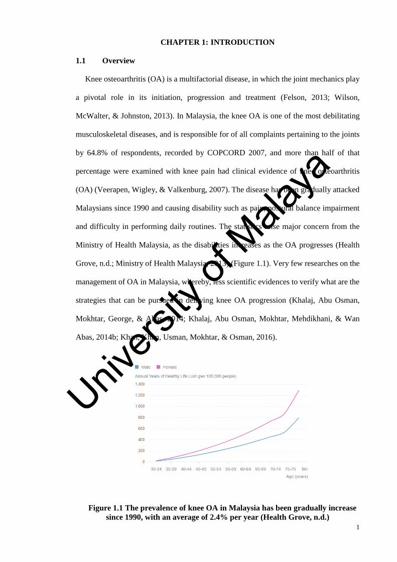
Figure 1.1 The prevalence of knee OA in Malaysia has been gradually increase since
1990, with an average of 2.4% per year (Health Grove, n.d.) .......................................... 1
Figure 2.1: Characteristics of knee OA which include muscle atrophy, synovitis (cause
radiological progression and pain), osteosclerosis and cartilage damage (Egloff et al.,
2012) ................................................................................................................................. 8
Figure 2.2: Visual Analog Scale (VAS) that is used for rating pain (Mannion, Balagué,
Pellisé, & Cedraschi, 2007) ............................................................................................. 13
Figure 2.3: The phases of walking gait. Figure is adopted from
http://advancedhealth.ca/clients/516/images/Chiro_gait_cycle.jpg ................................ 15
Figure 2.4: Graphical illustration for KAM during stance phase (Henriksen et al., 2013)
......................................................................................................................................... 17
Figure 2.5: Illustration for lever arm, and vGRF, resulting in KAM. Figure is adapted
from Turpin et al., (2012) ................................................................................................ 17
Figure 2.6: Sagittal kinematics that often investigated in knee OA researches (Maly,
Costigan, & Olney, 2008) ............................................................................................... 19
Figure 3.1: The method of measuring the knee circumference for the sizes of knee sleeves
......................................................................................................................................... 35
Figure 3.2: Knee sleeves used in the study; a) Simple knee Sleeve (Knee Sleeve A), and
b) Knee sleeve with patella cutout (Knee Sleeve B) (Drytex Basic Knee Sleeve, Donjoy,
USA) ............................................................................................................................... 35
Figure 3.3: Marker placement for this study, following Davis model (Henriksen et al.,
2012) ............................................................................................................................... 38
Figure 3.4: Biodex Stability System used in the study to assess postural ability (Biodex
Medical Systems, 1999) .................................................................................................. 40
Figure 3.5: The foot placement of participants that needs to be set by the investigator
before starting any trial using BSS (Biodex Medical Systems, 1999) ............................ 40
Figure 3.6: Study protocol .............................................................................................. 42
Figure 4.1: Participants involved in the study (following CONSORT Flow Diagram).. 46
Figure 4.2: WOMAC scores for knee pain of 17 participants, for simple knee sleeve (Knee
Sleeve A) and knee sleeve with patella cutout (Knee Sleeve B), respectively. .............. 48
Univers
ity of
Mala
ya
xiii
Figure 4.3: WOMAC scores for knee stiffness of 17 participants, for simple knee sleeve
(Knee Sleeve A) and knee sleeve with patella cutout (Knee Sleeve B), respectively. ... 49
Figure 4.4: WOMAC scores for functional performance of 17 participants, respectively,
for simple knee sleeve (Knee Sleeve A) and knee sleeve with patella cutout (Knee Sleeve
B) ..................................................................................................................................... 49
Figure 4.5: Knee adduction moment (KAM) during stance phase for all the participants.
(NS is the measurement at baseline; S is measurement immediately after knee sleeve use;
6 wks is measurement at six weeks effects) .................................................................... 52
Figure 4.6: OSI for PST (Static) for pre, immediate and post effects according to treatment
groups. Overall [n=17], Simple knee sleeve (Knee Sleeve A) [n=9], Knee sleeve with
patella cutout (Knee Sleeve B) [n=8]. ............................................................................. 57
Figure 4.7: OSI for PST (Dynamic) for pre, immediate and post effects according to
treatment groups. Overall [n=17], Simple knee sleeve (Knee Sleeve A) [n=9], Knee
sleeve with patella cutout (Knee Sleeve B) [n=8]. .......................................................... 57
Figure 4.8: OSI for ASL (Affected Knee - Static) for pre, immediate and post effects
according to treatment groups. Overall [n=17], Simple knee sleeve (Knee Sleeve A)
[n=9], Knee sleeve with patella cutout (Knee Sleeve B) [n=8]. ..................................... 59
Figure 4.9: OSI for ASL (Affected Knee - Dynamic) for pre, immediate and post effects
according to treatment groups. Overall [n=17], Simple knee sleeve (Knee Sleeve A)
[n=9], Knee sleeve with patella cutout (Knee Sleeve B) [n=8]. ..................................... 59
Figure 4.10: OSI for ASL (Unaffected Knee - Static) for pre, immediate and post effects
according to treatment groups. Overall [n=17], Simple knee sleeve (Knee Sleeve A)
[n=9], Knee sleeve with patella cutout (Knee Sleeve B) [n=8]. ..................................... 60
Figure 4.11: OSI for ASL (Unaffected Knee - Dynamic) for pre, immediate and post
effects according to treatment groups. Overall [n=17], Simple knee sleeve (Knee Sleeve
A) [n=9], Knee sleeve with patella cutout (Knee Sleeve B) [n=8]. The asterisk (*) denotes
significant reduction between T0 – T1 effects.................................................................. 60
Figure 4.12: OSI for FRT (Level 12 to Level 8) for pre, immediate and post effects
according to treatment groups. Overall [n=17], Simple knee sleeve (Knee Sleeve A)
[n=9], Knee sleeve with patella cutout (Knee Sleeve B) [n=8] ...................................... 61
Univers
ity of
Mala
ya
xiv
LIST OF TABLES
Table 2.1: Knee OA grading based on ACR classification (Table is adapted from the
Ministry of Health Malaysia, 2013). ................................................................................. 9
Table 2.2: Knee OA grading based on Kellgren-Lawrence classification (Kirkley et al.,
1999). ................................................................................................................................ 9
Table 2.3: Kinematics and kinetics parameters used in the studies ................................ 22
Table 2.4: Studies' characteristics for proprioception and balance ................................. 26
Table 4.1: Brief participants' profiles (n = 17). ............................................................... 46
Table 4.2: WOMAC overall scores (n = 17) ................................................................... 48
Table 4.3: Knee adduction moment (KAM) measurement of the participants for pre,
immediate, and post effects scores .................................................................................. 51
Table 4.4: Vertical Ground Reaction Force (vGRF) summary ....................................... 52
Table 4.5: Kinematics parameters and walking speed results of the participants (n = 17)
......................................................................................................................................... 55
Univers
ity of
Mala
ya

xv
LIST OF SYMBOLS AND ABBREVIATIONS
OA : Osteoarthritis
KOA : Knee Osteoarthritis
BMI : Body Mass Index
ACL : Anterior Cruciate Ligament
KAM : Knee Adduction Moment
ACR : American College of Rheumatology
UMMC : University of Malaya Medical Centre
APSI : Anterior-Posterior Stability Index
MLSI : Medial-Lateral Stability Index
COP : Center of Pressure
vGRF : Vertical Ground Reaction Force
ROM : Range of Motion
WOMAC : Western Ontario McMaster Universities Osteoarthritis Index
K&L : Kellgren-Lawrence
CI : Confidence Interval
CONSORT : Consolidated Standards of Reporting Trials
SD : Standard Deviation
Univers
ity of
Mala
ya
xvi
LIST OF APPENDICES
Appendix A: Medical Ethics 97
Appendix B: Participant Consent Form 98
Appendix C: Participant’s Log Book on Daily Usage 99
Appendix D: WOMAC Score® (Malay version) 104
Appendix E: Patient Information Sheet 110
Appendix F: Lifestyle Questionnaire 113
Appendix G: Results – Participants’ Feedback on Log Book 115
Appendix H: Results – WOMAC on Knee Pain 116
Appendix I: Results – WOMAC on Knee Stiffness 117
Appendix J: Results – WOMAC on Knee Functional Performance 118
Appendix K: Results – WOMAC Overall Score 119
Appendix L: Results – Postural Stability Test (PST) Static 120
Appendix M: Results – Postural Stability Test (PST) Dynamic 121
Appendix N: Results – Athlete Single Leg Test (ASL) Static – Affected
Knee
122
Appendix O: Results – Athlete Single Leg Test (ASL) Dynamic – Affected
Knee
123
Appendix P: Results – Athlete Single Leg Test (ASL) Static – Unaffected
Knee
124
Appendix Q: Results – Athlete Single Leg Test (ASL) Dynamic –
Unaffected Knee
125
Appendix R: Results – Fall Risk Test (FRT) 126
Appendix S: Results – Gait Parameters 127
Appendix T: Method – Plug-In-Gait Modelling (using Vicon Nexus 2.5.1) 136
Univers
ity of
Mala
ya
1
CHAPTER 1: INTRODUCTION
1.1 Overview
Knee osteoarthritis (OA) is a multifactorial disease, in which the joint mechanics play
a pivotal role in its initiation, progression and treatment (Felson, 2013; Wilson,
McWalter, & Johnston, 2013). In Malaysia, the knee OA is one of the most debilitating
musculoskeletal diseases, and is responsible for of all complaints pertaining to the joints
by 64.8% of respondents, recorded by COPCORD 2007, and more than half of that
percentage were examined with knee pain had clinical evidence of knee osteoarthritis
(OA) (Veerapen, Wigley, & Valkenburg, 2007). The disease has been gradually attacked
Malaysians since 1990 and causing disability such as pain, postural balance impairment
and difficulty in performing daily routines. The statistics raise major concern from the
Ministry of Health Malaysia, as the disabilities increases as the OA progresses (Health
Grove, n.d.; Ministry of Health Malaysia, 2013) (Figure 1.1). Very few researches on the
management of OA in Malaysia, whereby, less scientific evidences to verify what are the
strategies that can be pursued in delaying knee OA progression (Khalaj, Abu Osman,
Mokhtar, George, & Abas, 2014; Khalaj, Abu Osman, Mokhtar, Mehdikhani, & Wan
Abas, 2014b; Khan, Khan, Usman, Mokhtar, & Osman, 2016).
Figure 1.1 The prevalence of knee OA in Malaysia has been gradually increase
since 1990, with an average of 2.4% per year (Health Grove, n.d.)
Univers
ity of
Mala
ya

2
Knee OA also known as a disease of mechanics but not many studies have been
conducted on the biomechanical characteristics of OA. Interestingly, patients with the
disease have abnormal gait parameters such as reduced knee flexion (Al-Zahrani &
Bakheit, 2002; Andriacchi & Mündermann, 2006), increased knee adduction moment
(KAM) (Landry, McKean, Hubley-Kozey, Stanish, & Deluzio, 2007), and greater
impulsive forces (Liikavainio et al., 2016). These abnormalities are believed to have
adverse effects on knee joint loading (Lewek, Rudolph, & Snyder-Mackler, 2004).
However, correction or rectification of these abnormal biomechanics has been successful
in mitigating the disease progression and alleviating pain – the most common and
disabling symptom in knee OA (Bennell et al., 2011).
Many treatment approaches have been used to help improve functionality in knee OA
patients, including the use of orthoses (Johann Beaudreuil et al., 2009). Orthoses such as
knee braces, foot insoles and knee sleeves are widely used – especially for joint support
and compression (Wilson, Mazahery, Koh, & Zhang, 2010). Knee sleeves are often used
to assist and stabilize movements leading to pain reduction in the joints. Besides, knee
sleeves provide better biomechanical balance between the joint structures, and
consequently help in reducing pain (Bryk et al., 2011).
The effects of knee sleeves are mainly functional and neuromuscular in nature
because they do not usually offer rigid support and therefore their physical restraining
effects on the skeletal system are minimal. Knee sleeves are made from sock-like elastic
material which provides compression and warmth to the targeted area to improve
functional performances (Sasek, 2015). It is relatively cheaper, lighter and less rigid than
knee brace, thus, making it a popular option for patients with knee problems. Moreover,
there has been no report on serious side effects attributed to knee sleeves, which indicates
that they are relatively safe.
Univers
ity of
Mala
ya

3
Knee sleeves have been shown to have various clinical effects such as pain relief,
improved proprioception, functional performances of the knee, and stability (Bryk et al.,
2011; Chuang et al., 2007; Collins et al., 2012). However, the benefits of knee sleeves
from the biomechanical aspects are less clear. Some researchers believed that the
compression exerted by knee sleeves stimulates the mechanoreceptors around the knee
joint, leading to improvement in proprioception and balance (Bottoni, Herten, Kofler,
Hasler, & Nachbauer, 2013; Ramsey, Briem, Axe, & Synder-Mackler, 2011; Wilson et
al., 2010). Although knee sleeves are generally found to be beneficial for knee OA, it is
unclear which type of sleeves confer the best clinical and biomechanical benefits.
There have not been many studies on the effects of knee sleeves pertaining to knee
forces and loading in knee OA, probably because of the elastic nature of the sleeves
(Collins et al., 2014; Collins, Blackburn, Olcott, Yu, & Weinhold, 2011; Giotis et al.,
2011; Schween, Gehring, & Gollhofer, 2015). In their study, Schween et al. used knee
adduction moment (KAM) to represent medial joint loading, and found that knee sleeves
can significantly reduce joint loading by 10.1%, immediately after application. Since
most of these studies investigated only the immediate effects of the sleeve use, the long-
term benefits of knee sleeve use is still unclear. Practically, it is important to establish if
the effect of knee sleeve persist beyond immediate application in order to justify its
extended use in patients with knee OA.
1.2 Problem Statement
Many treatment approaches that have been introduced to alleviate the pain have not
proven to be satisfactory. Knee sleeves have been widely prescribed to alleviate the pain,
but with limited evidences. Pain management is vital for knee OA individuals as pain
causes difficulty doing daily routines. The studies that have been conducted on knee
Univers
ity of
Mala
ya
4
sleeves use have thus far focused on the immediate effects and without comparing
different designs of knee sleeve.
The research intends to answer the following questions: 1) Can knee sleeves help to
reduce pain in people with early unilateral knee OA in six weeks? 2) Can knee sleeves
improve postural stability in people with early unilateral knee OA in six weeks? 3) Do
knee sleeves change the knee joint mechanics in people with early unilateral knee OA in
six weeks? And 4) Do the different types of knee sleeves produce different effects?
1.3 Objective
This study is undertaken: 1) to determine the biomechanical effects of knee sleeves on
early unilateral knee OA following six weeks of application, and 2) to determine the
difference in the effects for two different types of knee sleeves. To achieve the objectives,
our primary outcome is the patient-centered parameter – pain, stiffness, and difficulty
performing daily activities. Our secondary outcome - the biomechanical effect comprises
of: 1) Gait parameters – knee adduction moment (KAM), ground reaction force (GRF),
knee sagittal plane kinematics, and walking speed; and 2) Postural stability – Single-
stance standing balance and dual-limb standing balance.
This study will investigate whether knee sleeves could modify any of the
biomechanical parameters to achieve the following aims: 1) to help clinicians
comprehend the indications and properties of the knee sleeves, and 2) to help fill the gap
on information pertaining to the biomechanical aspects of knee sleeves. If proven to be
effective, knee sleeves could be used in patients with early knee OA to alleviate pain.
We chose a six-week testing duration as this is the generally accepted duration for any
academic study and is generally longer than immediate duration (Birmingham et al.,
Univers
ity of
Mala
ya
5
2008; Hunter et al., 2011). Besides, we are considering the logistics of the project: short
duration for project, and no administrative assistance in managing the patient's affairs.
1.4 Hypothesis Statement
In this study, we aim to determine the effects of knee sleeves of two different designs
on the knees of people with early knee OA based on three aspects – pain, gait, and postural
stability. In this context, we have formulated the following hypotheses to guide us in
answering questions pertaining to the efficiency of knee sleeves used in knee
osteoarthritis:
Hypothesis 1 – pertaining to pain in knee OA:
H0: The application of the knee sleeves would not alleviate pain, reduce knee
stiffness and improve knee functional performance;
H1: The application of the knee sleeves would alleviate pain, reduce knee stiffness
and improve knee functional performance.
Hypothesis 2 – pertaining to gait in knee OA:
H0: The application of the knee sleeves would not increase walking speed, decrease
ground reaction force (GRF) loading rates and KAM in people with knee OA;
H1: The application of the knee sleeves would increase walking speed, decrease
ground reaction force (GRF) loading rates and KAM in people with knee OA.
Hypothesis 3 – pertaining to postural stability:
H0: The application of the knee sleeves would not improve postural stability in
people with knee OA;
Univers
ity of
Mala
ya
6
H1: The application of the knee sleeves would improve postural stability in people
with knee OA.
1.5 Dissertation Structure
This dissertation consists of six chapters.
Chapter 1 provides a general overview of this dissertation and discusses the
background of the study, objectives, problem statement and the hypotheses formulated
for the study. Chapter 2 presents the literature review pertaining to knee sleeves use in
treatment of knee osteoarthritis, their limitations, and findings. Chapter 3 is the
methodology used in the study, which include discussion on the recruitment of
participants for the study, and the various processes involved. Chapter 4 presents the
results and discusses the findings vis-à-vis the hypotheses. Chapter 5 discusses the study
findings presented in Chapter 4 and focuses on the core aspects of the study that include
the kinetics and kinematics parameters, postural stability, knee pain, knee stiffness, and
knee functional performance. Chapter 6 presents the conclusion to the study, and
highlights the problems encountered in the study, the contribution of the study, and
suggestions for future researches.
1.6 Summary
This chapter introduced the reader to the main message, aim, and objectives of this
dissertation. Univers
ity of
Mala
ya

7
CHAPTER 2: LITERATURE REVIEW
2.1 Introduction
This chapter discusses various findings from the literature review on the topics
relevant to this research. It includes background on knee osteoarthritis (OA) and the
management strategies in dealing with the pain and other symptoms associated with the
disease. Subsection 2.2 discusses the definitions of OA; subsection 2.3 discusses the
management and treatment of knee OA; subsection 2.4 discusses the orthoses for knee
OA; subsection 2.5 discusses the studies on knee sleeves use; and subsection 2.6
summarizes the whole chapter.
2.2 Osteoarthritis: Definition
Osteoarthritis (OA) is a multifactorial disease in which the joints of the body become
damaged and painful. The prevalence of knee OA in Malaysia was estimated to be 10%
to 20% of the elderly population and is said to affect mainly female, with a male to female
ratio of 2:3 among Malays, 1:1 among Chinese, and 3:6 among Indians, respectively and
the ratio increases gradually every year (Foo et al., 2017; Fransen et al., 2011). Knee OA
results from the breakdown of the tissues of the knee joint (UK Arthritis Research, 2013).
Joint abnormalities such as cartilage degradation, muscle weakening, inappropriate
mechanical stress and ligament tear are some of the risk factors which initiate the disease
(Figure 2.1) (Egloff, Hügle, & Valderrabano, 2012). Besides, there are several other
factors that could affect the progress of the disease.
There are some non-modifiable risk factors such as advancing age, gender and genetic
influence. The female has a higher likelihood of having arthritis due to the wider range
of motion of the pelvis and the hip (Kaufman, Hughes, Morrey, Morrey, & An, 2001;
McKean et al., 2007). As for age, there is higher prevalence of knee OA in the older
population due to the decrease in bone strength and density (Bagge, Bjelle, Edén, &
Univers
ity of
Mala
ya

8
Svanborg, 1991). Genetic influences that lead to abnormal joint morphology, and obesity
or injuries can amplify the effect of abnormal mechanical stress (Guilak, 2011).
Figure 2.1: Characteristics of knee OA which include muscle atrophy, synovitis
(cause radiological progression and pain), osteosclerosis and cartilage damage
(Egloff et al., 2012)
The modifiable risk factors include previous knee injury, obesity (BMI >30 kg/m2),
overweight BMI (BMI 25 – 30 kg/m2), and malalignment of the knee (Ministry of Health
Malaysia, 2013).
Patients are diagnosed to have clinical knee OA if they fulfill the American College of
Rheumatology (ACR) 1986 Criteria that include both clinical and radiographic criteria
and a myriad of symptoms that include knee pain; osteophytes seen on x-ray; at least 50
years old; and knee stiffness of less than 30 minutes, or crepitus (Table 2.1). Kellgren-
Lawrence (K&L) Grading System is a commonly-used radiographic classification to
identify and grade the severity of radiographic OA (Kellgren & Lawrence, 1957) (Table
2.2). The World Health Organization (WHO) adopted the grading as the standard for
epidemiological studies of OA. The reliability of K&L grading system showed good to
very good reliability (K between 0.6 and 0.8 and above 0.8) (Schiphof, de Klerk, Koes,
& Bierma-Zeinstra, 2008).
Univers
ity of
Mala
ya

9
Pain and stiffness of the knee joint are the primary indicators of the presence of knee
OA and are usually very mild at the beginning and progresses as the disease increases in
severity (Foxworth, 2007). The pain and stiffness are often much worse when the patient
rises from a seated position. Patients with knee OA also often present with inflammation
and swelling on the knee joint. Besides, the other minor symptoms include gait
disturbance, clicking or grinding sensation on the arthritic joint, and also instability
(Ministry of Health Malaysia, 2013).
Table 2.1: Knee OA grading based on ACR classification (Table is adapted from
the Ministry of Health Malaysia, 2013).
Table 2.2: Knee OA grading based on Kellgren-Lawrence classification (Kirkley
et al., 1999).
Grade Narrowing of
Joint Space
Osteophytes Sclerosis Deformation of
Joint Contour
I Doubtful Possible lipping None None
II Possible Definite None None
III Definite Moderate,
multiple
Some Possible
IV Marked Large Severe Definite
Univers
ity of
Mala
ya

10
2.3 Management on Knee OA and the Challenges
Owing to the irreversible nature of knee OA, physicians focus their efforts on
arresting the functional decline of the osteoarthritic knee, reducing pain, and thus, giving
quality of life to their patients. The treatment approaches include both pharmacological
and non-pharmacological interventions, and surgery in severe cases (Egloff et al., 2012;
Sasek, 2015). Today, surgical intervention is recommended in severe cases.
The most common approach to manage knee OA is to combine pharmacological
therapy and non-pharmacological approaches (Foxworth, 2007). Pharmacological
treatment involves oral treatment which consists of simple analgesics such as
paracetamol, and also nutraceuticals such as glucosamine and chondroitin. Chondroitin
sulphate and glucosamine sulphate may be beneficial as modifying agents while
analgesics are mainly used to control pain (Bijlsma & Knahr, 2007).
Non-pharmacological treatment involves education of knee OA patients regarding the
disease itself, self-management and on how to cope with the pain and disabilities. Obese
patients are also advised to reduce their weight as this helps to minimize joint loading,
especially on the knee (Christensen, Bartels, Astrup, & Bliddal, 2006; Guilak, 2011; M
Henriksen et al., 2013). Khalaj et al. (2014) recommended exercise as an effective way
in reducing pain, reducing body weight, improving mobility, and improving muscle
strength.
Other than exercises, the European League Against Rheumatism (EULAR)
recommends that patients with knee OA should use external assistive devices such as
knee brace, knee sleeve and also orthosis when indicated (Jordan et al., 2003). Beaudreuil
et al. (2009) systematically reviewed the clinical guidelines on OA management and
found that one-quarter to one-third of the physicians are likely to prescribe knee sleeves,
while 65% to 74% are unlikely to do so.
Univers
ity of
Mala
ya

11
2.4 Orthosis for Knee OA
Orthotic devices are used to give external strength and support the joint, align
deformities or improve function of movable parts of the body. The main purpose of
prescribing these devices in OA is primarily to reduce pain, improve physical function
and, hopefully, delay disease progression. Insoles, elastic knee brace, rigid knee brace,
knee sleeve, and knee orthosis are often been prescribed by health professionals as a
management of knee OA (Beaudreuil et al., 2016; Beaudreuil et al., 2009; Jones et al.,
2013). Knee brace is effective in altering the deformities of the knee, depending on the
varus or valgus alignment of the device, by getting the knee back to its neutral position –
in a way, to unload the excessive forces on the knee. For insoles, it reduced KAM but no
lessening in pain (Jones, Chapman, Forsythe, Parkes, & Felson, 2014). But, an extended
usage of laterally-wedged insoles for one-month is effective in reducing pain (Turpin et
al., 2012). Knee brace and foot insoles have been extensively experimented for a longer
term – from eight-week to 12-month longitudinal studies (Hinman et al., 2008; Hurley,
Murdock, Stanish, & Hubley-Kozey, 2012; Knoop et al., 2013; Toda & Tsukimura, 2004;
Turpin et al., 2012).
While there are good quality evidence to support the positive clinical outcomes of knee
braces, the laboratory evidence that demonstrates beneficial biomechanical outcomes of
knee sleeves is still not fully explored and unconvincing (Johann Beaudreuil et al., 2009;
Moyer et al., 2015). Beaudreuil et al. also stated that clinicians and consumers tend to use
the knee sleeves indiscriminately, hence, it is important to assess the effects of knee
sleeves more objectively.
Knee braces have been well investigated in high quality randomized controlled studies,
while knee sleeves or rest orthosis have not been much researched (Johann Beaudreuil et
al., 2009). This could be due to the assumption that the more rigid knee brace would be
Univers
ity of
Mala
ya

12
more likely to alter abnormal joint mechanics than the flexible knee sleeve (Mohd Sharif,
Goh, Usman, & Wan Safwani, 2017). However, the bulkiness of the brace can cause
discomfort and inconvenience to the user and from a review, in longer follow-up studies
(1 to 2 years) many patients stopped their brace or insole treatment (Bottoni et al., 2013;
Brouwer et al., 2009). For some, the simple and flexible knee sleeves then become the
more practical and more acceptable treatment option.
However, it is difficult to conclude whether knee sleeves have beneficial effects
because of the limited research evidences. Based on current evidence, knee sleeves are
effective in relieving pain in knee osteoarthritis, and they have been generally associated
with effecting subjective improvement to the disease (Johann Beaudreuil et al., 2009).
They also effect improvement in gait parameters and balance control, but no improvement
in knee alignment alteration. Therefore, Beaudreuil et al. suggested more comprehensive
investigations on the knee sleeves and their effects on knee OA.
2.5 Past Studies on Knee Sleeves
From the literature review covering the period 2005 until 2015, it was found that seven
studies had reported the use of knee sleeves for knee OA (Bryk et al., 2011; Chuang et
al., 2007; Collins et al., 2010, 2011, 2012, 2014; Schween et al., 2015). In these studies,
various assessment parameters were used and duration of the tests were generally or
immediate.
2.5.1 Clinical Assessment on Knee Sleeves
Apart from the biomechanical parameters, pain experienced by patients is used to
determine the efficacy and effectiveness of knee sleeves. This subsection will also discuss
some of the adverse effects of knee sleeves reported in past studies.
Univers
ity of
Mala
ya
13
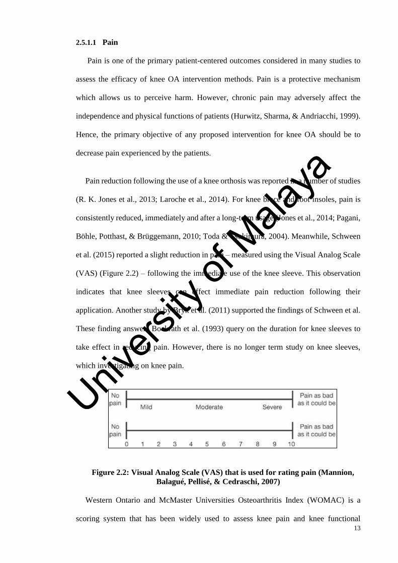
2.5.1.1 Pain
Pain is one of the primary patient-centered outcomes considered in many studies to
assess the efficacy of knee OA intervention methods. Pain is a protective mechanism
which allows us to perceive harm. However, chronic pain may adversely affect the
independence and physical functions of patients (Hurwitz, Sharma, & Andriacchi, 1999).
Hence, the primary objective of any proposed intervention for knee OA should be to
decrease pain experienced by the patients.
Pain reduction following the use of a knee orthosis was reported in a number of studies
(R. K. Jones et al., 2013; Laroche et al., 2014). For knee brace and foot insoles, pain is
consistently reduced, immediately and after a long-term usage (Jones et al., 2014; Pagani,
Böhle, Potthast, & Brüggemann, 2010; Toda & Tsukimura, 2004). Meanwhile, Schween
et al. (2015) reported a slight reduction in pain – measured using the Visual Analog Scale
(VAS) (Figure 2.2) – following the immediate use of the knee sleeve. This observation
indicates that knee sleeves can effect immediate pain reduction following their
application. Another study by Bryk et al. (2011) supported the findings of Schween et al.
These finding answers Bockrath et al. (1993) query on the duration for knee sleeves to
take effect in reducing pain. However, there is no longer term study on knee sleeves,
which investigating on knee pain.
Figure 2.2: Visual Analog Scale (VAS) that is used for rating pain (Mannion,
Balagué, Pellisé, & Cedraschi, 2007)
Western Ontario and McMaster Universities Osteoarthritis Index (WOMAC) is a
scoring system that has been widely used to assess knee pain and knee functional
Univers
ity of
Mala
ya
14
performance in OA (Jones et al., 2013; Laroche et al., 2014; Pagani et al., 2010). Higher
WOMAC score indicates more severe knee pain, stiffness, and other physical
dysfunctions (Turpin et al., 2012). The WOMAC score is derived from the VAS scale
(Figure 2.2), in which in a 10-cm line (scale), the initial number 0, positioned at the left
end of the line, indicates ‘no pain’ and a final number 10, positioned at the right end of
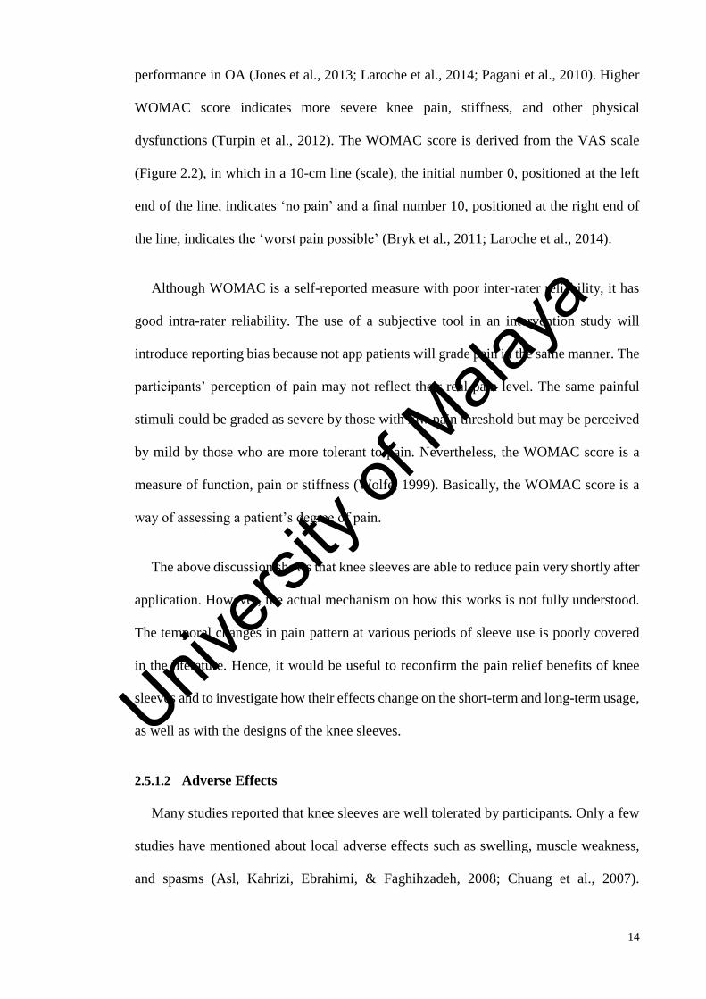
the line, indicates the ‘worst pain possible’ (Bryk et al., 2011; Laroche et al., 2014).
Although WOMAC is a self-reported measure with poor inter-rater reliability, it has
good intra-rater reliability. The use of a subjective tool in an intervention study will
introduce reporting bias because not app patients will grade pain in the same manner. The
participants’ perception of pain may not reflect their real pain level. The same painful
stimuli could be graded as severe by those with low pain threshold but may be perceived
by mild by those who are more tolerant to pain. Nevertheless, the WOMAC score is a
measure of function, pain or stiffness (Wolfe, 1999). Basically, the WOMAC score is a
way of assessing a patient’s degree of pain.
The above discussion shows that knee sleeves are able to reduce pain very shortly after
application. However, the actual mechanism on how this works is not fully understood.
The temporal changes in pain pattern at various periods of sleeve use is poorly covered
in the literature. Hence, it would be useful to reconfirm the pain relief benefits of knee
sleeves and to investigate how their effects change on the short-term and long-term usage,
as well as with the designs of the knee sleeves.
2.5.1.2 Adverse Effects
Many studies reported that knee sleeves are well tolerated by participants. Only a few
studies have mentioned about local adverse effects such as swelling, muscle weakness,
and spasms (Asl, Kahrizi, Ebrahimi, & Faghihzadeh, 2008; Chuang et al., 2007).
Univers
ity of
Mala
ya
15
Moreover, there has been no report that knee sleeve has caused any serious discomfort to
participants.
2.5.2 Gait
Studies have also been conducted on the biomechanical effects of knee sleeves.
Excessive loading on the knee has been acknowledged as the main cause for the onset of
knee OA. By enhancing the proprioceptive ability and also decreasing the loading rate on
the knee, knee sleeves could aid in altering gait and delaying disease progression (Collins
et al., 2011; Van Tiggelen, Coorevits, & Witvrouw, 2008). In this section, we focused on
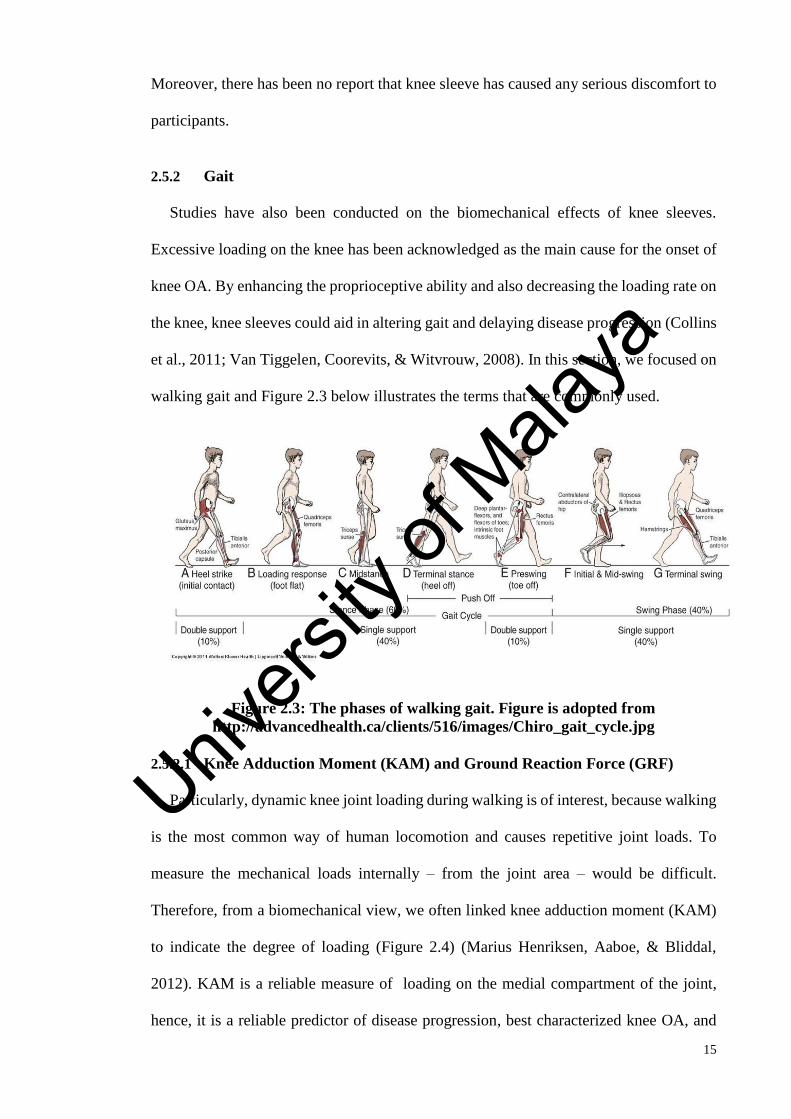
walking gait and Figure 2.3 below illustrates the terms that are commonly used.
Figure 2.3: The phases of walking gait. Figure is adopted from
http://advancedhealth.ca/clients/516/images/Chiro_gait_cycle.jpg
2.5.2.1 Knee Adduction Moment (KAM) and Ground Reaction Force (GRF)
Particularly, dynamic knee joint loading during walking is of interest, because walking
is the most common way of human locomotion and causes repetitive joint loads. To
measure the mechanical loads internally – from the joint area – would be difficult.
Therefore, from a biomechanical view, we often linked knee adduction moment (KAM)
to indicate the degree of loading (Figure 2.4) (Marius Henriksen, Aaboe, & Bliddal,
2012). KAM is a reliable measure of loading on the medial compartment of the joint,
hence, it is a reliable predictor of disease progression, best characterized knee OA, and
Univers
ity of
Mala
ya

16
closely related to pain, walking speed and body mass OA (Baert et al., 2013; Creaby,
Bennell, & Hunt, 2012; Khalaj et al., 2014b; Levinger et al., 2012).
Reducing KAM has become the objective of early and conservative treatment
approaches in attempting to reduce pain, maintaining function and arresting disease
progression (Heiden, Lloyd, & Ackland, 2009; R. K. Jones et al., 2013). Often, increased
KAM is the cause of pain in patients with knee OA (Thorp et al., 2003). In another study,
Heiden et al. suggested that pain is a protective mechanism that leads to a self-selected
reduction in KAM during gait and is inversely correlated with KAM. As such, some
treatment approaches involve modifying gait in order to minimize KAM and to alleviate
pain in knee OA. These include reducing gait speed and applying toe-out gait (Farrokhi,
O’Connell, Gil, & Kelley Fitzgerald, 2013; Jenkyn, Erhart, & Andriacchi, 2011). As well
as orthosis, the devices – knee brace, knee supporters, foot insoles – are meant to unload
the medial joint loading (Deshaies, 2002). Number of studies identified the positive
effects of knee braces and foot insoles, and two studies compared the effects between
these two orthoses and found that foot insole is more effective in reducing knee moments
and is comfortable to be used by knee OA individuals (Dessery, Belzile, Turmel, &
Corbeil, 2014; Jones et al., 2013; Lamberg, Streb, Werner, Kremenic, & Penna, 2015;
Maleki et al., 2014; Shelburne, Torry, Steadman, & Pandy, 2008).
For knee sleeve, from our literature search, only two studies were investigated the
effects on gait – focusing on frontal and sagittal plane kinematics and kinetics. The studies
reported the positive effect of knee sleeves on KAM and frontal plane kinematics during
walking following the immediate use of knee sleeves (Collins et al., 2014; Schween et
al., 2015). Schween et al. reported that KAM is significantly reduced by 10.1%, while
Collins et al. found that KAM is reduced, but not significantly. They also reported
improvements on knee adduction angle and found significant reduction during terminal
Univers
ity of
Mala
ya

17
stance while Schween et al. obtained similar result during walking while wearing the knee
sleeves.
Figure 2.4: Graphical illustration for KAM during stance phase (Henriksen et al.,
2013)
Figure 2.5: Illustration for lever arm, and vGRF, resulting in KAM. Figure is
adapted from Turpin et al., (2012)
In addition, KAM was also reported to be closely correlated with the loading rate or
the ground reaction force (GRF). However, the projection of GRF may deviate from the
center of the body due to malalignment. Therefore, when there is an increase in the GRF,
combined with an increased lever-arm distance between the knee joint center and the GRF
vector, they will result in higher KAM (Figure 2.5) (Duffell, Southgate, Gulati, &
McGregor, 2014; Turpin et al., 2012). Hence, reducing KAM is aimed at reducing pain,
vGRF
Knee lever arm KAM
Univers
ity of
Mala
ya

18
maintaining function and arresting disease progression, and similarly, reducing GRF
would also bring the same benefits to knee OA patients (Jones et al., 2013).
Collins et al. (2011) applied additional stochastic electrical stimulation to the knee
sleeve, and found significant reduction in the GRF. This finding shows that electrical
stimulation applied to the knee sleeves can change the GRF. However, in all these studies,
only the immediate effects of the knee sleeve use were observed. To date, there has been
no study on the effects of long-term use of knee sleeves on gait biomechanics.
2.5.2.2 Knee Extension and Walking Speed
GRF is correspondingly highly affected by the initial contact of the heel. Greater
flexion angle could also be an attempt to stabilize the joint to lessen knee pain. This
additional knee flexion serves as a shock absorption mechanism as the body weight is
transferred from the opposite limb (Foxworth, 2007). However, this claim is
controversial. Another study reported that high knee flexion during heel strike may create
larger GRF projection. The study suggested that to reduce knee medial loading, knee
flexion during heel strike must be smaller to generate smaller GRF (Paquette, 2012).
Therefore, heel repositioning during heel strike must be carefully considered in defining
knee flexion at contact and also the GRF.
The conflicting findings can be resolved by considering them from the biomechanical
aspect. When the knee flexes within a certain range, it will immediately transfer the load.
Schipplein and Andriacchi (1991) found that knee flexion ranging from 35o to 40o would
reduce the impact of GRF due to the gradual deceleration of the vertical velocity.
Riskowski, Mikesky, Bahamonde, Alvey, and Burr (2005) also stated that large GRF may
cause faster degradation of the cartilage, and this will quicken the thinning of the
meniscus over time.
Univers
ity of
Mala
ya
19
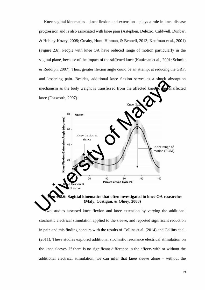
Knee sagittal kinematics – knee flexion and extension – plays a role in knee disease
progression and is also associated with knee pain (Astephen, Deluzio, Caldwell, Dunbar,
& Hubley-Kozey, 2008; Creaby, Hunt, Hinman, & Bennell, 2013; Kaufman et al., 2001)
(Figure 2.6). People with knee OA have reduced range of motion particularly in the
sagittal plane, because of the impact of the stiffened knee (Kaufman et al., 2001; Schmitt
& Rudolph, 2007). Thus, greater flexion angle could be an attempt at reducing the GRF,
and lessening pain. Besides, additional knee flexion serves as a shock absorption
mechanism as the body weight is transferred from the affected knee to the unaffected
knee (Foxworth, 2007).
Figure 2.6: Sagittal kinematics that often investigated in knee OA researches
(Maly, Costigan, & Olney, 2008)
Two studies assessed knee flexion and knee extension by varying the additional
stochastic electrical stimulation applied to the sleeve, and reported significant reduction
in pain and this finding concurs with the results of Collins et al. (2014) and Collins et al.
(2011). These studies explored additional stochastic resonance electrical stimulation on
the knee sleeves. If there is no significant difference in the effects with or without the
additional electrical stimulation, we can infer that knee sleeve alone – without the
Knee flexion at
heel strike
Knee flexion at
stance
Knee flexion at
swing
Knee range of
motion (ROM)
Univers
ity of
Mala
ya

20
additional stochastic electrical stimulation – is beneficial to those with knee OA or knee-
related problems.
It was found that test subjects exhibit slower walking speed during the pain adaptation
stage (da Silva, Cliquet Junior, Zorzi, & Batista de Miranda, 2012). Walking speed clearly
represents the health level of a person and it is inversely correlated with the pain levels
(Astephen Wilson, Deluzio, Dunbar, Caldwell, & Hubley-Kozey, 2011). In fact, walking
at a reduced speed has been found to be the best method for reducing adduction moment
(Mündermann, Dyrby, Hurwitz, Sharma, & Andriacchi, 2004). Also, from walking speed,
we should also assess the pain level. Since pain is a protective mechanism, people tend to
walk slower in a painful condition. However, in the only study that assessed gait velocity
in conjunction with the usage of knee sleeve, Collins et al. (2011) found that there is no
significant difference in the mean forward velocity between the treatment group (with
stochastic electrical stimulation) and the control group (no stochastic electrical
stimulation). If knee sleeves can help people to improve their gait speed, then it can be
inferred that they can also help to improve knee functions and postural stability.
Therefore, further studies on the long-term use of knee sleeves would give clearer and
more substantive findings of their benefits.
2.5.2.3 The Gait Analysis
Many of the studies on motion analysis used the universally recommended Helen
Hayes (Davis model) marker placement protocol (Davis, 1988; Giotis et al., 2011;
Schween et al., 2015). However, other studies did not specify their markings (Collins et
al., 2014; Collins et al., 2011). The optimum sampling rates ranged from 100Hz to 200Hz
for kinematics, and 1,000Hz to 1,440Hz for kinetics. The parameters used are presented
in Table 2.3.
Univers
ity of
Mala
ya

21
It is advisable to adopt the Davis model for marker placement and at the same time
provide more cameras for motion capture. If there are too few cameras, the distance
between cameras may lead to lower image resolution and inaccuracy in detecting the
markers in some frames (Kirtley, 2006). These studies, however, involved only one
design of knee sleeve, thus, further research using different knee sleeve designs would be
useful.
Univers
ity of
Mala
ya

22
Table 2.3: Kinematics and kinetics parameters used in the studies
Author,
Year
Motion
Capture
System
Procedure/Protocol Results
Collins
et al.
(2013)
MotionStar
electromagnetic
tracking system
Walking speed is
controlled. Walking,
5 trials, barefooted
Test duration:
Immediate
Knee adduction moment: 0.84%
reduced to 0.80% (with 75%
electrical stimulation applied),
no significant difference
Knee adduction angle: Reduced
from 3.9o ±5.6 to 2.9o ±6.4
(weight acceptance)
Reduced from 5.0o ±5.7 to 4.6o
±6.3 (mid-stance), no significant
difference
Knee flexion angle: Significantly
increased from 25.4o ±10.0 to
26.8o ±9.1 (weight acceptance)
and from 5.0o ±5.7 to 4.6o ±6.3
(mid-stance)
Schween
et al.
(2015)
Vicon V-mx Walking speed is
controlled. Walking,
10 trials, using
special shoe
Test duration:
Immediate
Knee adduction moment:
Significantly reduced (p < 0.05)
from 0.854 to 0.780 Nm•kg-1
(10.1% reduction)
Knee adduction impulse:
Significantly reduced (p < 0.05)
from 0.243 to 0.219Nm•s•kg-1
(12.9% reduction)
Knee adduction angle:
Significantly reduced (p < 0.05)
from 11.5o to 9.6o
Collins
et al.
(2011)
i. Flock of Birds
ii. electromagnetic
tracking system
iii. Walking speed is
controlled. Walking,
5 trials, barefooted
Test duration:
Immediate
Knee flexion angle: E75:S
increased from 12.40o ±8.28 to
14.67o ±8.13 (Significant
improvement in NE:S and E75:S
(p < 0.05))
Giotis et
al.
(2011)
Vicon No reported walking
speed. Doing
activities: jumping
and landing
Test duration:
Immediate
Tibial rotation: Descending:
Reduced from 17.1o ±7.7 to
16.1o ±4.5. Ascending: Reduced
from 14.0o ±3.3 to 12.2o ±3.5
(No significant difference)
NE:S : no electrical stimulation applied to knee sleeve; E75:S: 75% electrical stimulation applied to knee
sleeve
Univers
ity of
Mala
ya

23
2.5.3 Balance and Postural Stability
Aside from gait parameters, intervention study on knee OA patients also often assesses
their postural stability. This is because these patients often experience balancing
impairment and knee instability probably due to the decreased sensitivity of the
proprioception and somatosensory receptors resulting in limited functional independence
(Elbaz et al., 2010; Khalaj, Abu Osman, Mokhtar, Mehdikhani, & Wan Abas, 2014a;
O’Connell, Farrokhi, & Kelley Fitzgerald, 2015). This dysfunctional condition could lead
to injuries following a fall and aggravate any existing balancing problem of these patients.
Besides, knee OA patients often have impairment of proprioceptive acuity or weakness
in the quadriceps muscles when compared with those without knee OA. These are the
consequences of instability in those with knee OA (Park, Ko, Hong, Ok, & Lee, 2013).
As equally important as pain, knee instability also causes significant disability for
people with knee OA in performing their daily activities such as climbing stairs, bending
and reaching for things, and walking. In Malaysia, people tend to do outdoor activities
such as outdoor exercises, playing tennis and badminton (Eng Hoe, 2009). Therefore,
knee support is highly needed to overcome the difficulty. To date, however, there has not
been much attention on the sensorimotor control parameters of patients with early knee
OA. For knee brace, significant improvement in proprioception has been reported, but no
significant changes in postural control in flat surface, after an immediate use of knee brace
(Birmingham et al., 2001). Another study found In the literature search, there were four
studies on knee sleeve use in knee OA, which specifically focused on knee proprioception
and balance (Bryk et al., 2011; Chuang et al., 2007; Collins et al., 2010; Collins et al.,
2012) (Table 2.4).
Chuang et al. (2007) reported significant reduction in the stability index in both static
and dynamic balance tests after patients wore the sleeves. Reduction in the stability index
Univers
ity of
Mala
ya

24
denotes improvement in postural stability. They recorded 28% and 7% to 8%
improvement in static balance and dynamic balance, respectively. Their findings were
supported by Schween et al. (2015) who measured instability using self-stability
approach, and reported significant immediate improvement in postural stability following
the use of knee sleeve. Only one study used sway velocity and reported no significant
difference in the sagittal and coronal planes despite using five different levels of
stochastic electrical stimulation (Collins et al., 2012).
Sanchez-Ramirez et al. (2013) reported that postural stability index can help
physicians in assessing the muscle strength of the lower extremities as they represent the
balance ability. Thus, instrumented balance systems have been widely used in hospitals
and rehabilitation centers to monitor the progress of the patients’ balancing ability. An
instrumented balance system can be set to several test protocols and to several dynamic
levels. The system also provides visual feedback to ensure that patients control their
balance in the same way they control their balance on the real surface. Measuring balance
using a balance system is relatively easy and produces reliable results (Karimi, Ebrahimi,
Kahrizi, & Torkaman, 2008).
Postural stability can be tested dynamically and statically. Dynamic tests assess
balance control during voluntary movement, such as walking or rising from a chair. Static
tests assess the ability to maintain an upright position under varied situations, such as with
the eyes closed, or with expected or unexpected disturbance in motion (Hassan, Mockett,
& Doherty, 2001). Many past studies considered these two conditions in balance
assessment in order to obtain more cogent findings for these two conditions, and to have
a better understanding of the postural stability of the patients (Giuliamarta Bottoni,
Kofler, Hasler, Giger, & Nachbauer, 2014; Chuang et al., 2007; Khalaj, Abu Osman,
Mokhtar, Mehdikhani, et al., 2014a).
Univers
ity of
Mala
ya

25
2.5.3.1 Functional Tests
Besides assessing postural stability, functional tests are often conducted on people with
knee OA to evaluate their sensorimotor control and to assess their ability to perform daily
activities such as walking, sitting down and standing up to walk, stair climbing, and
jumping (Bryk et al., 2011). These activities are relatively easy to carry out and the
performance of these activities is assessed based on the total time taken to complete each
task or activity. Physicians deem the reduction in total time to complete an activity as an
improvement in the patients’ sensorimotor control. These functional tests are relevant to
knee OA as they assess the performance of daily activities, and the test outcomes largely
reflect a person’s physical ability and current health condition (Bryk et al., 2011).
For people with knee OA, the functional tests used include Timed-up and Go (TUG)
test and Stair Climb Power Test (SCPT) (Bryk et al., 2011). Bryk et al. found significant
improvement in TUG test, but no significant improvement in SCPT. However, other
studies that involved healthy subjects, found significant improvement in both TUG test
and SCPT, suggesting that knee sleeves improve proprioception of healthy knees (Kwon
et al., 2014). Van Tiggelen et al. (2008) also found significant improvement in
proprioception in healthy knees, both in fatigued state and in non-fatigued state. In
conclusion, knee sleeves can effect improvement in the functions of osteoarthritic knee
because they improve proprioception, and give additional strength and stabilizing effect
to the knees (Bryk et al., 2011). Univers
ity of
Mala
ya
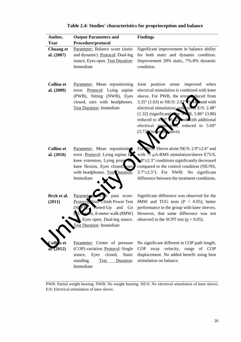
26
Table 2.4: Studies' characteristics for proprioception and balance
Author,
Year
Output Parameters and
Procedure/protocol
Findings
Chuang et
al. (2007)
Parameter: Balance score (static
and dynamic). Protocol: Dual-leg
stance, Eyes open. Test Duration:
Immediate
Significant improvement in balance ability
for both static and dynamic condition.
Improvement 28% static, 7%-8% dynamic
condition.
Collins et
al. (2009)
Parameter: Mean repositioning
error. Protocol: Lying supine
(PWB), Sitting (NWB), Eyes
closed, ears with headphones.
Test Duration: Immediate
Joint position sense improved when
electrical stimulation is combined with knee
sleeve. For PWB, the error reduced from
3.35° (1.63) to NE/S: 2.87° (1.41), and with
electrical stimulation; reduced to E/S: 2.48°
(1.32) (significant). For NWB, 5.86° (3.80)
reduced to 4.96° (3.52) and with additional
electrical stimulation, reduced to 5.69°
(3.73) (no significance).
Collins et
al. (2010)
Parameter: Mean repositioning
error. Protocol: Lying supine for
knee extension, Lying prone for
knee flexion, Eyes closed, ears
with headphones. Test Duration:
Immediate
For PWB: Sleeve alone NE/S: 2.9°±2.6° and
with 75 μA-RMS stimulation/sleeve E75/S,
3.0°±2.3° conditions significantly decreased
compared to the control condition (NE/NS,
3.7°±2.5°). For NWB: No significant
difference between the treatment conditions.
Bryk et al.
(2011)
Parameters: VAS pain score.
Protocol: Stair Climb Power Test
(SCPT), Timed-Up and Go
(TUG) test, 8-meter walk (8MW)
test. Eyes open, Dual-leg stance.
Test Duration: Immediate
Significant difference was observed for the
8MW and TUG tests (P < 0.05); better
performance in the group with knee sleeves.
However, that same difference was not
observed in the SCPT test (p > 0.05).
Collins et
al. (2012)
Parameter: Center of pressure
(COP) variation. Protocol: Single
stance, Eyes closed, Static
standing. Test Duration:
Immediate
No significant different in COP path length,
COP sway velocity, range of COP
displacement. No added benefit using heat
stimulation on balance.
PWB: Partial weight bearing; NWB: No weight bearing; NE/S: No electrical stimulation of knee sleeve;
E/S: Electrical stimulation of knee sleeve.
Univers
ity of
Mala
ya
27
2.5.4 Relationship between Gait and Balance
Gait and balance are two functions that are derived from sensorimotor control and
they are affected by the strength of the lower extremities of an individual. Farrokhi et al.
(2013) investigated the relationship between instability and joint moments, and suggested
that in order to increase stability, knee OA patients tend to add more compressive forces
on the knee which lead to an increase in knee extension moment. Mechanically, the forces
will undoubtedly create additional forces on the knee, resulting in a better balance but
causing further damage to the knee. With the aid of knee sleeves, Collins et al. (2014)
found that individuals with the poorest proprioceptive acuity will have the greatest
improvement in knee flexion kinematics.
The above discussion on the relationship between gait and balance, and the effects of
knee sleeves with regard to both parameters, show that knowledge on these issues is still
at the preliminary stage. Wolfson (2001) believed, that there is correlation between gait
and balance. He suggested that diseases such as knee OA – which involves motor
association cortex and subcortical white matter – are often associated with gait
disturbance and significantly influence the balance ability of an individual. Similarly,
Jones et al. (2014) suggested that any structural changes within the knee would also affect
a person’s balance control.
2.5.5 Types of Knee Sleeves
The mixed clinical and biomechanical conclusions in the past studies could be caused
by the use of non-standardized or different knee sleeve designs. Although knee sleeves
are generally beneficial for knee OA, it is unclear which type or design of sleeve is more
effective for each of the parameters mentioned above. Some of the studies used only
simple knee sleeves, while other studies used semi-rigid knee sleeves such as those with
patella reinforcement, as well as those with thin metal bars at the side (Asl et al., 2008;
Univers
ity of
Mala
ya
28
Baltaci et al., 2011; Bryk et al., 2011; Chuang et al., 2007; Schween et al., 2015). These
differences in knee sleeve designs could change the joint mechanics, such as exerting
more force on the patella, and more compression on the knee.
Different sleeve designs, materials, and the constriction characteristics might produce
different changes and stimulation on the knee, and have different influences on knee
proprioception as the compression could improve coordination by restricting the range of
motion (Baltaci et al., 2011; Bottoni et al., 2013). Sleeves with patella cutout could restrict
and stabilize the movement of the patella which is useful for knee problems that is caused
by abnormal patella tracking (movement). The thin metal bars, if present, may help in
lateral stabilization for knees that has collateral ligament laxities. The results from the
studies suggest that the additional restriction imposed by the compressive design of knee
sleeves might help in minimizing proprioception deficits, thereby, enhancing the level of
stability (Bottoni et al., 2014).
Giotis et al. (2011) studied two different sleeve designs: 1) a knee sleeve with a thin
reinforced metal on the medial and lateral sides, and 2) a simple knee sleeve with patella
cutout. The reinforced knee sleeve helps in limiting the range of tibial rotation that
typically occurs in anterior cruciate ligament (ACL)-deficient knee. Excessive tibial
rotation could degrade the soft tissues of the knee (Kanamori et al., 2002). Asl et al. (2008)
also showed that the use of a reinforced knee sleeve effects more improvements in
replicating target angles as compared to the use of a simple knee sleeve. The use of knee
sleeves with reinforcements at the sides to support the knee and also the patella could
minimize soft tissues damage caused by excessive tibial rotation in those with ACL
injuries.
Bottoni et al. (2014) also reported the use of a simple knee sleeve – without fixation
straps or rigid side bars – but is still able to reduce the stability index, significantly. This
Univers
ity of
Mala
ya
29
shows that a simple knee sleeve also gives a slight compression to the joint area, and does
not affect the range of knee motion. Bottoni et al. believed that the fixation straps or rigid
side bars, in fact, cause discomfort around the knee and may also disturb the signal
generation of the skin receptors, and thus, affect balance control, adversely.
Collins et al. (2010, 2011, 2012, 2014) studied additional stochastic resonance
electrical stimulation on the sleeve, and found only minimal improvement in gait and
proprioception but no additional improvement in balance. Thus, stochastic electrical
stimulation on the knee sleeve does not help in improving stability.
The use of different types of knee sleeves seems to effect varied improvements to gait
and balance control. Although sleeves with patella cutout might restrict the movement of
the patella, they do help in patella stabilization and reinforcement. It appears that knee
sleeve can act through different mechanisms to improve knee proprioception. This is
because knee sleeves – irrespective of designs – have all demonstrated evidence of
benefits on knee pain, gait and postural stability (Baltaci et al., 2011; Bottoni et al., 2013).
On the materials of knee sleeve, manufacturer often used either Neoprene or Drytex.
To make a good choice, the user should consider the activities he might dealing with, and
his perspiration rate. Neoprene are ideal in giving uniform compression and mostly used
in OA to reduce the pain. Nevertheless, it is also durable and suitable in colder climates
(Shapiro, 2010). However, Neoprene does not work well in perspiration, which Drytex
would be a better choice since Drytex allows breathability and not allergenic as Neoprene
(DonJoy USA, 2016; Shapiro, 2010).
2.5.6 Limitations
Most studies on knee sleeve investigated only the immediate effects and the long term
effect is largely unknown. It would be useful to bridge this gap in understanding and to
Univers
ity of
Mala
ya

30
establish if the effects of knee sleeve could be sustained and improved with time. Besides,
the immediate effects are not necessarily an accurate reflection of the potential benefits
of knee sleeves. This is because the immediate measurements do not consider the effect
of neuromuscular adaptation in knee sleeve use. The initial beneficial effect such as found
by Bottoni et al. (2013) following 30-minute of knee sleeve use may only represent the
partial effects of knee sleeves. Due to the lack of past research, it is unclear if any benefits
that is observed with following short term use can continue to increase or remain the same
if knee sleeve is worn for longer period of time such as for weeks or months. Hence, it is
useful to conduct serial measurements of biomechanical or clinical effect a longer
duration of knee sleeve use. The type of knee sleeve used in any study, as mentioned in
Subsection 2.5.5, could be one of the factors that could lead to difference in the study
results. The different types of knee sleeves used, make it difficult to decide which type of
knee sleeves effect greater improvement on the target population.
Another factor that hampers definitive conclusion on the effects of knee sleeves is due
to different designs of knee sleeve used. Knee sleeves can be of different rigidities. A
simple knee sleeve is more likely to give partial functional improvement when compared
to a complex and more rigid knee sleeve which could change the joint mechanics. Until
more studies are done to directly compare different types of knee sleeves, the relative
efficacy of different knee sleeves is unknown.
The use of different experimental protocols – each with its own method of measuring
– makes it difficult to generalize any findings pertaining to knee sleeve effects on
balancing. Some studies used the balance scores or index, and one study used the center
of pressure (COP) parameter to identify postural sway using force plates, and only focuses
on static condition. On the other hand, Chuang et al. (2007) obtained more specific
information because they used a balance machine which can measure the stability index
Univers
ity of
Mala
ya

31
directly, and can assess the postural stability on the dynamic condition as well. This shows
that it is more reliable to evaluate postural stability using a balance machine, compared
to force plates (Arnold & Schmitz, 1998).
2.6 Summary
The beneficial effects of knee sleeves were observed immediately in some studies -
on pain; on the joint kinetics and kinematics – KAM and knee flexion angle (Bryk et al.,
2011; Collins et al., 2014; Collins et al., 2011; Schween et al., 2015).
However, the efficacy of knee sleeves from their biomechanical aspect is still unclear.
Although knee sleeves have proven to be effective in improving the functional
parameters, there is still a gap in understanding the biomechanical mechanism involved
because many of the study protocols are not standardized. Moreover, there is no serial
measurement of each parameter over long-term testing. Thus, it is difficult to generalize
the results due to lack of homogeneity in the study designs, protocols, participants
population, comparator groups, type of knee sleeves used, and the parameters. However,
further work is needed to confirm this hypothesis, due to the lack of homogeneity and
rigor of existing studies. Nevertheless, from the studies reviewed, it can be concluded that
knee sleeves effect positive changes on the knee movement, can effect functional
improvements to knee problems, thereby, helping people to have a better walking pattern.
Based on the findings, more rigorous experimental design should be adopted to obtain
more reliable results. More longitudinal studies with randomization on the testing
conditions would provide more reliable outcomes pertaining to the benefits and
understanding of knee sleeves use from the biomechanical aspect.
Univers
ity of
Mala
ya
32
CHAPTER 3: METHODOLOGY
3.1 Introduction
This chapter discusses the experimental procedures in more detail. It begins by
discussing the study design and ends with procedure on data collection for gait kinetics
and kinematics, postural stability and pain, as well as the statistical tests that are applied
on the collected data.
3.2 Study Design
A comparative study was performed. It was calculated that samples of 17 participants
in total would provide 80% statistical power. In this study, participants do not know there
is a comparator group. The participants were handed the knee sleeve by quasi-
randomization, which means each participant received only one knee sleeve and the
randomization sequence is alternate. Then, the sizes of the knee sleeve were then set
according to the knee circumference of the participants, whenever possible (Refer to
Section 3.4). All participants gave written informed consent before the trial began. This
study adopts a parallel-group randomized clinical trial design, which involves three
different measurements over six weeks. Each group receives knee sleeves of two different
designs. This study was approved by the Medical Ethics Committee of the University of
Malaya Medical Centre (UMMC) (MECID No 201410-626). This study also adheres to
the principles of the Declaration of Helsinki of the World Medical Association. The
testing duration was set to six weeks because it is generally longer, and considering the
logistic factors of the project, including the administration of patients and short duration
of project. The testing duration was confirmed via consultation from a specialist from
Sports Medicine Unit, UMMC.
Univers
ity of
Mala
ya
33
3.3 Participants
Participants (patients) with early unilateral knee OA who are registered in UMMC
were selected. The participants had been diagnosed based on the American College of
Rheumatology (ACR) criteria. These diagnoses were also confirmed radiographically.
Participants were included if they were aged between 30 and 60, and had suffered from
unilateral knee pain. Other inclusion criteria include: 1) Kellgren–Lawrence (K&L) scale
of disease severity of grade 1 or 2; 2) functionally independent people; and 3) no known
allergy to knee sleeves. Both male and female participants were recruited for the trial.
The limb considered in the study was decided based on the radiography results.
Radiographic severity of the OA was categorized – based on the K&L scale – by a
specialist from Sports Medicine Unit, UMMC. In the K&L scale, disease severity is rated
from 0 to 4; where 0 = none, 1 = doubtful, 2 = mild, 3 = moderate, and 4 = severe; by
observing the bony changes such as narrowing of joint space, and presence of osteophytes
(Altman et al., 1986). In this study, a participant is selected if his/her rating is 1 or 2.
Exclusion criteria include participants: 1) receiving any intra-articular injection within
the last six months; 2) who had prior knee surgery e.g., total knee replacement or fracture
fixation; 3) with other balance problems e.g., neurological conditions, inner ear problem;
and 4) with back, hip or ankle injuries that could affect the physical testing outcomes.
3.4 Intervention
This study is aimed to evaluating and comparing the biomechanical effects of two
different knee sleeves – simple knee sleeve (without patella cutout), and knee sleeve with
patella cutout (Drytex Basic Knee Support, DonJoy, USA) (Figure 3.2). The simple knee
sleeve exerts even compression throughout the knee area, while the sleeve with patella
cutout provides slight reinforcement on the patella, aimed at controlling the movement of
the patella. These two types of sleeves were chosen because the material is suitable in a
Univers
ity of
Mala
ya
34
warmer climate and for rigorous activity usage due to its breathability. Both are elastic
knee sleeves made from Drytex® - an alternative material to neoprene (Shapiro, 2010).
Drytex is a combination of nylon core and polyester lycra fabric that allows breathability
and good airflow (DonJoy USA, 2016).
The suitable size of the sleeve for a user is based on the measurement of the
circumference at six inches above the middle of the knee cap of the user in a seated
position (Figure 3.1). The knee sleeve sizes were individualized for each participant.
Randomization of intervention was implemented based on quasi-randomization. Each
individual is alternately randomized to one treatment group. As the study only involved
two male participants, we handed the first male participant the simple knee sleeve and the
second male participant the knee sleeve with patella cutout.
To ensure that the knee sleeves are used correctly, participants were taught the way to
apply the knee sleeve – to grasp the edges of the sleeves at the larger opening and roll the
sleeve to cover the knee. Participants were instructed to wear the sleeve daily for as long
as it could be tolerated, since past studies mentioned the compliance for knee bracing and
orthosis were ranged from 3-12 hours per day (Johann Beaudreuil et al., 2009; Bennell,
Kean, Wrigley, & Hinman, 2013; Toda & Tsukimura, 2004). They were then asked to
register in a log book the number of hours of usage per day. Participants were told they
could put on or take the knee sleeve off according to their needs and schedule, but it has
to be worn daily for six weeks. The log book was given to each participant on their first
lab visit.
Univers
ity of
Mala
ya

35
Figure 3.1: The method of measuring the knee circumference for the sizes of
knee sleeves
Figure 3.2: Knee sleeves used in the study; a) Simple knee Sleeve (Knee Sleeve
A), and b) Knee sleeve with patella cutout (Knee Sleeve B) (Drytex Basic Knee
Sleeve, Donjoy, USA)
3.5 Adherence
The participants were regularly monitored to ensure that they strictly follow the
instructions on how to wearing of the knee sleeves. Compliance was confirmed via the
recorded daily usage in their log book during the second visit.
3.5.1 Log Book
Along with the knee sleeve, participants were given a log book (Appendix C) each –
on their first lab visit – to record their total hours of usage for the entire six weeks. They
were also advised to record any adverse effects attributed to the knee sleeve usage. All
participants were also asked to report on the overall knee sleeve usage experiences.
a) b)
6”
Knee center
Univers
ity of
Mala
ya
36
3.6 Instrumentations
During each visit, gait analysis was conducted, while walking on a flat surface, on a
self-selected speed and on a controlled speed (Refer Section 3.6.2); and the Western
Ontario and McMaster Universities Arthritis Index (WOMAC) was used to assess the
pain levels of the participants.
3.6.1 WOMAC® Score
Clinical outcomes were assessed using the WOMAC scores at each stage of the trial.
WOMAC is a pain score to assess level of pain, stiffness, and physical functions in
patients with knee OA. The participants may choose the WOMAC either in English or
Malay (Bellamy, 2014) (See Appendix D).
WOMAC consists of 24 items divided into three subscales: 1) Pain (5 items): during
walking, using stairs, in bed, sitting or lying, and standing; 2) Stiffness (2 items): after
waking and later in the day; 3) Physical Functions (17 items): stair use, rising from sitting,
standing, bending, walking, getting in / out of a car, shopping, putting on / taking off
socks, rising from bed, lying in bed, getting in / out of bath, sitting, getting on / off toilet
bowl, heavy household duties, light household duties (Bellamy, 2014). Total score for
WOMAC would be 2,400 mm (based on VAS 100 mm scores for each question).
Participants have to answer the questions before, immediately after, and after six weeks
of the knee sleeve usage. Participants were also given a VAS score ruler to guide them
on answering the WOMAC questionnaire.
3.6.2 Gait Analysis
Kinetics data were collected at 1,000Hz using two Kistler force plates (Kistler
Instruments, Winterthur, Switzerland) to measure the ground reaction forces and joint
moments. The kinematics data were collected at 100Hz using the Vicon motion analysis
system with five infrared cameras (Vicon, Oxford, UK). The system was calibrated at
Univers
ity of
Mala
ya
37
every session to ensure that the output from the cameras and force plates are appropriately
synchronized and accurate. The standard Vicon Plug-in-Gait model for measuring the
lower extremity was used. Sixteen (16) markers were placed on the lower extremity
following the Helen Hayes standard marker set (Davis, 1997). The markers were placed
on the lateral side of both legs, anterior superior iliac spines, posterior superior iliac
spines, femur, lateral epicondyles, tibia, lateral malleoli, distal head of the second
metatarsals, and on the heels (Laroche et al., 2014). Figure 3.3 illustrates the marker
placement for this study.
Each participant performed twelve complete walking trials and the mean scores were
used for analysis. Each gait assessment consisted of six trials at the self-selected speed
and six trials at the controlled speed. All participants performed barefoot walking trials.
At their self-selected walking speed, the participants were asked to walk leisurely at their
own comfortable walking speed. The controlled walking speed was monitored using a
timer to ensure that the speed reflects the average walking speed of a normal person,
which is estimated at 1.3 ± 0.2 m/s (Wikipedia, n.d.). The participants were asked to
practise walking within a 3-meter walking range and within 2 seconds to obtain the
controlled speed. After the practice session, the speeds in the actual trials were monitored.
Trial speeds that exceeded the set speed range were ignored. The speed was controlled to
obtain an optimum value on moments and forces for all participants. Meanwhile, the self-
selected speed trials are done to obtain the walking speed parameter. Following the
baseline data collection, the participants were given a knee sleeve to be worn on the
studied limb. The immediate-effects were measured after a 10-minute adaptation period,
during which participants practised walking.
Univers
ity of
Mala
ya
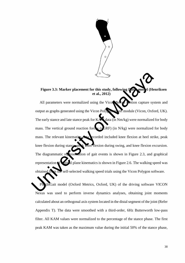
38
Figure 3.3: Marker placement for this study, following Davis model (Henriksen
et al., 2012)
All parameters were normalized using the Vicon Nexus motion capture system and
output as graphs generated using the Vicon Polygon report module (Vicon, Oxford, UK).
The early stance and late stance peak for KAM data (in Nm/kg) were normalized for body
mass. The vertical ground reaction forces (vGRF) (in N/kg) were normalized for body
mass. The relevant kinematics data recorded included knee flexion at heel strike, peak
knee flexion during stance, peak knee flexion during swing, and knee flexion excursion.
The diagrammatic representation of gait events is shown in Figure 2.3, and graphical
representation of sagittal plane kinematics is shown in Figure 2.6. The walking speed was
obtained from the self-selected walking speed trials using the Vicon Polygon software.
PlugInGait model (Oxford Metrics, Oxford, UK) of the driving software VICON
Nexus was used to perform inverse dynamics analyses, obtaining joint moments
calculated about an orthogonal axis system located in the distal segment of the joint (Refer
Appendix T). The data were smoothed with a third-order, 6Hz Butterworth low-pass
filter. All KAM values were normalized to the percentage of the stance phase. The first
peak KAM was taken as the maximum value during the initial 50% of the stance phase,
Univers
ity of
Mala
ya
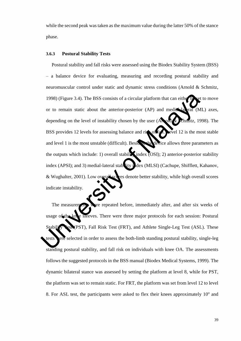
39
while the second peak was taken as the maximum value during the latter 50% of the stance
phase.
3.6.3 Postural Stability Tests
Postural stability and fall risks were assessed using the Biodex Stability System (BSS)
– a balance device for evaluating, measuring and recording postural stability and
neuromuscular control under static and dynamic stress conditions (Arnold & Schmitz,
1998) (Figure 3.4). The BSS consists of a circular platform that can either be set to move
or to remain static about the anterior-posterior (AP) and medial-lateral (ML) axes,
depending on the level of instability chosen by the user (Arnold & Schmitz, 1998). The
BSS provides 12 levels for assessing balance and risk of fall. Level 12 is the most stable
and level 1 is the most unstable (difficult). Besides, the device allows three parameters as
the outputs which include: 1) overall stability index (OSI); 2) anterior-posterior stability
index (APSI); and 3) medial-lateral stability index (MLSI) (Cachupe, Shifflett, Kahanov,
& Wughalter, 2001). Low overall scores denote better stability, while high overall scores
indicate instability.
The measurements were repeated before, immediately after, and after six weeks of
usage of the knee sleeves. There were three major protocols for each session: Postural
Stability Test (PST), Fall Risk Test (FRT), and Athlete Single-Leg Test (ASL). These
tests were selected in order to assess the both-limb standing postural stability, single-leg
standing postural stability, and fall risk on individuals with knee OA. The assessments
follows the suggested protocols in the BSS manual (Biodex Medical Systems, 1999). The
dynamic bilateral stance was assessed by setting the platform at level 8, while for PST,
the platform was set to remain static. For FRT, the platform was set from level 12 to level
8. For ASL test, the participants were asked to flex their knees approximately 10o and
Univers
ity of
Mala
ya
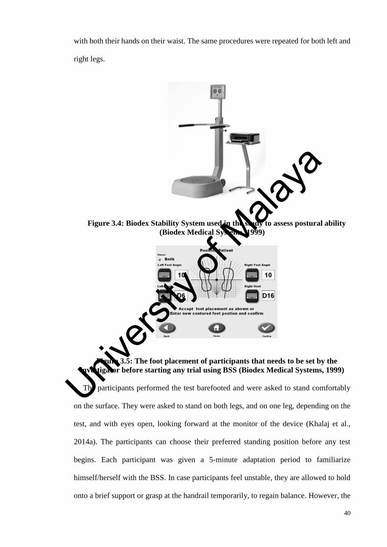
40
with both their hands on their waist. The same procedures were repeated for both left and
right legs.
Figure 3.4: Biodex Stability System used in the study to assess postural ability
(Biodex Medical Systems, 1999)
Figure 3.5: The foot placement of participants that needs to be set by the
investigator before starting any trial using BSS (Biodex Medical Systems, 1999)
The participants performed the test barefooted and were asked to stand comfortably
on the surface. They were asked to stand on both legs, and on one leg, depending on the
test, and with eyes open, looking forward at the monitor of the device (Khalaj et al.,
2014a). The participants can choose their preferred standing position before any test
begins. Each participant was given a 5-minute adaptation period to familiarize
himself/herself with the BSS. In case participants feel unstable, they are allowed to hold
onto a brief support or grasp at the handrail temporarily, to regain balance. However, the
Univers
ity of
Mala
ya
41
test is considered invalid if the participant is unable to remain stable, and holds on to a
support longer than three seconds.
In the static balance test, the participants were asked to maintain the point at the center
of the screen as level as possible. Balance and risk of fall were assessed using three trials
for a period of 20 seconds with 5 seconds rest in between. Participants were given 2
minutes rest in between two testing positions to avoid the effect of fatigue and pain from
affecting the scores. Similar procedures were repeated for dynamic balance test and fall
risk test. Our study included single-leg standing tests for both affected knee and
unaffected knee to make necessary adjustment in view of the theory of leg dominance
(Duffell et al., 2014). The standing position during postural stability tests was self-
selected (Figure 3.5).
3.7 Procedures
The trial consists of two visits. In the first visit, participants’ baseline measurements
were recorded. After immediate application of the knee sleeve, the measurements were
repeated. In the second visit, which was scheduled six weeks after sleeve application, the
post six-week effects were recorded. The same procedures were repeated for immediate
and post six-week effects (Figure 3.6). Standardized instructions were given to the
participants before the trial begins. Participants were asked to fill in the consent form,
responded to the WOMAC score for the baseline measurement, and their anthropometrics
data were collected – height, weight, and age. The instructions of knee sleeve usage and
using the log book were also explained before the trial begins. Next, they were instructed
to run a gait analysis, followed by postural stability tests (Figure 3.6).
Univers
ity of
Mala
ya
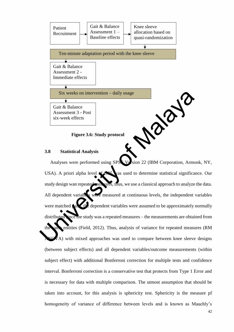
42
3.8 Statistical Analysis
Analyses were performed using SPSS Version 22 (IBM Corporation, Armonk, NY,
USA). A priori alpha level of 0.05 was used to determine statistical significance. Our
study design wan repeated measures, thus, we use a classical approach to analyze the data.
All dependent variables were measured at continuous levels, the independent variables
were matched pairs, the dependent variables were assumed to be approximately normally
distributed since the study was a repeated measures – the measurements are obtained from
the same entities (Field, 2012). Thus, analysis of variance for repeated measures (RM
ANOVA) with mixed approaches was used to compare between knee sleeve designs
(between subject effects) and all dependent variables/outcome measurements (within
subject effect) with additional Bonferroni correction for multiple tests and confidence
interval. Bonferroni correction is a conservative test that protects from Type 1 Error and
is necessary for data with multiple comparison. The utmost assumption that should be
taken into account, for this analysis is sphericity test. Sphericity is the measure pf
homogeneity of variance of difference between levels and is known as Mauchly’s
Six weeks on intervention – daily usage
Patient
Recruitment
Gait & Balance
Assessment 2 -
Immediate effects
Gait & Balance
Assessment 3 - Post
six-week effects
Ten-minute adaptation period with the knee sleeve
Gait & Balance
Assessment 1 –
Baseline effects
Figure 3.6: Study protocol
Knee sleeve
allocation based on
quasi-randomization
Univers
ity of
Mala
ya

43
sphericity test (W). The violation of this assumption can impact the results drawn from
the analysis – loss of statistical power. If W>0.05, the sphericity assumption is satisfied
and no correction should be made. But if the W<0.05, the assumption is violated and
correction is necessary – the Geisser-Greenhouse (G-G) or Huynh-Feldt epsilon (€)
correction is used (Field, 2012). Descriptive statistics were used to analyze the
demographics of the participants. The data were considered missing if the participants did
not managed to complete all trials and the data were not included in the final results of
this study.
3.9 Summary
In summary, all the procedures stated above were conducted to obtain the outcomes.
The procedures started with participant recruitment, ethical approval, followed by
participants’ selection, intervention allocation, followed by data collection which consists
of the experiment and the intervention period, and lastly, data analysis. The next chapter
will present the results from all the trials that were conducted.
Univers
ity of
Mala
ya

44
CHAPTER 4: RESULTS
4.1 Introduction
This chapter presents the results of all the tests for knee pain, knee stiffness, knee
function, gait, and postural stability. It also presents the quantitative data collected using
procedures mentioned in the previous chapter. All measurements are quantified in mean
± standard deviation (SD) format, if not stated otherwise. A few often-used terms
pertaining to when the test measurement is taken are defined as follows: 1) T0 or pre –
indicates the measurement taken at baseline, 2) T1 or immediate – indicates the
measurement taken after the immediate use of knee sleeve, 3) T2 or post – indicates the
measurement taken following six weeks of knee sleeve use, 4) T0 - T2 or pre-post –
indicates the measurement taken at baseline compared to measurement taken after six
weeks of knee sleeve use, 5) T0 - T1 or pre-immediate – indicates the measurement taken
at baseline compared to the immediate reading after immediate use of knee sleeve, and 6)
T1 - T2 or immediate-post – indicates the measurement taken after the immediate use of
knee sleeve compared to the measurement made after six weeks. The assumption of
Mauchly’s test of sphericity is met for all dependent variables (W > 0.05) (Refer
Appendix G to Appendix S).
4.2 Participants
Nineteen participants (including two male participants) with unilateral knee OA (of
K&L grade 1 and grade 2) were recruited from UMMC for this study. Figure 4.1 shows
that two participants dropped out from the trial - one had to stop wearing the sleeve after
seven days due to severe pain which aggravated the participant’s condition, and another
dropped out due to work commitment. Those who remain (17 participants) have managed
to perform all required tests. All participants have the same baseline characteristics and
the two groups are treated in the same manner. Table 4.1 shows the summary profiles of
the participants that include their age, gender, height, weight, and BMI. All values
Univers
ity of
Mala
ya

45
obtained from the various tests are reported as mean ± standard deviation (SD), unless
stated otherwise.
4.2.1 Lifestyle of Participants
Six participants (31.6%) out of the 19 participants had previous injuries due to one of
the following reasons: accidents and falls (10.6%); ligament tear (10.5%); and muscle
tear (10.5%). Their injuries were not caused by any sports activities. Other information
on the participants include: none are smokers; 11 participants never consumed nor had
been prescribed analgesics for the knee pain; six of them are undergoing a rehabilitation
program; 16 participants are planning to lose weight to alleviate their knee pain; 17
participants reported that the knee pain had disrupted their daily routines; eight
participants fear that exercise will aggravate their knee pain; 14 participants are working;
none of them are sportsmen; and all participants have gone through tertiary education –
diploma or degree holders.
From the log book given to each participant (Appendix C), the hours of knee sleeve
use and the adverse effects were tabulated and averaged. From Appendix G, the knee
sleeves were used an average of 3.7 ± 2.4 hours per day, calculated based on an average
of 3.7 ± 2.7 hours for simple knee sleeve and 3.7 ± 2.3 hours for knee sleeve with patella
cutout. Overall, 82.4% (14 out of 17 participants) stated that the sleeve is helpful.
Meanwhile, the adverse effects reported include poor fitting (47.1%), skin irritation
(47.1%), and discomfort (41.2%).
Univers
ity of
Mala
ya

46
Table 4.1: Brief participants' profiles (n = 17).
Group All (n = 17) Simple knee
sleeve (n = 9)
Knee sleeve with
patella cutout (n =
8)
p-values
(between
groups)
Age (year) 47.2 (10.2) 46.9 (11.3) 47.6 (9.6) 0.888
Gender 15 F, 2 M 8 F, 1 M 7 F, 1 M -
Height (m) 1.58 (0.1) 1.59 (0.1) 1.58 (0.1) 0.860
Weight (kg) 66.82 (13.7) 72.44 (15.7) 60.50 (7.8) 0.081
BMI (kg/m2) 26.69 (6.1) 28.90 (7.1) 24.23 (3.7) 0.134
Data presented as Mean (SD). BMI = Body-mass index, F = Female, M = Male. p > 0.05 denotes no significant
difference among participants at baseline.
Figure 4.1: Participants involved in the study (following CONSORT Flow
Diagram)
Analysed (n=9)
- Excluded from analysis (n=0) Analysed (n=8)
- Excluded from analysis – drop-
outs (n=2)
Analysis
Work commitment (n=1)
Discontinued intervention due
to severe pain on the knee (n=
1)
Lost to follow-up (n=0).
Follow-Up
Allocated knee sleeve A (n=9) Allocated knee sleeve B (n=10)
Allocation
Assessed for eligibility
(n=80)
Excluded (n= 61)
- Not meeting inclusion
criteria (n=30)
- Declined to participate
(n=31)
Enrollment
Univers
ity of
Mala
ya
47
4.3 WOMAC® Scores
We used WOMAC to measure pain level. All participants (17) have completed the
WOMAC questionnaire. Table 4.2 shows the WOMAC scores for T0, T1 and T2
measurements. Appendix K shows the SPSS results for descriptive statistics and pairwise
comparison on points of measurement and different types of knee sleeves. The table
shows that the WOMAC scores at T0 – T1 were significantly reduced; mean difference =
17.958, p = 0.034, 95% CI (1.233, 34.684) (Refer Appendix K). Significant decrease in
overall WOMAC scores was observed for T0 - T2; mean difference = 28.888, p = 0.012,
95% CI (5.914, 51.862) and for T1 - T2; mean difference = 10.930, p = 0.048, 95% CI
(0.097, 21.762) for both sleeves. There was no significant difference in all points of
measure between the two interventions (p > 0.05). Following six-week of use, there is
marked reduction of 49% in the WOMAC scores after simple knee sleeve application
when compared to reduction of 26% in the scores after knee sleeve with patella cutout
application, compared to baseline. Overall, 26.0% reduction in WOMAC scores for pre-
immediate effects, and 42.9% for pre-post effects of knee sleeve use.
Appendix H shows the tables of results on WOMAC score – pain. From the tables, the
mean difference for T0 – T1 readings is 3.29, p = 0.155, 95% CI (-0.900, 7.481). For T0 -
T2 measurement, the mean difference is 5.414, p = 0.055, 95% CI (-0.089, 10.917).
Between simple knee sleeve and knee sleeve with patella cutout, there is no significant
differences at all points of measure (p = 0.122). Figure 4.2 shows the graphs for both
types of knee sleeves. We partially reject the null hypothesis, and partially accept the
alternative hypothesis (H1), that, the application of the knee sleeves would alleviate knee
pain for pre-post measurement.
Univers
ity of
Mala
ya

48
Table 4.2: WOMAC overall scores (n = 17)
All Simple knee sleeve Knee sleeve with patella
cutout
T0 697.4 (50.96) 936.8 (52.11) 428.1 (35.60)
T1 514.4 (46.15)* 699.1 (40.38) 306.6 (45.52)
T2 398.2 (42.31)*,† 471.9 (45.04) 315.3 (40.32)
* indicates significant difference compared to baseline (p<0.05). † indicates significant
reduction compared to immediate effects (p<0.05). WOMAC pain score is measured in
100mm. Total score is 2,400mm. Data presented as mean ± SD (standard deviation).
Figure 4.2: WOMAC scores for knee pain of 17 participants, for simple knee
sleeve (Knee Sleeve A) and knee sleeve with patella cutout (Knee Sleeve B),
respectively.
WOMAC scores for Pain
Univers
ity of
Mala
ya

49
Figure 4.3: WOMAC scores for knee stiffness of 17 participants, for simple knee
sleeve (Knee Sleeve A) and knee sleeve with patella cutout (Knee Sleeve B),
respectively.
Figure 4.4: WOMAC scores for functional performance of 17 participants,
respectively, for simple knee sleeve (Knee Sleeve A) and knee sleeve with patella
cutout (Knee Sleeve B)
WOMAC scores for Difficulty Performing Daily Activities
WOMAC scores for Stiffness
Univers
ity of
Mala
ya

50
Appendix I shows the WOMAC scores on knee stiffness and Figure 4.3 shows the
graphs for both types of knee sleeves. In the measurement of knee stiffness, the mean
difference of the T0 - T1 scores is 2.62, p = 0.022, 95% CI (0.347, 4.894). For T0 - T2
measurement, the mean difference is 2.690, p = 0.018, 95% CI (0.421, 4.959). For the
intervention groups – simple knee sleeve and knee sleeve with patella cutout – no
significant differences was found (p = 0.443). Figure 4.3 shows the graphs for the
different types of knee sleeves on knee stiffness. We reject the null hypothesis, and accept
the alternative hypothesis (H1), that, the application of the knee sleeves would alleviate
knee stiffness for pre-immediate and pre-post measurements.
Appendix J shows the WOMAC scores on functional performance and Figure 4.4
shows the graphs for both types of knee sleeves. In measuring the knee functional
performance, the mean difference for T0 - T1 is 12.048, p = 0.061, 95% CI (-0.477, -
24.573). For T0 – T2, the mean difference is 20.784, p = 0.014, 95% CI (3.834, 37.734).
At all points of measurement, there is no significant difference was found for simple knee
sleeve and knee sleeve with patella cutout (mean difference = 27.901, p = 0.079). We
reject the null hypothesis, and accept the alternative hypothesis (H1) for knee functional
parameters – indicating that there are benefits from using the knee sleeves after six weeks.
4.4 Gait Analysis
All reported values in this subsection are shown in Appendix S.
4.4.1 Knee Adduction Moment (KAM)
Table 4.3 shows the results for KAM measurement. There is no significant difference
in the KAM during early stance; mean difference = 0.107; p = 0.744, 95% CI (-0.133,
0.347) or late stance; mean difference = 0.04; p = 1.00; 95% CI (-0.182, 0.191) for T0 -
T1 effects. There is reduction between T0 – T2 effects on first peak KAM; mean difference
= 0.273; p = 0.061, 95% CI (-0.011, 0.557), and significant reduction on second peak
Univers
ity of
Mala
ya
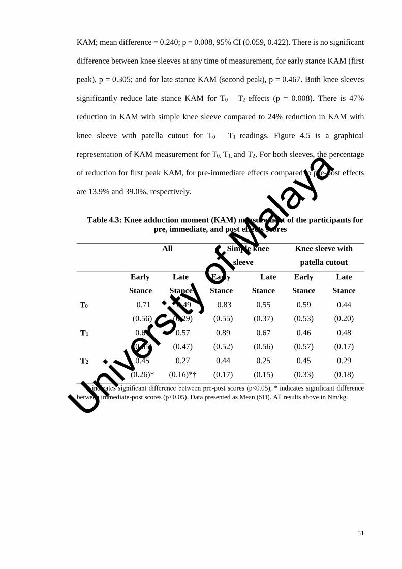
51
KAM; mean difference = 0.240; p = 0.008, 95% CI (0.059, 0.422). There is no significant
difference between knee sleeves at any time of measurement, for early stance KAM (first
peak), p = 0.305; and for late stance KAM (second peak), p = 0.467. Both knee sleeves
significantly reduce late stance KAM for T0 – T2 effects (p = 0.008). There is 47%
reduction in KAM with simple knee sleeve compared to 24% reduction in KAM with
knee sleeve with patella cutout for T0 – T1 readings. Figure 4.5 is a graphical
representation of KAM measurement for T0, T1, and T2. For both sleeves, the percentage
of reduction for first peak KAM, for pre-immediate effects compared to pre-post effects
are 13.9% and 39.0%, respectively.
Table 4.3: Knee adduction moment (KAM) measurement of the participants for
pre, immediate, and post effects scores
All Simple knee
sleeve
Knee sleeve with
patella cutout
Early
Stance
Late
Stance
Early
Stance
Late
Stance
Early
Stance
Late
Stance
T0 0.71
(0.56)
0.49
(0.29)
0.83
(0.55)
0.55
(0.37)
0.59
(0.53)
0.44
(0.20)
T1 0.65
(0.65)
0.57
(0.47)
0.89
(0.52)
0.67
(0.56)
0.46
(0.57)
0.48
(0.17)
T2 0.45
(0.26)*
0.27
(0.16)*†
0.44
(0.17)
0.25
(0.15)
0.45
(0.33)
0.29
(0.18)
† indicates significant difference between pre-post scores (p<0.05), * indicates significant difference
between immediate-post scores (p<0.05). Data presented as Mean (SD). All results above in Nm/kg.
Univers
ity of
Mala
ya

52
Figure 4.5: Knee adduction moment (KAM) during stance phase for all the
participants. (NS is the measurement at baseline; S is measurement immediately
after knee sleeve use; 6 wks is measurement at six weeks effects)
Table 4.4: Vertical Ground Reaction Force (vGRF) summary
All Simple knee sleeve Knee sleeve with
patella cutout
1st Peak 2nd Peak 1st Peak 2nd Peak 1st Peak 2nd Peak
T0 13.45
(5.33)
12.41
(5.04)
12.86
(5.48)
12.05
(5.51)
11.96
(4.82)
11.00
(3.93)
T1 13.91
(5.70)
12.97
(4.91)
14.80
(6.09)
13.92
(4.91)
13.30
(5.24)
12.21
(4.59)
T2 9.95
(2.26)*
9.36
(1.98)*
9.79
(2.60)
9.42
(2.70)
10.20
(2.12)
9.29
(1.31)
* indicates significant difference between immediate-post values (p<0.05). Data
presented as Mean (SD), 1st Peak indicates the maximum loading rate during early stance,
and 2nd Peak indicates the maximum loading rate during late stance.
-0.3
-0.1
0.1
0.3
0.5
0.7
0 10 20 30 40 50 60Ad
duct
ion M
om
ent
(Nm
/kg)
% Gait Cycle
S
NS
6 wks
Univers
ity of
Mala
ya
53
4.4.2 Vertical Ground Reaction Forces (vGRF)
Appendix S and Table 4.4 show the results of vertical ground reaction force (vGRF)
which measures loading rate. There is significant reduction between T1 – T2 effects in
early stance (first vGRF peak); mean difference = 4.673; p = 0.038, 95% CI (0.233, 9.112)
and in late stance (second vGRF peak); mean difference = 3.840; p = 0.041, 95% CI
(0.138, 7.542) in vGRF for both sleeves. No significant difference between T0 – T2 scores
for first peak loading rate; mean difference = 3.622; p = 0.092, 95% CI (-0.471, 7.714)
and second peak loading rate; mean difference = 2.889; p = 0.148, 95% CI (-0.751, 6.528).
No difference in using knee sleeves of different designs in first peak and second peak, at
any point of measure (p = 0.306, p = 0.207).
4.4.3 Knee Flexion and Walking Speed
Table 4.5 shows the results of kinematics of knee flexion and walking speed.
Significant difference for knee flexion at heel strike; mean difference = 4.834, p = 0.012,
95% CI (1.000, 8.668) and knee flexion during stance; mean difference = 5.119, p =
0.020, 95% CI (0.735, 9.502) observed between T0 – T2 assessments. No significant
findings in the flexion range of motion; mean difference = 5.841, p = 0.094, 95% CI (-
0.784, 12.465) and knee flexion during swing; mean difference = 1.154, p = 1.000, 95%
CI (-5.321, 7.630) between pre-post six-week effects. There is no significant difference
in knee flexion at contact, during stance phase, and during swing phase between T0 – T1
effect (p = 0.457, p = 0.426, p = 0.460). There is no significant difference between both
sleeves in all sagittal plane parameters, at all points of measurements (p > 0.05)
(Appendix S).
The results in Appendix S also show that there is no significant difference in walking
speed between T0 – T1 effects of knee sleeve usage; mean difference = -0.011, p = 1.000,
95% CI (-0.081, 0.060). However, walking speed increased significantly after six weeks
Univers
ity of
Mala
ya

54
of knee sleeve usage (T0 – T2); mean difference = -0.090, p = 0.026, 95% CI (-0.170, -
0.010). There is no significant difference in walking speed when using either simple knee
sleeve or knee sleeve with patella cutout; mean difference = 0.044, p = 0.542, 95% CI (-
0.107, 0.196). Based on the results discussed above, on gait, alternative Hypothesis 2
(H1) is accepted – the application of the knee sleeves would increase walking speed,
decrease ground reaction force (GRF) loading rates and KAM in people with knee OA.
Overall, there is increment of 1.2% after the immediate use of knee sleeve, and 7.9%
increment following six weeks of knee sleeves use, in walking speed.
Univers
ity of
Mala
ya

55
Table 4.5: Kinematics parameters and walking speed results of the participants (n = 17)
† indicates significant difference between pre-post scores (p<0.05), * indicates significant difference between immediate-post scores (p<0.05). Data presented as Mean (SD). T0 =
baseline measurement, T1 = immediate measurement, and T2 = post six-week effects of knee sleeve usage.
All Simple knee sleeve Knee sleeve with patella cutout
T0 T1 T2 T0 T1 T2 T0 T1 T2
Walking Speed
(m/s)
1.05
(0.18)
1.05 (0.16) 1.15
(0.16)†
1.08
(0.17)
1.12
(0.16)
1.17
(0.14)
1.12
(0.20)
0.98
(0.14)
1.13
(0.18)
Flexion ROM (o) 55.8
(11.76)
54.4 (9.41) 54.5
(10.79)
57.4
(13.96)
53.9
(7.35)
54.07
(13.79)
53.9
(9.26)
54.9
(11.83)
54.9
(6.96)
Knee Flexion at
heel strike (o)
18.8
(6.30)
15.9 (9.03) 14.0
(8.06)†
18.3
(6.29)
13.2
(8.69)
14.4
(7.64)
19.4
(6.68)
19.1
(8.89)
13.6
(9.01)
Peak knee flexion
during stance (o)
27.2
(7.78)
22.3
(10.00)
22.2
(10.01)†
26.1
(7.09)
18.9
(8.79)
22.0
(9.02)
28.4
(8.83)
26.0
(10.48)
22.3
(11.67)
Peak knee flexion
during swing (o)
69.5
(14.32)
65.3
(11.75)
63.6
(15.47)
71.3
(18.22)
64.0
(12.02)
64.5
(18.93)
67.4
(8.95)
66.9
(12.06)
62.5
(11.61)
Univers
ity of
Mala
ya
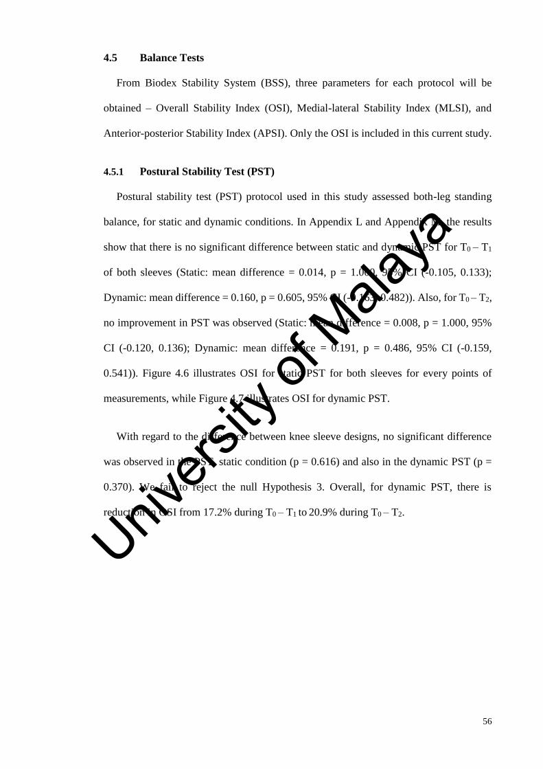
56
4.5 Balance Tests
From Biodex Stability System (BSS), three parameters for each protocol will be
obtained – Overall Stability Index (OSI), Medial-lateral Stability Index (MLSI), and
Anterior-posterior Stability Index (APSI). Only the OSI is included in this current study.
4.5.1 Postural Stability Test (PST)
Postural stability test (PST) protocol used in this study assessed both-leg standing
balance, for static and dynamic conditions. In Appendix L and Appendix M, the results
show that there is no significant difference between static and dynamic PST for T0 – T1
of both sleeves (Static: mean difference = 0.014, p = 1.000, 95% CI (-0.105, 0.133);
Dynamic: mean difference = 0.160, p = 0.605, 95% CI (-0.163, 0.482)). Also, for T0 – T2,
no improvement in PST was observed (Static: mean difference = 0.008, p = 1.000, 95%
CI (-0.120, 0.136); Dynamic: mean difference = 0.191, p = 0.486, 95% CI (-0.159,
0.541)). Figure 4.6 illustrates OSI for static PST for both sleeves for every points of
measurements, while Figure 4.7 illustrates OSI for dynamic PST.
With regard to the difference between knee sleeve designs, no significant difference
was observed in the PST, static condition (p = 0.616) and also in the dynamic PST (p =
0.370). We fail to reject the null Hypothesis 3. Overall, for dynamic PST, there is
reduction in OSI from 17.2% during T0 – T1 to 20.9% during T0 – T2.
Univers
ity of
Mala
ya

57
Figure 4.6: OSI for PST (Static) for pre, immediate and post effects according to
treatment groups. Overall [n=17], Simple knee sleeve (Knee Sleeve A) [n=9], Knee
sleeve with patella cutout (Knee Sleeve B) [n=8].
Figure 4.7: OSI for PST (Dynamic) for pre, immediate and post effects
according to treatment groups. Overall [n=17], Simple knee sleeve (Knee Sleeve A)
[n=9], Knee sleeve with patella cutout (Knee Sleeve B) [n=8].
0
0.1
0.2
0.3
0.4
0.5
0.6
Overall Knee Sleeve A Knee Sleeve B
Sta
bil
ity I
nd
ex
Pre Immediate Post
0
0.2
0.4
0.6
0.8
1
1.2
1.4
Overall Knee Sleeve A Knee Sleeve B
Sta
bil
ity I
nd
ex
Pre Immediate PostUniv
ersity
of M
alaya

58
4.5.2 Athlete Single Leg (ASL) Test
The ASL test was used to test the single-leg standing – for the affected knee and the
unaffected knee. For the affected knee, the results from our study show that there is no
significant difference between the static and the dynamic ASL tests on both sleeves for
T0 – T1 effects (Static: mean difference = -0.080, p = 1.000, 95% CI (-0.893, 0.734);
Dynamic: mean difference = 0.248, p = 1.000, 95% CI (-0.462, 0.958)) after the
immediate usage of both sleeves (refer Appendix N and Appendix O). After six weeks of
knee sleeve application, no improvement was observed (Static: mean difference = 0.392,
p = 0.167, 95% CI (-0.117, 0.900); Dynamic: mean difference = 0.276, p = 1.000, 95%
CI (-0.690, 1.243)). Based on the p-values, we fail to reject the null Hypothesis 3 (H0).
Figure 4.8 illustrates the OSI for ASL static test for the affected knee, for each knee
sleeve for every point of measurements. From the figure, no significant difference is
observed between the interventions for static test; mean difference = 0.200, p = 0.392,
95% CI (-0.284, 0.685). Figure 4.9 shows the graphical illustration for ASL dynamic test
for the affected knee. From Appendix O, there are 14.1% and 15.8% reduction in OSI,
for pre-immediate and pre-post effects, respectively.
Appendix P and Appendix Q show the results of the ASL test on the unaffected knee,
for the static and the dynamic tests, respectively. The results reveal no significant
difference in the static and the dynamic ASL tests for both types of sleeves for pre and
immediate effects on the affected knee (Static: mean difference = 0.106, p = 1.000, 95%
CI (-0.372, 0.584); Dynamic: mean difference = 0.219, p = 1.000, 95% CI (-0.434, 0.872))
after the immediate usage of both types of sleeves. For T0 – T2 effects, no improvement
was observed during the static test but a significant reduction in the OSI during the
dynamic ASL test (Static: mean difference = 0.300, p = 0.209, 95% CI (-0.268, 0.898);
Dynamic: mean difference = 0.744, p = 0.047, 95% CI (0.009, 1.479)).
Univers
ity of
Mala
ya

59
Figure 4.8: OSI for ASL (Affected Knee - Static) for pre, immediate and post
effects according to treatment groups. Overall [n=17], Simple knee sleeve (Knee
Sleeve A) [n=9], Knee sleeve with patella cutout (Knee Sleeve B) [n=8].
Figure 4.9: OSI for ASL (Affected Knee - Dynamic) for pre, immediate and post
effects according to treatment groups. Overall [n=17], Simple knee sleeve (Knee
Sleeve A) [n=9], Knee sleeve with patella cutout (Knee Sleeve B) [n=8].
In comparing the types of knee sleeves, no significant difference is observed in the
static test in the mean difference (p = 0.480) (Figure 4.10) and also for the dynamic test;
mean difference = -0.171, p = 0.527. Figure 4.10 and Figure 4.11 show the graphical
illustration of the ASL test on the unaffected knee, for the static and the dynamic tests,
respectively.
0
0.2
0.4
0.6
0.8
1
1.2
1.4
1.6
1.8
2
Overall Knee Sleeve A Knee Sleeve B
Sta
bil
ity I
nd
ex
Pre Immediate Post
0
0.2
0.4
0.6
0.8
1
1.2
1.4
1.6
1.8
2
Overall Knee Sleeve A Knee Sleeve B
Sta
bil
ity I
nd
ex
Pre Immediate Post
Univers
ity of
Mala
ya
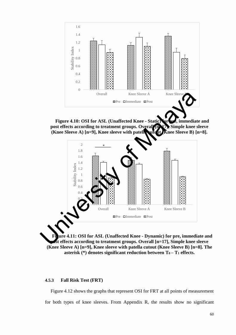
60
Figure 4.10: OSI for ASL (Unaffected Knee - Static) for pre, immediate and
post effects according to treatment groups. Overall [n=17], Simple knee sleeve
(Knee Sleeve A) [n=9], Knee sleeve with patella cutout (Knee Sleeve B) [n=8].
Figure 4.11: OSI for ASL (Unaffected Knee - Dynamic) for pre, immediate and
post effects according to treatment groups. Overall [n=17], Simple knee sleeve
(Knee Sleeve A) [n=9], Knee sleeve with patella cutout (Knee Sleeve B) [n=8]. The
asterisk (*) denotes significant reduction between T0 – T1 effects.
4.5.3 Fall Risk Test (FRT)
Figure 4.12 shows the graphs that represent OSI for FRT at all points of measurement
for both types of knee sleeves. From Appendix R, the results show no significant
0
0.2
0.4
0.6
0.8
1
1.2
1.4
1.6
Overall Knee Sleeve A Knee Sleeve B
Sta
bil
ity I
nd
ex
Pre Immediate Post
0
0.2
0.4
0.6
0.8
1
1.2
1.4
1.6
1.8
2
Overall Knee Sleeve A Knee Sleeve B
Sta
bil
ity I
nd
ex
Pre Immediate Post
*
Univers
ity of
Mala
ya

61
difference in the OSI between the two knee sleeves of different designs, regardless of
when the measurement were made; mean difference = 0.146, p = 0.398, 95% CI (-0.211,
0.503). For T0 – T1 scores, no significant reduction in OSI was observed; mean difference
= 0.131, p = 0.142, 95% CI (-0.032, 0.293 and similar changes were observed for T0 – T2;
mean difference = 0.067, p = 1.000, 95% CI (-0.177, 0.311).
As a summary for postural stability, from all the p-values, we fail to reject the null
hypothesis pertaining to postural stability (Hypothesis 3). Thus, the application of the
knee sleeves would not improve postural stability in our study participants.
Figure 4.12: OSI for FRT (Level 12 to Level 8) for pre, immediate and post
effects according to treatment groups. Overall [n=17], Simple knee sleeve (Knee
Sleeve A) [n=9], Knee sleeve with patella cutout (Knee Sleeve B) [n=8]
4.6 Summary
The results from all the measurements show significant improvements on our targeted
parameters: 1) pain level is reduced after the six weeks of knee sleeve application, 2)
significant increase in walking speed, 3) reduction in knee joint loading rates and KAM,
and 4) no significant reduction in postural stability assessment – except on single-leg
standing task. Between the intervention groups, there is no significant difference between
any of the targeted parameters, regardless of when the measurement were made.
0
0.1
0.2
0.3
0.4
0.5
0.6
0.7
0.8
0.9
1
Overall Knee Sleeve A Knee Sleeve B
Sta
bil
ity I
nd
ex
Pre Immediate Post
Univers
ity of
Mala
ya

62
CHAPTER 5: DISCUSSION
5.1 Introduction
This chapter discusses the study findings presented in the previous chapter, interprets
the results, and compares the findings with that of past studies. This chapter also discusses
the core aspects of the study under appropriate subsections as follows: 1) kinetics and
kinematics parameters; 2) postural stability; and 3) knee pain, stiffness, and functions.
This chapter also states the limitations of this current study, and the clinical implications
of the knee sleeves application in knee OA. A few often-used terms pertaining to when
the test measurement is taken are defined as follows: 1) pre – indicates the measurement
taken at baseline, 2) immediate – indicates the measurement taken after the immediate
use of knee sleeve, and 3) post – indicates the measurement taken following six weeks of
knee sleeve use.
5.2 Significance Findings
Overall, knee sleeves used in the current study have been effective in reducing pain
(based on WOMAC score) (42.9%), early stance peak KAM (38.9%), late stance peak
KAM (47.0%), GRF loading rates (first peak (27.1%), second peak (23.8%)), and
increased walking speed (9.5%) after six weeks of sleeve application. This study found a
significant reduction in WOMAC scores – on pain (44.7%), on stiffness (39.8%), and on
difficulty performing daily activities (42.1%). Significant differences in single-leg
postural stability test for unaffected leg (45.6%). No significant differences between the
knee sleeves in all tested parameters (p>0.05).
5.3 Effects of Knee Sleeve on Pain, Stiffness, and Physical Functions of the Knee
This present study also assessed the effects of knee sleeves on knee pain, stiffness and
dysfunctions associated with knee OA. In particular, it was aimed at determining how
pain in knee OA can be alleviated following six weeks of knee sleeve use. Pain has been
Univers
ity of
Mala
ya

63
the most patient-reported parameter in knee OA, therefore, it is important to explore the
patient-centered outcome pertaining to the beneficial effects of the knee sleeve.
To confirm the findings from the previous studies, WOMAC was used to evaluate
pain, stiffness and knee functions among the participants. WOMAC has been used
extensively in knee OA studies and has proven to be a reliable health instrument (Wolfe,
1999). From the scores, we would be able to assess the level of pain, stiffness and
difficulty in performing daily activities. Also, we tested using the knee sleeve alone –
without external stochastic stimulation or additional reinforcing features applied to the
sleeve – in order to generalize the effects of knee sleeves on the clinical symptoms of
knee OA.
5.3.1 Knee Pain
The findings from this study show that pain is significantly reduced following
immediate and post six-week of knee sleeve application. Statistically, the results from the
study show that a knee sleeve can reduce knee pain significantly immediately in 26.2%
of participants after the application of the sleeve, and can reduce knee pain significantly
in 42.9% of participants after six weeks of application of the sleeve. No significant
difference in knee pain reduction was observed between using knee sleeves of different
designs. This level of pain relief was larger than that observed by Hassan et al. (6–11%)
in a study that used a common elastic bandage, and observed in Mazzuca et al. (16%) in
a study that used knee sleeve (with heat retention properties) and simple knee sleeve,
following a four-week treatment period, in patients with knee OA (Hassan, Mockett, &
Doherty, 2002; Mazzuca, Page, Meldrum, Brandt, & Petty-Saphon, 2004). This reduction
is lower than a study which assessed the effects of Cingal Hyaluronic Acid on patients
with stage I-III knee OA, with a 70% reduction after 12 weeks (Hangody et al., 2017).
Univers
ity of
Mala
ya

64
Although knee sleeves have been claimed to have antalgic effects on people with knee
OA, there have not been many studies that assess the effects in more details (Bryk et al.,
2011). Previous studies suggested that knee sleeves might improve heat retention and
blood flow in knee OA, and this has been consistently supported by many studies (Bryk
et al., 2011; Mazzuca et al., 2004; Schween et al., 2015). Taping – which works in the
same way as the knee sleeve – relieves pain by improving blood circulation in the target
joint (Kwon et al., 2014).
With regard to the additional stochastic stimulation on the knee sleeves, we compared
our findings with the study of Collins et al. (2011). They found reduction in the pain,
denotes a presence of pain-relieving effect due to the additional stochastic stimulation on
the sleeves. This suggests that the additional warmth or external heat stimulus might give
additional benefit to the sleeve in reducing pain.
5.3.2 Knee Stiffness
Our findings show that most participants have a stiffed knee upon waking up. In the
average baseline measurement, high rate of knee stiffness was recorded, but the rate
reduced significantly after using the knee sleeve for six weeks (39.8%) – indicating the
benefits of the knee sleeve. This findings agreed with a previous study, Collins et al.
(2011) which found improvement in knee stiffness, using Knee Injury and Osteoarthritis
Outcome Score (KOOS) score. Our study found no significant difference in the effects
attributed to knee sleeve of different designs used in our study.
5.3.3 Functional Performance of the Knee
There are 14 questions in WOMAC pertaining to knee functions. These questions
attempt to determine a participant’s difficulty in performing these functions because of
the pain (Appendix D). It is well-known that knee pain can adversely affect the functional
activities of the knee (Foxworth, 2007). Our study found significant reduction in
Univers
ity of
Mala
ya
65
WOMAC scores – pertaining to difficulty in performing daily activities – following the
use of knee sleeves for six weeks (42.1%). This indicates an improvement in our
participants’ knee functions for them to perform the daily activities, following six weeks
of knee sleeves use.
Our findings are consistent with the findings of Bryk et al. (2011), who also found
statistically significant reduction of knee pain (p < 0.001), following the use of knee
sleeves. The other studies discussed above, only assessed the immediate effects of knee
sleeve use, but there has been no study that assesses their long-term effects. In our study,
we also assessed the effects of knee sleeve use over a six-week period, and found that the
knee sleeves reduce pain and stiffness, as well as improve functional activities of the
osteoarthritic knee.
We took precautions to prevent external factors such as fatigue and depression of
participants from influencing the outcomes of the trials by ensuring that they have
adequate rest and that they only rate the knee pain of the affected limb.
5.4 Effects of Knee Sleeve on Gait
The present study shows that the use of knee sleeves of two different designs: 1) simple
knee sleeve, and 2) simple knee sleeve with a patella cutout, for six weeks could improve
the knee mechanics in people with knee OA. Based on our findings, we advocate extended
usage of the knee sleeves to improve symptoms associated with knee OA especially knee
joint loading.
5.4.1 Knee Adduction Moment (KAM) and Ground Reaction Force (GRF)
The knee joint loading in knee OA individuals are said to be altered due to
proprioceptive deficits and mechanical alteration due to breakdown of cartilage (A.
Collins et al., 2014). Besides, KAM is considered a reliable parameter for identifying
Univers
ity of
Mala
ya
66
knee OA progression (Sharma et al., 1998). Our study shows further reduction in first
peak KAM after prolonged use of the knee sleeves – 39.0% reduction after six weeks of
use, and 13.9% reduction after immediate use compared to 10.1% reduction in first peak
KAM after immediate use (Schween et al., 2015). This clearly shows that prolonged knee
sleeves usage effects greater improvement to symptoms of knee OA, evident after the
treatment period.
Moreover, our findings highlight the efficacy of knee sleeve – without any stochastic
resonance electrical stimulation – in which Collins et al. (2011, 2014) have found no
significant in KAM. This adds the current configuration that stochastic resonance
stimulation did not enhance any effects on knee loading. Our study has established a
similar findings, in which we used the same population of knee OA individuals, with a
mild to moderate level of severity.
However, our study found a higher peak for late stance KAM, after the immediate
usage of knee sleeve, compared to the baseline. This finding denotes a higher loading
during the late stance of gait, after wearing the knee sleeve. We advocate the results due
to the adaptation of our participants with the knee sleeve while walking. After six weeks
of usage, there is a significant reduction in late stance KAM, denotes the treatment effect,
with a 44.9% reduction (p<0.005).
KAM is also positively correlated to GRF (Duffell et al., 2014; Jenkyn et al., 2011).
Our study shows that GRF loading rate during gait decreases significantly when using
both types of knee sleeves over a six-week period, together with KAM reduction. These
findings are similar to the findings of Collins et al. (2011). The authors explained that the
higher loading rate in people with knee OA causes faster cartilage degradation, but
following the use of knee sleeves, the loading rate is reduced. However, the actual
association between knee sleeve use and any mechanical change in joint loading is still
Univers
ity of
Mala
ya
67
not clearly understood. Some studies suggested that the change could be due to reduced
pain or improved proprioceptive ability brought about by the knee sleeves (Perlau, Frank,
& Fick, 1995; Van Tiggelen, Coorevits, & Witvrouw, 2008).
A study investigated on peak pressure and contact area of knee bracing when applied
to the knee (Wilson et al., 2010). They found a significant decrease in peak pressure and
significant increase contact area on the patellofemoral, when knee bracing is applied –
suggest to improve the abnormal patella tracking which lead to patellofemoral contact
pressure. Though, knee sleeve did not significantly reduce the peak pressure, but only has
a significant increment on the contact area. These finding suggests that while the
compression applied by sleeve-type braces increases contact area, it also increases contact
force, leading to no decrease in patellofemoral contact pressure.
Knee sleeves could also improve proprioceptive ability which will lead to more normal
gait pattern. It was previously suggested that the compression exerted by the knee sleeve
improves sensory stimulation and joint position sense (Schween et al., 2015; Van
Tiggelen et al., 2008). Thus, the improvements observed in our study could be due to the
extra support provided by the sleeve, which in turn, facilitates movement of the pained
knee or diseased knee. As a result, the knee gains more strength as those with knee OA
feel more confident in moving their knees.
5.4.2 Knee Range of Motion – Knee Flexion Angle
With regard to the knee range of motion, however, we did not found any significant
changes in the current study (55.8o to 54.5o). People with knee OA usually walks slower
with a reduced range of motion (Foroughi, Smith, & Vanwanseele, 2009). These
impairments could be troublesome to knee OA individuals as their movement is restricted
(Fitzgerald, Piva, & Irrgang, 2004). Knee range of motion is said to decrease with
increasing OA severity (Nagano et al., 2012). Therefore, since our findings did not found
Univers
ity of
Mala
ya
68
any significant observations, we suggest that simple knee sleeve could not improve nor
impede the range of motion of our participants.
A lower knee flexion angle at heel strike was recorded in this study and is significantly
reduced (18.8o to 14.0o). This finding contrasts the results obtained by Collins et al. (2011)
and the statement that knee OA individuals have a greater knee flexion at heel strike to
effectively reducing the GRF. However, the range of knee flexion is still within the
normal range for early knee OA (Nagano et al., 2012). In addition, no significant changes
is found in the range of knee extension, indicating that the range of motion neither degrade
nor improve after a six-week trial. This could be because our participants comprised those
diagnosed with early knee OA – their knees are not yet stiff and still have good flexion
range or our method in calculating knee kinematics is insufficiently accurate. Our study
also found negative results comparing to other similar studies – on peak knee flexion
during stance (Collins et al., 2011; Nagano et al., 2012). It is possible that avoidance gait
to pain or decreased knee stability induced by decreased knee extension strength
negatively affects the knee flexion angle (Nagano et al., 2012).
5.4.3 Walking Speed
In terms of walking speed, the findings indicate increase in walking speed following
the treatment period. Walking speed may decrease as the severity levels of knee OA is
increases (Pagani et al., 2010). Previous studies mentioned that in order to compensate
for pain, knee OA sufferers often reduce their walking speed to reduce the impact on the
joint (Amin et al., 2004; Foxworth, 2007). But our study found a significant reduction in
pain, together with the significant increase in walking speed. These suggest the positive
effects of the knee sleeves.
There is no studies investigated on the change in walking speed after the application
of knee sleeve. Thus, we compare our results with other bracing. Pagani et al. found no
Univers
ity of
Mala
ya

69
significant improvement in walking speed in their study’s participants after an immediate
use of flexible knee brace and knee brace with 4o valgus alignment – but, this could be
due their participants were among those with a grade IV knee OA (severe). Another study
uses grade II and grade III knee OA, and found significant increment in the walking speed
(4.4%), after four-week use of lateral-wedged foot insoles and knee brace. The study
found no significant difference in both types of intervention (Jones et al., 2013).
Supported with another study which found 10.2% increment in gait speed after five-week
usage of knee brace on knee OA with grade II and above (Laroche et al., 2014). While,
we found 9.5% increment in walking speed for grade I and grade II knee OA – with the
six-week intervention period. From these findings, it is agreeable that every level of
severity has to be managed appropriately i.e. knee brace is appropriate to moderate level
of severity; knee sleeve is appropriate for mild level of severity.
5.5 Effect of Knee Sleeve on Postural Stability
Biomechanically, whenever the movement of the knee is restricted, it would lead to
instability (Fitzgerald et al., 2004). Thus, our study was also aimed at determining
whether there is any improvement in postural stability of participants with early knee OA
after six weeks of knee sleeve application. It is believed that the knee sleeves can improve
proprioceptive ability (Herrington, Simmonds, & Hatcher, 2005). The results of our study
revealed that the use of knee sleeves of two different designs on a longer-term basis could
improve postural stability in participants with knee OA – a reduction, albeit not
significantly, in the overall stability index (OSI) in all tests. Also, there was no significant
difference in the effects when knee sleeves of two different designs were used. Knee
sleeves used in the current study effect reduction in the OSI following the stability tests,
and this indicates better stability.
Univers
ity of
Mala
ya

70
In our study, we used Biodex Stability System (BSS) to assess postural stability.
Arnold & Schmitz (1998) also used BSS and deemed it a good postural stability
measuring device that produces highly reliable measure of OSI. In addition, BSS is a user-
friendly machine and is easy to handle. It provides a screen monitor to display visual input
to the users to aid them in controlling their posture. The series of tests available in BSS
are also reliable and provide accurate scores to represent balance (Cachupe et al., 2001).
In the trial, we only considered the OSI. The OSI is a composite of the medial-lateral
stability index (MLSI) and anterior-posterior stability index (APSI), and is sensitive
enough to detect changes in any direction of the postural sway, and represents the overall
score for stability (Arnold & Schmitz, 1998). High stability index indicates higher sway
of motion, which means that the participant has problem in balancing.
In this study, different protocols on postural stability were assessed which involve both
static and dynamic tests (single-leg standing – using the Athlete Single-Leg (ASL) Test
and both-leg standing), and also the fall risk test. Findings from this study showed that
there is no significant changes on postural stability except the unaffected limb during a
single-leg standing dynamic test. There is no significant effect observed in other tests on
postural stability after six weeks of knee sleeve usage. However, we believe that even a
small reduction in OSI is indicative of the positive effects of the interventions.
5.5.1 Both-Leg Standing of Postural Stability
Standing balance is often part of the physical evaluation of lower-limb neuromuscular
function. In comparing our findings with that from previous studies, we note that Chuang
et al. (2007) found significant immediate improvement upon application of the knee
sleeve – 28% reduction in the stability index for the static test, and 7% - 8% reduction in
the stability index for the dynamic tests. Hassan et al. (2002) found significant
improvement in postural sway after applying L-bandage. Although our study did not
Univers
ity of
Mala
ya

71
produce similar findings, the single-leg test results also show reduction in the stability
index. The discrepancy between our findings and other studies could be due to the
different methods and tests used, as well as the different severity levels of the disease
among the participants or study subjects.
As discussed above, our test results show reduction in the stability index and this
indicates better balance ability after six weeks of knee sleeve usage. The full effects of
knee sleeve were not observed during the intervention period, probably because our
sample size was too small to be able to detect occurrence of type-2 error.
The results of previous studies to determine the underlying mechanism of knee sleeve
in postural stability suggest that the compression exerted by the knee sleeve improves
both proprioceptive ability and joint position sense, as well as postural stability
(Birmingham et al., 1998; McNair, Stanley, & Strauss, 1996). Our study outcomes also
suggest that our participants gain increased confidence regarding their postural stability
following the use of the knee sleeve. Many knee OA patients experience postural
instability, caused mainly by impaired proprioception of the knee. This, in turn, could
lead to alteration in gait patterns, which could usually be reversed by improvement in
postural stability (Schween et al., 2015). Kirkley et al. (1999) found that a neoprene knee
sleeve used by knee OA patients – regardless of their disease severity – can lead to a
modest decrease in the symptoms. This supports our findings that prolonged usage of the
knee sleeve could improve postural stability on our study participants.
With regard to pain associated with postural stability in knee OA, our study shows that
there is a reduction in the WOMAC scores in the tests, which indicates alleviation or
decrease in pain levels following the use of the knee sleeves for six weeks. However, over
the same period, we did not observe any significant change in postural stability. Pain is
seemingly related to balance, because we often observed that increased pain can lead to
Univers
ity of
Mala
ya
72
instability. To cope with pain associated with knee OA, people adapt in various ways such
as adopting certain standing styles, and walking patterns (Kim et al., 2011). It is well-
known that people with knee OA have impairment in position sense (static)
proprioception (Walker, Amstutz, & Rubinfeld, 1976). In this context, it is clear that there
is close correlation between postural stability and pain. In our study, however, we found
no significant reduction in the stability index, or in other words, no significant change in
postural stability. Hence, our study outcome is not able to reaffirm the aforementioned
correlation between postural stability and pain.
Owing to the limitations of our study, the results of the postural stability tests were not
sufficiently comprehensive. One limitation is the absence of functional tests. BSS is a
reliable tool but if it is complemented by relevant functional tests, it would provide a more
comprehensive understanding of postural stability. Besides, more levels of dynamic
testing should be applied. This study used a few levels of dynamic tests – level 8 (both-
leg standing) and level 4 (single-leg standing), guided by the proposed protocol in the
BSS manual. Besides, BSS produces relatively reliable values with only one decimal
point for the stability index, therefore, there is little difference between pre and post
measurements.
In addition, our measurements did not consider the brief loss of balance which was
allowed during the trials. It was obvious that all the participants have different degrees of
postural stability. Also, we did not take into consideration other parameters such as APSI
and MLSI. In our study, the choice of foot position was a self-made decision, hence, there
was no balance constraint. We suggest that balance should be controlled by the
participants depending on their own comfortable standing style.
This is the first study to assess the use of knee sleeves of different designs on both leg
testing and single-leg testing over a six-week period by using a balance machine. The
Univers
ity of
Mala
ya
73
findings from this study would be beneficial to clinicians who are always exploring ways
of managing pain in knee OA patients, and improving their quality of life. People with
balance problems inevitably lose confidence in their postural stability because balance
control indicates good physical functioning of the lower limbs to prevent incidence of
falling (Hunt, McManus, Hinman, & Bennell, 2010).
5.5.2 Single-Leg Standing Test of Postural Stability
Single-leg testing is conducted to evaluate balance as a whole and is related to the
modifiable factors, such as, lower extremity alignment, knee pain, and quadriceps
strength in people with knee OA (Hunt et al., 2010). Logically, falls may occur in
situations where only one foot is in contact with the surface. For example, people who
loses stability may trip while stepping over an obstacle. As people with knee OA often
experience instability, single-leg balance retraining is often part of many rehabilitation
programs and is crucial for them. Single-leg testing also allows us to evaluate the effects
on the affected limb without any help from the unaffected limb (Hunt et al., 2010). The
results in the current study show statistically significant changes to the unaffected limb in
the dynamic test after six weeks use of the knee sleeve. Unaffected limb in this context
represents the non-pained knee. The findings, however, could suggest that there would be
a significant compensatory stabilizing effect on both sides of the limb (Duffell et al.,
2014).
From the single-leg test that we conducted, the results show no significant reduction
in the stability index after six weeks of knee sleeves application. A possible explanation
is that the participants were at the early stage of knee OA, hence, most of them were able
to balance themselves properly – the disease has not affected their balance yet (Duffell et
al., 2014). The single-leg standing balance test, however, may not provide information
that assists clinicians in determining clinical change or functional level for knee OA
Univers
ity of
Mala
ya

74
patients, therefore, the need to evaluate both-leg standing in this individuals is necessary
(Harrison, Duenkel, Dunlop, & Russell, 1994).
5.5.3 Fall Risk
The risk of falls in people with knee OA is less well studied (Levinger et al., 2011). In
our study, however, we did not find any significant difference in all the three preset
schedules to take measurement: pre (baseline), immediate, and post 6-week of knee sleeve
use. We believe that it is possible for a type-2 error to occur under such circumstance
because at the early stages of our statistical analysis for pre and immediate effects of knee
sleeve use – with 15 participants – we observed significant reduction in the OSI. Fall risk
is one of the major concerns of people with knee OA because it could further aggravate
symptoms of the disease. Since people with knee OA have an impaired postural stability,
they need a device that can strengthen their weakened knee besides making them more
aware of the risk of falling. The risk of falling in people with knee OA can emanate from
many factors, such as, muscle weakness, poor proprioceptive acuity, and postural
imbalance (Sturnieks et al., 2004). Therefore, we should constantly strive to drastically
reduce the risk of falling among people with knee OA to avert more serious consequences.
No study related to knee sleeve identifying on fall risk. However, a study investigated
the effects of hyaluronic acid injection on mild-to-moderate level of knee OA. They found
significant reduction in the stability index after five dosage of injection (within six weeks)
(Khalaj et al., 2014). Comparing to our study, we did not found significant reduction in
the OSI. This can relate to the type of treatment. Hyaluronic acid injections are effective
in preserving knee pain, which consecutively help improving postural balance (Ministry
of Health Malaysia, 2013).
Univers
ity of
Mala
ya
75
5.6 Participants’ Lifestyle
Feedback to the questionnaire from our participants indicate that most of their lifestyle
requires them to be active. Such lifestyle may lead to faster degradation of the cartilage
of their knee joints. However, active people are less likely to develop knee OA at an early
age. Moreover, knee tissues and cartilage are more likely to remain in good condition if
people – of any lifestyle – learn to adapt impact loads (Nyland et al., 2015). None of our
participants are athletes which suggests that most of them lead sedentary lifestyle which
could be the reasons for the different responses to our targeted parameters. Besides, none
of them are smokers, thus, we believe that their knee OA is not attributed to smoking.
Only six participants had undergone a rehabilitation program. Six participants (31.6%)
out of 19 participants, had previous injuries, which might have involved the need for
various kinematics and kinetics treatments, and these injuries could have initiated the
knee OA, as mentioned earlier in Chapter 2. Other injuries reported included ligament
tear, falls and dislocation of patella, which could also have initiated their knee OA.
5.7 Participants’ Compliance and Adverse Effects of Knee Sleeve
The validity of the trial results and outcomes depends also on participants’ compliance
with the study protocols. Based on the data in the log book (subsection 4.3), the total daily
compliance for both knee sleeves application is 3.7 ± 2.4 hours.
The most reported problem regarding the knee sleeves is the poor fitting. The
participants were urged to adapt to the sleeves as best as they could as individually
customized sleeves were not available. There were also reports on skin irritation and
discomfort.
Univers
ity of
Mala
ya
76
5.8 Clinical Implications
Our results have important clinical implications on early knee OA. The use of knee
sleeves in people with early knee OA, can improve the functional performance of the knee
in their daily activities and delay disease progression. Using the knee sleeves for six
weeks can bring about pain reduction, improvement in joint moments and loading rates.
Our study also shows that there is no significant difference in the effects between the use
of simple knee sleeve and knee sleeve with patella cutout – both of different designs. Our
study is the first to evaluate and compare the biomechanical efficacy of the two different
designs of knee sleeves in a six-week trial on patients with early knee OA.
Any improvement in postural stability is crucial in preventing people from falling and
also to enable them to carry out their daily activities. This is because falls could lead to
serious health consequences and further aggravate the loading problems of the knee joint.
Any balance deficits people may have, are likely to affect their overall bodily functions
and inevitably their quality of life (Hsieh et al., 2013). For such people, any form of
treatment or intervention – such as the use of knee sleeves – would be of invaluable
benefits in their daily life.
The extensive use of knee sleeve today is largely attributed to the intense promotion
by the manufacturers. Its lower cost, compared to other orthotic aids, makes it affordable
to most knee OA sufferers. It is a simple aid and relatively easy to use. Other than
effecting physical functions improvement, knee sleeves are now a source of emotional
and therapeutic support to knee OA sufferers because of their beneficial properties.
Overall, the results from this study suggest that different designs of knee sleeves are
not significantly different in their effect on osteoarthritic knees.
Univers
ity of
Mala
ya

77
5.9 Summary
It can be said that patients’ perception of pain can result in altered knee mechanics and
postural stability. Knee sleeves could be working in this way to relieve pain and reduce
stiffness in the knee of people with knee OA. They help to reduce medial loading and also
the GRF. They also improve joint position sense, resulting in improved postural stability,
and knee functional performance. In summary, this chapter has highlighted new findings
from our study and compared them to that from previous studies – highlighting both
similarities and differences. This chapter also discusses the principles and relationships
of the parameters considered in this study. The clinical implications are also discussed.
The next chapter concludes the present study and suggests further researches on this same
topic.
Univers
ity of
Mala
ya
78
CHAPTER 6: CONCLUSION
6.1 Introduction
This study was undertaken to evaluate and compare the biomechanical effects of
knee sleeves of different designs on early unilateral knee OA. Our trials involved
observing the immediate effects of knee sleeves, and the effects after six weeks
application. We aim to determine whether knee sleeves can modify any of the
biomechanical parameters for the following reasons: 1) Help clinicians to comprehend
the indications and properties of the knee sleeves, and 2) Help fill the gap on
information pertaining to the biomechanical aspects of knee sleeves. The outcomes
from this study and achievement of the objectives would encourage wider use of knee
sleeves as an affordable and non-invasive intervention to relieve symptoms, especially
pain, in early knee OA.
The results from the trial show that application of the knee sleeves over six weeks
has many positive effects such as reduction in pain level, improvement in knee joint
moments and loading rates. This is the first study to focus on evaluating and comparing
the biomechanical mechanism of knee sleeves of two different designs used in a six-
week trial.
6.2 Limitations
Although the current study provides new information on the beneficial effects of
knee sleeves for our participants, this study has its limitations. We only compared the
immediate effects and the effects after six weeks of knee sleeve use with the baseline,
and within the same group of participants. The difference or similarity in the effects
would have been clearer if there had been a control group to compare. Besides, we
had focused on only one stage of knee OA – early knee OA – thus, our findings are
Univers
ity of
Mala
ya

79
only relevant to early knee OA. With regard to knee sleeve application, we did not
consider slippage of the knee sleeves during the trials. The slippage rate must be
considered to ensure minimal deviation errors and to confirm that the sleeve remains
intact throughout the trial. The grip of the knee sleeve might change after the walking
trial, and this could affect the results as well. Besides, feedback from our participants
indicate some problems with the sleeves such as the sleeves slipping downwards after
using them for some time. Owing to time constraint and other participant-related
factors, we were unable to recruit more participants for each intervention. However,
to reduce the error, we added three point of measurements for each participant – pre,
immediate, and post six-week effects.
6.3 Contribution of Research
From our findings, we can conclude that the two types of knee sleeves used in this
study are able to decrease pain and to improve knee functions in people with early
knee OA following six weeks of use. The knee sleeves also help in reducing joint
loading and increasing walking speed. The positive effects observed are attributed to
the reduction in pain and the extra support brought about by the sleeves during the six-
week treatment period. The knee sleeves could be used by people with early knee OA
on a longer term to bring about better improvement to their condition. Our findings
also indicate that the reduction in loading rates is probably due to the decreased KAM
during early stance and late stance, as well as marked reduction in pain levels which
is probably due to the increased feeling of stability from improvement in
proprioception following six weeks of knee sleeve application. The findings further
indicate that knee sleeves can improve postural stability and reduce risk of falling,
based on the reduction in the stability index.
Univers
ity of
Mala
ya

80
The improvements on the immediate effects agreed with similar previous studies,
which had investigated the immediate effects of knee sleeve – such as KAM, pain and
vGRF. These findings give additional merits to the knee sleeve as one of the treatment
approach to people with early knee OA. We also tested the postural stability on single-
leg standing, which has insufficient findings from past studies. Though our results did
not find any significant findings on the parameter, the idea to investigate the shingle-
leg standing balance should lead to future works that could focus on the importance of
the assessment. Our study also justifies the improvements that can be addressed to the
people with knee OA to have an easy intervention to easing their knee pain.
6.4 Future Research
Although the results of our study seem promising, future studies should evaluate
various designs of knee sleeves, and also include participants with different levels of
severities in knee OA. This is because knee sleeves are relatively easy and convenient
to use, besides being a cost-effective treatment option for early knee OA.
More researches should be undertaken on other pertinent aspects of knee sleeves
use in knee OA, such as: the experimental protocol; different designs of knee sleeves;
other gait parameters; different stages of knee OA; the underlying mechanism of knee
sleeves; and knee braces and knee sleeves with reinforcements. Further, the materials
and mechanical aspects of other knee supports, and the functional tests to give better
understanding of proprioception, balance and stability, use of other gait parameters
such as tibial rotation and knee flexion moment, to provide more information from
different aspects of gait, other gait studies such as ankle, knee and hip kinematics and
kinetics in all planes, and tests for high-speed walking, and wearing shoes as the
intervention method for outdoor tests.
Univers
ity of
Mala
ya

81
Future studies should explore the relationships between gait parameters of sagittal
plane with pain and knee functional performance, and also disease progression (Chang
et al., 2015). They mentioned that from sagittal biomechanics, it appears more relevant
to characterize the disease progression of knee OA. Besides, future studies should also
consider investigating the transverse plane biomechanics, as the past studies have put
more focus on the frontal and sagittal biomechanical parameters.
Future studies should also consider the role of quadriceps muscle strength in
people with knee OA, and whether increased muscle contraction leads to higher risk
of muscle stiffness or more joint loading. The correlation between these parameters
will provide further insights on knee OA. Besides, research should be conducted to
understand the long-term effect of altered muscle activity on the onset and progression
of knee OA.
To have a better understanding of the study population, more functional tests such
as Timed-up and Go (TUG), 8-metre Walking Test (8MW) and Stair Climb Power
Test (SCPT) should also be conducted (Bryk et al., 2011).
Because of our study limitations, further testing should involve the gender factor
because of the anthropometric difference and the influence of gender on the efficacy
of knee sleeves. Also, other factors such as BMI and participants’ lifestyle should be
explored, as those factors, combined, might provide different effects when using the
knee sleeve. These efforts will provide a more comprehensive understanding of the
effects of knee sleeves.
In terms of experimental protocols, future studies should control the foot position
during standing and more dynamic levels should be included. Besides, postural
Univers
ity of
Mala
ya

82
stability can also be improved with functional tests to fully comprehend balance ability
of knee OA population. Studies should also be undertaken on the adaptation periods
during trials, and provision for rest in between the tests, are advisable to prevent fatigue
among participants. The measurement of APSI and MLSI should be further explored
to obtain a better understanding of postural stability, besides the OSI. The standard
deviation obtained together with the OSI results should also be taken into
consideration. The BSS should be used to conduct a thorough and more accurate
evaluation of postural stability.
Further investigations should consider the BMI of the participants because a
person’s BMI can influence the level of loading (Runhaar, Koes, Clockaerts, &
Bierma-Zeinstra, 2011). In addition, specific parts of an osteoarthritic knee should be
considered in the inclusion criteria during recruitment of participants – such as the
medial part or lateral part of the knee. This is because Weidow et al. (2005) reported
that the pelvic and hip anatomy and biomechanical measurements of the medial and
lateral parts of the osteoarthritic knee are different.
6.5 Conclusion
Our study also indicates the need to further explore the use of knee sleeves of
different designs to evaluate their true effects. We conclude that knee sleeves effect
both biomechanical and clinical improvements to the symptoms of early knee OA
following knee sleeve use over a six-week trial. The study outcomes have met the
research objectives in helping clinicians to comprehend the indications and properties
of the knee sleeves, and helping researchers to have better insight on the biomechanical
action of knee sleeves.
Univers
ity of
Mala
ya
83
REFERENCES
Al-Zahrani, K. S., & Bakheit, A. M. O. (2002). A study of the gait characteristics of
patients with chronic osteoarthritis of the knee. Disability and Rehabilitation,
24(5), 275–280. http://doi.org/10.1080/09638280110087098
Altman, R., Asch, E., Bloch, D., Bole, G., Borenstein, D., Brandt, K., … Wolfe, F.
(1986). Development of criteria for the classification and reporting of
osteoarthritis: Classification of osteoarthritis of the knee. Arthritis & Rheumatism,
29(8), 1039–1049.
Amin, S., Luepongsak, N., McGibbon, C. a, LaValley, M. P., Krebs, D. E., & Felson,
D. T. (2004). Knee adduction moment and development of chronic knee pain in
elders. Arthritis & Rheumatism, 51(3), 371–376. http://doi.org/10.1002/art.20396
Andriacchi, T. P., & Mündermann, A. (2006). The role of ambulatory mechanics in
the initiation and progression of knee osteoarthritis. Current Opinion in
Rheumatology, 18(5), 514–518.
http://doi.org/10.1097/01.bor.0000240365.16842.4e
Arnold, B. L., & Schmitz, R. J. (1998). Examination of balance measures produced by
the biodex stability system. Journal of Athletic Training, 33(4), 323–327.
Asl, J. M., Kahrizi, S., Ebrahimi T., E., & Faghihzadeh, S. (2008). The effect of short-
term usage of rigid neoprene knee sleeve and soft neoprene knee sleeve on knee
joint position sense perception after reconstruction surgery of anterior cruciate
ligament. World Journal of Sport Sciences, 1(1), 42–47.
Astephen, J. L., Deluzio, K. J., Caldwell, G. E., Dunbar, M. J., & Hubley-Kozey, C.
L. (2008). Gait and neuromuscular pattern changes are associated with
differences in knee osteoarthritis severity levels. Journal of Biomechanics, 41(4),
868–876. http://doi.org/10.1016/j.jbiomech.2007.10.016
Astephen Wilson, J. L., Deluzio, K. J., Dunbar, M. J., Caldwell, G. E., & Hubley-
Kozey, C. L. (2011). The association between knee joint biomechanics and
neuromuscular control and moderate knee osteoarthritis radiographic and pain
severity. Osteoarthritis & Cartilage, 19(2), 186–93.
http://doi.org/10.1016/j.joca.2010.10.020
Baert, I. A. C., Jonkers, I., Staes, F., Luyten, F. P., Truijen, S., & Verschueren, S. M.
P. (2013). Gait characteristics and lower limb muscle strength in women with
early and established knee osteoarthritis. Clinical Biomechanics, 28(1), 40–47.
http://doi.org/10.1016/j.clinbiomech.2012.10.007
Bagge, E., Bjelle, A., Edén, S., & Svanborg, A. (1991). Osteoarthritis in the elderly:
Clinical and radiological findings in 79 and 85 year olds. Annals of the Rheumatic
Diseases, 50(8), 535–9. Retrieved from
http://www.pubmedcentral.nih.gov/articlerender.fcgi?artid=1004482&tool=pmc
entrez&rendertype=abstract
Univers
ity of
Mala
ya

84
Baltaci, G., Aktas, G., Camci, E., Oksuz, S., Yildiz, S., & Kalaycioglu, T. (2011). The
effect of prophylactic knee bracing on performance: Balance, proprioception,
coordination, and muscular power. Knee Surgery, Sports Traumatology,
Arthroscopy, 19(10), 1722–1728. http://doi.org/10.1007/s00167-011-1491-3
Beaudreuil, J., Bendaya, S., Faucher, M., Coudeyre, E., Ribinik, P., Revel, M., &
Rannou, F. (2009). Clinical practice guidelines for rest orthosis, knee sleeves, and
unloading knee braces in knee osteoarthritis. Joint Bone Spine, 76, 629–636.
http://doi.org/10.1016/j.jbspin.2009.02.002
Beaudreuil, J., Coudreuse, J. M., Guyen, C. N., Deat, P., Chabaud, A., Pereira, B., …
Coudeyre, E. (2016). An algorithm to improve knee orthosis prescription for
osteoarthritis patients. Annals of Physical and Rehabilitation Medicine, 59, e156.
http://doi.org/10.1016/j.rehab.2016.07.347
Bellamy, N. (2014). WOMAC Osteoarthritis Index.
Bennell, K. L., Bowles, K.-A., Wang, Y., Cicuttini, F., Davies-Tuck, M., & Hinman,
R. S. (2011). Higher dynamic medial knee load predicts greater cartilage loss over
12 months in medial knee osteoarthritis. Annals of the Rheumatic Diseases,
70(10), 1770–1774. http://doi.org/10.1136/ard.2010.147082
Bennell, K. L., Kean, C. O., Wrigley, T. V., & Hinman, R. S. (2013). Effects of a
modified shoe on knee load in people with and those without knee osteoarthritis.
Arthritis & Rheumatism, 65(3), 701–709. http://doi.org/10.1002/art.37788
Bijlsma, J. W. J., & Knahr, K. (2007). Strategies for the prevention and management
of osteoarthritis of the hip and knee. Best Practice & Research Clinical
Rheumatology, 21(1), 59–76. http://doi.org/10.1016/j.berh.2006.08.013
Biodex Medical Systems, I. (1999). Balance System Sd Operation / Service Manual,
6339, 117.
Birmingham, T. B., Bryant, D. M., Giffin, J. R., Litchfield, R. B., Kramer, J. F.,
Donner, A., & Fowler, P. J. (2008). A randomized controlled trial comparing the
effectiveness of functional knee brace and neoprene sleeve use after anterior
cruciate ligament reconstruction. The American Journal of Sports Medicine,
36(C), 648–655. http://doi.org/10.1177/0363546507311601
Birmingham, T. B., Kramer, J. F., Inglis, J. T., Mooney, C. a, Murray, L. J., Fowler,
P. J., & Kirkley, S. (1998). Effect of a neoprene sleeve on knee joint position
sense during sitting open kinetic chain and supine closed kinetic chain tests. The
American Journal of Sports Medicine, 26(4), 562–566.
Birmingham, T. B., Kramer, J. F., Kirkley, A., Inglis, J. T., Spaulding, S. J., &
Vandervoort, a a. (2001). Knee bracing for medial compartment osteoarthritis:
effects on proprioception and postural control. Rheumatology, 40, 285–289.
Retrieved from http://www.ncbi.nlm.nih.gov/pubmed/11285375
Bottoni, G., Heinrich, D., Kofler, P., Hasler, M., & Nachbauer, W. (2014). Effect of
Univers
ity of
Mala
ya

85
knee supports on knee joint position sense after uphill and downhill walking. A
test using a hiking simulation method. Journal of Ergonomics, 4(2), 2–5.
http://doi.org/10.4172/2165-7556.1000132
Bottoni, G., Herten, A., Kofler, P., Hasler, M., & Nachbauer, W. (2013). The effect of
knee brace and knee sleeve on the proprioception of the knee in young non-
professional healthy sportsmen. The Knee, 20, 490–492.
http://doi.org/10.1016/j.knee.2013.05.001
Bottoni, G., Kofler, P., Hasler, M., Giger, A., & Nachbauer, W. (2014). Effect of knee
braces on balance ability wearing ski boots (a pilot study). Procedia Engineering,
72, 327–331. http://doi.org/10.1016/j.proeng.2014.06.057
Brouwer, R. W., Van Raaij, T. M., Jakma, T. T., Verhagen, A. P., Verhaar, J. A., &
Bierma-zeinstra, Si. M. (2009). Braces and orthoses for treating osteoarthritis of
the knee (Intervention Review). The Cochrane Collaboration, (1).
http://doi.org/10.1002/14651858.CD004020.pub2.Copyright
Bryk, F. F., Jesus, J. F. De, Fukuda, T. Y., Moreira, E. G., Marcondes, F. B., & Santos,
M. G. Dos. (2011). Immediate effect of the elastic knee sleeve use on individuals
with osteoarthritis. Revista Brasileira de Reumatologia, 51(5), 434–446.
Retrieved from http://www.ncbi.nlm.nih.gov/pubmed/21952996
Cachupe, W. J. C., Shifflett, B., Kahanov, L., & Wughalter, E. H. (2001). Reliability
of Biodex Balance System measures. Measurement in Physical Education and
Exercise Science, 5(2), 97–108. http://doi.org/10.1207/S15327841MPEE0502_3
Chang, A. H., Chmiel, J. S., Almagor, O., Moisio, K. C., Belisle, L., Zhang, Y., …
Sharma, L. (2015). Association between knee sagittal plane dynamic joint
stiffness during gait and disease severity and pain in knee osteoarthritis (OA).
Osteoarthritis & Cartilage, 23(2015), A94–A95.
http://doi.org/10.1016/j.joca.2015.02.803
Christensen, R., Bartels, E. M., Astrup, A., & Bliddal, H. (2006). Effect of weight
reduction in obese patients diagnosed with knee osteoarthritis: A systematic
review and meta-analysis. Annals of the Rheumatic Diseases, 66(4), 433–439.
http://doi.org/10.1136/ard.2006.065904
Chuang, S., Huang, M., Chen, T., Weng, M., Liu, C., & Chen, C. (2007). Effect of
knee sleeve on static and dynamic balance in patients with knee osteoarthritis.
The Kaohsiung Journal of Medical Sciences, 23(8), 405–411.
Collins, A., Blackburn, T., Olcott, C., Jordan, J. M., Yu, B., & Weinhold, P. (2014). A
kinetic and kinematic anlaysis of the effect of stochastic resonance electrical
stimulation and knee sleeve during gait in osteoarthritis of the knee. Journal of
Applied Biomechanics, IN PRESS, 104–112. http://doi.org/10.1123/jab.2012-
0257
Collins, A. T., Blackburn, J. T., Olcott, C. W., Jordan, J. M., Yu, B., & Weinhold, P.
S. (2012). The assessment of postural control with stochastic resonance electrical
Univers
ity of
Mala
ya
86
stimulation and a neoprene knee sleeve in the osteoarthritic knee. Archives of
Physical Medicine and Rehabilitation, 93(July), 1123–1128.
http://doi.org/10.1016/j.apmr.2011.12.006
Collins, A. T., Blackburn, J. T., Olcott, C. W., Miles, J., Jordan, J., Dirschl, D. R., &
Weinhold, P. S. (2010). Stochastic resonance electrical stimulation to improve
proprioception in knee osteoarthritis. The Knee, 18(5), 317–322.
http://doi.org/10.1016/j.knee.2010.07.001
Collins, A. T., Blackburn, J. T., Olcott, C., Yu, B., & Weinhold, P. (2011). The impact
of stochastic resonance electrical stimulation and knee sleeve on impulsive
loading and muscle co-contraction during gait in knee osteoarthritis. Clinical
Biomechanics, 26(8), 853–858.
http://doi.org/10.1016/j.clinbiomech.2011.04.011
Creaby, M. W., Bennell, K. L., & Hunt, M. A. (2012). Gait differs between unilateral
and bilateral knee osteoarthritis. Archives of Physical Medicine and
Rehabilitation, 93(5), 822–827. http://doi.org/10.1016/j.apmr.2011.11.029
Creaby, M. W., Hunt, M. a., Hinman, R. S., & Bennell, K. L. (2013). Sagittal plane
joint loading is related to knee flexion in osteoarthritic gait. Clinical
Biomechanics, 28(8), 916–920.
http://doi.org/10.1016/j.clinbiomech.2013.07.013
da Silva, H. G. P. V., Cliquet Junior, A., Zorzi, A. R., & Batista de Miranda, J. (2012).
Biomechanical changes in gait of subjects with medial knee osteoarthritis. Acta
Ortopedica Brasileira, 20(3), 150–6. http://doi.org/10.1590/S1413-
78522012000300004
Davis, R. B. (1988). Clinical gait analysis. IEEE Engineering in Medicine and Biology
Magazine, (September), 389–406. http://doi.org/10.1016/B978-0-12-396961-
3.00025-1
Davis, R. B. (1997). Reflections on clinical gait analysis. Journal of Electromyography
and Kinesiology, 7(4), 251–257. http://doi.org/10.1016/S1050-6411(97)00008-4
Deshaies, L. D. (2002). Upper extremity orthoses. Occupational Therapy for Physical
Dysfunction. 5th Edition. Baltimore MD: Lippincott, Williams & Wilkins, 313–
349.
Dessery, Y., Belzile, É. L., Turmel, S., & Corbeil, P. (2014). Comparison of three knee
braces in the treatment of medial knee osteoarthritis. The Knee.
http://doi.org/10.1016/j.knee.2014.07.024
DonJoy USA. (2016). Drytex Knee Support. Retrieved from
http://www.donjoy.eu/en_US/54838Drytex-Knee-Support-.html#characteristics
Duffell, L. D., Southgate, D. F. L., Gulati, V., & McGregor, A. H. (2014). Balance and
gait adaptations in patients with early knee osteoarthritis. Gait & Posture, 39(4),
1057–1061. http://doi.org/10.1016/j.gaitpost.2014.01.005
Univers
ity of
Mala
ya

87
Egloff, C., Hügle, T., & Valderrabano, V. (2012). Biomechanics and pathomechanisms
of osteoarthritis. Swiss Medical Weekly, 142(July), w13583.
http://doi.org/10.4414/smw.2012.13583
Elbaz, A., Mor, A., Segal, G., Debbi, E., Haim, A., Halperin, N., & Debi, R. (2010).
APOS therapy improves clinical measurements and gait in patients with knee
osteoarthritis. Clinical Biomechanics, 25(9), 920–5.
http://doi.org/10.1016/j.clinbiomech.2010.06.017
Eng Hoe, W. (2009). Malaysia : Health and exercise statistics at a glance, 39–41.
Farrokhi, S., O’Connell, M., Gil, A., & Kelley Fitzgerald, G. (2013). Alterations in
sagittal-plane knee joint kinematics and kinetics during gait in knee osteoarthritis
patients with complaints of instability. Osteoarthritis & Cartilage, 21(2013), S88.
http://doi.org/10.1016/j.joca.2013.02.190
Felson, D. T. (2013). Osteoarthritis as a disease of mechanics. Osteoarthritis &
Cartilage, 21(1), 10–5. http://doi.org/10.1016/j.joca.2012.09.012
Field, A. (2012). Repeated Measures ANOVA. Retrieved November 29, 2017, from
https://www.google.com/url?sa=t&rct=j&q=&esrc=s&source=web&cd=3&cad
=rja&uact=8&ved=0ahUKEwiGoPK8suPXAhVKKY8KHWHfArsQFghCMAI
&url=http%3A%2F%2Fhealth.uottawa.ca%2Fbiomech%2Fcourses%2Fapa610
1%2FRepeated%2520Measures%2520ANOVA.pdf&usg=AOvVaw0zx76BOZ
U5tVQE8K1U
Fitzgerald, G. K., Piva, S. R., & Irrgang, J. J. (2004). Reports of joint instability in
knee osteoarthritis: its prevalence and relationship to physical function. Arthritis
& Rheumatism, 51(6), 941–946. http://doi.org/10.1002/art.20825
Foo, C., Manohar, A., Rampal, L., Lye, M., Sherina, M., & Zubaidah, J. (2017). Knee
pain and functional disability of knee osteoarthritis patients seen at Malaysian
tertiary hospitals. Malaysian Journal of Medicine & Health Sciences, June: in
p(June), 7–15.
Foroughi, N., Smith, R., & Vanwanseele, B. (2009). The association of external knee
adduction moment with biomechanical variables in osteoarthritis: A systematic
review. The Knee, 16(5), 303–309. http://doi.org/10.1016/j.knee.2008.12.007
Foxworth, J. L. (2007). Effects of shock absorbing insoles on knee pain, funtional
mobility, and lower extremity biomechanics in persons with symptomatic knee
osteoarthritis.
Fransen, M., Bridgett, L., March, L., Hoy, D., Penserga, E., & Brooks, P. (2011). The
epidemiology of osteoarthritis in Asia. International Journal of Rheumatic
Diseases, 14(2), 113–21. http://doi.org/10.1111/j.1756-185X.2011.01608.x
Giotis, D., Tsiaras, V., Ristanis, S., Zampeli, F., Mitsionis, G., Stergiou, N., &
Georgoulis, A. D. (2011). Knee braces can decrease tibial rotation during pivoting
that occurs in high demanding activities. Knee Surgery, Sports Traumatology,
Univers
ity of
Mala
ya

88
Arthroscopy, 19(8), 1347–1354. http://doi.org/10.1007/s00167-011-1454-8
Guilak, F. (2011). Biomechanical factors in osteoarthritis. Best Practice & Research
Clinical Rheumatology, 25(6), 815–23. http://doi.org/10.1016/j.berh.2011.11.013
Hangody, L., Szody, R., Lukasik, P., Zgadzaj, W., Lénárt, E., Dokoupilova, E., …
Szendroi, M. (2017). Intraarticular Injection of a Cross-Linked Sodium
Hyaluronate Combined with Triamcinolone Hexacetonide (Cingal) to Provide
Symptomatic Relief of Osteoarthritis of the Knee: A Randomized, Double-Blind,
Placebo-Controlled Multicenter Clinical Trial. Cartilage, 194760351770373.
http://doi.org/10.1177/1947603517703732
Harrison, E. L., Duenkel, N., Dunlop, R., & Russell, G. (1994). Evaluation of single-
leg standing following anterior cruciate ligament surgery and rehabilitation.
Physical Therapy, 74(3), 245–252.
Hassan, B. S., Mockett, S., & Doherty, M. (2001). Static postural sway,
proprioception, and maximal voluntary quadriceps contraction in patients with
knee osteoarthritis and normal control subjects. Annals of the Rheumatic
Diseases, 60(6), 612–618. http://doi.org/10.1136/ard.60.6.612
Hassan, B. S., Mockett, S., & Doherty, M. (2002). Influence of elastic bandage on knee
pain, proprioception, and postural sway in subjects with knee osteoarthritis.
Annals of the Rheumatic Diseases, 61(1), 24–28.
Health Grove. (n.d.). Osteoarthritis in Malaysia. Retrieved November 25, 2017, from
http://global-disease-burden.healthgrove.com/l/76335/Osteoarthritis-in-
Malaysia
Heiden, T. L., Lloyd, D. G., & Ackland, T. R. (2009). Knee joint kinematics , kinetics
and muscle co-contraction in knee osteoarthritis patient gait. Clinical
Biomechanics, 24(10), 833–841.
http://doi.org/10.1016/j.clinbiomech.2009.08.005
Henriksen, M., Aaboe, J., & Bliddal, H. (2012). The relationship between pain and
dynamic knee joint loading in knee osteoarthritis varies with radiographic disease
severity. A cross sectional study. The Knee, 19(4), 392–398.
http://doi.org/10.1016/j.knee.2011.07.003
Henriksen, M., Hunter, D. J., Dam, E. B., Messier, S. P., Andriacchi, T. P., Lohmander,
L. S., … Christensen, R. (2013). Is increased joint loading detrimental to obese
patients with knee osteoarthritis? A secondary data analysis from a randomized
trial. Osteoarthritis & Cartilage, 21(12), 1865–75.
http://doi.org/10.1016/j.joca.2013.10.003
Herrington, L., Simmonds, C., & Hatcher, J. (2005). The effect of a neoprene sleeve
on knee joint position sense. Research in Sports Medicine, 13, 37–46.
http://doi.org/10.1080/15438620590922077
Hinman, R. S., Payne, C., Metcalf, B. E. N. R., Wrigley, T. I. M. V, Bennell, K. I. M.
Univers
ity of
Mala
ya

89
L., Wrigley, T. V, & Ben-, K. L. (2008). Lateral wedges in knee osteoarthritis:
What are their immediate clinical and biomechanical effects and can these predict
a three-month clinical outcome? Arthritis & Rheumatism, 59(3), 408–415.
http://doi.org/10.1002/art.23326
Hsieh, R.-L., Lee, W.-C., Lo, M.-T., & Liao, W.-C. (2013). Postural stability in
patients with knee osteoarthritis: comparison with controls and evaluation of
relationships between postural stability scores and International Classification of
Functioning, Disability and Health components. Archives of Physical Medicine
and Rehabilitation, 94(2), 340–6. http://doi.org/10.1016/j.apmr.2012.09.022
Hunt, M. a., McManus, F. J., Hinman, R. S., & Bennell, K. L. (2010). Predictors of
single-leg standing balance in individuals with medial knee osteoarthritis.
Arthritis Care & Research, 62(4), 496–500. http://doi.org/10.1002/acr.20046
Hunter, D. J., Harvey, W., Gross, K. D., Felson, D., McCree, P., Li, L., … Bennell, K.
(2011). A randomized trial of patellofemoral bracing for treatment of
patellofemoral osteoarthritis. Osteoarthritis & Cartilage, 19(7), 792–800.
http://doi.org/10.1016/j.joca.2010.12.010
Hurley, S. T., Murdock, G. L. H., Stanish, W. D., & Hubley-Kozey, C. L. (2012). Is
there a dose response for valgus unloader brace usage on knee pain, function, and
muscle strength? Archives of Physical Medicine and Rehabilitation, 93(3), 496–
502. http://doi.org/10.1016/j.apmr.2011.09.002
Hurwitz, D., Sharma, L., & Andriacchi, T. (1999). Effect of knee pain on joint loading
in patients with osteoarthritis. Current Opinion in Rheumatology, 11, 422–426.
Jenkyn, T. R., Erhart, J. C., & Andriacchi, T. P. (2011). An analysis of the mechanisms
for reducing the knee adduction moment during walking using a variable stiffness
shoe in subjects with knee osteoarthritis. Journal of Biomechanics, 44(7), 1271–
6. http://doi.org/10.1016/j.jbiomech.2011.02.013
Jones, L. D., Bottomley, N., Harris, K., Jackson, W., Price, A. J., & Beard, D. J. (2014).
The clinical symptom profile of early radiographic knee arthritis: a pain and
function comparison with advanced disease. Knee Surgery, Sports Traumatology,
Arthroscopy, 24(1), 161–168. http://doi.org/10.1007/s00167-014-3356-z
Jones, R. K., Chapman, G. J., Forsythe, L., Parkes, M. J., & Felson, D. T. (2014). The
relationship between reductions in knee loading and immediate pain response
whilst wearing lateral wedged insoles in knee osteoarthritis. Journal of
Orthopaedic Research, 32(9), 1147–1154. http://doi.org/10.1002/jor.22666.The
Jones, R. K., Nester, C. J., Richards, J. D., Kim, W. Y., Johnson, D. S., Jari, S., …
Tyson, S. F. (2013). A comparison of the biomechanical effects of valgus knee
braces and lateral wedged insoles in patients with knee osteoarthritis. Gait &
Posture, 37(3), 368–72. http://doi.org/10.1016/j.gaitpost.2012.08.002
Jordan, K. M., Arden, N. K., Doherty, M., Bannwarth, B., Bijlsma, J. W. J., Dieppe,
P., & Gunther, K. (2003). EULAR Recommendations 2003: An evidence based
Univers
ity of
Mala
ya
90
approach to the management of knee osteoarthritis: Report of a Task Force of the
Standing Committee for International Clinical Studies Including Therapeutic
Trials (ESCISIT). Annals of the Rheumatic Diseases, 62(12), 1145–1155.
http://doi.org/10.1136/ard.2003.011742
Kanamori, A., Zeminski, J., Rudy, T. W., Li, G., Fu, F. H., & Woo, S. L. Y. (2002).
The effect of axial tibial torque on the function of the anterior cruciate ligament:
A biomechanical study of a simulated pivot shift test. Arthroscopy, 18(4), 394–
398. http://doi.org/10.1053/jars.2002.30638
Karimi, N., Ebrahimi, I., Kahrizi, S., & Torkaman, G. (2008). Reliability of postural
balance evaluation using the biodex balance system in subjects with and without
low back pain. JPMI - Journal of Postgraduate Medical Institute, 22(2), 95–101.
Kaufman, K. R., Hughes, C., Morrey, B. F., Morrey, M., & An, K. N. (2001). Gait
characteristics of patients with knee osteoarthritis. Journal of Biomechanics,
34(7), 907–915. http://doi.org/10.1016/S0021-9290(01)00036-7
Kellgren, J. H., & Lawrence, J. S. (1957). Radiological assessment of osteo-arthrosis.
Annals of the Rheumatic Diseases, 16, 494–502.
Khalaj, N., Abu Osman, N. A., Mokhtar, A. H., George, J., & Abas, W. A. B. W.
(2014). Effect of intra-articular hyaluronic injection on postural stability and risk
of fall in patients with bilateral knee osteoarthritis. The Scientific World Journal,
2014. http://doi.org/10.1155/2014/815184
Khalaj, N., Abu Osman, N. A., Mokhtar, A. H., Mehdikhani, M., & Wan Abas, W. A.
B. (2014a). Balance and risk of fall in individuals with bilateral mild and
moderate knee osteoarthritis. PLoS ONE, 9(3), e92270.
http://doi.org/10.1371/journal.pone.0092270
Khalaj, N., Abu Osman, N. a, Mokhtar, A. H., Mehdikhani, M., & Wan Abas, W. a B.
(2014b). Effect of exercise and gait retraining on knee adduction moment in
people with knee osteoarthritis. Journal of Engineering in Medicine, 228(2), 190–
199. http://doi.org/10.1177/0954411914521155
Khan, S. J., Khan, S. S., Usman, J., Mokhtar, A. H., & Osman, N. A. A. (2016). A
Pilot Study on Physical Performance Measures: What is Better for Knee
Osteoarthritis Patients, Orthosis or Gait Modifications? Movement, Health &
Exercise Journal.
Kim, H.-S., Yun, D. H., Yoo, S. D., Kim, D. H., Jeong, Y. S., Yun, J.-S., … Choi, S.
H. (2011). Balance control and knee osteoarthritis severity. Annals of
Rehabilitation Medicine, 35(5), 701. http://doi.org/10.5535/arm.2011.35.5.701
Kirkley, A., Webster-Bogaert, S., Litchfield, R., Amendola, A., MacDonald, S.,
McCalden, R., & Fowler, P. (1999). The effect of bracing on varus gonarthrosis.
The Journal of Bone and Joint Surgery, 81(4), 539–48. Retrieved from
http://jbjs.org/content/81/4/539.abstract
Univers
ity of
Mala
ya
91
Kirtley, C. (2006). The tempora-spatial parameters. In Clinical Gait Analysis.
Knoop, J., Dekker, J., van der Leeden, M., van der Esch, M., Thorstensson, C. a,
Gerritsen, M., … Steultjens, M. P. M. (2013). Knee joint stabilization therapy in
patients with osteoarthritis of the knee: A randomized controlled trial.
Osteoarthritis & Cartilage, 21(8), 1025–34.
http://doi.org/10.1016/j.joca.2013.05.012
Kwon, H., Park, D., Jeong, J. R., & Jung, K. (2014). The effect of silicone sleeve and
taping on balance and strength in anterior cruciate ligament reconstruction
patients. The Journal of Korean Society of Physical Therapy, 26(3), 147–155.
Lamberg, E. M., Streb, R., Werner, M., Kremenic, I., & Penna, J. (2015). The 2- and
8-week effects of decompressive brace use in people with medial compartment
knee osteoarthritis. Prosthetics and Orthotics International.
http://doi.org/10.1177/0309364615589537
Landry, S. C., McKean, K. a, Hubley-Kozey, C. L., Stanish, W. D., & Deluzio, K. J.
(2007). Knee biomechanics of moderate OA patients measured during gait at a
self-selected and fast walking speed. Journal of Biomechanics, 40(8), 1754–61.
http://doi.org/10.1016/j.jbiomech.2006.08.010
Laroche, D., Morisset, C., Fortunet, C., Gremeaux, V., Maillefert, J.-F., & Ornetti, P.
(2014). Biomechanical effectiveness of a distraction-rotation knee brace in
medial knee osteoarthritis: preliminary results. The Knee, 21(3), 710–716.
http://doi.org/10.1016/j.knee.2014.02.015
Levinger, P., Menz, H. B., Morrow, A. D., Feller, J. A., Bartlett, J. R., & Bergman, N.
R. (2012). Foot kinematics in people with medial compartment knee
osteoarthritis. Rheumatology, 51(August), 2191–2198.
http://doi.org/10.1093/rheumatology/kes222
Levinger, P., Menz, H. B., Wee, E., Feller, J. a, Bartlett, J. R., & Bergman, N. R.
(2011). Physiological risk factors for falls in people with knee osteoarthritis
before and early after knee replacement surgery. Knee Surgery, Sports
Traumatology, Arthroscopy, 19(7), 1082–9. http://doi.org/10.1007/s00167-010-
1325-8
Lewek, M. D., Rudolph, K. S., & Snyder-Mackler, L. (2004). Control of frontal plane
knee laxity during gait in patients with medial compartment knee osteoarthritis.
Osteoarthritis & Cartilage, 12(9), 745–751.
http://doi.org/10.1016/j.joca.2004.05.005
Liikavainio, T., Isolehto, J., Helminen, H. J., Perttunen, J., Lepola, V., Kiviranta, I., …
Komi, P. V. (2016). Loading and gait symmetry during level and stair walking in
asymptomatic subjects with knee osteoarthritis: Importance of quadriceps femoris
in reducing impact force during heel strike? The Knee, 14(3), 231–238.
http://doi.org/10.1016/j.knee.2007.03.001
Maleki, M., Arazpour, M., Joghtaei, M., Hutchins, S. W., Aboutorabi, a., & Pouyan,
Univers
ity of
Mala
ya

92
a. (2014). The effect of knee orthoses on gait parameters in medial knee
compartment osteoarthritis: A literature review. Prosthetics and Orthotics
International. http://doi.org/10.1177/0309364614547411
Maly, M. R., Costigan, P. A., & Olney, S. J. (2008). Mechanical factors relate to pain
in knee osteoarthritis. Clinical Biomechanics, 23(6), 796–805.
http://doi.org/10.1016/j.clinbiomech.2008.01.014
Mannion, A. F., Balagué, F., Pellisé, F., & Cedraschi, C. (2007). Pain measurement in
patients with low back pain. Nature Clinical Practice Rheumatology, (3), 610–
618. Retrieved from
http://www.nature.com/nrrheum/journal/v3/n11/full/ncprheum0646.html
Mazzuca, S. a, Page, M. C., Meldrum, R. D., Brandt, K. D., & Petty-Saphon, S. (2004).
Pilot study of the effects of a heat-retaining knee sleeve on joint pain, stiffness,
and function in patients with knee osteoarthritis. Arthritis & Rheumatism, 51(5),
716–721. http://doi.org/10.1002/art.20683
McKean, K. a, Landry, S. C., Hubley-Kozey, C. L., Dunbar, M. J., Stanish, W. D., &
Deluzio, K. J. (2007). Gender differences exist in osteoarthritic gait. Clinical
Biomechanics, 22(4), 400–9. http://doi.org/10.1016/j.clinbiomech.2006.11.006
McNair, P. J., Stanley, S. N., & Strauss, G. R. (1996). Knee bracing: Effects on
proprioception. Archives of Physical Medicine and Rehabilitation, 77(March),
287–289. http://doi.org/10.1016/S0003-9993(96)90114-8
Ministry of Health Malaysia. (2013). Management of osteoarthritis.
Mohd Sharif, N. A., Goh, S. L., Usman, J., & Wan Safwani, W. K. Z. (2017).
Biomechanical and functional efficacy of knee sleeves: A literature review.
Physical Therapy in Sport, 28, 44–52. http://doi.org/10.1016/j.ptsp.2017.05.001
Mortaza, N., Ebrahimi, I., Jamshidi, A. A., Abdollah, V., Kamali, M., Abas, W. A. B.
W., & Osman, N. A. A. (2012). The effects of a prophylactic knee brace and two
neoprene knee sleeves on the performance of healthy athletes: A crossover
randomized controlled trial. PLoS ONE, 7(11), 1–6.
http://doi.org/10.1371/journal.pone.0050110
Moyer, R. F., Birmingham, T. B., Bryant, D. M., Giffin, J. R., Marriott, K. a., & Leitch,
K. M. (2015). Biomechanical effects of valgus knee bracing: a systematic review
and meta-analysis. Osteoarthritis & Cartilage, 23(2), 178–188.
http://doi.org/10.1016/j.joca.2014.11.018
Mündermann, A., Dyrby, C. O., Hurwitz, D. E., Sharma, L., & Andriacchi, T. P.
(2004). Potential strategies to reduce medial compartment loading in patients with
knee osteoarthritis of varying severity: Reduced walking speed. Arthritis &
Rheumatism, 50(4), 1172–1178. http://doi.org/10.1002/art.20132
Nagano, Y., Naito, K., Saho, Y., Torii, S., Ogata, T., Nakazawa, K., … Fukubayashi,
T. (2012). Association between in vivo knee kinematics during gait and the
Univers
ity of
Mala
ya

93
severity of knee osteoarthritis. The Knee, 19(5), 628–32.
http://doi.org/10.1016/j.knee.2011.11.002
Nyland, J., Wera, J., Henzman, C., Miller, T., Jakob, R., & Caborn, D. N. M. (2015).
Preserving knee function following osteoarthritis diagnosis: A sustainability
theory and social ecology clinical commentary. Physical Therapy in Sport, 16(1),
3–9. http://doi.org/10.1016/j.ptsp.2014.07.003
O’Connell, M., Farrokhi, S., & Kelley Fitzgerald, G. (2015). The role of knee joint
moments and knee impairments on self-reported knee pain during gait in patients
with knee osteoarthritis. Clinical Biomechanics.
http://doi.org/10.1016/j.clinbiomech.2015.10.003
Pagani, C. H. F., Böhle, C., Potthast, W., & Brüggemann, G.-P. (2010). Short-term
effects of a dedicated knee orthosis on knee adduction moment, pain, and function
in patients with osteoarthritis. Archives of Physical Medicine and Rehabilitation,
91(12), 1936–1941. http://doi.org/10.1016/j.apmr.2010.09.003
Paquette, M. R. (2012). Effects of increased step width on knee joint biomechanics in
healthy and knee osteoarthritis older adults during stair descent.
Park, H. J., Ko, S., Hong, H. M., Ok, E., & Lee, J. I. (2013). Factors related to standing
balance in patients with knee osteoarthritis. Annals of Rehabilitation Medicine,
37(3), 373–378. http://doi.org/10.5535/arm.2013.37.3.373
Perlau, R., Frank, C., & Fick, G. (1995). The effect of elastic bandages on human knee
proprioception in the uninjured population. The American Journal of Sports
Medicine, 23(2), 251–255. http://doi.org/10.1177/036354659502300221
Ramsey, D. K., Briem, K., Axe, M. J., & Synder-Mackler, L. (2011). A mechanical
hypothesis for the effectiveness of knee bracing for medial compartment knee
osteoarthritis. Bone Joint Surgery, 89(11), 2398–2407.
http://doi.org/10.2106/JBJS.F.01136.A
Riskowski, J. L., Mikesky, A. E., Bahamonde, R. E., Alvey, T. V., & Burr, D. B.
(2005). Proprioception, gait kinematics, and rate of loading during walking: Are
they related? Journal of Musculoskeletal Neuronal Interactions, 5(4), 379–387.
Runhaar, J., Koes, B. W., Clockaerts, S., & Bierma-Zeinstra, S. M. a. (2011). A
systematic review on changed biomechanics of lower extremities in obese
individuals: a possible role in development of osteoarthritis. Obesity Reviews : An
Official Journal of the International Association for the Study of Obesity, 12(12),
1071–82. http://doi.org/10.1111/j.1467-789X.2011.00916.x
Sanchez-Ramirez, D. C., Van Der Leeden, M., Knol, D. L., Van Der Esch, M., Roorda,
L. D., Verschueren, S., … Dekker, J. (2013). Association of postural control with
muscle strength, proprioception, self-reported knee instability and activity
limitations in patients with knee osteoarthritis. Journal of Rehabilitation
Medicine, 45(2), 192–197. http://doi.org/10.2340/16501977-1087
Univers
ity of
Mala
ya

94
Sasek, C. (2015). An update on primary care management of Knee Osteoarthritis.
Journal of the American Academy of Physician Assistants, 37–43.
http://doi.org/10.1097/01.JAA.0000458853.38655.02
Schiphof, D., de Klerk, B. M., Koes, B. W., & Bierma-Zeinstra, S. (2008). Good
reliability, questionable validity of 25 different classification criteria of knee
osteoarthritis: a systematic appraisal. Journal of Clinical Epidemiology, 61(12),
1205–1215.e2. http://doi.org/10.1016/j.jclinepi.2008.04.003
Schipplein, D., & Andriacchi, T. P. (1991). Interaction between active and passive
knee stabilizers during level walking. Journal of Orthopaedic Research, 9, 113–
119.
Schmitt, L. C., & Rudolph, K. S. (2007). Influences on knee movement strategies
during walking in persons with medial knee osteoarthritis. Arthritis Care and
Research, 57(6), 1018–1026. http://doi.org/10.1002/art.22889
Schween, R., Gehring, D., & Gollhofer, A. (2015). Immediate effects of an elastic knee
sleeve on frontal plane gait biomechanics in knee osteoarthritis. Plos One, 10,
e0115782. http://doi.org/10.1371/journal.pone.0115782
Shapiro, K. (2010). DonJoy’s Drytex Knee Braces Vs Neoprene Knee Braces.
Retrieved November 25, 2017, from http://ezinearticles.com/?DonJoys-Drytex-
Knee-Braces-Vs-Neoprene-Knee-Braces&id=4691727
Sharma, L., Hurwitz, D. E., Thonar, E. J., Sum, J. A., Lenz, M. E., Dunlop, D. D., …
Andriacchi, T. P. (1998). Knee adduction moment, serum hyaluronan level, and
disease severity in medial tibiofemoral osteoarthritis. Arthritis & Rheumatism,
41(7), 1233–1240. http://doi.org/10.1002/1529-0131(199807)41:7<1233::AID-
ART14>3.0.CO;2-L
Shelburne, K. B., Torry, M. R., Steadman, J. R., & Pandy, M. G. (2008). Effects of
foot orthoses and valgus bracing on the knee adduction moment and medial joint
load during gait. Clinical Biomechanics, 23(6), 814–21.
http://doi.org/10.1016/j.clinbiomech.2008.02.005
Sturnieks, D. L., Tiedemann, A., CHapman, K., Munro, B. J., Murray, S. M., & Lord,
S. R. (2004). Physiological risk factors for falls in older people with lower limb
arthritis. Rheumatology, 31(October 2015), 2272–9.
http://doi.org/10.1046/j.1532-5415.2001.49107
Thorp, L., Sumner, D., Block, J., Moisio, K., Shott, S., & Wimmer, M. (2003). Knee
adduction angular momentum is a more sensitive predictor of severity of
radiographic knee osteoarthritis than peak joint moments. In 52nd Annual
Meeting of the Orthopaedic Research Society.
Toda, Y., & Tsukimura, N. (2004). A six-month follow-up of a randomized trial
comparing the efficacy of a lateral-wedge insole with subtalar strapping and an
in-shoe lateral-wedge insole in patients with varus deformity osteoarthritis of the
knee. Arthritis & Rheumatism, 50(10), 3129–3136.
Univers
ity of
Mala
ya
95
http://doi.org/10.1002/art.20569
Turpin, K. M., De Vincenzo, A., Apps, A. M., Cooney, T., MacKenzie, M. D., Chang,
R., & Hunt, M. a. (2012). Biomechanical and clinical outcomes with shock-
absorbing insoles in patients with knee osteoarthritis: immediate effects and
changes after 1 month of wear. Archives of Physical Medicine and Rehabilitation,
93(3), 503–8. http://doi.org/10.1016/j.apmr.2011.09.019
UK Arthritis Research. (2013). Osteoarthritis in general practice.
Van Tiggelen, D., Coorevits, P., & Witvrouw, E. (2008). The effects of a neoprene
knee sleeve on subjects with a poor versus good joint position sense subjected to
an isokinetic fatigue protocol. Journal of the Canadian Academy of Sport
Medicine, 18(3), 259–265. http://doi.org/10.1097/JSM.0b013e31816d78c1
Van Tiggelen, D., Coorevits, P., & Witvrouw, E. (2008). The use of a neoprene knee
sleeve to compensate the deficit in knee joint position sense caused by muscle
fatigue. Scandinavian Journal of Medicine and Science in Sports, 18, 62–66.
http://doi.org/10.1111/j.1600-0838.2007.00649.x
Veerapen, K., Wigley, R. D., & Valkenburg, H. (2007). Musculoskeletal pain in
Malaysia: A COPCORD survey. Journal of Rheumatology, 34(1), 207–213.
http://doi.org/0315162X-34-207 [pii]
Walker, P., Amstutz, H. C., & Rubinfeld, M. (1976). Canine tendon studies. II.
Biomechanical evaluation of normal and regrown canine tendons. Journal of
Biomedical Materials Research, 10(23), 61–76.
Wikipedia. (n.d.). Preferred walking speed. Retrieved March 2, 2015, from
https://en.wikipedia.org/wiki/Preferred_walking_speed
Wilson, D. R., McWalter, E. J., & Johnston, J. D. (2013). The measurement of joint
mechanics and their role in osteoarthritis genesis and progression. Rheumatic
Disease Clinics of North America. http://doi.org/10.1016/j.rdc.2012.11.002
Wilson, N. A., Mazahery, B. T., Koh, J. L., & Zhang, L.-Q. (2010). Effect of bracing
on dynamic patellofemoral contact mechanics. Journal of Rehabilitation
Research and Development, 47(6), 531–541.
http://doi.org/10.1682/JRRD.2009.12.0204
Wolfe, F. (1999). Determinants of WOMAC function, pain and stiffness scores:
Evidence for the role of low back pain, symptom counts, fatigue and depression
in osteoarthritis, rheumatoid arthritis and fibromyalgia. Rheumatology, 38(4),
355–361. http://doi.org/10.1093/rheumatology/38.4.355
Wolfson, L. (2001). Gait and balance dysfunction: A model of the interaction of age
and disease. The Neuroscientist : A Review Journal Bringing Neurobiology,
Neurology and Psychiatry, 7(2), 178–183.
http://doi.org/10.1177/107385840100700212
Univers
ity of
Mala
ya
96
LIST OF PUBLICATIONS AND PAPERS PRESENTED
Mohd Sharif, N. A., Wan Safwani, W. K. Z., Usman, J., Goh, S. L., Abdul Karim, S.,
Mohamed, N. A., & Khan, S. J. (2017). Biomechanical effects of different knee
sleeves in OA following a 6-week application. Manuscript submitted for
publication.
Mohd Sharif, N. A., Goh, S. L., Usman, J., & Wan Safwani, W. K. Z. (2017).
Biomechanical and functional efficacy of knee sleeves: A literature review.
Physical Therapy in Sport, 28, 44–52. http://doi.org/10.1016/j.ptsp.2017.05.001
Mohd Sharif, N. A., (2016, September). Effect of different knee sleeves on balance and
risk of fall in early knee osteoarthritis (OA): Preliminary results of a randomized
trial. Paper presented at the Annual Meeting of 3rd International Conference on
Movement, Health & Exercise, 28th – 30th September 2016, Melaka, Malaysia.
Univers
ity of
Mala
ya

97
APPENDIX
Appendix A – Medical Ethics
Univers
ity of
Mala
ya

98
Appendix B - Participant Consent Form
Patient Name:
Intervention Risks
The nature and purpose of the procedure necessary for my condition has been explained
to me. I am aware that the practice of medicine is not exact science and no guarantee about
the outcome. I have been given explanation on the medical risks and consequences of these
interventions. I understand that there are general risks associated with these non-invasive
procedures which may include discomfort and pain.
Intervention Benefits
I also know the benefits which may include:
Less pain on the knee.
Improving my balance.
Reducing load on my knee.
Consent for Intervention
I consent to participate in the 6 weeks intervention period. I also consent to come to the
Motion Analysis Lab and Body Performance Lab in Biomedical Engineering Department
two times during the intervention. I also consent to participate to record the daily usage of
knee sleeve in the log book and pain score.
I understand the intervention protocol and agree to adhere to it during my daily routines.
I consent to the taking of photographs for the purpose of medical study or research and
the initial reproduction or publication of the photographs in any manner, providing my
identity is not revealed. I also consent to the presence of observers, technical
representatives and participants in the laboratory.
Consent for use of body part
I allow the researchers to use my knee for the procedures and I consent to take part in
this research.
Signatures
My signature below means that:
I have read and understood this consent form.
I have been given all the information I asked about the procedures, risks and other options.
All my questions were satisfactorily answered.
I agree to everything explained above.
Sincerely,
____________________________
Univers
ity of
Mala
ya

99
Appendix C – Participant’s Log Book on Daily Usage
INSTRUCTIONS FOR PARTICIPANT
ARAHAN KEPADA PESERTA
The data provided in this log book is very important for this research. Please fill
in the data very carefully and honestly.
Buku log ini sangat penting untuk mendapatkan satu set data untuk projek ini. Sila
isi dengan baik dan jujur.
The log book needs to be filled daily. Please do not fill in after gaps of one or
more days. If you forget to fill in the data on that day, please leave the page
blank.
Buku log ini perlu diisi setiap hari selama projek berjalan. Jika anda terlupa untuk
mengisi data untuk satu hari, sila teruskan ke hari yang berikutnya.
Guidelines for Using Knee Sleeve
Garis Panduan Menggunakan Knee Sleeve
Please wear the sleeve carefully as demonstrated to you during your first lab
visit.
Sila pakai knee sleeve ini dengan cara yang betul sebagaimana yang telah
ditunjukkan kepada anda.
The sleeve must be worn at least 4 hours per day. Please use the sleeve during
the hours in which you are most active (daytime/ work/ doing home chores) or as
much as needed. The more you use it, the more beneficial it will be to you and
the study.
Knee sleeve ini perlu dipakai dengan kadar minimum selama 4 jam sehari. Sila
pakai knee sleeve ini ketika anda sedang melakukan aktiviti yang memerlukan
pergerakan (seperti semasa kerja ataupun ketika melakukan kerja-kerja rumah)
atau selama mana yang anda mahu. Lebih lama anda memakai knee sleeve ini,
lebih banyak ia dapat membantu anda dan projek ini.
Please do not use the sleeve while sleeping or when bathing.
Anda tidak dinasihatkan untuk memakai ketika tidur atau ketika mandi.
Only air dry the sleeve. If you wish to wash it, reduce the amount of detergent as
it can cause irritation due to the residuals.
Hanya angin keringkan knee sleeve ini. Tetapi jika anda mahu membasuh, sila
pastikan tiada saki-baki sabun kerana ia akan menyebabkan iritasi pada kulit.
Week 1, Day 1 will be your first visit. Week 6, Day 7 will be your final visit.
Minggu 1, Hari 1 ialah temujanji pertama. Minggu 6, Hari 7 ialah temujanji
terakhir.
Univers
ity of
Mala
ya

100
Guidelines to Use the Log Book
Garis Panduan Menggunakan Buku Log
Please report any discomfort or any pain that you have felt/experienced while
wearing the sleeve.
Sila laporkan sebarang ketidakselesaan ataupun kesakitan yang anda alami
semasa memakai knee sleeve.
You may write in Malay or English, whichever you prefer.
Anda boleh menulis buku log ini dalam Bahasa Melayu atau Bahasa Inggeris,
mengikut kesesuaian anda.
This is how you answer the questions for the pain section. Just state the average
pain on that day while using the knee sleeve.
Berikut ialah contoh jawapan yang boleh anda isi dalam bahagian “kesakitan”.
Univers
ity of
Mala
ya

101
Date
Tarikh
Hours of usage for today
Jumlah jam anda menggunakan
knee sleeve hari ini
Average pain score for today?
Purata kesakitan anda alami hari
ini?
Notes / Problems / Others
Nota / Masalah / Lain-lain
Wee
k 1
/
Min
ggu
1
Day 1
Day 2
Day 3
Day 4
Day 5
Day 6
Day 7
Wee
k 2
/
Min
ggu
2
Day 1
Day 2
Day 3
Day 4
Day 5
Day 6
Day 7
Wee
k 3
/
Min
ggu
3
Day 1
Day 2
Day 3
Day 4
Day 5
Day 6
Day 7
Wee
k
4 /
Min
ggu
4
Day 1
Day 2
Day 3
Day 4
Univers
ity of
Mala
ya

102
Day 5
Day 6
Day 7
Wee
k 5
/
Min
ggu
5
Day 1
Day 2
Day 3
Day 4
Day 5
Day 6
Day 7
Wee
k 6
/
Min
ggu
6
Day 1
Day 2
Day 3
Day 4
Day 5
Day 6
Day 7
Univers
ity of
Mala
ya

103
Overall Knee Sleeve Usage Experiences /
Pengalaman Keseluruhan dengan Pengunaan Knee Sleeve
1. Do you think the knee sleeve helps you in your daily routines during these 6
weeks? Adakah anda rasa knee sleeve tersebut membantu anda menjalani
hidupan harian selama 6 minggu ini?
□ Yes / Ya
□ No / Tidak
2. Adverse Effects: Please tick the appropriate box for any adverse effects you feel
while wearing the knee sleeve. Sila tandakan mana-mana kotak yang bersesuaian
dengan sebarang kesan sampingan ketika memakai knee sleeve.
□ Too tight / Terlalu ketat
□ Not fitting properly / Tidak mengikut saiz lutut saya
□ Pain in knee joint / Sakit di bahagian lutut
□ Skin irritation and redness / Gatal-gatal dan kemerahan di bahagian kulit
□ Discomfort / Kurang selesa
□ Joint swelling / Sendi membengkak
□ None / Tiada
□ Others / Lain-lain :
_______________________________________________________
Univers
ity of
Mala
ya

104
Appendix D – WOMAC® Score (Malay version)
Univers
ity of
Mala
ya

105
Univers
ity of
Mala
ya
106
Univers
ity of
Mala
ya

107
Univers
ity of
Mala
ya

108
Univers
ity of
Mala
ya
109
Univers
ity of
Mala
ya

110
Appendix E – Patient Information Sheet
Patient Information Sheet / Maklumat Untuk Pesakit
You are invited to participate in our research study. However, before you decide whether
you want to take part or not, it is important that you completely understand the whole idea
of this research. It is important that you read the following information in order to make
an informed decision. If you have any questions on this study, never hesitate to ask our
team members. Please make sure that you are satisfied before you decide to take part or
not. Thank you for your time and consideration of this invitation.
Anda telah dijemput untuk melibatkan diri dalam projek kami. Sebelum anda bersetuju
untuk mengambil bahagian, anda dinasihatkan agar memahami protokol eksperimen
untuk kajian ini dengan membaca kertas kerja ini. Jika anda mempunyai sebaran
pertanyaan tentang projek ini, jangan takut untuk bertanya kepada para penyelidik. Sila
pastikan anda berpuas hati dengan keterangan yang diberikan sebelum anda bersetuju.
Terima kasih atas masa dan kerjasama anda dengan jemputan ini.
Research Title / Tajuk Kajian: Do individuals with symptomatic early osteoarthritis
demonstrate functional improvement and reduced pain after 6 weeks of knee sleeve
usage?
Adakah individu yang mempunyai osteoarthritis (OA) lutut di peringkat awal
menunjukkan penambahbaikan dan kurang rasa sakit selepas 6 minggu menggunakan
knee sleeve?
A Brief Introduction about the Research / Pengenalan Ringkas tentang Kajian:
Knee osteoarthritis (OA) is one common joint disease that most occurred among people.
One of the common approach used to improve the symptoms is knee sleeves. However,
the benefit of these sleeves, both early and delayed, have not been convincingly
demonstrated.
Osteoarthritis (OA) lutut merupakan sakit sendi yang selalu dialami oleh kebanyakan
orang. Salah satu cara untuk mengurangkan kesakitan ialah dengan penggunaan sarung
tangan. Walaubagaimanapun, kebaikan knee sleeve ini masih belum dikenalpasti
sehabis-habisnya.
Some Q&A’s and Facts about this study / Soal Jawab tentang OA lutut:
Q: What is the purpose of this study? Apakah tujuan kajian ini?
A: The purpose of the study is to establish if / Tujuan kajian ini adalah untuk memaparkan
jika: Knee sleeves can help improve symptoms, function and gait of patients with knee
OA after 6 weeks. / Knee sleeve mampu mengurangkan symptom dan menambahbaik
fungsi harian dan tahap sakit pesakit OA lutut selepas 6 minggu penggunaan.
1. Any difference on the functional improvement of using different designs of knee
sleeves. / Ada sebarang perbezaan yang positif terhadap memperbaiki kualiti aktiviti
harian menggunakan dua knee sleeve yang berbeza.
Univers
ity of
Mala
ya

111
Q: What are the procedures to be followed? / Apakah yang patut saya lakukan?
A: If you are agreeable, you will be expected to go through the following steps / Jika anda
bersetuju, kami perlukan bantuan anda untuk mengikuti beberapa langkah di bawah:
1. You will be asked / Anda akan disuruh:
a. Day 1 / Hari Pertama: To participate without wearing any knee sleeve in order to
collect a baseline data. You have to come to the lab on this day. / Untuk mengambil
bahagian tanpa memakai sebarang sarung lutut untuk mengumpul data asas. Anda perlu
untuk datang ke makmal pada hari ini.
b. Day 1 to Week 6 / Hari pertama hingga minggu keenam: To wear a knee sleeve
on your knee. Knee sleeves will be provided for you, which you have to consent to wear
it daily for 6 weeks’ time (as long as you can). / Memakai sarung lutut pada lutut anda
yang sakit. Sarung lutut akan disediakan untuk anda. Anda dikehendaki memakai setiap
hari untuk jangka masa 6 minggu (selama mana yang anda selesa).
c. Day 7 (of Week 6) / Hari ketujuh: Data collection for post-intervention effect.
You have to come to the lab on this day. / Pengumpulan data untuk kesan selepas
pemakaian sarung lutut. Anda perlu untuk datang ke makmal pada hari ini.
2. Within the research period, you will be required to / Dalam tempoh kajian, anda
diperlukan untuk:
a. Attend two lab experiments for two times. On Day 1, and Day 7, Week 6 at the
Faculty of Engineering, University of Malaya. Transportation will be provided. /
Menghadiri dua eksperimen makmal untuk dua kali. Pada hari pertama, dan Hari ke-7,
Minggu ke-6 di Fakulti Kejuruteraan, Universiti Malaya. Pengangkutan akan disediakan.
b. Participate to jot down a log book on your condition (daily) and record a pain
score (every lab visit). The pain score and log book will be provided. / Mengambil
bahagian untuk mencatat buku log tentang jumlah jam pemakaian (harian) dan
merekodkan skor kesakitan (setiap lawatan makmal). Buku log dan ‘pain score’ akan
disediakan.
3. In the Motion Analysis Laboratory, you will be assessed on your gait. The analysis
is about how you walk, which tells how your joints worked (quantitatively). For that
purpose, your weight and height will be recorded and you will be fitted in dark and fit
attire especially on your lower limbs. The attire is provided. But you may bring your own
attire if you want. Loose fitting attire is not recommended. Then, few reflective markers
will be attached on your attire (leg area) using adhesive tapes. You will have to perform
3 meters walking at your self-selected speed. You will need to repeat the activity for 5
valid trials. Rest interval between 5 to 10 minutes will be given after each trial or activity,
and upon request. The lab will be repeated during your next lab visit.
Di Makmal Analisa Gerakan, kami akan membuat analisa gaya berjalan anda. Analisa
ini adalah tentang bagaimana anda berjalan, dan bagaimana sendi anda bekerja (secara
kuantitatif). Untuk tujuan itu, berat badan dan ketinggian anda akan direkodkan dan
anda akan memakai pakaian gelap dan ketat. Pakaian akan disediakan. Tetapi anda
boleh menggunakan pakaian anda jika ada. Pakaian longgar tidak digalakkan.
Kemudian, beberapa penanda reflektif akan dilekatkan pada pakaian anda (kawasan
kaki) menggunakan pita pelekat. Anda perlu berjalan sepanjang 3 meter menggunakan
kelajuan yang anda suka dan juga kelajuan yang dikawal. Anda perlu untuk mengulangi
aktiviti selama 5 ujian sah. Selang rehat antara 5 hingga 10 minit akan diberikan selepas
setiap percubaan atau aktiviti, dan atas permintaan. Protokol makmal ini akan diulang
semasa lawatan makmal anda yang seterusnya.
Univers
ity of
Mala
ya
112
4. In the Body Performance Laboratory, you will be assessed on your balance. For
this experiment, you just have to stand on a platform to perform several test which will
define your balance. There will be 3 tests which either one-leg stand or both legs stand,
open or closed eyes and we will set the platform to be stable or unstable. Each trial will
take 10 seconds with a 5 seconds rest. If you feel imbalance, there are handrails which
you can hold on to. The lab will be repeated during your next lab visit.
Di makmal Prestasi Tubuh, anda akan dinilai dengan kestabilan anda. Untuk tujuan ini,
anda hanya perlu untuk berdiri di atas platform untuk melakukan beberapa ujian yang
akan menentukan kestabilan anda. Akan ada 3 ujian yang menggunakan pendirian satu
kaki dan kedua-dua kaki berdiri, mata terbuka dan tertutup dan juga platform yang stabil
dan tidak stabil. Setiap percubaan akan mengambil masa 10 saat dengan rehat 5 saat.
Jika anda rasa tidak seimbang, terdapat susur tangan yang anda boleh berpegang
kepada. Protokol makmal ini akan diulang semasa lawatan makmal anda yang
seterusnya.
Q: What are the benefits of this study to the investigator? / Apakah kebaikan yang
dapat diperoleh oleh para penyelidik melalui kajian ini?
A: For healthcare providers to decide if there is any benefit in prescribing knee sleeves. /
Bagi penyedia penjagaan kesihatan untuk menentukan jika terdapat apa-apa faedah
dalam menetapkan lengan lutut.
Q: What are the benefits of this study to the patient? / Apakah kebaikan yang dapat
diperoleh oleh pesakit melalui kajian ini?
A: The intervention might help you find a way to ease your daily routines especially on
balance and pain. Besides, we can help to see whether the intervention help you as much
as possible. Apart from that, from the tests, you are able to know and improve your gait
and balance. Besides, the outcomes from the intervention may also assist your healthcare
provider in identifying a possible way to help in managing your OA progression. /
Pemakaian sarung lutut ini mungkin akan membantu anda mencari jalan untuk
mengurangkan kesakitan dan menambah kestabilan anda. Selain itu, kami boleh
membantu untuk melihat sama ada sarung lutut ini dapat membantu anda sebanyak yang
mungkin. Selain itu, anda dapat mengetahui cara anda berjalan dan juga kestabilan
anda. Selain itu, hasil daripada sarung lutut ini boleh membantu pembekal penjagaan
kesihatan anda dalam mengenal pasti cara yang mungkin untuk membantu dalam
menguruskan perkembangan OA anda.
Q: Is there any possible drawbacks to the patient during this study? / Adakah
terdapat apa-apa kesan sampingan terhadap pesakit semasa kajian ini?
A: Since you are expected to perform only movements of daily routine, your injury risk
in this study will not be much different from your daily predisposition. There is no
invasive method used in this study, so there should be no concern about procedure related
complications. However, there may be possibility of skin irritation for patients who have
sensitive skin. If you do, please stop using the sleeve and contact the investigators. / Oleh
kerana anda melakukan pergerakan biasa dan rutin harian sahaja, risiko kecederaan
anda dalam kajian ini tidak akan banyak berbeza daripada kecenderungan harian anda.
Tidak ada kaedah berbahaya yang digunakan dalam kajian ini, jadi tidak perlu bimbang
tentang komplikasi prosedur. Walau bagaimanapun, mungkin ada kemungkinan untuk
pesakit mengalami gatal-gatal pada lutut jika kulit sensitif. Jika anda mengalaminya, sila
berhenti menggunakan sarung lutut dan segera menghubungi kami.
Univers
ity of
Mala
ya

113
Appendix F – Lifestyle Questionnaire
LIFESTYLE QUESTIONNAIRE
This questionnaire (2 pages) is designed to help the project investigators understand your
lifestyle. Answer the questions without thinking about them too much because your first
response is the most accurate. Circle the answer that best reflects your current lifestyle.
Patient’s Name: ____________________________
Gender: Male / Female
Age: _______________________
Eating Habits
1 How many meals you eat a day? 3 - 5
meals
per day
2 meals
per day
1 meal per
day
Inconstant
2 Do you want to lose weight? Yes Planning
to
No Don’t
know
3 Do you try to eat a healthy
balanced diet every day?
Yes Planning
to
No Don’t
know
Physical Activity Section
1 Do you exercise regularly? Yes No
2 How often do you take the
stairs?
Always Often Seldom Never
3 How many times a week do you
exercise for at least 30 minutes?
Always Often Seldom Never
4 Do you organize your time so as
to include exercise?
Always Often Seldom Never
5 Does the following statement
apply to you? - “I don’t exercise
because I’m afraid of my knee
getting hurt.”
Always Often Seldom Never
6 When you have joint problems,
do you find alternatives for
adapting your exercise program
and staying active?
Always Often Seldom Never
7 How often do you get around on
foot?
Always Often Seldom Never
Medication Section
1 Do you take any medicine or
drugs (on medication) such as
acetaminophen or glucosamine
to reduce the pain on your knee?
Always Often Seldom Never
Univers
ity of
Mala
ya

114
2 Do you take your drugs as
prescribed by your doctor?
Always Often Seldom Never
Smoking Section
1 Do you smoke? (If No, ignore
the following questions.)
Yes No
2 How often do you smoke? Every
day
Smoke
but not
every
day
Smoke
occasionally
3 If you smoke every day, how
many cigarettes per day?
More
than 10
5 – 10 Less than 5
Career Section
1 Are you working? Yes No
2 Please name your occupation:
3 Your highest level of education:
4 How is your working lifestyle? Office At site At home
5 Do your work require you to be
active (go here and there?)
Yes No
6 When you are having joint pain,
does it restrict your work?
Yes
No
Rehabilitation Section
1 Do you go for rehabilitation? Yes Planning
to go
No Don’t
know
2 If yes, how frequent? Every 2
weeks
Once a
month
Once in
three
months
Whenever
necessary
3 Do you think it helps for people
with knee pain?
Very
helpful
Helpful Not helpful Don’t
know
Previous Injury Section
1 Do you have any previous
injury?
Yes No
2 If yes, please describe the
injury:
Thank you for your participation.
Univers
ity of
Mala
ya

115
Appendix G: Results – Participants’ Feedback on Log Book
Table 1: Hours of Knee Sleeve Usage
Sleeve Hours of Usage
Simple knee
sleeve
Mean 3.700
N 9
Std. Deviation 2.7295
Knee sleeve with
patella cutout
Mean 3.713
N 8
Std. Deviation 2.2618
Total Mean 3.706
N 17
Std. Deviation 2.4419
Table 2: Knee sleeve is helpful (Yes) / not helpful (No)
Yes/No
Frequency Percent Valid Percent
Cumulative
Percent
Valid Yes 14 73.7 82.4 82.4
No 3 15.8 17.6 100.0
Total 17 89.5 100.0
Missing System 2 10.5
Total 19 100.0
Univers
ity of
Mala
ya
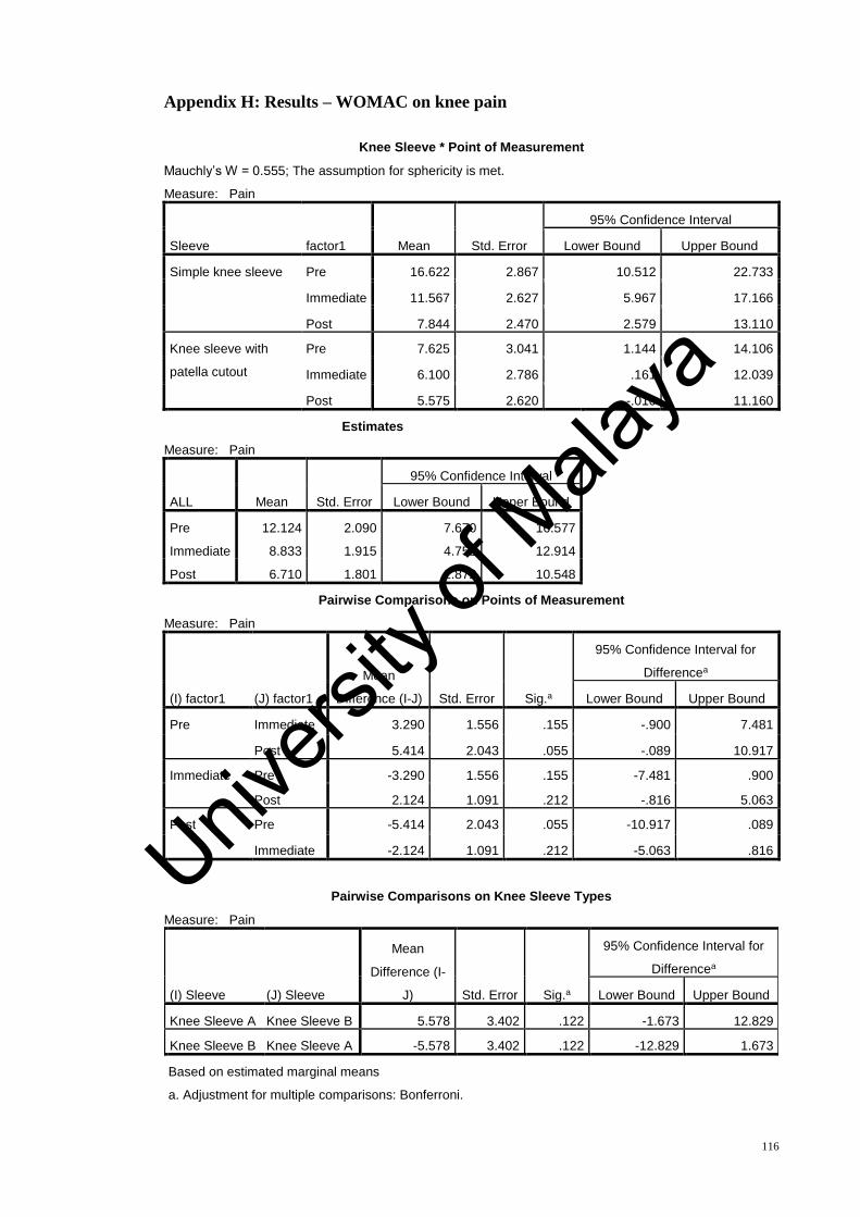
116
Appendix H: Results – WOMAC on knee pain
Knee Sleeve * Point of Measurement
Mauchly’s W = 0.555; The assumption for sphericity is met.
Measure: Pain
Sleeve factor1 Mean Std. Error
95% Confidence Interval
Lower Bound Upper Bound
Simple knee sleeve Pre 16.622 2.867 10.512 22.733
Immediate 11.567 2.627 5.967 17.166
Post 7.844 2.470 2.579 13.110
Knee sleeve with
patella cutout
Pre 7.625 3.041 1.144 14.106
Immediate 6.100 2.786 .161 12.039
Post 5.575 2.620 -.010 11.160
Estimates
Measure: Pain
ALL Mean Std. Error
95% Confidence Interval
Lower Bound Upper Bound
Pre 12.124 2.090 7.670 16.577
Immediate 8.833 1.915 4.752 12.914
Post 6.710 1.801 2.872 10.548
Pairwise Comparisons on Points of Measurement
Measure: Pain
(I) factor1 (J) factor1
Mean
Difference (I-J) Std. Error Sig.a
95% Confidence Interval for
Differencea
Lower Bound Upper Bound
Pre Immediate 3.290 1.556 .155 -.900 7.481
Post 5.414 2.043 .055 -.089 10.917
Immediate Pre -3.290 1.556 .155 -7.481 .900
Post 2.124 1.091 .212 -.816 5.063
Post Pre -5.414 2.043 .055 -10.917 .089
Immediate -2.124 1.091 .212 -5.063 .816
Pairwise Comparisons on Knee Sleeve Types
Measure: Pain
(I) Sleeve (J) Sleeve
Mean
Difference (I-
J) Std. Error Sig.a
95% Confidence Interval for
Differencea
Lower Bound Upper Bound
Knee Sleeve A Knee Sleeve B 5.578 3.402 .122 -1.673 12.829
Knee Sleeve B Knee Sleeve A -5.578 3.402 .122 -12.829 1.673
Based on estimated marginal means
a. Adjustment for multiple comparisons: Bonferroni.
Univers
ity of
Mala
ya
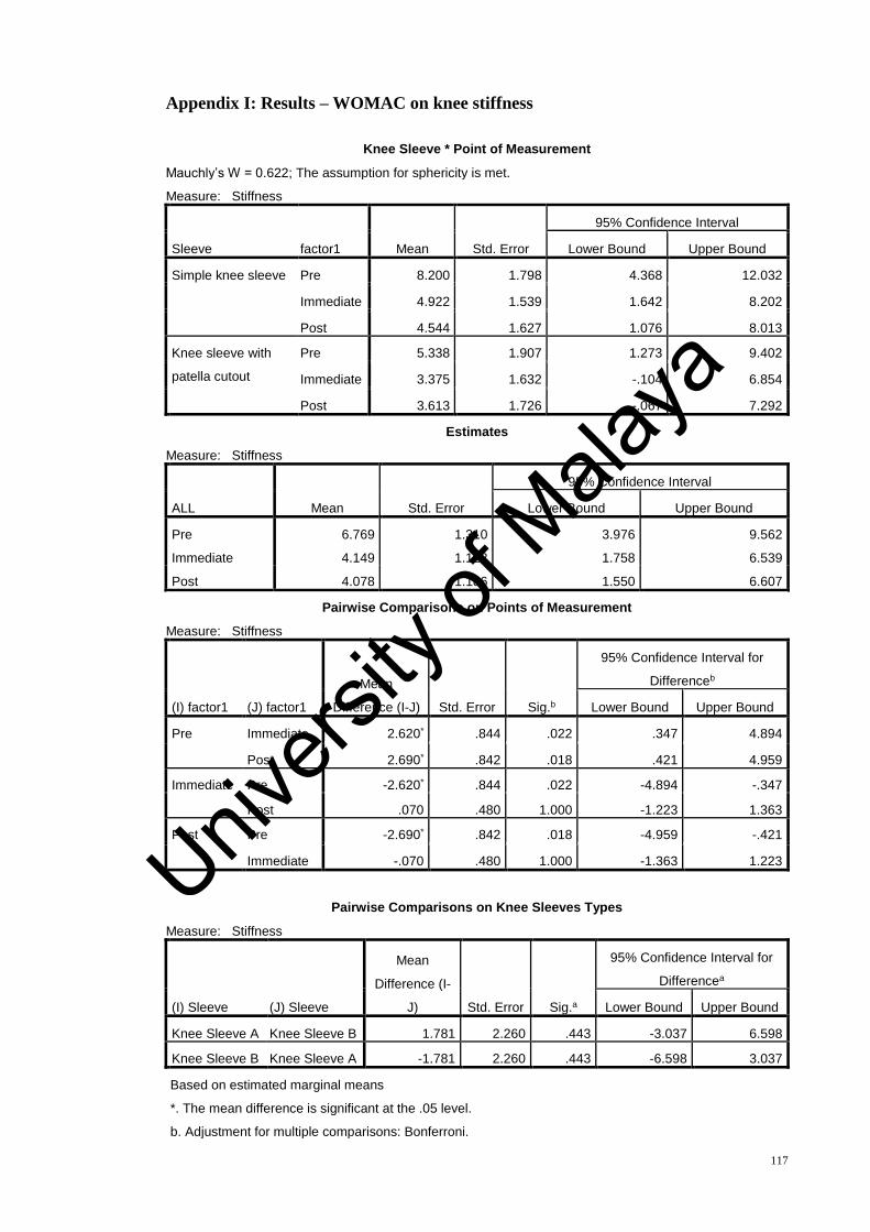
117
Appendix I: Results – WOMAC on knee stiffness
Knee Sleeve * Point of Measurement
Mauchly’s W = 0.622; The assumption for sphericity is met.
Measure: Stiffness
Sleeve factor1 Mean Std. Error
95% Confidence Interval
Lower Bound Upper Bound
Simple knee sleeve Pre 8.200 1.798 4.368 12.032
Immediate 4.922 1.539 1.642 8.202
Post 4.544 1.627 1.076 8.013
Knee sleeve with
patella cutout
Pre 5.338 1.907 1.273 9.402
Immediate 3.375 1.632 -.104 6.854
Post 3.613 1.726 -.067 7.292
Estimates
Measure: Stiffness
ALL Mean Std. Error
95% Confidence Interval
Lower Bound Upper Bound
Pre 6.769 1.310 3.976 9.562
Immediate 4.149 1.122 1.758 6.539
Post 4.078 1.186 1.550 6.607
Pairwise Comparisons on Points of Measurement
Measure: Stiffness
(I) factor1 (J) factor1
Mean
Difference (I-J) Std. Error Sig.b
95% Confidence Interval for
Differenceb
Lower Bound Upper Bound
Pre Immediate 2.620* .844 .022 .347 4.894
Post 2.690* .842 .018 .421 4.959
Immediate Pre -2.620* .844 .022 -4.894 -.347
Post .070 .480 1.000 -1.223 1.363
Post Pre -2.690* .842 .018 -4.959 -.421
Immediate -.070 .480 1.000 -1.363 1.223
Pairwise Comparisons on Knee Sleeves Types
Measure: Stiffness
(I) Sleeve (J) Sleeve
Mean
Difference (I-
J) Std. Error Sig.a
95% Confidence Interval for
Differencea
Lower Bound Upper Bound
Knee Sleeve A Knee Sleeve B 1.781 2.260 .443 -3.037 6.598
Knee Sleeve B Knee Sleeve A -1.781 2.260 .443 -6.598 3.037
Based on estimated marginal means
*. The mean difference is significant at the .05 level.
b. Adjustment for multiple comparisons: Bonferroni.
Univers
ity of
Mala
ya

118
Appendix J: Results – WOMAC on knee functional performance
Knee Sleeve * Point of Measurement
Mauchly’s W = 0.384; The assumption for sphericity is met.
Measure: Difficulty Performing Daily Activities
Sleeve factor1 Mean Std. Error
95% Confidence Interval
Lower Bound Upper Bound
Simple knee sleeve Pre 68.856 11.316 44.736 92.975
Immediate 53.422 10.463 31.120 75.724
Post 34.800 10.692 12.010 57.590
Knee sleeve with
patella cutout
Pre 29.850 12.003 4.267 55.433
Immediate 21.188 11.098 -2.467 44.842
Post 22.338 11.341 -1.835 46.510
Estimates
Measure: Difficulty
ALL Mean Std. Error
95% Confidence Interval
Lower Bound Upper Bound
Pre 49.353 8.248 31.773 66.933
Immediate 37.305 7.626 21.050 53.560
Post 28.569 7.793 11.958 45.180
Pairwise Comparisons on Points of Measurement
Measure: Difficulty Performing Daily Activities
(I) factor1 (J) factor1
Mean
Difference (I-J) Std. Error Sig.b
95% Confidence Interval for
Differenceb
Lower Bound Upper Bound
Pre Immediate 12.048 4.650 .061 -.477 24.573
Post 20.784* 6.292 .014 3.834 37.734
Immediate Pre -12.048 4.650 .061 -24.573 .477
Post 8.736* 2.870 .025 1.006 16.466
Post Pre -20.784* 6.292 .014 -37.734 -3.834
Immediate -8.736* 2.870 .025 -16.466 -1.006
Pairwise Comparisons on Knee Sleeves Types
Measure: Difficulty Performing Daily Activities
(I) Sleeve (J) Sleeve
Mean
Difference (I-
J) Std. Error Sig.a
95% Confidence Interval for
Differencea
Lower Bound Upper Bound
Knee Sleeve A Knee Sleeve B 27.901 14.777 .079 -3.596 59.398
Knee Sleeve B Knee Sleeve A -27.901 14.777 .079 -59.398 3.596
Based on estimated marginal means
*. The mean difference is significant at the .05 level, b. Adjustment for multiple comparisons: Bonferroni.
Univers
ity of
Mala
ya

119
Appendix K: Results – WOMAC Overall Score
Descriptive Statistics
Knee Sleeve Mean Std. Deviation N
WOMAC ALL Pre Simple knee sleeve 93.6778 52.11487 9
Knee sleeve with
patella cutout 42.8125 35.59942 8
Total 69.7412 50.96370 17
WOMAC ALL
Immediate
Simple knee sleeve 69.9111 40.38290 9
Knee sleeve with
patella cutout 30.6625 45.51800 8
Total 51.4412 46.14767 17
WOMAC ALL Post Simple knee sleeve 47.1889 45.03500 9
Knee sleeve with
patella cutout 31.5250 40.31730 8
Total 39.8176 42.31043 17
Mauchly’s W = 0.401; the assumption for sphericity is met.
Pairwise Comparisons on Points of Measurement
Measure: WOMAC Overall
(I) factor1 (J) factor1
Mean
Difference (I-J) Std. Error Sig.b
95% Confidence Interval for
Differenceb
Lower Bound Upper Bound
Pre Immediate 17.958* 6.209 .034 1.233 34.684
Post 28.888* 8.529 .012 5.914 51.862
Immediate Pre -17.958* 6.209 .034 -34.684 -1.233
Post 10.930* 4.021 .048 .097 21.762
Post Pre -28.888* 8.529 .012 -51.862 -5.914
Immediate -10.930* 4.021 .048 -21.762 -.097
Based on estimated marginal means
*. The mean difference is significant at the .05 level.
b. Adjustment for multiple comparisons: Bonferroni.
Pairwise Comparisons on Knee Sleeves Types
Measure: WOMAC Overall
(I) Knee
Sleeve
(J) Knee
Sleeve
Mean
Difference (I-
J) Std. Error Sig.a
95% Confidence Interval for
Differencea
Lower Bound Upper Bound
Knee Sleeve A Knee Sleeve B 35.259 19.831 .096 -7.010 77.529
Knee Sleeve B Knee Sleeve A -35.259 19.831 .096 -77.529 7.010
Based on estimated marginal means
a. Adjustment for multiple comparisons: Bonferroni.
Univers
ity of
Mala
ya

120
Appendix L: Results – Postural Stability Test (PST) Static
Descriptive Statistics
Knee Sleeve Mean Std. Deviation N
PST Static Pre Simple knee sleeve .467 .2291 9
Knee sleeve with patella cutout .463 .2722 8
Total .465 .2422 17
PST Static
Immediate
Simple knee sleeve .489 .2667 9
Knee sleeve with patella cutout .413 .1458 8
Total .453 .2154 17
PST Static Post
Simple knee sleeve .489 .2205 9
Knee sleeve with patella cutout .425 .1282 8
Total .459 .1805 17
Mauchly’s W = 0.922; the assumption for sphericity is met.
Pairwise Comparisons on Points of Measurement
Measure: PST Static
(I) factor1 (J) factor1
Mean
Difference (I-J) Std. Error Sig.a
95% Confidence Interval for
Differencea
Lower Bound Upper Bound
Pre Immediate .014 .044 1.000 -.105 .133
Post .008 .048 1.000 -.120 .136
Immediate Pre -.014 .044 1.000 -.133 .105
Post -.006 .037 1.000 -.106 .094
Post Pre -.008 .048 1.000 -.136 .120
Immediate .006 .037 1.000 -.094 .106
Based on estimated marginal means
a. Adjustment for multiple comparisons: Bonferroni.
Pairwise Comparisons on Knee Sleeves Types
Measure: PST Dynamic
(I) Knee
Sleeve
(J) Knee
Sleeve
Mean
Difference (I-
J) Std. Error Sig.a
95% Confidence Interval for
Differencea
Lower Bound Upper Bound
Knee Sleeve A Knee Sleeve B .048 .094 .616 -.152 .249
Knee Sleeve B Knee Sleeve A -.048 .094 .616 -.249 .152
Based on estimated marginal means
a. Adjustment for multiple comparisons: Bonferroni.
Univers
ity of
Mala
ya
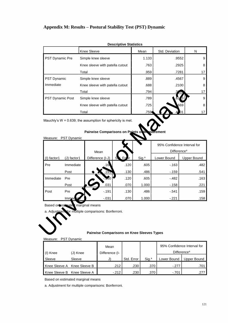
121
Appendix M: Results – Postural Stability Test (PST) Dynamic
Descriptive Statistics
Knee Sleeve Mean Std. Deviation N
PST Dynamic Pre Simple knee sleeve 1.133 .9552 9
Knee sleeve with patella cutout .763 .2925 8
Total .959 .7281 17
PST Dynamic
Immediate
Simple knee sleeve .889 .4567 9
Knee sleeve with patella cutout .688 .2100 8
Total .794 .3665 17
PST Dynamic Post Simple knee sleeve .789 .6153 9
Knee sleeve with patella cutout .725 .1669 8
Total .759 .4501 17
Mauchly’s W = 0.639; the assumption for sphericity is met.
Pairwise Comparisons on Points of Measurement
Measure: PST Dynamic
(I) factor1 (J) factor1
Mean
Difference (I-J) Std. Error Sig.a
95% Confidence Interval for
Differencea
Lower Bound Upper Bound
Pre Immediate .160 .120 .605 -.163 .482
Post .191 .130 .486 -.159 .541
Immediate Pre -.160 .120 .605 -.482 .163
Post .031 .070 1.000 -.158 .221
Post Pre -.191 .130 .486 -.541 .159
Immediate -.031 .070 1.000 -.221 .158
Based on estimated marginal means
a. Adjustment for multiple comparisons: Bonferroni.
Pairwise Comparisons on Knee Sleeves Types
Measure: PST Dynamic
(I) Knee
Sleeve
(J) Knee
Sleeve
Mean
Difference (I-
J) Std. Error Sig.a
95% Confidence Interval for
Differencea
Lower Bound Upper Bound
Knee Sleeve A Knee Sleeve B .212 .230 .370 -.277 .701
Knee Sleeve B Knee Sleeve A -.212 .230 .370 -.701 .277
Based on estimated marginal means
a. Adjustment for multiple comparisons: Bonferroni.
Univers
ity of
Mala
ya

122
Appendix N: Results – Athlete Single Leg Test (ASL) Static – Affected Knee
Descriptive Statistics
Knee Sleeve Mean Std. Deviation N
ASL Static Affected
Knee Pre
Simple knee sleeve 1.333 .7665 9
Knee sleeve with patella cutout 1.200 .5155 8
Total 1.271 .6440 17
ASL Static Affected
Knee Immediate
Simple knee sleeve 1.656 1.4475 9
Knee sleeve with patella cutout 1.038 .3159 8
Total 1.365 1.0920 17
ASL Static Affected
Knee Post
Simple knee sleeve .800 .3317 9
Knee sleeve with patella cutout .950 .1604 8
Total .871 .2687 17
Mauchly’s W = 0.700; the assumption for sphericity is met.
Pairwise Comparisons on Points of Measurement
Measure: ASL Static Affected Knee
(I) factor1 (J) factor1
Mean
Difference (I-J) Std. Error Sig.a
95% Confidence Interval for
Differencea
Lower Bound Upper Bound
Pre Immediate -.080 .302 1.000 -.893 .734
Post .392 .189 .167 -.117 .900
Immediate Pre .080 .302 1.000 -.734 .893
Post .472 .230 .175 -.149 1.092
Post Pre -.392 .189 .167 -.900 .117
Immediate -.472 .230 .175 -1.092 .149
Based on estimated marginal means
a. Adjustment for multiple comparisons: Bonferroni.
Pairwise Comparisons on Knee Sleeves Types
Measure: ASL Static Affected Knee
(I) Knee
Sleeve
(J) Knee
Sleeve
Mean
Difference (I-
J) Std. Error Sig.a
95% Confidence Interval for
Differencea
Lower Bound Upper Bound
Knee Sleeve A Knee Sleeve B .200 .227 .392 -.284 .685
Knee Sleeve B Knee Sleeve A -.200 .227 .392 -.685 .284
Based on estimated marginal means
a. Adjustment for multiple comparisons: Bonferroni.
Univers
ity of
Mala
ya

123
Appendix O: Results – Athlete Single Leg Test (ASL) Dynamic – Affected Knee
Descriptive Statistics
Knee Sleeve Mean Std. Deviation N
ASL Dynamic
Affected Knee
Pre
Simple knee sleeve 1.833 1.1435 9
Knee sleeve with patella cutout 1.588 .6244 8
Total 1.718 .9167 17
ASL Dynamic
Affected Knee
Immediate
Simple knee sleeve 1.700 1.0747 9
Knee sleeve with patella cutout 1.225 .4950 8
Total 1.476 .8628 17
ASL Dynamic
Affected Knee
Post
Simple knee sleeve 1.656 1.2953 9
Knee sleeve with patella cutout 1.213 .5194 8
Total 1.447 1.0044 17
Mauchly’s W = 0.892; the assumption for sphericity is met.
Pairwise Comparisons on Points of Measurement
Measure: ASL Dynamic Affected Knee
(I) factor1 (J) factor1
Mean
Difference (I-J) Std. Error Sig.a
95% Confidence Interval for
Differencea
Lower Bound Upper Bound
Pre Immediate .248 .263 1.000 -.462 .958
Post .276 .359 1.000 -.690 1.243
Immediate Pre -.248 .263 1.000 -.958 .462
Post .028 .275 1.000 -.711 .768
Post Pre -.276 .359 1.000 -1.243 .690
Immediate -.028 .275 1.000 -.768 .711
Based on estimated marginal means
a. Adjustment for multiple comparisons: Bonferroni.
Pairwise Comparisons on Knee Sleeves Types
Measure: ASL Dynamic Affected Knee
(I) Knee
Sleeve
(J) Knee
Sleeve
Mean
Difference (I-
J) Std. Error Sig.a
95% Confidence Interval for
Differencea
Lower Bound Upper Bound
Knee Sleeve A Knee Sleeve B .388 .292 .204 -.235 1.011
Knee Sleeve B Knee Sleeve A -.388 .292 .204 -1.011 .235
Based on estimated marginal means
a. Adjustment for multiple comparisons: Bonferroni.
Univers
ity of
Mala
ya
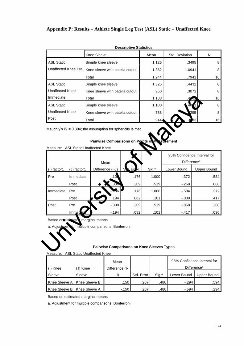
124
Appendix P: Results – Athlete Single Leg Test (ASL) Static – Unaffected Knee
Descriptive Statistics
Knee Sleeve Mean Std. Deviation N
ASL Static
Unaffected Knee Pre
Simple knee sleeve 1.125 .3495 8
Knee sleeve with patella cutout 1.362 1.0941 8
Total 1.244 .7941 16
ASL Static
Unaffected Knee
Immediate
Simple knee sleeve 1.325 .4432 8
Knee sleeve with patella cutout .950 .3071 8
Total 1.138 .4161 16
ASL Static
Unaffected Knee
Post
Simple knee sleeve 1.100 .4811 8
Knee sleeve with patella cutout .788 .2295 8
Total .944 .3983 16
Mauchly’s W = 0.394; the assumption for sphericity is met.
Pairwise Comparisons on Points of Measurement
Measure: ASL Static Unaffected Knee
(I) factor1 (J) factor1
Mean
Difference (I-J) Std. Error Sig.a
95% Confidence Interval for
Differencea
Lower Bound Upper Bound
Pre Immediate .106 .176 1.000 -.372 .584
Post .300 .209 .519 -.268 .868
Immediate Pre -.106 .176 1.000 -.584 .372
Post .194 .082 .101 -.030 .417
Post Pre -.300 .209 .519 -.868 .268
Immediate -.194 .082 .101 -.417 .030
Based on estimated marginal means
a. Adjustment for multiple comparisons: Bonferroni.
Pairwise Comparisons on Knee Sleeves Types
Measure: ASL Static Unaffected Knee
(I) Knee
Sleeve
(J) Knee
Sleeve
Mean
Difference (I-
J) Std. Error Sig.a
95% Confidence Interval for
Differencea
Lower Bound Upper Bound
Knee Sleeve A Knee Sleeve B .150 .207 .480 -.294 .594
Knee Sleeve B Knee Sleeve A -.150 .207 .480 -.594 .294
Based on estimated marginal means
a. Adjustment for multiple comparisons: Bonferroni.
Univers
ity of
Mala
ya

125
Appendix Q: Results – Athlete Single Leg Test (ASL) Dynamic – Unaffected Knee
Descriptive Statistics
Knee Sleeve Mean Std. Deviation N
ASL Dynamic
Unaffected Knee Pre
Simple knee sleeve 1.475 .7960 8
Knee sleeve with patella cutout 1.788 1.1192 8
Total 1.631 .9520 16
ASL Dynamic
Unaffected Knee
Immediate
Simple knee sleeve 1.350 .7091 8
Knee sleeve with patella cutout 1.475 .8828 8
Total 1.413 .7762 16
ASL Dynamic
Unaffected Knee Post
Simple knee sleeve .850 .3338 8
Knee sleeve with patella cutout .925 .4652 8
Total .888 .3931 16
Mauchly’s W = 0.894; the assumption for sphericity is met.
Pairwise Comparisons on Points of Measurement
Measure: ASL Dynamic Unaffected Knee
(I) factor1 (J) factor1
Mean
Difference (I-J) Std. Error Sig.b
95% Confidence Interval for
Differenceb
Lower Bound Upper Bound
Pre Immediate .219 .240 1.000 -.434 .872
Post .744* .270 .047 .009 1.479
Immediate Pre -.219 .240 1.000 -.872 .434
Post .525 .202 .063 -.025 1.075
Post Pre -.744* .270 .047 -1.479 -.009
Immediate -.525 .202 .063 -1.075 .025
Based on estimated marginal means
*. The mean difference is significant at the .05 level.
b. Adjustment for multiple comparisons: Bonferroni.
Pairwise Comparisons on Knee Sleeves Types
Measure: ASL Dynamic Unaffected Knee
(I) Knee
Sleeve
(J) Knee
Sleeve
Mean
Difference (I-
J) Std. Error Sig.a
95% Confidence Interval for
Differencea
Lower Bound Upper Bound
Knee Sleeve A Knee Sleeve B -.171 .263 .527 -.736 .394
Knee Sleeve B Knee Sleeve A .171 .263 .527 -.394 .736
Based on estimated marginal means
a. Adjustment for multiple comparisons: Bonferroni.
Univers
ity of
Mala
ya

126
Appendix R: Results – Fall Risk Test (FRT)
Descriptive Statistics
Knee Sleeve Mean Std. Deviation N
FRT Pre Simple knee sleeve .844 .4275 9
Knee sleeve with patella cutout .675 .2375 8
Total .765 .3517 17
FRT Immediate Simple knee sleeve .733 .3708 9
Knee sleeve with patella cutout .525 .1832 8
Total .635 .3081 17
FRT Post Simple knee sleeve .722 .6180 9
Knee sleeve with patella cutout .663 .2825 8
Total .694 .4763 17
Mauchly’s W = 0.787; the assumption for sphericity is met.
Pairwise Comparisons on Points of Measurement
Measure: FRT
(I) factor1 (J) factor1
Mean
Difference (I-J) Std. Error Sig.a
95% Confidence Interval for
Differencea
Lower Bound Upper Bound
Pre Immediate .131 .060 .142 -.032 .293
Post .067 .091 1.000 -.177 .311
Immediate Pre -.131 .060 .142 -.293 .032
Post -.063 .073 1.000 -.261 .134
Post Pre -.067 .091 1.000 -.311 .177
Immediate .063 .073 1.000 -.134 .261
Based on estimated marginal means
a. Adjustment for multiple comparisons: Bonferroni.
Pairwise Comparisons on Knee Sleeves Types
Measure: FRT
(I) Knee
Sleeve
(J) Knee
Sleeve
Mean
Difference (I-
J) Std. Error Sig.a
95% Confidence Interval for
Differencea
Lower Bound Upper Bound
Knee Sleeve A Knee Sleeve B .146 .167 .398 -.211 .503
Knee Sleeve B Knee Sleeve A -.146 .167 .398 -.503 .211
Based on estimated marginal means
a. Adjustment for multiple comparisons: Bonferroni.
Univers
ity of
Mala
ya

127
Appendix S: Results – Gait Parameters
a) Knee Adduction Moment
i) First Peak KAM
Descriptive Statistics
Knee Sleeve Mean Std. Deviation N
Pre 1st Peak KAM
(Nmm/kg)
Simple knee sleeve .8283 .55787 9
Knee sleeve with patella cutout .6023 .49374 8
Total .7219 .52515 17
Imm 1st Peak KAM
(Nmm/kg)
Simple knee sleeve .8396 .54267 9
Knee sleeve with patella cutout .3767 .54922 8
Total .6218 .57961 17
Post 1st Peak KAM
(Nmm/kg)
Simple knee sleeve .4070 .16813 9
Knee sleeve with patella cutout .4776 .31548 8
Total .4402 .24289 17
Mauchly’s W = 0.964; the assumption for sphericity is met.
Pairwise Comparisons on Points of Measurement
Measure: FirstKAM
(I) factor1 (J) factor1
Mean
Difference (I-J) Std. Error Sig.a
95% Confidence Interval for
Differencea
Lower Bound Upper Bound
Pre Immediate .107 .089 .744 -.133 .347
Post .273 .105 .061 -.011 .557
Immediate Pre -.107 .089 .744 -.347 .133
Post .166 .097 .326 -.096 .428
Post Pre -.273 .105 .061 -.557 .011
Immediate -.166 .097 .326 -.428 .096
Based on estimated marginal means
a. Adjustment for multiple comparisons: Bonferroni.
Pairwise Comparisons on Knee Sleeves Types
Measure: FirstKAM
(I) Knee
Sleeve
(J) Knee
Sleeve
Mean
Difference (I-
J) Std. Error Sig.a
95% Confidence Interval for
Differencea
Lower Bound Upper Bound
Knee Sleeve A Knee Sleeve B .206 .194 .305 -.207 .620
Knee Sleeve B Knee Sleeve A -.206 .194 .305 -.620 .207
Based on estimated marginal means
a. Adjustment for multiple comparisons: Bonferroni.
Univers
ity of
Mala
ya

128
ii) Second Peak KAM
Descriptive Statistics
Knee Sleeve Mean Std. Deviation N
Pre 2nd Peak KAM
(Nmm/kg)
Simple knee sleeve .5486 .37766 9
Knee sleeve with patella cutout .4627 .18316 8
Total .5082 .29656 17
Imm 2nd Peak
KAM (Nmm/kg)
Simple knee sleeve .6462 .53494 9
Knee sleeve with patella cutout .3569 .20588 8
Total .5101 .42870 17
Post 2nd Peak
KAM (Nmm/kg)
Simple knee sleeve .2193 .17749 9
Knee sleeve with patella cutout .3110 .17854 8
Total .2625 .17867 17
Mauchly’s W = 0.922; The assumption for sphericity is met.
Pairwise Comparisons on Points of Measurement
Measure: SecondKAM
(I) factor1 (J) factor1
Mean
Difference (I-J) Std. Error Sig.b
95% Confidence Interval for
Differenceb
Lower Bound Upper Bound
Pre Immediate .004 .069 1.000 -.182 .191
Post .240* .067 .008 .059 .422
Immediate Pre -.004 .069 1.000 -.191 .182
Post .236* .083 .037 .012 .460
Post Pre -.240* .067 .008 -.422 -.059
Immediate -.236* .083 .037 -.460 -.012
Based on estimated marginal means
*. The mean difference is significant at the .05 level.
b. Adjustment for multiple comparisons: Bonferroni.
Pairwise Comparisons on Knee Sleeves Types
Measure: SecondKAM
(I) Knee
Sleeve
(J) Knee
Sleeve
Mean
Difference (I-
J) Std. Error Sig.a
95% Confidence Interval for
Differencea
Lower Bound Upper Bound
Knee Sleeve A Knee Sleeve B .095 .127 .467 -.176 .365
Knee Sleeve B Knee Sleeve A -.095 .127 .467 -.365 .176
Based on estimated marginal means
a. Adjustment for multiple comparisons: Bonferroni.
Univers
ity of
Mala
ya

129
b) Vertical Ground Reaction Force (vGRF)
i) First Peak vGRF
Descriptive Statistics
Knee Sleeve Mean Std. Deviation N
Pre 1st Peak GRF
(N/kg)
Simple knee sleeve 15.2543 6.00736 9
Knee sleeve with patella cutout 12.0588 4.46837 8
Total 13.7505 5.42979 17
Imm 1st Peak GRF
(N/kg)
Simple knee sleeve 15.9436 6.54020 9
Knee sleeve with patella cutout 13.4713 4.89704 8
Total 14.7801 5.78764 17
Post 1st Peak GRF
(N/kg)
Simple knee sleeve 9.8267 2.06286 9
Knee sleeve with patella cutout 10.2428 1.96175 8
Total 10.0225 1.96398 17
Mauchly’s W = 0.550; the assumption for sphericity is met.
Pairwise Comparisons on Points of Measurement
Measure: FirstGRF
(I) factor1 (J) factor1
Mean
Difference (I-J) Std. Error Sig.b
95% Confidence Interval for
Differenceb
Lower Bound Upper Bound
Pre Immediate -1.051 .802 .630 -3.212 1.110
Post 3.622 1.519 .092 -.471 7.714
Immediate Pre 1.051 .802 .630 -1.110 3.212
Post 4.673* 1.648 .038 .233 9.112
Post Pre -3.622 1.519 .092 -7.714 .471
Immediate -4.673* 1.648 .038 -9.112 -.233
Based on estimated marginal means
*. The mean difference is significant at the .05 level.
b. Adjustment for multiple comparisons: Bonferroni.
Pairwise Comparisons on Knee Sleeves Types
Measure: FirstGRF
(I) Knee
Sleeve
(J) Knee
Sleeve
Mean
Difference (I-
J) Std. Error Sig.a
95% Confidence Interval for
Differencea
Lower Bound Upper Bound
Knee Sleeve A Knee Sleeve B 1.751 1.651 .306 -1.768 5.269
Knee Sleeve B Knee Sleeve A -1.751 1.651 .306 -5.269 1.768
Based on estimated marginal means
a. Adjustment for multiple comparisons: Bonferroni.
Univers
ity of
Mala
ya
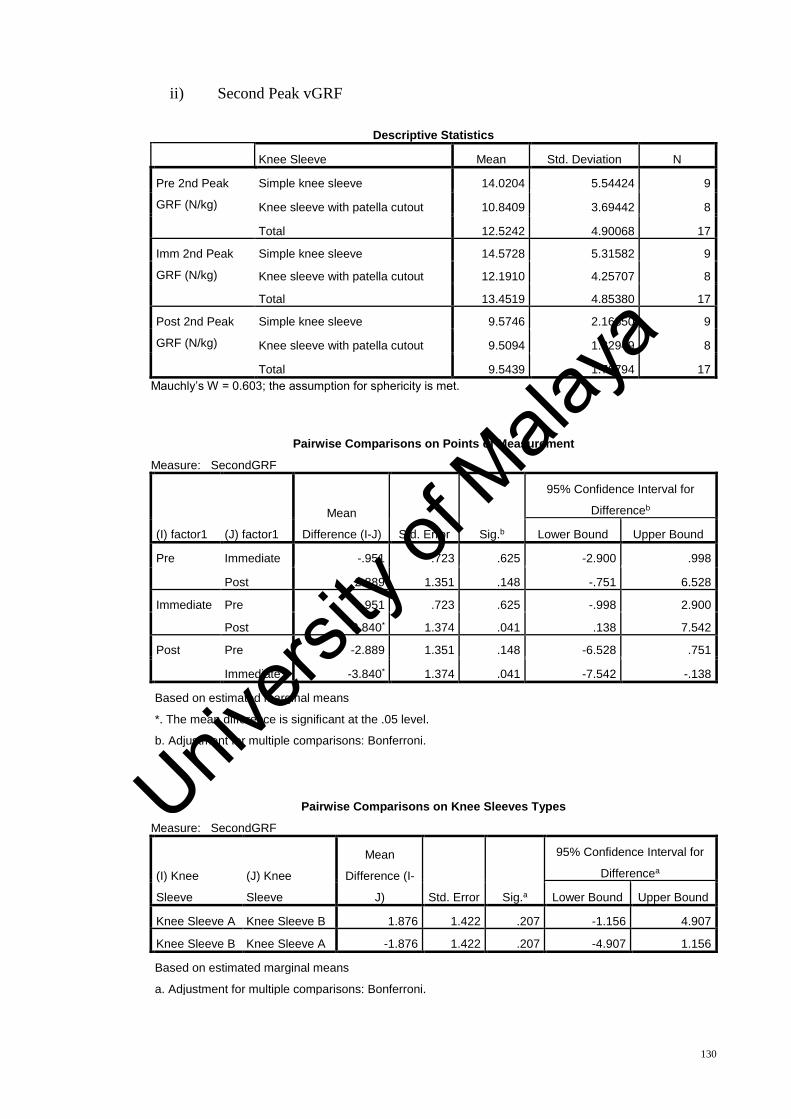
130
ii) Second Peak vGRF
Descriptive Statistics
Knee Sleeve Mean Std. Deviation N
Pre 2nd Peak
GRF (N/kg)
Simple knee sleeve 14.0204 5.54424 9
Knee sleeve with patella cutout 10.8409 3.69442 8
Total 12.5242 4.90068 17
Imm 2nd Peak
GRF (N/kg)
Simple knee sleeve 14.5728 5.31582 9
Knee sleeve with patella cutout 12.1910 4.25707 8
Total 13.4519 4.85380 17
Post 2nd Peak
GRF (N/kg)
Simple knee sleeve 9.5746 2.16850 9
Knee sleeve with patella cutout 9.5094 1.32949 8
Total 9.5439 1.76794 17
Mauchly’s W = 0.603; the assumption for sphericity is met.
Pairwise Comparisons on Points of Measurement
Measure: SecondGRF
(I) factor1 (J) factor1
Mean
Difference (I-J) Std. Error Sig.b
95% Confidence Interval for
Differenceb
Lower Bound Upper Bound
Pre Immediate -.951 .723 .625 -2.900 .998
Post 2.889 1.351 .148 -.751 6.528
Immediate Pre .951 .723 .625 -.998 2.900
Post 3.840* 1.374 .041 .138 7.542
Post Pre -2.889 1.351 .148 -6.528 .751
Immediate -3.840* 1.374 .041 -7.542 -.138
Based on estimated marginal means
*. The mean difference is significant at the .05 level.
b. Adjustment for multiple comparisons: Bonferroni.
Pairwise Comparisons on Knee Sleeves Types
Measure: SecondGRF
(I) Knee
Sleeve
(J) Knee
Sleeve
Mean
Difference (I-
J) Std. Error Sig.a
95% Confidence Interval for
Differencea
Lower Bound Upper Bound
Knee Sleeve A Knee Sleeve B 1.876 1.422 .207 -1.156 4.907
Knee Sleeve B Knee Sleeve A -1.876 1.422 .207 -4.907 1.156
Based on estimated marginal means
a. Adjustment for multiple comparisons: Bonferroni.
Univers
ity of
Mala
ya
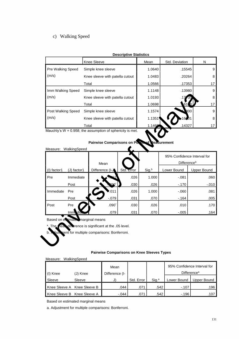
131
c) Walking Speed
Descriptive Statistics
Knee Sleeve Mean Std. Deviation N
Pre Walking Speed
(m/s)
Simple knee sleeve 1.0640 .15545 9
Knee sleeve with patella cutout 1.0483 .20264 8
Total 1.0566 .17353 17
Imm Walking Speed
(m/s)
Simple knee sleeve 1.1148 .13980 9
Knee sleeve with patella cutout 1.0193 .17735 8
Total 1.0698 .16108 17
Post Walking Speed
(m/s)
Simple knee sleeve 1.1574 .12800 9
Knee sleeve with patella cutout 1.1351 .16701 8
Total 1.1469 .14327 17
Mauchly’s W = 0.958; the assumption of sphericity is met.
Pairwise Comparisons on Points of Measurement
Measure: WalkingSpeed
(I) factor1 (J) factor1
Mean
Difference (I-J) Std. Error Sig.b
95% Confidence Interval for
Differenceb
Lower Bound Upper Bound
Pre Immediate -.011 .026 1.000 -.081 .060
Post -.090* .030 .026 -.170 -.010
Immediate Pre .011 .026 1.000 -.060 .081
Post -.079 .031 .070 -.164 .005
Post Pre .090* .030 .026 .010 .170
Immediate .079 .031 .070 -.005 .164
Based on estimated marginal means
*. The mean difference is significant at the .05 level.
b. Adjustment for multiple comparisons: Bonferroni.
Pairwise Comparisons on Knee Sleeves Types
Measure: WalkingSpeed
(I) Knee
Sleeve
(J) Knee
Sleeve
Mean
Difference (I-
J) Std. Error Sig.a
95% Confidence Interval for
Differencea
Lower Bound Upper Bound
Knee Sleeve A Knee Sleeve B .044 .071 .542 -.107 .196
Knee Sleeve B Knee Sleeve A -.044 .071 .542 -.196 .107
Based on estimated marginal means
a. Adjustment for multiple comparisons: Bonferroni.
Univers
ity of
Mala
ya
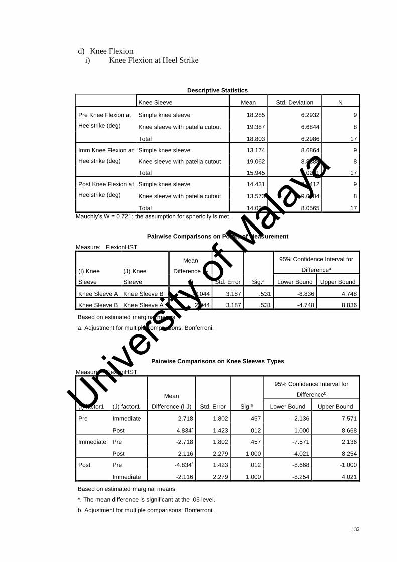
132
d) Knee Flexion
i) Knee Flexion at Heel Strike
Descriptive Statistics
Knee Sleeve Mean Std. Deviation N
Pre Knee Flexion at
Heelstrike (deg)
Simple knee sleeve 18.285 6.2932 9
Knee sleeve with patella cutout 19.387 6.6844 8
Total 18.803 6.2986 17
Imm Knee Flexion at
Heelstrike (deg)
Simple knee sleeve 13.174 8.6864 9
Knee sleeve with patella cutout 19.062 8.8888 8
Total 15.945 9.0261 17
Post Knee Flexion at
Heelstrike (deg)
Simple knee sleeve 14.431 7.6412 9
Knee sleeve with patella cutout 13.573 9.0104 8
Total 14.027 8.0565 17
Mauchly’s W = 0.721; the assumption for sphericity is met.
Pairwise Comparisons on Points of Measurement
Measure: FlexionHST
(I) Knee
Sleeve
(J) Knee
Sleeve
Mean
Difference (I-
J) Std. Error Sig.a
95% Confidence Interval for
Differencea
Lower Bound Upper Bound
Knee Sleeve A Knee Sleeve B -2.044 3.187 .531 -8.836 4.748
Knee Sleeve B Knee Sleeve A 2.044 3.187 .531 -4.748 8.836
Based on estimated marginal means
a. Adjustment for multiple comparisons: Bonferroni.
Pairwise Comparisons on Knee Sleeves Types
Measure: FlexionHST
(I) factor1 (J) factor1
Mean
Difference (I-J) Std. Error Sig.b
95% Confidence Interval for
Differenceb
Lower Bound Upper Bound
Pre Immediate 2.718 1.802 .457 -2.136 7.571
Post 4.834* 1.423 .012 1.000 8.668
Immediate Pre -2.718 1.802 .457 -7.571 2.136
Post 2.116 2.279 1.000 -4.021 8.254
Post Pre -4.834* 1.423 .012 -8.668 -1.000
Immediate -2.116 2.279 1.000 -8.254 4.021
Based on estimated marginal means
*. The mean difference is significant at the .05 level.
b. Adjustment for multiple comparisons: Bonferroni.
Univers
ity of
Mala
ya
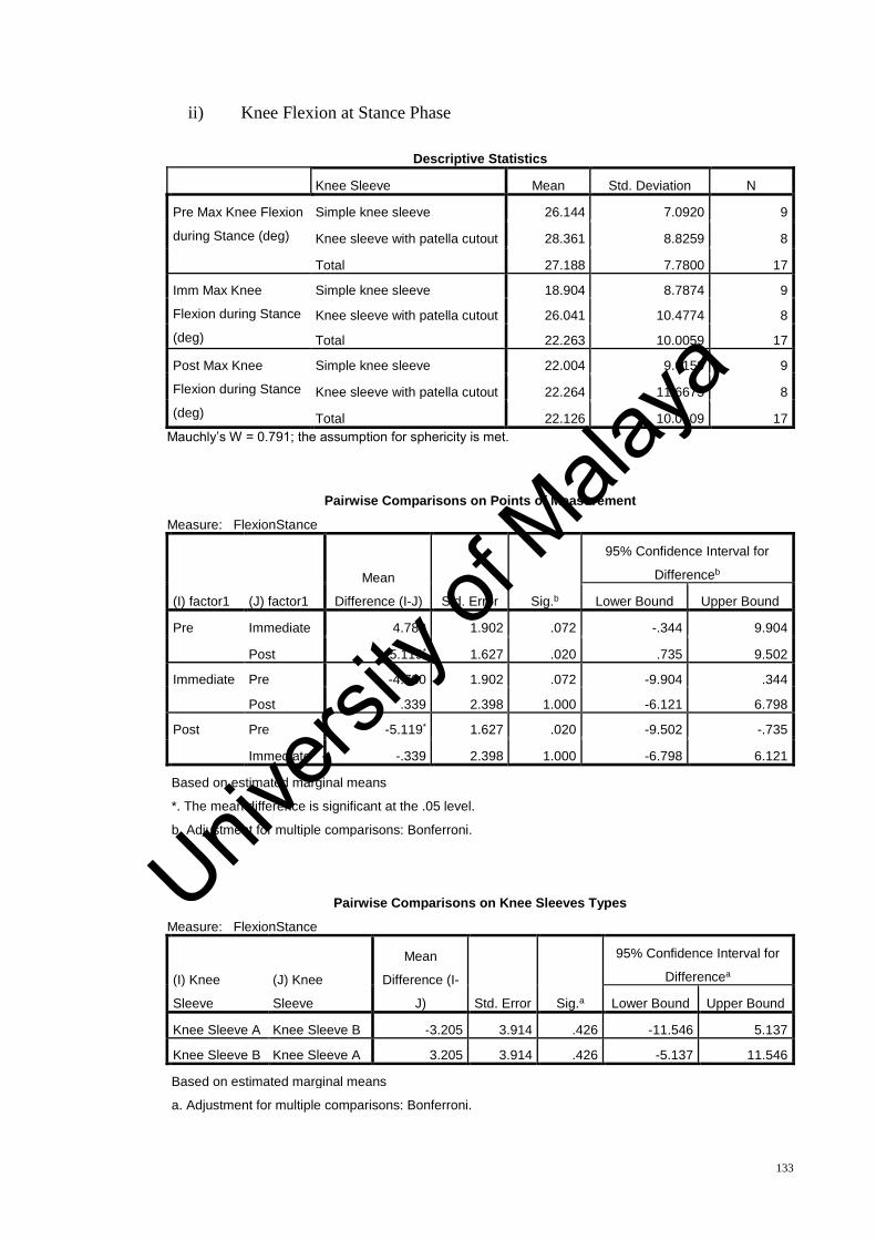
133
ii) Knee Flexion at Stance Phase
Descriptive Statistics
Knee Sleeve Mean Std. Deviation N
Pre Max Knee Flexion
during Stance (deg)
Simple knee sleeve 26.144 7.0920 9
Knee sleeve with patella cutout 28.361 8.8259 8
Total 27.188 7.7800 17
Imm Max Knee
Flexion during Stance
(deg)
Simple knee sleeve 18.904 8.7874 9
Knee sleeve with patella cutout 26.041 10.4774 8
Total 22.263 10.0059 17
Post Max Knee
Flexion during Stance
(deg)
Simple knee sleeve 22.004 9.0159 9
Knee sleeve with patella cutout 22.264 11.6675 8
Total 22.126 10.0109 17
Mauchly’s W = 0.791; the assumption for sphericity is met.
Pairwise Comparisons on Points of Measurement
Measure: FlexionStance
(I) factor1 (J) factor1
Mean
Difference (I-J) Std. Error Sig.b
95% Confidence Interval for
Differenceb
Lower Bound Upper Bound
Pre Immediate 4.780 1.902 .072 -.344 9.904
Post 5.119* 1.627 .020 .735 9.502
Immediate Pre -4.780 1.902 .072 -9.904 .344
Post .339 2.398 1.000 -6.121 6.798
Post Pre -5.119* 1.627 .020 -9.502 -.735
Immediate -.339 2.398 1.000 -6.798 6.121
Based on estimated marginal means
*. The mean difference is significant at the .05 level.
b. Adjustment for multiple comparisons: Bonferroni.
Pairwise Comparisons on Knee Sleeves Types
Measure: FlexionStance
(I) Knee
Sleeve
(J) Knee
Sleeve
Mean
Difference (I-
J) Std. Error Sig.a
95% Confidence Interval for
Differencea
Lower Bound Upper Bound
Knee Sleeve A Knee Sleeve B -3.205 3.914 .426 -11.546 5.137
Knee Sleeve B Knee Sleeve A 3.205 3.914 .426 -5.137 11.546
Based on estimated marginal means
a. Adjustment for multiple comparisons: Bonferroni.
Univers
ity of
Mala
ya

134
iii) Knee Flexion at Swing Phase
Descriptive Statistics
Knee Sleeve Mean Std. Deviation N
Pre Max Knee Flexion
during Swing (deg)
Simple knee sleeve 71.335 18.2152 9
Knee sleeve with patella cutout 67.438 8.9467 8
Total 69.501 14.3156 17
Imm Max Knee
Flexion during Swing
(deg)
Simple knee sleeve 63.994 12.0178 9
Knee sleeve with patella cutout 66.850 12.0596 8
Total 65.338 11.7474 17
Post Max Knee
Flexion during Swing
(deg)
Simple knee sleeve 64.548 18.9321 9
Knee sleeve with patella cutout 62.544 11.6082 8
Total 63.605 15.4670 17
Mauchly’s W = 0.919; the assumption for sphericity is met.
Pairwise Comparisons on Points of Measurement
Measure: FlexionSwing
(I) factor1 (J) factor1
Mean
Difference (I-J) Std. Error Sig.a
95% Confidence Interval for
Differencea
Lower Bound Upper Bound
Pre Immediate 3.965 2.665 .473 -3.214 11.143
Post 5.841 2.459 .094 -.784 12.465
Immediate Pre -3.965 2.665 .473 -11.143 3.214
Post 1.876 3.114 1.000 -6.513 10.265
Post Pre -5.841 2.459 .094 -12.465 .784
Immediate -1.876 3.114 1.000 -10.265 6.513
Based on estimated marginal means
a. Adjustment for multiple comparisons: Bonferroni.
Pairwise Comparisons on Knee Sleeves Types
Measure: FlexionSwing
(I) Knee
Sleeve
(J) Knee
Sleeve
Mean
Difference (I-
J) Std. Error Sig.a
95% Confidence Interval for
Differencea
Lower Bound Upper Bound
Knee Sleeve A Knee Sleeve B 1.015 6.173 .872 -12.143 14.173
Knee Sleeve B Knee Sleeve A -1.015 6.173 .872 -14.173 12.143
Based on estimated marginal means
a. Adjustment for multiple comparisons: Bonferroni.
Univers
ity of
Mala
ya
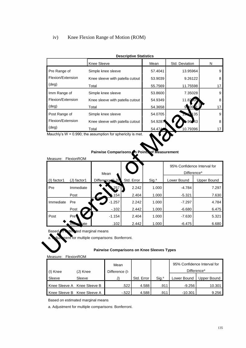
135
iv) Knee Flexion Range of Motion (ROM)
Descriptive Statistics
Knee Sleeve Mean Std. Deviation N
Pre Range of
Flexion/Extension
(deg)
Simple knee sleeve 57.4041 13.95964 9
Knee sleeve with patella cutout 53.9039 9.26122 8
Total 55.7569 11.75598 17
Imm Range of
Flexion/Extension
(deg)
Simple knee sleeve 53.8600 7.35028 9
Knee sleeve with patella cutout 54.9349 11.82905 8
Total 54.3658 9.40941 17
Post Range of
Flexion/Extension
(deg)
Simple knee sleeve 54.0705 13.79135 9
Knee sleeve with patella cutout 54.9287 6.96343 8
Total 54.4744 10.79396 17
Mauchly’s W = 0.990; the assumption for sphericity is met.
Pairwise Comparisons on Points of Measurement
Measure: FlexionROM
(I) factor1 (J) factor1
Mean
Difference (I-J) Std. Error Sig.a
95% Confidence Interval for
Differencea
Lower Bound Upper Bound
Pre Immediate 1.257 2.242 1.000 -4.784 7.297
Post 1.154 2.404 1.000 -5.321 7.630
Immediate Pre -1.257 2.242 1.000 -7.297 4.784
Post -.102 2.442 1.000 -6.680 6.475
Post Pre -1.154 2.404 1.000 -7.630 5.321
Immediate .102 2.442 1.000 -6.475 6.680
Based on estimated marginal means
a. Adjustment for multiple comparisons: Bonferroni.
Pairwise Comparisons on Knee Sleeves Types
Measure: FlexionROM
(I) Knee
Sleeve
(J) Knee
Sleeve
Mean
Difference (I-
J) Std. Error Sig.a
95% Confidence Interval for
Differencea
Lower Bound Upper Bound
Knee Sleeve A Knee Sleeve B .522 4.588 .911 -9.256 10.301
Knee Sleeve B Knee Sleeve A -.522 4.588 .911 -10.301 9.256
Based on estimated marginal means
a. Adjustment for multiple comparisons: Bonferroni.
Univers
ity of
Mala
ya
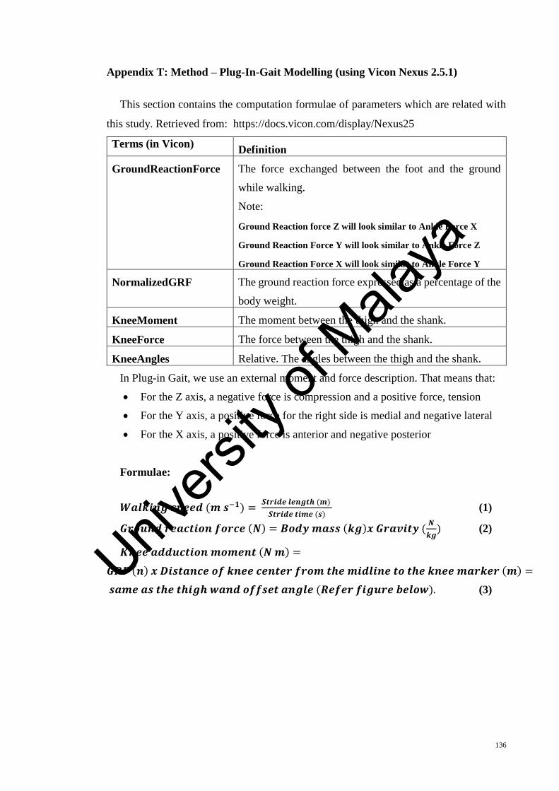
136
Appendix T: Method – Plug-In-Gait Modelling (using Vicon Nexus 2.5.1)
This section contains the computation formulae of parameters which are related with
this study. Retrieved from: https://docs.vicon.com/display/Nexus25
Terms (in Vicon) Definition
GroundReactionForce The force exchanged between the foot and the ground
while walking.
Note:
Ground Reaction force Z will look similar to Ankle Force X
Ground Reaction Force Y will look similar to Ankle Force Z
Ground Reaction Force X will look similar to Ankle Force Y
NormalizedGRF The ground reaction force expressed as a percentage of the
body weight.
KneeMoment The moment between the thigh and the shank.
KneeForce The force between the thigh and the shank.
KneeAngles Relative. The angles between the thigh and the shank.
In Plug-in Gait, we use an external moment and force description. That means that:
For the Z axis, a negative force is compression and a positive force, tension
For the Y axis, a positive force for the right side is medial and negative lateral
For the X axis, a positive force is anterior and negative posterior
Formulae:
𝑾𝒂𝒍𝒌𝒊𝒏𝒈 𝒔𝒑𝒆𝒆𝒅 (𝒎 𝒔−𝟏) = 𝑺𝒕𝒓𝒊𝒅𝒆 𝒍𝒆𝒏𝒈𝒕𝒉 (𝒎)
𝑺𝒕𝒓𝒊𝒅𝒆 𝒕𝒊𝒎𝒆 (𝒔) (1)
𝑮𝒓𝒐𝒖𝒏𝒅 𝒓𝒆𝒂𝒄𝒕𝒊𝒐𝒏 𝒇𝒐𝒓𝒄𝒆 (𝑵) = 𝑩𝒐𝒅𝒚 𝒎𝒂𝒔𝒔 (𝒌𝒈)𝒙 𝑮𝒓𝒂𝒗𝒊𝒕𝒚 (𝑵
𝒌𝒈) (2)
𝑲𝒏𝒆𝒆 𝒂𝒅𝒅𝒖𝒄𝒕𝒊𝒐𝒏 𝒎𝒐𝒎𝒆𝒏𝒕 (𝑵 𝒎) =
𝑮𝑹𝑭 (𝒏) 𝒙 𝑫𝒊𝒔𝒕𝒂𝒏𝒄𝒆 𝒐𝒇 𝒌𝒏𝒆𝒆 𝒄𝒆𝒏𝒕𝒆𝒓 𝒇𝒓𝒐𝒎 𝒕𝒉𝒆 𝒎𝒊𝒅𝒍𝒊𝒏𝒆 𝒕𝒐 𝒕𝒉𝒆 𝒌𝒏𝒆𝒆 𝒎𝒂𝒓𝒌𝒆𝒓 (𝒎) =
𝒔𝒂𝒎𝒆 𝒂𝒔 𝒕𝒉𝒆 𝒕𝒉𝒊𝒈𝒉 𝒘𝒂𝒏𝒅 𝒐𝒇𝒇𝒔𝒆𝒕 𝒂𝒏𝒈𝒍𝒆 (𝑹𝒆𝒇𝒆𝒓 𝒇𝒊𝒈𝒖𝒓𝒆 𝒃𝒆𝒍𝒐𝒘). (3)
Univers
ity of
Mala
ya
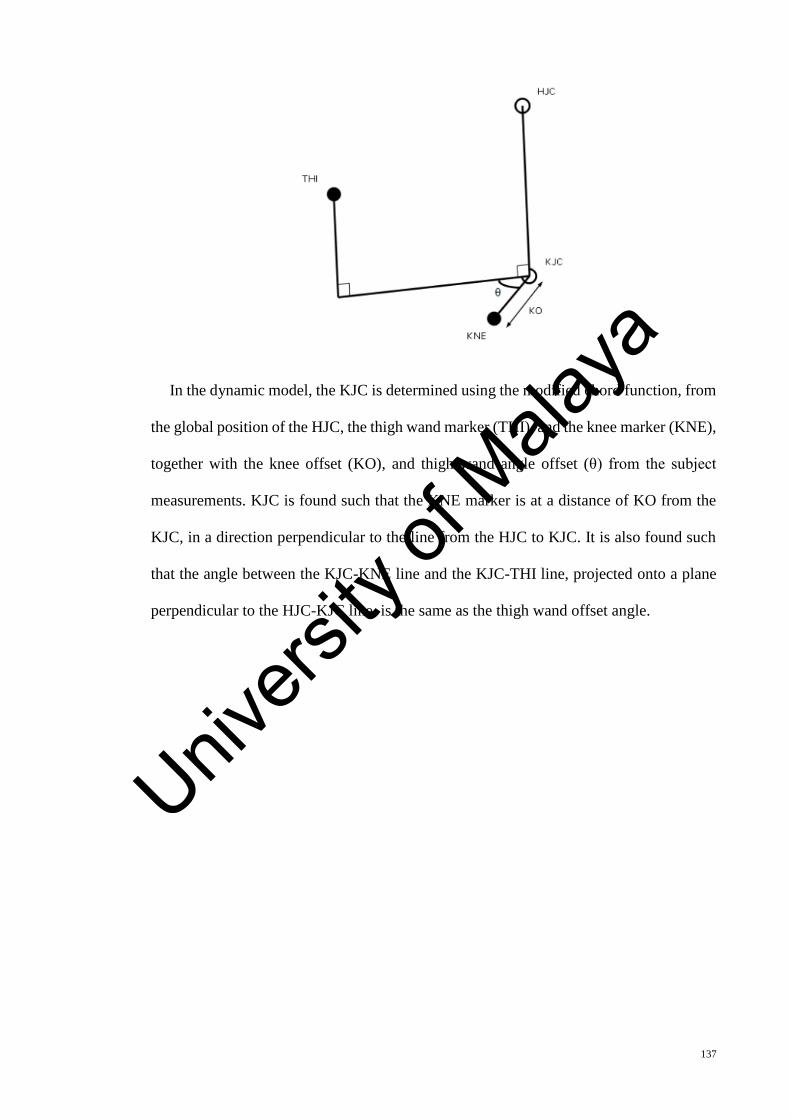
137
In the dynamic model, the KJC is determined using the modified chord function, from
the global position of the HJC, the thigh wand marker (THI), and the knee marker (KNE),
together with the knee offset (KO), and thigh wand angle offset (θ) from the subject
measurements. KJC is found such that the KNE marker is at a distance of KO from the
KJC, in a direction perpendicular to the line from the HJC to KJC. It is also found such
that the angle between the KJC-KNE line and the KJC-THI line, projected onto a plane
perpendicular to the HJC-KJC line, is the same as the thigh wand offset angle.
Univers
ity of
Mala
ya