IGCSE Human Biology Module Three: Human Physiology...
28
IGCSE Human Biology Module Three: Human Physiology B 1 Lesson Twelve The Nervous System Aims By the end of this lesson you should be able to: recall the plan of the nervous system and understand its functions recall the structure of sensory, motor and relay neurones, and understand the transmission of nerve impulses along and between neurones recall the structure of the spinal cord and describe the operation of spinal reflexes recall the main areas of the brain and their functions recall that there are receptors that respond to heat, chemical, mechanical and light energy, and o recall the structure of the eye, explain the principles of stereoscopic vision, and explain how the eye reacts to changes in light intensity and the need to focus near and distant objects o recall the structure of the ear, and describe its functions in balance and hearing Context This lesson covers elements (a) to (h), (l) and (m) of Section 5 (Coordination) of the Edexcel specification. It is quite long, but includes clear sub-sections on the different parts of the nervous system. Edexcel IGCSE Human Biology, chapter 5, pages 70-85. Oxford Open Learning
Transcript of IGCSE Human Biology Module Three: Human Physiology...
IGCSE Human Biology Module Three: Human Physiology B
1
Lesson
Twelve
The Nervous System
Aims By the end of this lesson you should be able to:
recall the plan of the nervous system and understand its
functions
recall the structure of sensory, motor and relay
neurones, and understand the transmission of nerve
impulses along and between neurones
recall the structure of the spinal cord and describe the
operation of spinal reflexes
recall the main areas of the brain and their functions
recall that there are receptors that respond to heat,
chemical, mechanical and light energy, and
o recall the structure of the eye, explain the
principles of stereoscopic vision, and explain how
the eye reacts to changes in light intensity and
the need to focus near and distant objects
o recall the structure of the ear, and describe its
functions in balance and hearing
Context
This lesson covers elements (a) to (h), (l) and (m) of Section 5
(Coordination) of the Edexcel specification. It is quite long, but
includes clear sub-sections on the different parts of the
nervous system.
Edexcel IGCSE Human Biology, chapter 5, pages 70-85.
Oxford Open Learning

Lesson Twelve The Nervous System
2
Introduction
One of the key characteristics of organisms is that they
respond to stimuli – if you prod them, they react! Biologists
call this characteristic sensitivity. In human beings it is
mainly produced by the nervous system.
To produce sensitivity, the nervous system needs several
different parts:
receptors that are sensitive to the stimulus (light,
sound etc.)
a central nervous system (CNS), consisting of the
brain and the spinal cord, which receives messages
from the receptors, decides on the appropriate response,
and coordinates it by sending messages to various
effectors
effectors, which produce the response. These are often
muscles, producing movement. Sometimes they are
glands, which release chemicals
nerves, which link the receptors, CNS and effectors
together, and carry messages between them in the form
of electrical impulses
In this lesson we will first study how the electrical impulses
are carried along nerves, then the structure and mode of
operation of the CNS, and finally the eye and ear as two
important receptors.
Log on to Twig and look at the film titled: Intro to the Brain
www.ool.co.uk/915mu
What does the brain look like and what does it actually
do? What specific functions do the brain stem,
cerebellum and cerebrum control?

IGCSE Human Biology Module Three: Human Physiology B
3
Activity 1
Suggest the response to each of these stimuli. In each case, what is
the effector?
(a) You step out into the road, and then hear a car horn blaring.
(b) You are hungry. You smell the aroma coming from a fish and chip
shop.
(c) An insect flies rapidly towards your eye.
Log on to Twig and look at the film titled: The Nervous System
www.ool.co.uk/1567jf
Our bodies contain an intricate network of nerves and
pathways which make up the Central Nervous System.
How are they linked? And how do they connect different
parts of our body to control bodily functions?
Nerves and Neurones
Look at figure 5.2 on page 71 of Edexcel IGCSE Human
Biology. The brain and spinal cord are outlined in red, while
some of the nerves are shown in yellow. Those running out
from the spinal cord are called spinal nerves, and those from
the brain are called cranial nerves.
Each nerve is thick enough to be visible to the naked eye, but
it is made up of a huge number of individual cells called
neurones running along its length. Every nerve contains two
sorts of neurone:

Lesson Twelve The Nervous System
4
sensory neurones which carry electrical impulses from
the receptors to the CNS
motor neurones which carry electrical impulses from
the CNS to the effectors
Any nerve will have lots of different impulses travelling along
it, in both directions, each in a separate neurone. There is also
a third sort of neurone, relay neurones, which carry impulses
between the sensory and motor neurones inside the CNS.
Log on to Twig and look at the film titled: Neurons as Cells
www.ool.co.uk/916yg
Everything our brain does, from controlling movement to
conscious thought, is achieved by the firing of electrical
signals called neurons.
Also look at the film titled: Neurons as Networks
www.ool.co.uk/966tc
Neurons, the brain's electrical signals, control how our
bodies work. Discover how they do this and why they are
implicit in learning new skills.
Neurone structure
Look at the diagram of a motor neurone in figure 5.3 on page
72 of the textbook. This neurone is a single cell, and has the
following parts:
a cell body, which contains a nucleus, mitochondria,
and the other usual organelles of an animal cell. It is
located in the CNS.
the cell membrane is drawn out into projections called
dendrons which end in hair-like dendrites, like twigs
on a tree. These make contact with other neurones in
the CNS at junctions called synapses, so electrical
impulses may be passed to it.
IGCSE Human Biology Module Three: Human Physiology B
5
one dendron is drawn out into a very long axon, which
runs the entire distance down a nerve to an effector
(here a muscle). The axon is surrounded by a myelin
sheath, with regular gaps in it. The myelin sheath is
made largely of lipid, and is an electrical insulator.
The far end of the axon is drawn out into several nerve
endings, rather like the dendrites at the near end.
These make contact with muscle fibres at special
synapses called neuromuscular junctions, and pass
the electrical impulses to them.
Figure 5.3 also gives a diagram of a sensory neurone. It has
essentially the same parts, but notice that:
the motor neurone has the cell body right at one end,
with dendrites on it, whereas
the sensory neurone has the cell body on a side stalk a
bit away from one end, with no dendrites on it.
Relay neurones vary in shape, but all have dendrites and no
long axon.
Get it right! The whole neurone (except for the myelin sheath) is a
single cell. The axon runs right along a nerve and may be a metre or
more long! Neurones are by far the longest cells in the body.
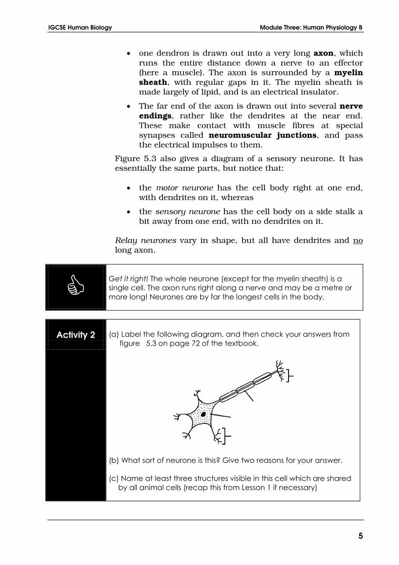
Activity 2
(a) Label the following diagram, and then check your answers from
figure 5.3 on page 72 of the textbook.
(b) What sort of neurone is this? Give two reasons for your answer.
(c) Name at least three structures visible in this cell which are shared
by all animal cells (recap this from Lesson 1 if necessary)
Lesson Twelve The Nervous System
6
Nervous impulses
When a receptor receives light, sound, or some other sort of
energy, it turns that energy into the form of electrical energy, a
process called transduction. The electrical energy is then shot
along the axons of its sensory neurones in short bursts called
nervous impulses or electrical impulses.
The myelin sheaths along the axons speed up the rate of
transmission of these impulses, and they typically travel at
about 100 metres per second (or ten times as fast as an
Olympic sprinter!) This high speed enables you to respond
quickly to stimuli, which is important for survival.
Activity 3
Estimate the time it would take for an electrical impulse to travel in a
neurone from the brain to the toe of a tall man.

IGCSE Human Biology Module Three: Human Physiology B
7
Synapses
The junction between two neurones is called a synapse. See
fig 5.15 on page 81 of Edexcel IGCSE Human Biology.
There is a small gap between the two neurones at a synapse
called the synaptic cleft. The electrical impulse is unable to
cross this gap, but its arrival stimulates the release of a
chemical called a neurotransmitter by the first neurone into
the synaptic cleft. This diffuses across the cleft and attaches
to dedicated sites on the cell membrane of the second
neurone. Its attachment stimulates a new electrical impulse
which sets off down the second neurone – the impulse has
crossed the synapse.
Synapses are important in several ways:
They slow up the process of transmission, increasing
reaction times.
Because neurotransmitter is only released by one of the
cells, and the dedicated sites are only on the other cell,
impulses can only travel in one direction across a
synapse. This keeps impulses heading in the right
direction along the neurones as well.
Each motor neurone can receive impulses from several
other neurones at synapses (see figure 5.16 on page 81
of the textbook). This makes free decision-making
possible in our brains – we do not always just react to
stimuli in an automatic fashion.
Several drugs (including caffeine and alcohol) alter
transmission at synapses, often by imitating or blocking
neurotransmitter molecules. This is how they change
our experience and/or performance.
Log on to Twig and look at the fact-pack titled: Spinal Cord
www.ool.co.uk/1084be
How the millions of nerves in the spinal cord help the brain
and body communicate.
Lesson Twelve The Nervous System
8
Reflexes and the Spinal Cord
The spinal cord is part of the central nervous system. It runs
from the base of the brain down inside the backbone, which
protects it from damage.
Structure of the spinal cord
The structure of the spinal cord, at the point where a spinal
nerve enters and leaves it, is shown in figure 5.13 on page 79
of Edexcel IGCSE Human Biology. Note the following parts:
an H-shaped central section called the grey matter.
This is where the cell bodies of the motor and relay
neurones are located;
an outer section called the white matter. This consists
mainly of neurone axons with their myelin sheaths;
a “roundabout” system linking the spinal cord to the
spinal nerve:
o the top branch of the roundabout, called the dorsal
root (towards your back), carries the sensory
neurones into the spinal cord. It has a bulge called
the dorsal root ganglion where the cell bodies of the
sensory neurones are located;
o the bottom branch, called the ventral root (towards
your front) carries the motor neurones out of the
spinal cord. It has no ganglion, because the cell
bodies of the motor neurones are in the grey matter
of the cord itself.
IGCSE Human Biology Module Three: Human Physiology B
9
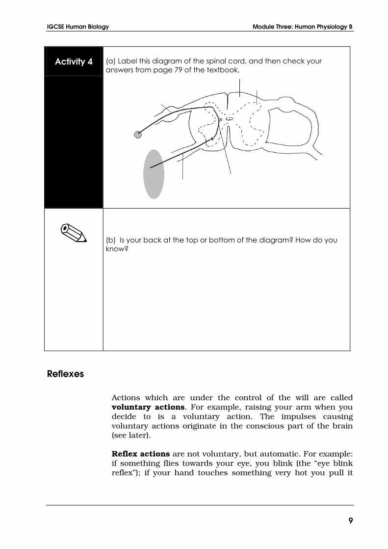
Activity 4
(a) Label this diagram of the spinal cord, and then check your
answers from page 79 of the textbook.
(b) Is your back at the top or bottom of the diagram? How do you
know?
Reflexes
Actions which are under the control of the will are called
voluntary actions. For example, raising your arm when you
decide to is a voluntary action. The impulses causing
voluntary actions originate in the conscious part of the brain
(see later).
Reflex actions are not voluntary, but automatic. For example:
if something flies towards your eye, you blink (the “eye blink
reflex”); if your hand touches something very hot you pull it
Lesson Twelve The Nervous System
10
away (the “withdrawal reflex”). These reactions “happen on
their own” - you do not need to think about them.
Reflex actions have survival value. That is, they decrease the
chance of damage to the body or death. They share the
following features:
they are rapid (useful for survival)
they are involuntary (we do not decide to do them and,
often, cannot stop them happening)
they are innate (you do not need to learn them)
Reflexes, or reflex actions, have these properties because they
are all based on an electrical impulse pathway called the
reflex arc.
The reflex arc
Look at figures 5.12 and 5.13 on page 79 of Edexcel IGCSE
Human Biology, which both show reflex arcs.
The stimulus generates an electrical impulse in a receptor.
This electrical impulse travels to the CNS in a sensory
neurone. At the CNS it transfers at a synapse to a relay
neurone and at a second synapse to a motor neurone. The
impulse travels down the motor neurone to a muscle (the
effector) and causes it to contract, producing the response.
Now:
because only two synapses are involved, the response
will be rapid (remember, it is synapses that slow down
impulse transmission)
because the conscious decision-making part of the brain
it not involved, the response will be involuntary
because the “wiring” of neurones is produced during
development without the need for any experience, the
response will be innate
The eye blink reflex mentioned above involves a cranial nerve
and the lower, non-conscious part of the brain. But most

IGCSE Human Biology Module Three: Human Physiology B
11
reflexes, called spinal reflexes, involve spinal nerves and the
spinal cord, not the brain at all. These include
the withdrawal reflex, and
the knee jerk reflex
Activity 5
Read about the knee-jerk reflex on page 80 of the textbook, and
then try it out as directed there. Can you get it to work?
The Brain
The human brain is a large swelling at the top of the spinal
cord, protected by the bony cranium or skull. The different
regions of the brain are specialised to do a large number of
different jobs - see figure 5.17 on page 82 of Edexcel IGCSE
Human Biology:
the medulla or brain stem, at the top of the spinal cord,
controls unconscious processes like the heartbeat and
breathing
the cerebellum, a cauliflower-like structure at the lower
back of the brain, coordinates balance
the hypothalamus, which we met in Lesson 11,
contains centres regulating body temperature and water
content
the pituitary gland releases a variety of hormones into
the bloodstream, including ADH which we also met in
Lesson 11
Lesson Twelve The Nervous System
12
however, by far the largest part of the human brain is
the cerebrum.
The cerebrum
The cerebrum is the location of our sensory experience,
thoughts and memory, and the place where our conscious
decisions and actions originate. It is divided into two halves
called the cerebral hemispheres. Its outer layer, called the
cerebral cortex, is folded over to give it a larger surface area,
which increases intelligence. It appears grey from the outside
because the grey matter, containing the cell bodies, is on the
outside rather than on the inside as in the spinal cord.
Different regions of the cerebrum are themselves specialized to
do different jobs: see figure 5.19 on page 83 of the textbook.
The sensory areas (green on the diagram) receive and
interpret impulses from the receptors (sense organs).
Each sense organ feeds its impulses into a different part
of the sensory area. This is why their impulses are
interpreted differently to give, for example, the different
experiences of sight and hearing. Notice:
o the vision area at the back of the brain
o the “main sensory area” where the receptors
scattered around the skin send their impulses,
each bit of skin to a different location
The motor areas (brown on the diagram) control the
movements of the various parts of the body. Again each
muscle has its own dedicated region of the motor area
which controls it
The association areas (yellow on the diagram) are where,
among other things:
o sensory experience is interpreted, “making sense”
of what we see and hear
o complicated actions like writing are coordinated
o experience is compared with stored memories to
produce “recognition”
o reasoning takes place
IGCSE Human Biology Module Three: Human Physiology B
13
The human cerebrum is more highly developed than that of
any other animal, which accounts for our high intelligence,
speech and technological capabilities.
Activity 6
Suggest what might happen if:
(a) the auditory nerve, coming from the ear, was “rewired” to feed
into the vision area at the back of the brain (do NOT try this at
home!).
(b) a person was given a mild electric shock in the part of the motor
area which controls the left leg
Receptors and Sense Organs
A receptor is a cell, or a group of cells, which is sensitive to a
stimulus. A sense organ is an organ dedicated to making one
or more receptors work. For example, the eye is a sense organ
which contains light-sensitive receptors (rod and cone cells) in
its retina.
Any receptor is sensitive to only one sort of stimulus. Human
beings have receptors sensitive to, among other things:
heat (temperature receptors in the skin – see Lesson 12)
light energy (rods and cones in the retina of the eye – see
below)
certain sorts of chemical (taste buds in the tongue;
olfactory organs in the nose)

Lesson Twelve The Nervous System
14
mechanical forces (cells detecting sound and gravity in
the inner ear – see below; pressure and pain receptors in
the skin – see Lesson 12)
The human body does not contain receptors sensitive to all
forms of energy. It has none sensitive to radio waves or X-rays
for example. These forms of energy may still cause damage,
however: X-rays can cause cancer even though we are
unaware of their presence.
Get it right! Experience occurs in the brain, not in the sense organs.
The brain interprets the incoming electrical impulses to produce our
experience of heat, light, sound and so on. If, for example, the vision
area of the brain is damaged, the victim will be blind even though
their eyes are in perfect working order.
Log on to Twig and look at the film titled: How we see 1: Eyes
www.ool.co.uk/944wc
The process of seeing is a collaboration between the
brain and the eye. How does the eye work to create
sight, from the lens and iris to the pupil, retina and optic
nerve?
The Eye
Study the diagram of the eye on page 73 of Edexcel IGCSE
Human Biology, and notice the following parts:
Light enters from the left through a thin, transparent
skin called the conjunctiva. It is refracted (bent) by the
curved, transparent cornea and the curved, transparent
lens, and brought to a focus on the retina at the back of
the eye, which contains the light-sensitive cells.
The retina cells are supplied with nutrients and oxygen
by blood vessels running through the choroid. The
choroid is also black to absorb the light which passes
through the retina. This would otherwise bounce around
inside the eye causing multiple images. The hard, white
IGCSE Human Biology Module Three: Human Physiology B
15
sclera outside this keeps the eyeball in shape. It is
continuous with the transparent cornea at the front.
The fovea lies on the axis of the eye. It is here that the
light falls when you look straight at something. It has
mainly light-sensitive cells called cones which give clear
colour vision but only work in bright light. The rest of
the retina has fewer cones and more rods. Rods work in
dim light, but give less clear black & white vision.
The optic nerve carries electrical impulses away from
the retina to the brain in sensory neurones. At the point
where it leaves the eye there are no rods or cones. This
is the blind spot – any object whose light falls here is
invisible.
The iris is a coloured (usually brown or blue) barrier in
front of the lens. Light passes through a hole in its
middle called the pupil to reach the retina. From the
outside the pupil looks like a black spot surrounded by
a coloured circular iris.
Get it right! The light is focused both by the cornea and by the lens. In
fact the cornea bends the light more than the lens.
Activity 7
Try out the exercise to locate your blind spot described in the last few
lines of page 75 of the textbook.
Lesson Twelve The Nervous System
16
Accommodation by the lens
By refracting (bending) the light, the cornea and lens between
them focus an inverted (upside down) image (picture) of
objects on the retina at the back of the eye. See figure 5.5 on
page 74 of the textbook to see how this is done. Notice that the
light from the top of the key ends up at the bottom of the image
and vice versa, making the image upside down.
To focus nearby objects the lens must be fatter than to focus
distant objects, so the lens changes its shape to bring near or
distant objects into focus. This change is called
accommodation. (See figure 5.7 on page 76 of the textbook
for this. Notice that the light from the nearby object is
spreading out as it reaches the eye, so must be refracted
more.) The change in lens shape is caused by the contraction
and relaxation of the ciliary muscles, which forms a ring
around the lens:
To focus a nearby object, the ciliary muscles contract
and form a smaller ring. This makes the suspensory
ligaments, which join the ciliary muscles to the lens, go
slack. The lens springs into its natural, rounded shape
and bends the light more strongly. You can feel the
ciliary muscles working if you look at a very close object
and try to focus it – you soon get eye strain!
To focus a distant object, the ciliary muscles relax and
return to their natural larger ring. This pulls the
suspensory ligaments taut, pulling the lens into a
thinner shape which does not bend the light so much.
As the ciliary muscles are doing no work, the eye is
relaxed looking at distant objects – no eye strain!
Log on to Twig and look at the film titled: How we see 2: Brain
www.ool.co.uk/945gk
Only a small part of what the eye 'sees' is in focus. To turn
the image on the retina into complete vision the eye
needs help from the brain.
IGCSE Human Biology Module Three: Human Physiology B
17
Activity 8
(a) Predict whether it should be possible to get both a near object
and a distant object in focus at the same time. Then try it out to
test your prediction.
(b) Hold an object as near to one of your eyes as you can while
keeping it still in focus. Hold it for a few seconds and feel the
strain in the eye. Which muscles are getting tired? Now relax
your eye. The strain will disappear, but the object will go out of
focus.
Response to changes in light intensity by the iris
The eye attempts to keep the intensity (brightness) of the light
reaching the eye as constant as possible. It achieves this by
contracting and relaxing muscles in the iris to change the size
of the pupil in its centre.
The iris contains two sets of muscles (see figure 5.6 on page
75 of the textbook). Its circular muscles make the pupil
smaller when they contract. Its radial muscles, spreading out
from the centre like the spokes of a wheel, pull the pupil wider
open as they contract.
When the light is bright the circular muscles contract
and the radial muscles relax. This makes the pupil
smaller, reducing the amount of light getting to the
retina.
When the light is dim the circular muscles relax and the
radial muscles contract. This makes the pupil larger,
increasing the amount of light getting to the retina.

Lesson Twelve The Nervous System
18
Activity 9
Observe the front of your eyes using a mirror in dim and bright light.
What happens to the pupil as you move from dim to bright light, and
why?
Stereoscopic vision
Because we have two eyes, with slightly different viewpoints,
the brain receives two slightly different pictures of any object it
looks at. The closer the object, the more different these two
pictures are.
The visual association area at the back of the brain uses this
information to construct a three-dimensional picture of the
world. Stereoscopic or binocular vision (vision with two eyes)
permits depth perception – we can see how far away objects
are.
This ability was developed during the period of our evolution
which we spent up trees. During this period we traded in the
ability to see behind us (achieved by having the eyes on the
sides of the head) for the ability to see how far away was the
branch we were about to leap for!
Activity 10
Try the exercise with the pencil described in the first paragraph of
page 76 of the textbook.

IGCSE Human Biology Module Three: Human Physiology B
19
The Ear
The human ear has two quite different functions:
it is sensitive to sound, and
it is an organ of balance
The structure of the ear is shown on page 77 of Edexcel IGCSE
Human Biology. Note the following parts.
The outer ear, filled with air (coloured blue in the
diagram), leads down the auditory canal to the
tympanum or ear drum. This vibrates backwards and
forwards rapidly when sound waves hit it.
The middle ear, also filled with air, transmits and
magnifies these vibrations through a complicated
arrangement of three ossicles, or ear bones: the
malleus (hammer), incus (anvil) and stapes (stirrup).
The middle ear connects to the back of the throat
through an air-filled tube called the Eustachian tube,
which opens when we swallow. This tube keeps the air
pressure the same on both sides of the tympanum, so
that it can vibrate freely.
The inner ear is filled with liquid. It contains receptors
responsible for hearing in a spiral structure called the
cochlea, and receptors responsible for balance in the
semicircular canals. The auditory nerve runs from
both of these structures to the brain, carrying nervous
impulses in sensory neurones.
Lesson Twelve The Nervous System
20
Log on to Twig and look at the film titled: How we hear
www.ool.co.uk/949ru
How our ears hear different frequencies, and how they
work with the brain to turn these into sounds which we
understand.
Sound and Hearing
Figure 5.9 on page 78 of the textbook shows the cochlea
uncoiled to make its function clearer.
The stapes transmits the magnified sound vibrations to the
liquid in the cochlea through a tough membrane called the
oval window. As this window moves rapidly back and forth
with the vibrations, the fluid in the cochlea does the same.
This repeatedly bends receptor cells in the organ of Corti
attached to the centre of the cochlea. When bent they generate
electrical impulses which travel to the brain.
Because liquid (unlike air) cannot be compressed, the cochlea
also has a second membrane called the round window. This
moves out as the oval window moves in, and vice versa to,
stop the cochlea bursting.
Receptor cells at different positions along the organ of Corti
vibrate strongly with different frequencies (pitches) of sound.
This enables the brain to sense the pitch of a sound, as well as
its loudness.
Balance
The inner ear also contains two different structures connected
with different aspects of balance. One detects movement of the
head, and the other the orientation of the head.
The three semicircular canals each have a swelling
called the ampulla, which contains a jelly-like cupula
connected to the wall by sensitive hair cells. When you
move your head the fluid in the canal moves. This moves
the cupula which bends the hair cells, generating
electrical impulses. Because the three canals are at right
angles, any movement of the head will move the fluid in
IGCSE Human Biology Module Three: Human Physiology B
21
at least one of the canals, so the brain is made aware of
the movement.
If you spin around for a long time, the fluid in some of
the canals carries on moving when you stop. As far as
the brain is concerned your head is still spinning round,
so you feel dizzy!
The utriculus and sacculus (see figure 5.8, page 77)
each contain a heavy otolith made of calcium carbonate
(chalk) attached to the wall by hair cells. The weight of
the otolith bends the hair cells, producing electrical
impulses. These give the brain information about the
orientation of the head: the impulses are different when
you are lying down to when you are standing up.
Now is the time to read through chapter 5, pages 70 - 85 of
Edexcel IGCSE Human Biology. They cover the same topics as
this lesson, so will add to your understanding of the material.
Keywords Sensitivity
Central nervous
system (CNS)
Spinal cord
Neurone
Synapse
Transduction
Reflex arc
Refracted
Accommodation
Ossicle
Semicircular
canal
Receptor
Effector
Impulse
Dendrite
Myelin sheath
Neurotransmitter
Cerebrum
Inverted
Tympanum
Organ of Corti
Cochlea

Lesson Twelve The Nervous System
22
Summary
Lesson Twelve: The Nervous System
NERVOUS SYSTEM ------ stimulus – receptor – sensory neurone –
CNS – motor neurone –
effector – response
NEURONES ------ sensory, motor, relay
------ electrical / nervous impulses
------ synapses
SPINAL CORD ------ structure
------ spinal reflexes
BRAIN ------ medulla, cerebellum, hypothalamus,
pituitary gland
------ cerebrum: sensory, motor and
association areas
SENSE ORGANS ------ eye: structure, accommodation, control
of light entering, stereoscopic vision
------ ear: hearing, balance
What you need to know
The structures of the different types of neurone
The structure of the spinal cord and the route of a reflex
arc
The structure of the brain and the functions of its
various parts
The structure of the eye and how each part contributes
to sight
The structure of the ear and how each part contributes
to either hearing or balance
IGCSE Human Biology Module Three: Human Physiology B
23
What you might be asked to do
Label diagrams of neurones, spinal cord, brain, eye and
ear
Explain impulse transmission along and between
neurones
Identify stimuli and responses
Explain the occurrence and importance of reflex actions
Explain how the eye copes with different light intensities
and the need to focus near and distant objects
Explain how the ear functions as an organ of hearing
and balance
Suggested Answers to Activities
Activity 1
(a) Response: you jump backwards. Effector: your leg
muscles.
(b) Response: your mouth waters. Effector: your salivary
glands.
(c) Response: you blink. Effector: your eyelid muscles.
Activity 2
(b) It is a motor neurone, because the cell body is right at
one end of the cell and has dendrites on it.
(c) Nucleus, cytoplasm and cell membrane.
Activity 3
Distance: about 2m.

Lesson Twelve The Nervous System
24
Speed: about 100m/s (metres per second)
Time: about 2/100 = 0.02s
Activity 4
(b) At the top. The dorsal root ganglion is drawn towards
the top, and “dorsal” means “back”.
Activity 6
(a) If you played the person music, they would hear
nothing, but would see flashing lights.
(b) Their left leg would twitch.
Activity 8
(a) No, it is not possible.
(b) The ciliary muscles are getting tired.
Activity 9
The pupil gets narrower in bright light, so that less light enters
and reaches the retina. (If the retina receives too much light it
can be damaged.)
IGCSE Human Biology Module Three: Human Physiology B
25
Tutor-marked Assignment F
Question 1
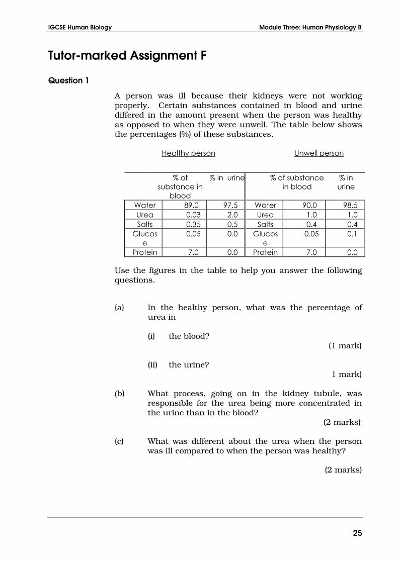
A person was ill because their kidneys were not working
properly. Certain substances contained in blood and urine
differed in the amount present when the person was healthy
as opposed to when they were unwell. The table below shows
the percentages (%) of these substances.
Healthy person Unwell person
% of
substance in
blood
% in urine % of substance
in blood
% in
urine
Water 89.0 97.5 Water 90.0 98.5
Urea 0.03 2.0 Urea 1.0 1.0
Salts 0.35 0.5 Salts 0.4 0.4
Glucos
e
0.05 0.0 Glucos
e
0.05 0.1
Protein 7.0 0.0 Protein 7.0 0.0
Use the figures in the table to help you answer the following
questions.
(a) In the healthy person, what was the percentage of
urea in
(i) the blood?
(1 mark)
(ii) the urine?
1 mark)
(b) What process, going on in the kidney tubule, was
responsible for the urea being more concentrated in
the urine than in the blood?
(2 marks)
(c) What was different about the urea when the person
was ill compared to when the person was healthy?
(2 marks)

Lesson Twelve The Nervous System
26
(d) (i) Which substance was not found in the urine
either when the person was healthy or when the
person was ill?
(1 mark)
(ii) Explain why this substance was not found in
the urine. (2 marks)
(e) (i) Which substance was not found in the urine of
the healthy person, but was found in the urine
when the person was ill?
(1 mark)
(ii) Explain how the kidney of a healthy person
avoids losing this substance in the urine.
(3 marks)
(iii) Describe a chemical test you could carry out to
show the presence of this substance in the
urine. (3 marks)
Total marks for Q1 = 16
Question 2
If the tendon below the knee is tapped with a hammer, the
lower leg jerks upwards. A student conducted an investigation
to find out how the speed of the hammer affected the distance
the lower leg moved.
Each trial was recorded on a video. A frame was taken every
33 milliseconds. The video was then played using single-frame
advance. The number of frames for the hammer to move to the
knee was found. The faster the speed, the smaller was the
number of frames. The video was also used to find the
distance moved by the toe.
The table below shows the results obtained:
IGCSE Human Biology Module Three: Human Physiology B
27
(a) (i) What was the student’s dependent variable?
(1 mark)
(ii) State one potential variable that the student
controlled. (1 mark)
(b) Explain the advantages of using the video camera for
this investigation (2 marks)
(c) Give a conclusion based on the results of the
investigation. (2 marks)
(d) Comment on the precision of the results for the
distance moved by the toe. (2 marks)
(e) Describe the pathway in the nervous system which
caused the toe to move when the tendon was struck.
(6 marks)
Total marks for Q2 = 14
Question 3
A cataract is an eye problem involving the lens of the eye
becoming cloudy. It can be treated by a simple operation
which removes the lens. After the operation, the patient is able
to see again, but will probably need to wear glasses.
(a) Explain why the eye can still form an image after the
lens has been removed. (2 marks)
(b) After the operation, the patient will be unable to
perform “accommodation”.
Explain what is meant by accommodation, and why
the patient will be unable to do it. (3 marks)
(c) Is the patient more likely to need glasses to see
nearby or distant objects? Explain your answer.
(3 marks)
(d) Will the operation affect the patient’s ability to see in
dim light? Explain your answer. (2 marks)
(total marks for Q3 = 10)
Lesson Twelve The Nervous System
28
Question 4
While running in the Olympic marathon, a runner produces a
lot of extra heat in their leg muscles and loses a lot of water by
breathing and sweating. Outline some of the processes in the
body which attempt to prevent the runner overheating and
dehydrating. Give your answer in full sentences.
(10 marks)
Total marks for TMA = 50