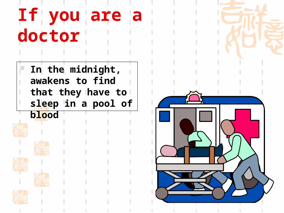
If you are a doctor In the midnight, awakens to find that they have to sleep in a pool of blood.
55
If you are a doctor In the midnight, awakens to find that they have to sleep in a pool of blood
-
Upload
nancy-houston -
Category
Documents
-
view
215 -
download
2
Transcript of If you are a doctor In the midnight, awakens to find that they have to sleep in a pool of blood.
If you are a doctor
In the midnight, awakens to find that they have to sleep in a pool of blood

How to diagnosis? How to management?
You

Antepartum Hemorrhage
Obstetrics & Gynecology Hospital of Fudan University
Xu Huan

Rationale (why we care…)
• 4-5% of pregnancies complicated by 3rd trimester bleeding
• Immediate evaluation needed• Significant threat to mother & fetus
(consider physiologic increase in uterine blood flow)• Consider causes of maternal & fetal death• Priorities in management (triage!)

Objectives We will be able to:
• Describe the approach to the patient with third-trimester bleeding
• Compare symptoms, physical findings, and diagnostic methods that differentiate bleeding etiologies
• Describe management and delivery options for 3rd trimester bleeding etiologies
• Describe potential maternal and fetal morbidity & mortality
• Describe management of postpartum hemorrhage• Apply knowledge in the discussion of clinical case
scenarios

Vaginal Bleeding: Differential diagnosis
• Common:• Abruption, previa, preterm labor, labor
• Less common: • Uterine rupture, fetal vessel rupture, lacerations/le
sions, cervical ectropion, polyps, vasa previa, bleeding disorders
• Unknown• NOT vaginal bleeding!!!
(happens more than you think!)

Other Etiologies
• Cervicitis• infection • Cervical erosion • Trauma • Cervical cancer • Foreign body • Bloody show/labor
Perinatal mortality and morbidity
• Previa• Decreased mortality from 30% to 1% over last 60 years• Now emergent cesarean delivery often possible• Risk of preterm delivery
• Abruption• Perinatal mortality rate 35%• Accounts for 15% of 3rd trimester stillbirths• Risk of preterm delivery• Most common cause of DIC in pregnancy
• Massive hemorrhage --> risk of ARF, Sheehan’s, etc.

Placenta previa

Definition
After 28 pregnant weeks placental implantation over the cervical os or in the lower uterine segment
It constitutes an obstruction of descent of the presenting part
Main cause of obstetrical hemorrhage(20%) Incidence
0.24%-1.57% (our country).

Risk factors & Associations
• Prior cesarean delivery/myomectomy• Prior previa (4-8% recurrence risk) • Previous abortion • Increased parity • Multiple pregnancy• Advanced maternal age • Abnormal presentation • Smoking

Etiology
Causes1. Endometrial abnormality1) Scared or poorly vascularized endometrium in the c
orpus.2) Curettage, Delivery, CS and infection of endometri
um2. Placental abnormality Large placenta (multiple pregnancy), succenturiate
lobe3. Delayed development of trophoblast

Classification
Complete
placenta previa
Partrial
placenta previa
Marginal placenta previa


Manifestation(1) Symptoms
• Painless vaginal bleeding (70%)• Spontaneous,After coitus• The most characteristic symptom• late pregnancy (after the 28th week) and delivery• Characteristics: sudden, painless and profuse
• Contractions• No symptoms
• Routine ultrasound finding
The mean gestational age of first bleed: 30 wks 1/3 before 30 weeks
Manifestation(2)
Anemia or shock
repeated bleeding→ anemia
heavy bleeding→ shock Abnormal fetal position
a high presenting part
breech presentation (often)

Physical Findings
• Bleeding on speculum exam• Cervical dilation
• Bleeding a sx related to PTL/normal labor• Abnormal position/lie• Non-reassuring fetal status • If significant bleeding:
• Tachycardia • Postural hypertension• Shock

Diagnosis(1)
History
1. Painless hemorrhage
2. At late pregnancy or delivery
3. History of curettage or CS

Diagnosis(1)
Signs
1. Abdominal findings
1) Uterus is soft, relaxed and nontender.
2) Contraction may be palpated.
3) A high presenting part can’t be pressed into the pelvic inlet. Breech presentation
4) Fetal heart tones maybe disappear (shock or abruption)

Diagnosis
Speculum examination
Rule out local causes of bleeding, such as cervical erosion or polyp or cancer.
Limited vaginal examination (seldom used)
Palpation of the vaginal fornices to learn if there is an intervening bogginess between the fornix and presenting part.
Rectal examination is useless and dangerous

Limited vaginal examination
Diagnosis(1)
• Ultrasound• abdominal 95% accurate to detect• transvaginal (TVUS) will detect almost all
• consider what placental location a TVUS may find that was missed on abdominal
• MRI• Check the placenta and membrane after delivery remember: no digital exams unless previa RULED O
UT!


Diagnosis
Before 20 weeks’ gestation,4-6% have some degree of placenta previa on ultrasonic examination
90% of these resolving by the third trimester Only 10% of complete placenta

Differential Diagnosis
Placental abruption
vagina bleeding with pain, tenderness of uterus. vasa previa
In cases of velamentous cord insertion fetal vessels cover cervical os
Abnormality of cervix
cervical erosion or polyp or cancer

vasa previa

Effects
obstetrical hemorrhage Placenta accreta, increta, and percreta Anemia and infection Premature labor or fetal death or fetal distress

Treatments
Expectant therapy
1. Rest: keep the bed
2. Controlling the contraction: MgSO4
3. Treatment of anemia
4. Preventing infection

Treatments
Termination of pregnancy1. CS1) total placenta previa (36th week), Partial placenta pr
evia (37th week) and heavy bleeding with shock2) Preventing postpartum hemorrhage: pitocin and PG3) Hysterectomy: Placenta accreta or uncontroled blee
ding

Treatments
2. Vaginal delivery
Marginal placenta previa
Vaginal bleeding is limited

Management
• Initial evaluation/diagnosis• Observe/admit to L&D• IV access, routine (maybe serial) labs • Continuous electronic fetal monitoring
• Continuous at least initally• May re-evaluate later if stable, no further bleeding
• Delivery???
Management
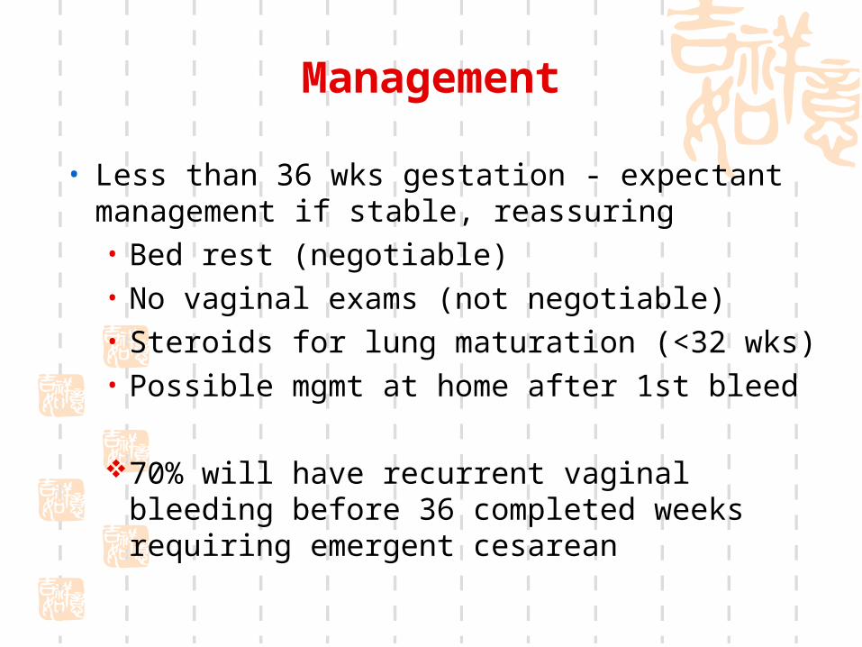
• Less than 36 wks gestation - expectant management if stable, reassuring• Bed rest (negotiable)• No vaginal exams (not negotiable) • Steroids for lung maturation (<32 wks)• Possible mgmt at home after 1st bleed
70% will have recurrent vaginal bleeding before 36 completed weeks requiring emergent cesarean

Management
• 36+ weeks gestation• Cesarean delivery if positive fetal lung maturity by amnioc
entesis• Delivery vs expectant mgmt if fetal lung immaturity• Schedule cesarean delivery at 37 weeks• Discussion/counseling regarding cesarean hysterectomy
Note: given stable maternal and reassuring fetal status, none of these management guidelines are absolute (this is why Obstetrics is so much fun!)

Other Considerations
• Placenta accreta, increta, percreta• Cesarean delivery may be necessary• History of uterine surgery increases risk• Must consider these diagnoses if previa present• Could require further evaluation, imaging (MRI cons
idered now)
NOT the delivery you want to do at 2 am

Abnormally adherent placentation. A. Placenta accreta. B. Placenta increta. C. Placenta percreta
A
B
C

Cesarean hysterectomy specimens with placenta percreta.

Cesarean hysterectomy specimens with placenta percreta.

Placental abruption

Definition
• abruptio placentae or placental abruption: placental separation from its implantation site before delivery (the normally implanted placenta )
• Incidence • complicates 0.5-1.5% of all pregnancies • recurrence risk
• 10% after 1st episode • 25% after 2nd episode

Risk factors & Associations
Cocaine maternal hypertension abdominal trauma smoking prior abruption preeclampsia multiple gestation
prolonged PROM uterine decompression short umbilical cord chorioamnionitis multiparity

Pathology
Placental separation is initiated by hemorrhage into the decidua basalis with formation of a decidual hematomaConcealed hemorrhageRevealed hemorrhage

revealed hemorrhage concealed hemorrhage

Total placental abruption with concealed hemorrhage and fetal death
Maternal-fetal risk
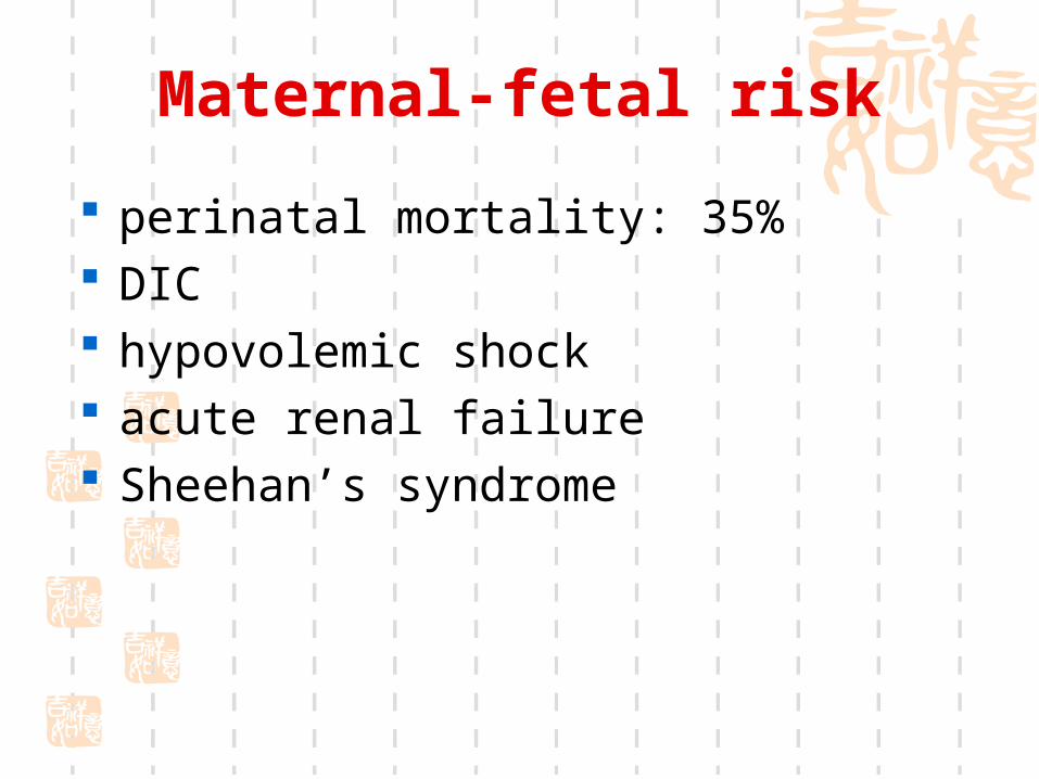
perinatal mortality: 35% DIC hypovolemic shock acute renal failure Sheehan’s syndrome


Symptoms
• Vaginal bleeding • Abdominal or back pain• Uterine contractions • Uterine tenderness

Physical Findings
• Vaginal bleeding• Uterine contractions • Hypertonus • Tetanic contractions • Non-reassuring fetal status or demise• Can be concealed hemorrhage


Laboratory Findings
• Anemia• may be out of proportion to observed blood lo
ss
• DIC• Can occur in up to 10% (30% if “severe”)• First, increase in fibrin split products • Followed by decrease in fibrinogen

Diagnosis
• Clinical scenario• Physical exam
• Not digital pelvic exams until rule out previa• Careful speculum exam
• Ultrasound• Can evaluate previa• Not accurate to diagnose abruption

Management
• Physical exam• Continuous electronic fetal monitoring • Ultrasound
• Assess viability, gestational age, previa, fetal position/lie
• Expectant management• vaginal vs cesarean delivery
• Available anesthesia, OR team for stat cesarean delivery

Partial placental abruption with adhered clot

Couvelaire Uterus