Identification of prognostic biomarkers in patients ... · Identification of prognostic biomarkers...
101
Identification of prognostic biomarkers in patients undergoing pulmonary metastasectomy from primary colorectal cancer Doctoral thesis at the Medical University of Vienna for obtaining the academic degree Doctor of Philosophy Submitted by Dr.med.univ. Thomas Sebastian Schweiger Supervisor: Assoc. Prof. Univ. Doz. Dr. Hendrik Jan Ankersmit, MBA Department of Thoracic Surgery Christian Doppler Laboratory for Cardiac and Thoracic Diagnosis and Regeneration Medical University of Vienna Waehringer Guertel 18-20 1090 Vienna, Austria Vienna, 06.03.2016
Transcript of Identification of prognostic biomarkers in patients ... · Identification of prognostic biomarkers...

Identification of prognostic biomarkers in patients
undergoing pulmonary metastasectomy from primary colorectal cancer
Doctoral thesis at the Medical University of Vienna
for obtaining the academic degree
Doctor of Philosophy
Submitted by
Dr.med.univ. Thomas Sebastian Schweiger
Supervisor:
Assoc. Prof. Univ. Doz. Dr. Hendrik Jan Ankersmit, MBA
Department of Thoracic Surgery
Christian Doppler Laboratory for Cardiac and Thoracic Diagnosis and Regeneration
Medical University of Vienna
Waehringer Guertel 18-20
1090 Vienna, Austria
Vienna, 06.03.2016

1
“When a plant goes to seed, its seeds are carried in all directions; but they can only live and
grow if they fall on congenial soil.”
Stephen Paget, English surgeon (1855 - 1926)
cited from Paget S, “The distribution of secondary growths in cancer of the breast” published in The Lancet (Volume 133, Issue 3421, 23 March 1889)

1
Declaration
The herein presented doctoral thesis was carried out at the Division of Thoracic Surgery
(Medical University of Vienna) and the Christian Doppler Laboratory for Diagnosis and
Regeneration of Thoracic Diseases (Medical University of Vienna). The collection of clinical
data during follow-up visits was kindly supported by Christoph Nikolowsky (Division of
Thoracic Surgery, Medical University of Vienna), Olaf Glück (Christian Doppler Laboratory for
Diagnosis and Regeneration of Thoracic Diseases, Medical University of Vienna), Christoph
Glogner (Division of Thoracic Surgery, Medical University of Vienna), Roland Mair (Division
of Thoracic Surgery, Medical University of Vienna) and Gerrit Lewik (Division of Thoracic
Surgery, Medical University of Vienna). Tissue preparation and immunohistochemical
staining were supported by Denise Traxler (Christian Doppler Laboratory for Diagnosis and
Regeneration of Thoracic Diseases, Medical University of Vienna). Immunohistochemical
staining for vimentin and KRAS/BRAF mutational analysis was conducted at the laboratory of
Balazs Hegedus (Thoracic Oncology Laboratory, Division of Thoracic Surgery, Medical
University of Vienna) and Balazs Döme (Thoracic Oncology Laboratory, Division of Thoracic
Surgery, Medical University of Vienna). Conduction of ELISA assays was supported by
Matthias Zimmermann (Christian Doppler Laboratory for Diagnosis and Regeneration of
Thoracic Diseases, Medical University of Vienna). Clinical data and specimen of colorectal
cancer liver metastases were kindly provided by Patrick Starlinger (Department of Surgery,
Medical University of Vienna) and Michael Bergmann (Department of Surgery, Medical
University of Vienna). Evaluation of the immunohistochemical stainings, data preparation,
interpretation and writing of the original articles was performed by Thomas Schweiger
(Christian Doppler Laboratory for Diagnosis and Regeneration of Thoracic Diseases, Division
of Thoracic Surgery, Medical University of Vienna) under supervision of Hendrik Jan
Ankersmit (Christian Doppler Laboratory for Diagnosis and Regeneration of Thoracic
Diseases, Division of Thoracic Surgery, Medical University of Vienna) and supported by
Peter Birner (Head of the Department, Department of Pathology, Medical University of
Vienna). The work was furthermore generously supported by Konrad Hoetzenecker (Division
of Thoracic Surgery, Medical University of Vienna) and Walter Klepetko (Head of the
Department, Division of Thoracic Surgery, Medical University of Vienna).

2
Table of contents
Declaration .......................................................................................................................... 1Table of contents ................................................................................................................ 2List of figures ....................................................................................................................... 4List of tables ........................................................................................................................ 5Abstract ............................................................................................................................... 6Zusammenfassung .............................................................................................................. 7Publications arising from this thesis .................................................................................. 8Abbreviations ...................................................................................................................... 9Acknowledgments ............................................................................................................. 10
CHAPTER ONE: INTRODUCTION ....................................................................................... 111. General introduction ....................................................................................................... 11
1.1 Epidemiology of CRC pulmonary metastases .......................................................... 111.2 Pathobiology of CRC lung metastases ..................................................................... 121.3 Clinical prognostic factors in pulmonary metastasectomy ........................................ 14
Carcinoembryonic antigen .......................................................................................... 14
Completeness of resection .......................................................................................... 14
Disease-free survival ................................................................................................... 15
Lymph node involvement ............................................................................................ 15
Number of nodules ...................................................................................................... 16
1.4 Molecular prognostic factors in pulmonary metastasectomy .................................... 18Prognostic factors in pulmonary metastasectomy: spotlight on molecular and radiological markers (published PDF) ......................................................................... 18
KRAS in metastatic colorectal cancer ......................................................................... 28
Heat shock protein 27 and fibroblasts ......................................................................... 29
1.7 Treatment of pulmonary metastases ........................................................................ 301.8 Aims of this thesis ..................................................................................................... 32
CHAPTER TWO: RESULTS .................................................................................................. 332. Prologue ......................................................................................................................... 33
2.1 EGFR, BRAF and KRAS status in patients undergoing pulmonary metastasectomy from primary colorectal carcinoma: a prospective follow-up study (published PDF) ...... 34
2.1.1. Interlude ............................................................................................................ 44
2.2 Stromal expression of heat-shock protein 27 is associated with worse clinical outcome in patients with colorectal cancer lung metastases (published PDF) ............... 45
CHAPTER THREE: DISCUSSION ........................................................................................ 613.1 General discussion ....................................................................................................... 613.9 Conclusion & future prospects ..................................................................................... 64
CHAPTER FOUR: MATERIALS & METHODS ...................................................................... 66

3
4.1 Materials ....................................................................................................................... 664.2 Methods ........................................................................................................................ 66
REFERENCES ...................................................................................................................... 70APPENDIX ............................................................................................................................. 82
Supplementary material “EGFR, BRAF and KRAS status in patients undergoing pulmonary metastasectomy from primary colorectal carcinoma: a prospective follow-up study” ........ 82
Supplementary Figure 1 ................................................................................................. 82Supplementary material “Stromal expression of heat-shock protein 27 is associated with worse clinical outcome in patients with colorectal cancer lung metastases.” ..................... 83
Supplementary Table 1 ................................................................................................... 83Supplementary Table 2 ................................................................................................... 84Supplementary Table 3 ................................................................................................... 85
CV .......................................................................................................................................... 86

4
List of figures
Figure 1: Overall survival of patients with CRC pulmonary metastases exhibiting sustained
lymphangiogenesis (left) or with evidence of lymphatic invasion (right) have a diminished
prognosis after pulmonary metastasectomy. The outcome is comparable to patients with
pathologically verified thoracic lymph node metastasis (Group B; dashed line). Adopted
from „Increased lymphangiogenesis in lung metastases from colorectal cancer is
associated with early lymph node recurrence and decreased overall survival“ (Schweiger
et al, 2015). ..................................................................................................................... 16
Figure 2: CT scan (A), PET scan (B) and merged PET-CT (C) showing strong 18F-FDG
uptake of two lung metastases in a patient with a history of colorectal cancer. ............. 17
Figure 3: KRAS mutational status as predictive marker for anti-EGFR therapy. Anti-EGFR
therapy, i.e. cetuximab or panitumumab, is capable to suppress the KRAS pathway
downstream to EGFR (left side). Tumors with activating mutations of KRAS are
characterized by constantly increased downstream signaling, e.g. ERK/MAPK pathway,
which leads to tumor progression (right side). Anti-EGFR targeted therapy upstream to
KRAS is considered being inefficient in a majority of these patients. ............................. 29

5
List of tables Table 1: Specification of antibodies and dilutions (IF= immunofluorescence; IHC=
immunohistochemistry) ................................................................................................... 69

6
Abstract
Pulmonary metastases (PM) contribute to the high cancer-related morbidity and mortality in
patients with solid malignancies including colorectal cancer (CRC). Despite advances in the
diagnosis and treatment of patients with early-stage CRC, the optimal treatment of patients
with distant metastases from CRC - especially PM - is currently unclear. To improve the
clinical outcome of these patients, the biology of metastatic CRC needs to be further
elucidated.
In this work we aimed to describe clinical as well as histopathological and genetic factors
contributing to the diminished prognosis in patients with an unfavorable tumor biology. On
DNA level, we assessed the mutation status of KRAS and BRAF, which are thought to play
an important role in CRC progression. Moreover, the expression of EGFR in PM was
described. In the second part of this work we focused on the tumor-surrounding stroma. To
the best of our knowledge, we could describe for the first time that Hsp27 is highly expressed
by activated fibroblasts adjacent to the disseminated tumor cells. Moreover, these activated
fibroblasts are a prognostic marker after pulmonary metastasectomy.
In this work we demonstrated that patients with PM from CRC are a highly heterogenous
group regarding the tumor biology and the outcome after pulmonary metastasectomy can be
prognosticated by the underlying tumor biology. The accurate description of PM based on
molecular markers might lead to an improved treatment of patients with metastatic CRC.

7
Zusammenfassung
Lungenmetastasen tragen zur hohen Morbidität und Mortalität von Patienten mit
Krebserkrankung, insbesondere dem colorectalen Carcinom (CRC), bei. Obwohl deutliche
Fortschritte in der Diagnose und Behandlung von Patienten CRC im Frühstadium der
Krebserkrankung gemacht werden, ist die optimale Behandlung von Patienten mit
Fernmetastasen – insbesondere Lungenmetastasen – derzeit unklar. Um die Behandlung
dieser Patienten weiter zu verbessern, ist es notwendig die Tumorbiologie des
metastasierten CRC besser zu verstehen.
Ziel dieser Arbeit war es histo-pathologische und genetische Faktoren zu identifizieren, die
zur schlechten Prognose von Patienten mit besonders aggressiven Tumoren beitragen. Auf
DNS-Ebene untersuchten wir Mutationen im KRAS- und BRAF-Gen, welche als wichtige
Faktoren im Voranschreiten des CRC angesehen werden. Außerdem haben wir die
Expression von EGFR in Lungenmetastasen untersucht.
Der zweite Teil unserer Arbeit fokussierte sich auf das tumor-assoziierte Stroma. Nach
unserem derzeitigen Wissenstand konnten wir als erste Gruppe beschreiben, dass Hsp27 in
aktivierten Fibroblasten in unmittelbarer Nachbarschaft zu Tumorzellen überexprimiert ist.
Desweiteren sind diese aktivierten Fibroblasten prognostisch in Patienten mit CRC
Lungenmetastasen.
In der vorliegenden Arbeit konnten wir zeigen, dass es große Unterschiede innerhalb der
biologischen Eigenschaften von CRC Lungenmetastasen gibt. Unterschiede im Überleben
dieser Patienten können durch die zugrunde liegende Tumorbiologie prognostiziert werden.
Basierend auf molekularen Markern könnte die genaue Beschreibung von
Lungenmetastasen zur Verbesserung der Behandlung von Patienten mit metastasiertem
CRC beitragen.

8
Publications arising from this thesis
Schweiger, T., Lang, G., Klepetko, W. & Hoetzenecker, K. Prognostic factors in pulmonary
metastasectomy: spotlight on molecular and radiological markers. Eur J Cardiothorac Surg
45, 408-416 (2014).
Schweiger, T., et al. EGFR, BRAF and KRAS status in patients undergoing pulmonary
metastasectomy from primary colorectal carcinoma: a prospective follow-up study. Ann Surg
Oncol 21, 946-954 (2014).
Schweiger, T., et al. Stromal expression of heat-shock protein 27 is associated with worse
clinical outcome in patients with colorectal cancer lung metastases. PLoS One 10, e0120724
(2015).

9
Abbreviations
95%CI... 95% confidence interval
alpha-SMA... alpha smooth muscle actin
CEA... carcinoembryonic antigen
COX-2 cyclooxygenase- 2
CRC... colorectal cancer
CT... computed tomography
CTC circulating tumor cells
CXCL... C-X-C motif ligand
DFI... disease-free interval
EGFR... epidermal growth factor receptor (EGFR).
EMT epithelial-mesenchymal transition
FAP... fibroblast-activation protein alpha Hsp27... heat shock protein 27
KRAS... Kirsten rat sarcoma viral oncogene homolog
MVD... microvessel density
Nd:YAG... neodymium-doped yttrium aluminium garnet
NFkappa-B... nuclear factor kappa-light-chain-enhancer of activated B-cells
NRAS Neuroblastoma RAS viral oncogene homolog
OR.... Odd’s ratio
PI3K... phosphatidylinositol 3-kinase
TGF beta... transforming growth factor beta
TKR... tyrosine kinase receptors
TRAIL-R2 Tumor necrosis factor-related apoptosis-inducing ligand receptor 2
VEGF... vascular endothelial growth factor
Wnt... Wingless-related integration site
10
Acknowledgments
Hiermit möchte ich mich bei Allen bedanken, die mich bei der Durchführung und
Fertigstellung dieser Arbeit unterstützt haben.
In erster Linie gilt mein Dank meinem Betreuer Univ. Doz. Dr. med. Hendrik Jan
Ankersmit, Leiter des Christian Doppler Labors für Diagnose und Regeneration von Herz-
und Thoraxerkrankungen. Durch seine Unterstützung wurde die Umsetzung dieses Projektes
ermöglicht. Nicht nur dadurch, dass er mich bei meinen Forschungsvorhaben unterstützt hat,
sondern auch, dass er mir dabei die nötige thematische Freiheit gelassen hat, hat zu meiner
persönlichen Entwicklung maßgeblich beigetragen. Einen herzlichen Dank für den – nach
Stephen Paget - „congenial soil“!
Weiters möchte ich mich bei Priv. Doz. Dr. Konrad Hoetzenecker für seine
Unterstützung bei der Aufarbeitung des Patientenkollektivs und die konstruktiven
Diskussionen über diese Arbeit bedanken. In diesem Zusammenhang sei auch dem Team
der Metastasen-Ambulanz der Abteilung für Thoraxchirurgie (Leiter: Prof. Dr. Walter
Klepetko) an der Medizinischen Universität Wien gedankt.
Bei Prof. Dr. Walter Klepetko und Ao.Univ.-Prof. Dr Shahrokh Taghavi möchte ich
mich zum Einen für die Unterstützung meiner wissenschaftlichen Tätigkeit im Klinikalltag und
zum Anderen für die hervorragende chirurgisch-akademischen Ausbildung bedanken.
Für die Bereitstellung von Ressourcen und finanziellen Mitteln möchte ich mich vor
allem bei der Hans und Blanca Moser Stiftung, sowie der Christian Doppler
Forschungsgesellschaft bedanken.
Zu guter Letzt gilt mein Dank meiner Familie, die mich bei der Umsetzung dieser
Arbeit stets motiviert und unterstützt hat.

11
CHAPTER ONE: INTRODUCTION
1. General introduction
In Europe, colorectal cancer (CRC) is the most commonly diagnosed type of cancer. In 2012,
the age-standardized incidence in male was 55.7/100.000 and in female 34.7/100.000,
resulting in an overall incidence of 43.5/100.000. Regarding the mortality, CRC is the second
most common cause of cancer-related deaths, following lung cancer (Ferlay et al, 2013).
Similar to other malignancies, distant metastasis is the cause of the high mortality of patients
with CRC (Vatandoust et al, 2015).
1.1 Epidemiology of CRC pulmonary metastases
Second to the liver, the lungs are the most common site of distant spreading of CRC. During
the course of disease, about 10-15% of all patients diagnosed with CRC will develop
pulmonary metastasis (Mitry et al, 2010; Pihl et al, 1987). Metastasis can occur either
synchronously, i.e. metastases are present at the time of diagnosis of the primary tumor, or
metachronously, which means that the metastases are diagnosed during follow-up
examinations. The median time between diagnosis of primary CRC and lung metastasis is
24.6 months, which is about 7 months longer than the average time to occurrence of liver
metastases (Manfredi et al, 2006). This reflects the model of a stepwise metastatic spread of
CRC, usually starting with the liver as first site of metastasis, afterwards involving the lung
and finally further distant organs like the brain or the bones. Compared to colon cancer,
patients with rectal cancer have a significantly higher risk of pulmonary metastases which is
true for both, synchronous (OR (95%CI) 2.80 (1.65 to 4.76)) and metachronous (OR (95%CI)
2.63 (1.69 to 4.08) metastatic spreading. This can be explained by tumor cells bypassing the
portal system through rectal veins and thus reaching the lungs as first filter organ. Patients
with isolated lung metastasis have a significantly better overall survival than patients with at
least one more site of metastatic spread, independently whether the lung metastases are
synchronous or metachronous (Mitry et al, 2010).

12
1.2 Pathobiology of CRC lung metastases
The metastatic cascade comprises (1) local invasion at the primary tumor site, (2)
intravasation into blood or lymphatic vessels, (3) circulating to the target organ, (4) arrest at
the metastatic site, (5) extravasation and (6) outgrowth at the secondary organ (Valastyan &
Weinberg, 2011).
Local invasion is characterized by the disintegration of the basement membrane, the
degradation of the surrounding extracellular matrix and the detachment of the tumor cells.
The process of detachment is called epithelial-mesenchymal-transition (EMT), which occurs
physiologically during embryogenesis (Kalluri & Weinberg, 2009). Mainly by downregulation
of E-cadherin by the transcription factor SNAIL, tumor cells can detach from the cell sheets
and invade in the surrounding parenchyma and get in close contact to vessels (Oda et al,
1998; Thiery et al, 2009).
The intravasation into lymphatic vessels and blood vessels is a further keystep in the
metastatic process. Evidence of lymphatic vessel invasion in the primary tumor is an
important clinical prognosticator in various malignancies including CRC (Ishii et al, 2009).
Our group could demonstrate that lymphatic invasion is of prognostic value in metastatic
CRC (Schweiger et al, 2015). The intravasation of tumor cells in tumor-adjacent vessels is
facilitated by the poor quality of intratumoral vessels, formed by excessive angiogenesis
(Carmeliet & Jain, 2011).
As soon as the tumor cells reach the lumen of vessels, these tumor cells en route to
metastatic organ sites are detectable in the peripheral blood and are termed circulating-
tumor cells (CTC). The presence of these cells, which are a pre-requisite for metastatic
disease, are also an negative prognostic marker in CRC (Romiti et al, 2014). CTC have to
overcome several factors to form distant metastases. First of all, normal cells detached from
their surrounding undergo anoikis. This programmed cell-death due to the loss of anchorage
needs to be suppressed (Guo & Giancotti, 2004). Additionally, CTC have to be resistant to
shear stress. Interestingly this seems to be mediated by coverage of the CTC with
thrombocytes. By the same mechanism CTC can escape cytotoxic immune cells until they
reach a distant capillary bed (Joyce & Pollard, 2009).
Whether the arrest of the tumor cell at the target site is a passive process (tumor
microemboli) or merely a site-specific process is currently under discussion. Hematogenous
dissemination is believed to be main route of metastasis to the lung, which is especially true
for CRC (Woodard et al, 1998). As previously mentioned, in patients with rectal cancer
metastases can be found more often in the lungs as initial metastatic site, compared to colon
cancer in general and especially compared to right-sided colon cancer (Mitry et al, 2010).
Thus, the first capillary bed encountered by a circulating tumor cell determined by vascular

13
anatomy is a key determinant of the metastatic process. However, there are limitations to this
theory, which were firstly described by the English surgeon Stephen Paget (1855 - 1926) and
the Viennese ophthalmologist Ernst Fuchs (1851 – 1930). Based on the observation that the
frequency of metastases at different target organ sites can not be explained solely by
anatomical considerations, the “seed and soil”- theory was proposed by Paget based on his
findings in patients with breast cancer and a work published seven years before by Fuchs on
patients with “uveal sarcoma” (Fuchs, 1882; Paget, 1889). According to this theory, the target
organ site is also determined by characteristics of the tumor cell (“seed”) and the
microenvironment of the secondary organ site (“soil”). Based on this hypothesis, the so-
called metastatic niche, corresponding to the “soil”, has gained the attention of today’s
oncological research (Psaila & Lyden, 2009). According to this model, the target organ forms
a conductive environment by various mechanisms. For instance, by up-regulation of matrix-
metalloproteinases, the extracellular matrix at the niche is degraded and pro-inflammatory
cytokines are released, facilitating the extravasation and outgrowth of metastatic cells
(Hiratsuka et al, 2002). Moreover, a site-specific colonization mediated by stromal-derived
factor-1 (released by the target organ) and CXC-chemokine receptor 4 (expressed by the
tumor cell) has been described (Feys et al, 2015).
The extravasation of intra-luminal CTC is the next step in the metastatic cascade. By the
disintegration of endothelial cells and pericytes, tumor cells can advance into the
parenchyma of the metastatic organ site and can therefore be seen as the opposite of
intravasation. The underlying mechanisms are a matter of current research. Proteins
increasing the permeability of capillaries like COX-2, VEGF and matrix-metalloproteinases
were demonstrated to be secreted by tumors (Gupta et al, 2007). Interestingly, by releasing
mediators capable to increase the permeability of the capillary bed, already the primary
tumor can prime the secondary organ site (“pre-metastatic niche”) to facilitate the
extravasation as soon as CTC arrive (Huang et al, 2009).
Finally, the metastatic tumor cells need to survive and grow at the distant organ site, despite
the distinct environment. It was proposed that also the microenvironment is already
modulated at an earlier stage of cancer progression (Psaila & Lyden, 2009). Cell proliferation
needs to outnumber cell death at the distant site to grow out to a micro- and further on to a
macro-metastasis. Increased neoangiogenesis is thought to be the prerequisite for the
outgrowth as it confers the supply with oxygen and nutrients (Klauber-DeMore et al, 2001).

14
1.3 Clinical prognostic factors in pulmonary metastasectomy
The pre-existing literature describes mainly clinical risk factors for a decreased outcome after
pulmonary metastasectomy. The most frequently discussed clinical prognostic factors are
summarized in the following.
Carcinoembryonic antigen
The blood level of the tumor marker carcinoembryonic antigen (CEA) is thought to reflect the
total tumor burden in CRC patients (Choi et al, 2005). Moreover, it was reported that an
increased serum CEA level before pulmonary metastasectomy prognosticates a worse
outcome after pulmonary metastasectomy. Suzuki et al. found a significantly decreased
disease-specific survival for patients with elevated pre-metastasectomy CEA levels (5-year
survival 57.0% vs. 30.9%) (Suzuki et al, 2015), without describing the used cut-off. In a
cohort of 199 patients undergoing PM, CEA levels > 5ng/mL were associated with a
significantly reduced overall survival (5-year survival 55% vs. 28%) (Zampino et al, 2014). In
another study cohort Pfannschmidt et al. also applied a 5 ng/mL cut-off and found a 5-year
survival of 48.3% for patients with low CEA levels and a 5-year survival rate of 22.7% for
patient with elevated serum levels (Pfannschmidt et al, 2003). Additionally, a large
multicenter study in Japan including 1030 patients confirmed these findings in univariate and
multivariate survival analysis (Iida et al, 2013). 5-year survival was 60.4% in the group with
normal CEA levels, compared to 43.4% in patients with increased serum levels.
Completeness of resection
A microscopic (R1) or even macroscopic (R2) incomplete resection is the most important and
most widely accepted clinical prognosticator for decreased outcome after pulmonary
metastasectomy. In an international survey of the European Society of Thoracic Surgeons,
98% of the participating surgeons consider an expected incomplete resection of pulmonary
metastases as absolute (74%) or relative (24%) contraindication for pulmonary
metastasectomy (Internullo et al, 2008). The reported 5-year survival drops to 8% -14% in
patients with incomplete resection compared to 5-year survival rates in the respective studies
of 47% - 55.8% for macroscopically and microscopically complete resection (Iida et al, 2013;
Zampino et al, 2014). Moreover, an international registry (International Registry of Lung
Metastases) including 5206 patients undergoing pulmonary metastasectomy confirmed the
completeness of resection as a valuable prognostic marker (Pastorino et al, 1997). Thus,
pulmonary metastasectomy should only be offered to patients if the pre-operative work-up

15
suggests that a complete resection of all pulmonary metastases is technically (and
functionally) feasible.
Disease-free survival
The disease-free interval (DFI), which describes the time between the diagnosis of the
primary tumor and distant metastases, is thought to reflect an aggressive tumor biology and
thus is another proposed prognostic factor in various malignancies. The International
Registry of Lung Metastases identified a DFI less than 36 months as prognosticator for
decreased overall survival after metastasectomy. In other studies on pulmonary
metastasectomy for metastatic CRC, the reported cut-off varies between 6 months and 60
months. Significant differences in survival dependent on the DFI could be demonstrated by
some of the studies (Hwang et al, 2010; Onaitis et al, 2009; Yedibela et al, 2006). Although it
appears to be legitimate to use the DFI as a measurable parameter for rapidly spreading
tumors, most of the studies assessing clinical prognostic markers in pulmonary
metastasectomy for CRC could not confirm the DFI as statistically significant (Iizasa et al,
2006; Kanemitsu et al, 2004; Melloni et al, 2006; Pfannschmidt et al, 2003; Saito et al, 2002;
Suzuki et al, 2015; Welter et al, 2007a; Zampino et al, 2014). This might be due to various
confounders like time of diagnosis of the primary tumor, different follow-up intervals, patients’
compliance, etc.. Thus, patients with a short DFI should not be excluded from surgery.
Excellent long-term outcome is achievable despite early evidence of pulmonary recurrence.
Lymph node involvement
In primary CRC, the evidence of lymph node metastases is an important prognostic factor
and thus part of the staging system (TNM staging) of primary CRC (Gertler et al, 2009).
Interestingly, in patients with pulmonary metastasis from CRC, the involvement of thoracic,
i.e. hilar and mediastinal lymph nodes, has gained special attention in the scientific
community as relatively novel prognostic factor. Patients with pulmonary CRC metastases
and evidence for thoracic LN metastases have a dramatically decreased prognosis. For
instance, in the Japanese multicenter study including 1030 patients, 5-year survival for
patients with pathologically proven thoracic lymph node metastasis was 37.3%, compared to
59.4% in patients without evidence for lymph node involvement. In the vast majority of other
studies, the 5-year survival drops to <20% as soon as thoracic lymph node metastases are
detected (Iizasa et al, 2006; Inoue et al, 2004; Pfannschmidt et al, 2003; Renaud et al, 2014;
Welter et al, 2007b; Zampino et al, 2014). Interestingly, the presence of lymph node
metastases has only prognostic implications, the resection of these lymph node metastases
does not add any survival benefit (Bolukbas et al, 2014; Renaud et al, 2014). These findings

16
suggest, that patients with CRC lung metastases and thoracic lymph node involvement,
which corresponds to “metastasis-from-metastasis”, do not benefit from surgery. Our group
could demonstrate in another recent study that evidence for infiltration to the lymphatic
system in the pulmonary metastases is capable to predict the occurrence of lymph node
metastases (Schweiger et al, 2015). Moreover, presence of lymphovascular invasion was
associated with a diminished prognosis after pulmonary metastasectomy (see Figure 1).
Figure 1: Overall survival of patients with CRC pulmonary metastases exhibiting sustained lymphangiogenesis (left) or with evidence of lymphatic invasion (right) have a diminished prognosis after pulmonary metastasectomy. The outcome is comparable to patients with pathologically verified thoracic lymph node metastasis (Group B; dashed line). Adopted from „Increased lymphangiogenesis in lung metastases from colorectal cancer is associated with early lymph node recurrence and decreased overall survival“ (Schweiger et al, 2015).
Number of nodules
Another proposed prognostic factor in patients undergoing pulmonary metastasectomy is the
number of resected metastases (see Figure 2). Most studies differentiate between singular
versus multiple metastases. Iida et al. described multiple metastases as significant poor
prognosticator regarding the overall survival after pulmonary metastasectomy in univariate
and multivariate analysis (5-year survival 42.0% versus 61.5%) (Iida et al, 2013). Also
Zampino et al. found a significantly different survival between patients with single and
multiple pulmonary metastases. 5-year survival was 51% for singular metastases compared
to 44% for multiple metastases. After 10-year almost twice as many patients with single
metastases were alive compared to the group of patients with multiple nodules (40% versus
23%) (Zampino et al, 2014). These recent works confirmed the prognostic impact of the
number of pulmonary metastases, which was also described by several works on pulmonary
metastasectomy for metastatic CRC in the past (Chen et al, 2009; Inoue et al, 2004;
Kanemitsu et al, 2004; Onaitis et al, 2009; Pfannschmidt et al, 2003; Rama et al, 2009; Saito
et al, 2002; Welter et al, 2007b; Yedibela et al, 2006). Moreover, the number of metastases
A B Lymphatic microvessel density Lymphovascular invasion
NR vs. 39 vs. 34 months NR vs. 36 vs. 34 months
No. at risk
LMVD low 25 17 4 1 1 1
LMVD high 31 16 6 1
Group B 15 9 5 3 1
No. at risk
- LVI 34 23 7 1 1 1
+ LVI 22 10 3 1
Group B 15 9 5 3 1

17
was also described as prognostic factor in the International Registry of Lung Metastases,
which found a 5-year survival for patients with single metastasis of 43%, compared to 34%
for patients with 2-3 metastases and 27% for patients with 4 or more metastases.
Figure 2: CT scan (A), PET scan (B) and merged PET-CT (C) showing strong 18F-FDG uptake of two lung metastases in a patient with a history of colorectal cancer.

18
1.4 Molecular prognostic factors in pulmonary metastasectomy
Prognostic factors in pulmonary metastasectomy: spotlight on molecular and radiological markers (published PDF)
Schweiger, T., Lang, G., Klepetko, W. & Hoetzenecker, K. Prognostic factors in pulmonary
metastasectomy: spotlight on molecular and radiological markers. Eur J Cardiothorac Surg
45, 408-416 (2014).

Prognostic factors in pulmonary metastasectomy: spotlight onmolecular and radiological markers
Thomas Schweigera,b, György Langa, Walter Klepetkoa and Konrad Hoetzeneckera,b,*
a Department of Thoracic Surgery, Medical University of Vienna, Vienna, Austriab Christian Doppler Laboratory for Cardiac and Thoracic, Diagnosis and Regeneration, Medical University of Vienna, Austria
* Corresponding author: Department of Thoracic Surgery, Medical University Vienna, Währinger Gürtel 18-20, 1090 Vienna, Austria. Tel: +43-1-404005644;fax +43-1-404005640; e-mail: [email protected] (K. Hoetzenecker).
Received 3 February 2013; received in revised form 5 April 2013; accepted 25 April 2013
Summary
Pulmonary metastasectomy is nowadays a common practice in thoracic surgery. Clinical prognostic markers for poor outcome after pulmon-ary metastasectomy have been proposed in the late 1970’s of the last century. Despite new insights in molecular mechanisms and advances inimaging techniques, neither molecular markers nor radiological features of metastases are used as prognosticators in routine clinical practice.Biomarkers associated with aggressive tumour behaviour came into the focus of clinical research and are the basis of personalized medicine.In addition, non-invasive imaging modalities like positron emission tomography and high-resolution computed tomography can providefurther information on tumour aggressiveness. Regardless of a myriad of studies assessing prognostic markers in primary tumours, little isknown about these markers in metastatic lesions. Furthermore, it has been emphasized that tumour biology of the primary might not reflectthe behaviour of the corresponding metastasis. Therefore, information on the biology of metastases is necessary to treat patients adequately.This work reviews potential prognostic biomarkers in patients with pulmonary metastases, grouped in soluble tumour markers, tumour sup-pressor genes/proto-oncogenes and proteins involved in cell adhesion, tumour growth, cell metabolism and tumour angiogenesis.
Keywords: Prognostic marker • Pulmonary metastasectomy • Lung metastasis •Molecular biology • Radiology
INTRODUCTION
Since the first case report on a surgically removed pulmonarynodule by Weinlechner [1] in 1882, pulmonary metastasectomyhas evolved into a standard procedure in patients with lung metas-tases. Widely accepted selection criteria are (i) a controlled or con-trollable primary tumour, (ii) the absence of extrathoracicmetastases, (iii) the patient must be able to tolerate the degree ofthe planned lung resection and (iv) the non-availability of anothersuperior effective therapy [2].
Although resection of pulmonary metastases is routinelyperformed in a variety of primary tumours, little is known on prog-nostic factors influencing the outcome of pulmonary metastasect-omy. In times of individualized patient treatment, this knowledgeis of great importance. Patients with a high risk of early tumour re-currence might benefit from pseudoadjuvant chemotherapy regi-mens. On the contrary, surveillance strategies may be sufficient forintermediate or low-risk patients. Moreover, a mapping of prog-nostic factors might help in the decision-making process whenoffering patients a remetastasectomy. A localized removal of pul-monary nodules is not recommendable if a recurrence withinweeks is expected. Important clinical prognostic factors have beenproposed by the International Registry of Lung Metastases (IRLM)including a short disease-free interval, multiple pulmonarynodules and an incomplete surgical resection [3]. However, con-tradicting studies can be found for other well-established clinicalmarkers in regard to their prognostic impact after pulmonary
metastasectomy [4, 5]. Thus, factors reflecting the tumour biologymight be more accurate in defining the prognosis of patients withpulmonary tumour spread [6]. This review focuses on the prognos-tic markers predicting the clinical outcome in patients undergoingpulmonary metastasectomy that can be identified by molecularbiological and radiological methods.Using tissue or body fluid samples of patients to predict the in-
dividual risk of a certain disease or the success of a therapeutic ap-proach has nowadays become routine in many diseases. Inaddition, biological markers are also of prognostic value in thecourse of some diseases [7]. The quest for reliable biomarkers is animportant field of today’s research in general and cancer researchin particular. Compared with the bulk of literature regardingmarkers in primary tumours, little data are available on biologicalmarkers in pulmonary metastasectomy. Currently, evidence in lit-erature is mostly based on small series of patients with primarycolorectal carcinoma (CRC), melanoma and sarcoma.In order to provide a clear overview, reviewed markers will be
grouped into soluble tumour markers, cell adhesion, tumourgrowth, tumour suppressor genes/proto-oncogenes, cell metabol-ism and tumour angiogenesis (Fig. 1).
Soluble tumour markers
Soluble forms of tumour-associated proteins can be measured inblood samples to substantiate a diagnosis and for surveillance
© The Author 2013. Published by Oxford University Press on behalf of the European Association for Cardio-Thoracic Surgery. All rights reserved.
European Journal of Cardio-Thoracic Surgery 45 (2014) 408–416 REVIEWdoi:10.1093/ejcts/ezt288 Advance Access publication 31 May 2013

purposes in the follow-up of patients with malignant disease.Some soluble factors have also been proposed to be capable ofpredicting the outcome of patients undergoing pulmonary metas-tasectomy.
Carcinoembryonic antigen. A heightened level ofcarcinoembryonic antigen (CEA) can be detected in patients withseveral types of cancer, inter alia CRC. Regarding its prognosticvalue in pulmonary metastasectomy, several contradicting studieswith small sample sizes have been performed [4, 5]. Recently, twolarge studies including 1030 and 1112 patients confirmed theprognostic value of increased prethoracotomy CEA level (cut-offvalue >5 ng/ml) for 5-year survival in multivariate analysis [8, 9].CEA expression persists during the metastatic process and hasbeen identified in 93.3% of pulmonary metastases of CRC byimmunohistochemistry [10]. Hence, it poses a helpful parameterin detecting the recurrence of disease.
CA19-9, alpha-fetoprotein and human chorionicgonadotropin. CA19-9 could be identified in 75% of pulmonarymetastases and in 73.3% of paired primary CRC with highconcordance in a work by Kayser et al. However, it did notinfluence the outcome [10]. Increased levels of alpha-fetoprotein(AFP; cut-off value ≥500 ng/ml), a marker associated withhepatocellular carcinoma (HCC), have been described as a negativeprognostic factor for survival in patients undergoing resection forpulmonary nodules from HCC [11, 12]. These findings were
confirmed by Kuo et al. [13], who found a significant association ofhigh AFP and worse disease-free survival applying a cut-off value of100 ng/ml. Although sample sizes of these three studies are small,they emphasize the potential value of serum markers to predictthe postoperative outcome of patients following pulmonarymetastasectomy. Besides AFP, des-gamma-carboxy prothrombin(DCP) is an established serum marker to monitor tumour burdenand recurrence in patients with primary HCC. Kitano et al. [14]demonstrated a prognostic effect of DCP in their study populationof 45 patients undergoing pulmonary metastasectomy fromprimary HCC (cut-off value: 40 mAU/ml) in univariate analysesregarding the disease-free survival and the overall survival. In germcell tumours AFP and human chorionic gonadotropin (hCG) areaccepted tumour markers. Elevated prethoracotomy serum levels ofeither AFP (cut-off value >15 μg/l) or hCG (cut-off value >5 Units/l)were significantly associated with reduced 5-year survival (50 vs87.2 months) in patients with pulmonary spread of primary germcell tumours [15].
Cell adhesion
The role of cell adhesion in cancer was first described in 1914 byBoveri. Since that time, the attention of metastasis research wasdrawn to molecules mediating cell–cell and cell–matrix adhesion[16]. Cell-adhesion molecules (CAMs) are capable of initiating
Figure 1: Potential prognostic biomarkers and radiological (*) markers in pulmonary metastases according to the classification chosen by the authors. Predictivemarkers for chemoresistance (§) have also been described in patients with pulmonary spreading of malignant disease.
REV
IEW
T. Schweiger et al. / European Journal of Cardio-Thoracic Surgery 409

intracellular signalling and play a key role in epithelial-mesenchymal transition, which is a key mechanism in the metas-tasic cascade [17]. Loss of cell adhesion can be determined bydemonstrating the presence of invasion of other structures withlight microscopy. Shiono et al. demonstrated in pulmonary metas-tases from primary CRC that vascular invasion as well asintra-alveolar cancer cell clusters was associated with diminishedprognosis after pulmonary metastasectomy in multivariate analysis(39.9 vs 85.8%; 46.9 vs 88.2%, respectively), and the presence of 10or more of such infiltrative cell clusters predicted early local recur-rence. Supporting this, lymphatic and pleural invasion werefurther variables with significant impact on 5-year survival in uni-variate analysis (47.0 vs 78.4 months; 49.5 vs 63.5 months) [18, 19].Expression of membrane-bound adhesion molecules like cadher-ins, immunoglobulin-like CAMs (Ig-CAMs), CD44 and integrinscan be determined by the utilization of immunohistochemistry.
Expression of cadherins. E(pithelial)-cadherin is one of themost extensively studied member of the cadherin-family. Itmediates cell–cell adhesion, and thus its down-regulation plays apivotal role in tumour dissemination [20]. In patients undergoingpulmonary metastasectomy subsequent to primary CRC, reducedexpression of E-cadherin in resected nodules was an independentpredictor for poor survival (5-year survival 74.5 vs 23.9 months).Loss of beta-catenin, which binds to E-cadherin at the intracellularside of the cell membrane, was also associated with worse 5-yearsurvival in univariate analysis [21].
Expression of immunoglobulin-like cell-adhesion molecules.The expression of members of the Ig-CAM family was investigatedin hepatoblastoma patients. Up-regulation of CEA-related CAM 1(CEACAM1, also known as CD66a) predicted early metastasis to thelung and was associated with a decreased 5-year survival (92 vs29%) [22]. CEACAM1 is also down-regulated in invasive HCC,however, data on expression levels in thoracic nodules and apossible prognostic value need to be elucidated [23].
Expression of CD44. The standard isoform of CD44 is expressedby many cell types and mediates cell–matrix adherence bybinding hyaluronic acid. As it is also a molecular marker for cancerstem cells, extensive research has been undertaken to understandthe role of this family of proteins in tumour progression andmetastasis [24, 25]. Different CD44 variants, which are the result ofalternative splicing of the CD44 gene, are expressed in tumourtissue. The up-regulation of CD44 seems to play an important rolein the homing of tumour cells to metastatic sites [25]. An increasedexpression of the CD44 gene in pulmonary metastases comparedwith corresponding primary CRC was found in a study by Kimet al. [26]. In a subsequent study, CD44 variant 9 expression wasfound in 88% of primary CRC with consecutive pulmonarydissemination, compared with 42% in patients without pulmonarydissemination. However, specimens from pulmonary noduleswere not examined and follow-up after pulmonary disseminationwas not described [27]. Additionally, an increased expression levelof CD44 variant 6 in patients with primary CRC was related to apoor 2-year survival after pulmonary metastasectomy [28].
Expression of integrins. Another group of proteins relevant tocell–matrix interaction are integrins. They consist of an alpha- anda beta-subunit and mediate their adhesive effect by bindingfibronectin and laminin [29]. Integrin alpha v was found to beexpressed in 85.7% of lung metastases from primary CRC and in52.4% of the paired primary tumours [30]. Yet, no data on thepost-metastasectomy follow-up were provided by this study.
Tumour growth
Sustained cell proliferation is the key characteristic of malignanttumours. It is a consequence of attributes described as the hall-marks of cancer by Hanahan and Weinberg [31], such as resistanceto cell death and up-regulation of proliferative signals.Proliferation can either be quantified by estimating the tumourgrowth radiologically or ex post by histological evaluation of theresected specimen.
Proliferation markers. Ki-67 (also known as MIB-1) is routinelyused in histopathology to determine the proliferation rate oftumours. A high expression level at the primary tumour site wasassociated with pulmonary recurrence in osteosarcoma patientsand mortality was increased in the strongly positive sub-groupcompared with patients with less proliferating osteosarcomas [32].In contrast to that, a study on pulmonary metastases and matchedprimary osteosarcomas showed that Ki-67 expression did notcorrelate with the time to first pulmonary spreading. Follow-upuntil pulmonary recurrence after metastasectomy was not givenand survival analysis dependent on Ki-67 was not described [33].Besides, in patients with primary melanoma, the number of Ki-67cells in the pulmonary nodules did not correlate with overallsurvival [34]. In patients with primary renal cell carcinoma (RCC)another marker for proliferation, the proliferating cell nuclearantigen (PCNA), was significantly higher in pulmonary metastasescompared with primary tumours of patients with or withoutmetastases. Moreover, mean PCNA expression was higher inpatients with poor survival [35]. Though highly proliferatingprimary CRC was linked to tumour grade, lymphnodeinvolvement, distant metastasis and an independent prognosticmarker for 5-year survival in multivariate analysis, specific dataafter pulmonary metastasectomy are still lacking [36, 37].
Tumour doubling time. Rapid tumour proliferation can also beestimated in vivo by imaging techniques, e.g. computedtomography (CT). The tumour doubling time (TDT), indicatingaggressive tumour biology, can vary between different types oftumours, within the same histological entities and even within thesame patient (e.g. with multiple metastases) [38]. In patients withpulmonary metastases from CRC, a TDT <100 days was predictivefor early pulmonary recurrence. Additionally, the overall survivalwas decreased in this sub-group with rapidly growing metastases;however, statistical significance was not reached [39, 40]. Inpatients with primary soft-tissue sarcoma, the TDT examined bychest X-ray was not associated with the patients’ outcome [41].Using CT for surveillance of patients with primary soft-tissuesarcoma, two studies identified the TDT of pulmonary nodules asa poor prognostic marker for overall survival [42, 43]. Moreover, ina series of 39 patients with primary osteosarcoma, the TDT of thepulmonary nodule was also predictive of poor overall survival,whereas this finding was not supported by Nakamura et al. in asmall series of 11 patients [42, 43]. Ollila et al. [44] could identify aTDT <60 days as a significant prognostic marker for poor survivalafter pulmonary metastasectomy from primary melanoma usingchest X-ray. Subsequently, the prognostic value and the cut-offvalue at 60 days were confirmed by Lee et al. It is noteworthy thatthe proliferative index determined by immunohistochemicalstaining for Ki-67 did not correlate with the radiologicallydetermined TDT in their patient cohort [34].
Markers of apoptosis. As tumour growth is the result of celldivision minus cell loss, antiapoptotic markers like bcl-2 or
T. Schweiger et al. / European Journal of Cardio-Thoracic Surgery410

proteins involved in apoptosis (e.g. caspases) might provideadditional information on the cell turnover. The presence of bcl-2in pulmonary metastases correlated with worse overall survival inpatients with primary CRC and was divergent from the expressionin the corresponding primary tumour [10]. This negativecorrelation between bcl-2 expression and patients’ prognosiscould not be confirmed in a series of osteosarcoma patients.Neither the expression level in the primary nor in the pulmonarymetastases had influence on the recurrence-free survival andoverall survival [45]. Caspase-3, a marker for cell apoptosis, wasassessed in pulmonary metastases from primary melanoma, butwas shown to have no effect on the clinical outcome afterpulmonary metastasectomy [34] (Tables 1 and 2).
Tumour suppressor genes/proto-oncogenes
In general, tumour suppressor genes encode proteins, which haverepressive functions on the cell-cycle. Proto-oncogenes are cellu-lar genes, which are titled as oncogenes as soon they are mutated,amplificated or rearranged, leading to up-regulated cell growth[46]. Genes are indicated by italic letters, whereas proteins aredenoted by normal letters. Alterations fo DNA or protein levelsmight have a diverging clinical impact.
Expression of p53. One of the most extensively studied tumoursuppressors is the TP53 gene, coding for the p53 protein, whichplays an essential role in the cell-cycle arrest and apoptosis ofdamaged cells [47]. Kayser et al. found positive p53 protein stainingin 26/60 pulmonary metastases and in 17/60 paired primary CRC,with a total of 45/60 concordant positive or negative stainings.Nevertheless, p53 expression was not associated withpost-metastasectomy outcome [10]. Danner et al. comparedchromosomal aberrations in the TP53 gene region in pairedprimary and metastatic tissue specimens from CRC. Gain mutationsat 20q, which correlates with altered TP53, were evident in 70 and54% of primary tumours and metastases, respectively. Furthermore,they could confirm the finding by Kayser et al., who found nosignificant correlation with recurrence-free survival after pulmonarymetastasectomy or overall survival [48]. In patients with primaryosteosarcoma, expression of p53 in the primary tissue failed topredict tumour progression [49]. However, in a more recent studyincluding patients with pulmonary nodules from primaryosteosarcoma, negative expression of p53 was significantlyassociated with poor survival after pulmonary metastasectomy inunivariate analysis (3-year post-metastasectomy survival rate 17 vs34%). It was concluded that the p53 might have a role as prognosticmarker only in the subset of patients presenting with pulmonarydissemination [45].
Chromosomal aberrations. SMAD2, SMAD4/DPC4 and DCC,other well-investigated tumour suppressor genes, are clustered onthe chromosome arm 18q, and a loss of 18q is associated withtumourigenesis [50]. In CRC patients, a loss of 18q was evident in 86%of pulmonary metastases, compared with 70% in correspondingprimary tumours. However, a significant relationship between theloss of 18q and the clinical outcome could not be established [48].
K-ras mutation status. In CRC, mutations of theproto-oncogene k-ras, which encodes a small guanosinetriphosphate-binding molecule involved in growth signallingpathways, are thought to be an early event in the progression fromadenoma to carcinoma. However, only one-third of primary
carcinomas have detectable alterations in this gene [46, 51]. K-rasmutation can more often be found in lung metastases (60%) thanin liver metastases (33.3%). Additionally, in patients with k-rasmutation in the primary CRC, the lung is more often the initial siteof metastasis compared with k-ras wild-type, thus k-ras might playan important role in metastatic dissemination, paticularly to thelung [52–54]. These findings were corroborated by data from theVICTOR study revealing that patients with k-ras mutation in theprimary tumour have a significantly higher risk of lung relapse inmultivariate analysis (hazard ratio: 1.9, 95% confidence interval:1.1–3.1), whereas this effect could not be observed regarding livermetastases [53]. In support of this, Cejas et al. found a significantlydecreased post-metastasectomy disease-free survival in patientswith k-ras mutation (12.0 vs 18.0 months) without differentiatingbetween lung and liver metastases.
Expression of Her2/neu. Another proto-oncogene, c-erbB2,encodes for the epidermal growth factor (EGF) homologon Her2/neu (alternatively named human EGF 2). Her2/neu is mostly knownfor its prognostic role and implications on treatment regimens inprimary breast cancer. It could be shown that expression of Her2/neu in pulmonary metastases of primary breast cancer wasassociated with poor 5-year survival after metastasectomy (22 vs74%). Additionally, loss of oestrogen receptor expression in thepulmonary metastases is associated with a worse post-metastasectomy 5-year survival in these patients (77 vs 12%) [55].Her2/neu expression status is also strongly associated withpulmonary-specific metastasis in osteosarcoma patients [49, 56, 57].Her2/neu could be detected immunohistochemically in 32% ofprimary osteosarcomas and in 53% of corresponding pulmonarymetastases. Moreover, Her2/neu correlated with early pulmonaryspreading (17.2 vs 31.8 months), but did not affect thepost-metastasectomy survival [45]. Zhou et al. [58] confirmed theseresults and provided additional evidence of c-erbB2 geneamplifications in some cases.
Expression of FOS-B and Ets-1. Addressing the prognostic valueof proto-oncogenes in pulmonary metastasectomy, Finkel–Biskis–Jinkins murine osteosarcoma viral oncogene homologue B (FOS-B),has been investigated. Initially, FOS-B has been associated withtumour progression in primary CRC [59]. When detected inpulmonary metastases of CRC, FOS-B significantly affected therecurrence-free survival as well as the overall survival in univariateanalysis, although this could not be confirmed in multivariate analysis[60]. Ets-1, an oncoprotein and transcription factor involved in matrixdegradation is expressed in tumour cells as well as in the tumour-surrounding stroma, facilitating tumour progression. The expressionlevel in pulmonary metastases correlated with correspondingprimary CRC. Furthermore, stromal expression of Ets-1 significantlyand independently predicted pulmonary recurrence of primary CRC[61]. Evaluating the prognostic value of Ets-1 regarding thepost-metastasectomy outcomewas not an aim of this study.
Cell metabolism
Glucose processing. An altered cell metabolism is common invarious cancerous tissues. It is necessary for a tumour to beprovided with sufficient amounts of carbohydrates, proteins, lipidsand nucleic acids in order to meet the increased demand. One ofthe most investigated metabolic phenotype in cancer is theexcessive glucose uptake of tumour tissue, the so-called Warburg
REV
IEW
T. Schweiger et al. / European Journal of Cardio-Thoracic Surgery 411

Table 1: Potential biomarkers in pulmonary metastases sorted by the entity of the corresponding primary tumour
Primary tumour Marker No. ofpatients
Outcome/result First author Year Reference
Breast ER 44 "OS Welter 2008 [55]HER-2/neu 31 #OS
Colorectal ASFC 87 #OS Shiono 2005 [18]ASFC 61 #Pulmonary recurrence-free survival Shiono 2005 [19]bcl-2 60 #OS Kayser 2002 [10]β-catenin 86 "OS (univariate) Shiono 2006 [21]BRCA1 80 "Outcome after PM and adjuvant chemotherapy Kaira 2011 [70]CA 19–9 (tissue) 60 n.s. Kayser 2002 [10]CD44 variant 6 20 n.s. Indinnimeo 2003 [28]CD44 variant 9 42 No follow-up Goi 2002 [27]CEA (no cut-off ) 1030 #OS Iida 2012 [8]CEA (>5 ng/ml) 1112 #OS Salah 2012 [9]CEA (tissue) 60 n.s. Kayser 2002 [10]Chromosomal aberrations 30 n.s. Danner 2011 [49]DPD 80 n.s. Kaira 2011 [70]E-cadherin 86 "OS Shiono 2006 [21]Ets-1 51 Prediction of lung metastases Sato 2002 [61]ERCC1 80 Predictive marker for "outcome after PM and
adjuvant chemotherapyKaira 2011 [70]
FOS-B 39 n.s. Pfannschmidt 2009 [60]IGF1R 86 n.s. Shiono 2006 [21]Integrin alpha v 21 No follow-up Sato 2003 [30]k-ras 17 #Post-metastasectomy disease-free survival (liver and
lung metastases combined)Cejas 2009 [54]
k-ras 45 No stratification depending on mutation in pulm met Tie 2011 [53]k-ras 27 No follow-up Kim 2012 [52]Lymphatic invasion 87 #OS (univariate) Shiono 2005 [18]MVD (CD34) 80 Predictive marker for #outcome after PM and
adjuvant chemotherapyKaira 2011 [70]
OPRT 80 Predictive marker for "outcome after PM andadjuvant chemotherapy
p53 86 n.s. Shiono 2006 [21]p53 60 n.s. Kayser 2002 [10]p53 80 Not predictive for better outcome after adjuvant
chemotherapyKaira 2011 [70]
Pleural invasion 87 #OS (univariate) Shiono 2005 [18]TS 80 Predictive marker for "outcome after PM and
adjuvant chemotherapyKaira 2011 [70]
Vascular invasion 87 #OS Shiono 2005 [18]VEGF-D 39 n.s. Pfannschmidt 2009 [60]VEGFα 49 #OS Tamura 2004 [79]
Hepatocellular AFP (≥500 ng/ml) 25 #OS Nakagawa 2006 [11]AFP (≥100 ng/ml) 34 #OS (univariate) Kuo 2007 [13]AFP (≥500 ng/ml) 20 #OS
#cancer-specific survivalOhba 2012 [12]
DCP (>40 mAU/ml) 45 #OS#DFS
Kitano 2012 [14]
Hepatoblastoma CEACAM1 19 #LMFS#OS
Tsukada 2009 [22]
Melanoma Caspase-3 20 n.s. Lee 2009 [34]Glut-1 20 #OSKi-67 20 n.s.MVD (CD31) 20 #OS
Testis Serum AFP (>15 μg/l)/hCG(>5 U/l)
41 #OS Pfannschmidt 2006 [15]
Osteosarcoma Ki-67 30 No follow-up Oda 2006 [33]bcl-2 19 n.s. Ferrari 2004 [45]p53 (IHC) 19 #3-year survival (univariate)HER-2/neu 19 Lung-specific recurrence-free survivalHER-2/neu 25 Lung-specific recurrence-free survival Zhou 2003 [58]P-glycoprotein 19 n.s. Ferrari 2004 [45]COX-2 36 #Disease-specific survival Rodriguez 2008 [74]
Renal cell PCNA 10 Increased in patients with survival <3 years Gotoh 1998 [35]Various EGFR 35 No follow-up Muehling 2010 [77]
Continued
T. Schweiger et al. / European Journal of Cardio-Thoracic Surgery412

effect [62]. Tumour cells generate adenosine triphosphate (ATP) toa high degree through aerobic glycolysis. Aerobic glycolysis is lessefficient in generating ATP, therefore resulting in an increasedglucose demand and consecutive uptake of glucose in tumourtissue. However, it has been speculated that aerobic glycolysis issuperior to oxidative phosphorylation in hypoxic tumour tissueand leads to clonal selection of these cells [63]. The increaseduptake of glucose via glucose transporters (GLUT) is also the basisfor clinical tumour imaging by 18[F]-fluorodeoxyglucose-positronemission tomography (FDG-PET). Hence, FDG uptake correlateswith the expression of GLUT1 [64]. Kaira et al. published a workcomparing 18[F]-FDG uptake and histological markers associatedwith glucose metabolism in patients with either NSCLC orpulmonary metastases from extrapulmonary malignancy and acontrol group with benign nodules. A 18[F]-FDG uptake beyondthe calculated cut-off value was evident in 60, 29, 36, 44, 66, 66,71 and 50% of pulmonary nodules of primary CRC (n = 80),sarcoma (n = 17), head and neck cancer (n = 14), breast cancer
(n = 9), genital cancers (n = 12), oesophageal cancer (n = 3),gastrointestinal cancer (n = 7) and others (n = 4), respectively.Furthermore, 18[F]-FDG uptake correlated with expression ofGLUT1, hexokinase I, HIF-1alpha vascular endothelial growthfactor (VEGF) and microvascular density in the pulmonarymetastases and the size of the pulmonary nodules. The 5-yearsurvival for all patients with confirmed metastases from anextrathoracic primary was 75.6 months. Assigning them to groupsaccording the 18[F]-FDG uptake, patients with highly positivepulmonary nodules were shown to have a significantly shortermedian survival time. This finding was, however, limited by thefact that patients with different primary tumours were includedin this analysis [65]. GLUT1 overexpression was also identified asa prognostic factor for poor overall survival after pulmonarymetastasectomy in patients with primary melanoma. A potentialcorrelation with 18[F]-FDG uptake was not assessed in this work[34]. Further studies are needed to elucidate the glucosemetabolism imaged by 18[F]-FDG uptake as a prognostic tool.
Table 1: Continued
Primary tumour Marker No. ofpatients
Outcome/result First author Year Reference
PDGFRVEGFRGLUT1 146 Correlation with 18[F]-FDG uptake Kaira 2011 [65]Hexokinase 1HIF-1VEGFMVD (CD34)
AFP: Alpha-fetoprotein; ASFC: aerogenous spread with floating cancer cell clusters; bcl-2: B-cell lymphoma 2; BRCA1: breast cancer susceptibility gene 1; CA:carbohydrate antigen; CD: cluster of differentiation; CEA: carcinoembryonic antigen; CEACAM: carcinomembryonic antigen-related cell-adhesion molecule 1(CD66a); COX-2: cyclooxygenase 2; DCP: des-gamma-carboxy prothrombin; DFS: disease-free-survival; DPD: dihydropirimidine dehydrogenase; EGFR:epidermal growth factor receptor; ER: oestrogen receptor; ERCC1: excision repair cross-complementation group 1; FDG: 18F-fluorodeoxyglucose; FOS-B:Finkel–Biskis–Jinkins murine osteosarcoma viral oncogene homologue B; GLUT: glucose transporter; hCG: human chorionic gonadotropin; HER1/neu: humanepidermal growth factor receptor 2/erb-B2; HIF-1: hypoxia-inducible factor 1; IGF1R: insulin-like growth factor 1 receptor; KRAS: Kirsten rat sarcoma viraloncogene homologue; LMFS: lung metastasis-free survival; MVD: microvessel density; n.s.: not significant; OPRT: orotate phosphoribosyltransferase; OS:5-year overall survival; PCNA: proliferating cell nuclear antigen; PDGFR: platelet-derived growth factor receptor; PM: pulmonary metastasectomy; TS:thymidylate synthase; VEGF: vascular endothelial growth factor; VEGFR: vascular endothelial growth factor receptor.
Table 2: Potential radiological markers in patients with pulmonary metastases sorted by the entity of the corresponding primarytumour
Primary tumour Modality Parameter No. of patients Outcome/result First author Year Reference
Colorectal CT TDT 65 #5-year intrapulmonary recurrence-free survival Tomimaru 2008 [40]Melanoma CXR TDT 20 #OS Lee 2009 [34]
CXR TDT 129 #OS Ollila 1998 [44]Osteosarcoma CT TDT 39 #OS Roth 1985 [42]
CT TDT 11 n.s. Nakamura 2011 [43]Soft-tissue sarcoma CXR TDT 21 n.s. Blomqvist 1993 [41]
CT TDT 67 #OS Roth 1985 [42]CT TDT 29 #OS Nakamura 2011 [43]
Various PET-CT FDG uptake 146 #OS; correlates with GLUT1, hexokinase I,HIF-1, VEGF and MVD
Kaira 2011 [65]
CT: computed tomography; CXR: chest X-ray; FDG: 18F-fluorodeoxyglucose; GLUT1: glucose transporter 1; HIF-1: hypoxia-inducible factor 1; MVD:microvessel density; n.s.: not significant; OS: 5-year overall survival; PET: positron emission tomography; TDT: tumour doubling time; VEGF: vascularendothelial growth factor.
REV
IEW
T. Schweiger et al. / European Journal of Cardio-Thoracic Surgery 413

Predictive markers indicating chemoresistance. Anotherimportant aspect with regard to cell metabolism lies in the changesleading to resistance to chemotherapeutics. In patients withpulmonary dissemination receiving an inter-disciplinary treatment,surgical removal of lung metastases is often accompanied byadjuvant chemotherapy. For that reason, markers predicting theresponse of patients to cytotoxic agents might affect the prognosisafter metastasectomy. In the following, we discuss interestingfindings regarding predictive markers in patients with pulmonarymetastases. Thymidylate synthase (TS) is an enzyme that is involvedin the metabolism of 5-fluorouracil (5-FU), pemetrexed andraltitrexed. An increased expression level of TS in primary CRC hasbeen described as predictive for 5-FU response and patients’prognosis. Jensen et al. [66] underlined the importance ofdifferentiating between tumour tissues from the primary tumoursite or the metastasis, as they possess divergent characteristicsregarding their pattern of chemoresistance. Supporting this, anincreased expression of TS was detected in the pulmonarymetastases of primary CRC, compared with the correspondingprimary tumour and/or liver metastases [67–69]. Patients with CRCreceiving 5-FU-based adjuvant chemotherapy after pulmonarymetastasectomy and sufficient expression of TS in lung metastasesbenefited most from adjuvant chemotherapy. In the same study,this could also been shown for other proteins, which are involved inchemoresistance (orotate phosphoribosyltransferase (OPRT),excision repair cross-complementation group 1 (ERCC1) and breastcancer susceptibility gene 1 (BRCA1)) [70]. In patients with primaryosteosarcoma, the expression of P-glycoprotein is significantlyincreased in pulmonary metastases compared with the primarytumours [45]. P-glycoprotein is a product of the multidrugresistance gene 1 and mediates the resistance to doxorubicin [71].Expression of P-glycoprotein in primary tumours or pulmonarymetastases is not associated with recurrence-free survival orsurvival after metastasectomy [45]. Cyclooxygenase-2 (COX-2) isinvolved in metastasis and resistance to chemoradiotherapy,particularly in patients with osteosarcoma [72, 73]. Increasedexpression of COX-2 in lung metastases predicts a worse outcomeof patients with primary osteosarcoma and lung metastases,whereas the expression level of COX-2 seems not to be capable toprognosticate the outcome [74, 75]. Therefore, COX-2 seems to be avaluable prognostic marker in the sub-population of patientsundergoing pulmonary metastasectomy.
In summary, existing data on the prognostic and predictiveeffect of cell metabolism markers is contradictory, and the contri-bution of these enzymes to the treatment outcome of patientswith metastatic disease needs to be further elucidated.
Tumour angiogenesis
Sufficient blood supply is crucial for the growth of a tumour.Pivotal proangiogenetic mediators are VEGF, EGF, platelet-derivedgrowth factor (PDGF) and their receptors (VEGFR, EGFR and EGFR,respectively) [76]. The increased activity of the proangiogeneticpathways leads to strongly vascularized tumours. This hypervascu-larization can be determined histologically and/or radiologically.
Receptor tyrosine kinases and ligands. The expression level ofreceptor tyrosine kinases has been described in various primarytumours and paired pulmonary metastases, irrespective of theirpotential role as biomarkers [77, 78]. An increased expression ofVEGFα in pulmonary nodules compared with corresponding
primary CRC could be determined by reverse transcriptionpolymerase chain reaction [26]. Additionally, this increasedexpression of VEGF in CRC pulmonary metastases was found be anindependent prognostic marker for poor 5-year survival inmultivariate analysis (18 vs 46.1%) [79]. In metastatic osteosarcoma,the expression of VEGF in the metastatic lesion was comparable tothat of the primary tumour. Nevertheless, the high expression levelof VEGF in the primary tumour was associated with decreasedsurvival in univariate analysis, whereas expression of VEGF in themetastatic lesion did not affect the outcome [33]. In a further workassessing pulmonary metastases from primary CRC, expression ofVEGF-D, a VEGF sub-type that mediates lymphangiogenesis, wasnot associated with recurrence-free survival or overall survival [60].Interestingly, VEGF expression was shown to correlate with18[F]-FDG uptake in pulmonary metastases from primary epithelialcancers [65]. This finding emphasizes the potential of PET-imagingto reflect tumour biology. Another growth factor involved inangiogenesis is the insulin-like growth factor. The increasedexpression of the corresponding receptor (IGF1R) seems not haveprognostic value in metastatic CRC [21].
Microvessel density. Microvessel density can be determinedimmunohistochemically by staining of e.g. CD31 or CD34 andcalculating the number of vessels per area. Pulmonary metastasesfrom primary CRC with low microvessel density are associatedwith better outcomes after pulmonary metastasectomy and5-FU-based adjuvant chemotherapy, whereas this is unaffected bythe expression of VEGF [70]. As mentioned above, microvesseldensity correlates with 18[F]-FDG uptake in various tumours [65].Hyper-vascularized pulmonary metastases from primarymelanoma are associated with worse survival after pulmonarymetastasectomy [34].
CONCLUSION
Currently, the prognosis of cancer patients is based on the TNMclassification. As this system is of limited value for patients under-going pulmonary metastasectomy [4], further prognostic markersare warranted. The most prominent literature on prognosticfactors following pulmonary metastasectomy was published 1997,reviewing over 5000 data sets from the IRLM [3]. The risk factorsdescribed in this publication are solely based on clinical observa-tions. However, these factors correspond to tumour aggressive-ness, characterized by a rapid, invasive and diffuse pattern ofmetastasis (short disease-free interval, incomplete resection andnumber of nodules). Each molecular and radiological marker ofour review can be linked to the tumour phenotype described bythe IRLM. Two examples illustrate this relationship: E-cadherin ex-pression and markers of tumour angiogenesis. A loss of the epithe-lial marker E-cadherin is a key factor in the process of a diffusetumour spreading. It enables the disaggregation of cancer cellsfrom one another, which is the first step of the metastatic cascade.Thus, E-cadherin expression may be directly linked to the numberof nodules and the disease-free interval. Supporting this,decreased levels of E-cadherin are still evident in metastatic tissue.On the other hand, angiogenesis is considered an essential com-ponent of tumour metastasis. Most of the disseminated tumourcells undergo apoptosis or remain in a silenced state on the sec-ondary site, because sufficient vascularization limits the outgrowthof micrometastases to clinically detectable macrometastases.Therefore, the up-regulation of TRK-pathways as described in this
T. Schweiger et al. / European Journal of Cardio-Thoracic Surgery414

review may contribute to the number of metastasis—one of theIRLM risk factor for poor survival.
Methods in molecular biology and radiological imaging techni-ques have provided clinicians with a myriad of novel prognosticmarkers in cancer research. The majority of the findings reviewedhere underline the divergent biology of primary tumour and cor-responding pulmonary metastases. Compared with the existing lit-erature on prognostic markers in primary tumours, only fewstudies have focused on the subset of patients with pulmonarymetastases, which widely consists of a small series of highlyselected patients. The best evidence for prognostic significancethat is already clinically applicable are the findings on CEA (pre-metastasectomy serum level ≥5 ng/ml) in patients with primaryCRC. Detection of CEA is easy and reproducible by commerciallyavailable assays and has pioneered the use of novel biomarkers inpulmonary metastasectomy. Besides this promising serum marker,non-invasive methods in radiology/nuclear medicine, e.g. PET-CTmight be an important prognostic tool in the future. All other sug-gested markers of this review are still in a preclinical or even ex-perimental phase and therefore a transition to a clinicalapplication is currently not possible. It is important to notice thatidentifying poor prognostic markers in patients with pulmonarymetastases remains a desirable aim, but should not lead to an in-crease in the long-term survival of our patients solely by strongerpatient selection. Rather than withholding surgery from patientswith a putative poor prognosis, aggressive treatment regimenswith a multidisciplinary approach should be applied, whereas inlow-risk patients, cost-intensive and burdening treatment and sur-veillance strategies might be unnecessary.
Conflict of interest: none declared.
REFERENCES
[1] Weinlechner J. Zur Kasuistik der Tumoren der Brustwand und derenBehandlung (Resektion der Rippen, Eröffnung der Brusthöhle, partielleEntfernung der Lunge). Wien Med Wochenschr 1882;32:589–91.
[2] Monteiro A, Arce N, Bernardo J, Eugenio L, Antunes MJ. Surgical resectionof lung metastases from epithelial tumors. Ann Thorac Surg 2004;77:431–7.
[3] Long-term results of lung metastasectomy: prognostic analyses based on5206 cases. The International Registry of Lung Metastases. J ThoracCardiovasc Surg 1997;113:37–49.
[4] Pfannschmidt J, Dienemann H, Hoffmann H. Surgical resection of pulmon-ary metastases from colorectal cancer: a systematic review of publishedseries. Ann Thorac Surg 2007;84:324–38.
[5] Gonzalez M, Poncet A, Combescure C, Robert J, Ris HB, Gervaz P. Riskfactors for survival after lung metastasectomy in colorectal cancer patients:a systematic review and meta-analysis. Ann Surg Oncol 2012;20:572–79.
[6] Kanemitsu Y, Kato T, Hirai T, Yasui K. Preoperative probability model forpredicting overall survival after resection of pulmonary metastases fromcolorectal cancer. Br J Surg 2004;91:112–20.
[7] Verberne CJ, Wiggers T, Vermeulen KM, de Jong KP. Detection of recur-rences during follow-up after liver surgery for colorectal metastases: bothcarcinoembryonic antigen (CEA) and imaging are important. Ann SurgOncol 2012;20:475–63.
[8] Iida T, Nomori H, Shiba M, Nakajima J, Okumura S, Horio H et al.Prognostic factors after pulmonary metastasectomy for colorectal cancerand rationale for determining surgical indications: a retrospective analysis.Ann Surg 2012;257:1059–64.
[9] Salah S, Watanabe K, Welter S, Park JS, Park JW, Zabaleta J et al. Colorectalcancer pulmonary oligometastases: pooled analysis and construction of aclinical lung metastasectomy prognostic model. Ann Oncol 2012;23:2649–55.
[10] Kayser K, Zink S, Andre S, Schuring MP, Hecker E, Klar E et al. Primary colo-rectal carcinomas and their intrapulmonary metastases: clinical, glyco-,immuno- and lectin histochemical, nuclear and syntactic structure analysis
with emphasis on correlation with period of occurrence of metastases andsurvival. Apmis 2002;110:435–46.
[11] Nakagawa T, Kamiyama T, Nakanishi K, Yokoo H, Kamachi H, MatsushitaM et al. Pulmonary resection for metastases from hepatocellular carcin-oma: factors influencing prognosis. J Thorac Cardiovasc Surg 2006;131:1248–54.
[12] Ohba T, Yano T, Yoshida T, Kawano D, Tsukamoto S, Shoji F et al. Results ofa surgical resection of pulmonary metastasis from hepatocellular carcin-oma: prognostic impact of the preoperative serum alpha-fetoprotein level.Surg Today 2012;42:526–31.
[13] Kuo SW, Chang YL, Huang PM, Hsu HH, Chen JS, Lee JM et al. Prognosticfactors for pulmonary metastasectomy in hepatocellular carcinoma. AnnSurg Oncol 2007;14:992–7.
[14] Kitano K, Murayama T, Sakamoto M, Nagayama K, Ueno K, Murakawa Tet al. Outcome and survival analysis of pulmonary metastasectomy forhepatocellular carcinoma. Eur J Cardiothorac Surg 2012;41:376–82.
[15] Pfannschmidt J, Zabeck H, Muley T, Dienemann H, Hoffmann H.Pulmonary metastasectomy following chemotherapy in patients with tes-ticular tumors: experience in 52 patients. Thorac Cardiovasc Surg 2006;54:484–8.
[16] Boveri T. Zur Frage der Entstehung maligner Tumoren. Jena: G. Fischer,1914.
[17] Gao D, Vahdat LT, Wong S, Chang JC, Mittal V. Microenvironmental regula-tion of epithelial-mesenchymal transitions in cancer. Cancer Res 2012;72:4883–9.
[18] Shiono S, Ishii G, Nagai K, Yoshida J, Nishimura M, Murata Y et al.Histopathologic prognostic factors in resected colorectal lung metastases.Ann Thorac Surg 2005;79:278–82; discussion 83.
[19] Shiono S, Ishii G, Nagai K, Yoshida J, Nishimura M, Murata Y et al.Predictive factors for local recurrence of resected colorectal lung metasta-ses. Ann Thorac Surg 2005;80:1040–5.
[20] Birchmeier W, Behrens J. Cadherin expression in carcinomas: role in theformation of cell junctions and the prevention of invasiveness. BiochimBiophys Acta 1994;1198:11–26.
[21] Shiono S, Ishii G, Nagai K, Murata Y, Tsuta K, Nitadori J et al.Immunohistochemical prognostic factors in resected colorectal lung me-tastases using tissue microarray analysis. Eur J Surg Oncol 2006;32:308–9.
[22] Tsukada M, Wakai T, Matsuda Y, Korita PV, Shirai Y, Ajioka Y et al. Loss ofcarcinoembryonic antigen-related cell adhesion molecule 1 expressionpredicts metachronous pulmonary metastasis and poor survival in patientswith hepatoblastoma. J Pediatr Surg 2009;44:1522–8.
[23] Cruz PV, Wakai T, Shirai Y, Yokoyama N, Hatakeyama K. Loss of carcinoem-bryonic antigen-related cell adhesion molecule 1 expression is an adverseprognostic factor in hepatocellular carcinoma. Cancer 2005;104:354–60.
[24] Jaggupilli A, Elkord E. Significance of CD44 and CD24 as cancer stem cellmarkers: an enduring ambiguity. Clin Dev Immunol 2012;2012:708036.
[25] Jacobs PP, Sackstein R. CD44 and HCELL: preventing hematogenous me-tastasis at step 1. FEBS Lett 2011;585:3148–58.
[26] Kim SH, Choi SJ, Cho YB, Kang MW, Lee J, Lee WY et al. Differential geneexpression during colon-to-lung metastasis. Oncol Rep 2011;25:629–36.
[27] Goi T, Koneri K, Katayama K, Hirose K, Yamaguchi A. Evaluation of clinico-pathological factors and the correlation between the adhesion moleculeCD44 variant 9 expression and pulmonary metastases from colorectalcancers. Int Surg 2002;87:130–6.
[28] Indinnimeo M, Cicchini C, Giarnieri E, Stazi A, Mingazzini PL, Stipa V.Evaluation of CD44 variant 6 expression and clinicopathological factors inpulmonary metastases from colon carcinoma. Oncol Rep 2003;10:1875–7.
[29] Rambaruth ND, Dwek MV. Cell surface glycan-lectin interactions in tumormetastasis. Acta Histochem 2011;113:591–600.
[30] Sato T, Konishi K, Maeda K, Yabushita K, Miwa A. Integrin alpha v, c-erbB2and DNA ploidy in lung metastases from colorectal cancer.Hepatogastroenterology 2003;50:27–30.
[31] Hanahan D, Weinberg RA. Hallmarks of cancer: the next generation. Cell2011;144:646–74.
[32] Hernandez-Rodriguez NA, Correa E, Sotelo R, Contreras-Paredes A,Gomez-Ruiz C, Green L et al. Ki-67: a proliferative marker that may predictpulmonary metastases and mortality of primary osteosarcoma. CancerDetect Prev 2001;25:210–5.
[33] Oda Y, Yamamoto H, Tamiya S, Matsuda S, Tanaka K, Yokoyama R et al.CXCR4 and VEGF expression in the primary site and the metastatic site ofhuman osteosarcoma: analysis within a group of patients, all of whomdeveloped lung metastasis. Mod Pathol 2006;19:738–45.
[34] Lee JH, Gulec SA, Kyshtoobayeva A, Sim MS, Morton DL. Biological factors,tumor growth kinetics, and survival after metastasectomy for pulmonarymelanoma. Ann Surg Oncol 2009;16:2834–9.
REV
IEW
T. Schweiger et al. / European Journal of Cardio-Thoracic Surgery 415

[35] Gotoh A, Hanioka K, Shirakawa T, Wada Y, Gohji K, Okada H et al.Assessment of proliferating cell nuclear antigen expression and prognosisin patients with renal cell carcinoma with pulmonary metastases. Int J Urol1998;5:214–8.
[36] Valera V, Yokoyama N, Walter B, Okamoto H, Suda T, Hatakeyama K.Clinical significance of Ki-67 proliferation index in disease progression andprognosis of patients with resected colorectal carcinoma. Br J Surg 2005;92:1002–7.
[37] Lumachi F, Orlando R, Marino F, Chiara GB, Basso SM. Expression of p53and Ki-67 as prognostic factors for survival of men with colorectal cancer.Anticancer Res 2012;32:3965–7.
[38] Chojniak R, Younes RN. Pulmonary metastases tumor doublingtime: assessment by computed tomography. Am J Clin Oncol 2003;26:374–7.
[39] Sadahiro S, Suzuki T, Ishikawa K, Nakamura T, Tanaka Y, Ishizu K et al.Estimation of the time of pulmonary metastasis in colorectal cancerpatients with isolated synchronous liver metastasis. Jpn J Clin Oncol 2005;35:18–22.
[40] Tomimaru Y, Noura S, Ohue M, Okami J, Oda K, Higashiyama M et al.Metastatic tumor doubling time is an independent predictor of intrapul-monary recurrence after pulmonary resection of solitary pulmonary me-tastasis from colorectal cancer. Dig Surg 2008;25:220–5.
[41] Blomqvist C, Wiklund T, Tarkkanen M, Elomaa I, Virolainen M.Measurement of growth rate of lung metastases in 21 patients with boneor soft-tissue sarcoma. Br J Cancer 1993;68:414–7.
[42] Roth JA, Putnam JB Jr, Wesley MN, Rosenberg SA. Differing determinantsof prognosis following resection of pulmonary metastases from osteogenicand soft tissue sarcoma patients. Cancer 1985;55:1361–6.
[43] Nakamura T, Matsumine A, Matsubara T, Asanuma K, Uchida A, Sudo A.Clinical impact of the tumor volume doubling time on sarcoma patientswith lung metastases. Clin Exp Metastasis 2011;28:819–25.
[44] Ollila DW, Stern SL, Morton DL. Tumor doubling time: a selection factorfor pulmonary resection of metastatic melanoma. J Surg Oncol 1998;69:206–11.
[45] Ferrari S, Bertoni F, Zanella L, Setola E, Bacchini P, Alberghini M et al.Evaluation of P-glycoprotein, HER-2/ErbB-2, p53, and Bcl-2 in primarytumor and metachronous lung metastases in patients with high-gradeosteosarcoma. Cancer 2004;100:1936–42.
[46] Walther A, Johnstone E, Swanton C, Midgley R, Tomlinson I, Kerr D.Genetic prognostic and predictive markers in colorectal cancer. Nat RevCancer 2009;9:489–99.
[47] Levine AJ. p53, the cellular gatekeeper for growth and division. Cell 1997;88:323–31.
[48] Danner BC, Gerdes JS, Jung K, Sander B, Enders C, Liersch T et al.Comparison of chromosomal aberrations in primary colorectalcarcinomas to their pulmonary metastases. Cancer Genet 2011;204:122–8.
[49] Gorlick R, Huvos AG, Heller G, Aledo A, Beardsley GP, Healey JH et al.Expression of HER2/erbB-2 correlates with survival in osteosarcoma. J ClinOncol 1999;17:2781–8.
[50] Diep CB, Kleivi K, Ribeiro FR, Teixeira MR, Lindgjaerde OC, Lothe RA. Theorder of genetic events associated with colorectal cancer progression in-ferred from meta-analysis of copy number changes. Genes ChromosomesCancer 2006;45:31–41.
[51] Watanabe T, Kobunai T, Yamamoto Y, Matsuda K, Ishihara S, Nozawa Ket al. Heterogeneity of KRAS status may explain the subset of discordantKRAS status between primary and metastatic colorectal cancer. Dis ColonRectum 2011;54:1170–8.
[52] Kim MJ, Lee HS, Kim JH, Kim YJ, Kwon JH, Lee JO et al. Different metastaticpattern according to the KRAS mutational status and site-specific discord-ance of KRAS status in patients with colorectal cancer. BMC Cancer 2012;12:347.
[53] Tie J, Lipton L, Desai J, Gibbs P, Jorissen RN, Christie M et al. KRAS mutationis associated with lung metastasis in patients with curatively resected colo-rectal cancer. Clin Cancer Res 2011;17:1122–30.
[54] Cejas P, Lopez-Gomez M, Aguayo C, Madero R, de Castro Carpeno J,Belda-Iniesta C et al. KRAS mutations in primary colorectal cancer tumorsand related metastases: a potential role in prediction of lung metastasis.PLoS One 2009;4:e8199.
[55] Welter S, Jacobs J, Krbek T, Totsch M, Stamatis G. Pulmonary metastases ofbreast cancer. When is resection indicated? Eur J Cardiothorac Surg 2008;34:1228–34.
[56] Akatsuka T, Wada T, Kokai Y, Kawaguchi S, Isu K, Yamashiro K et al. ErbB2expression is correlated with increased survival of patients with osteosar-coma. Cancer 2002;94:1397–404.
[57] Onda M, Matsuda S, Higaki S, Iijima T, Fukushima J, Yokokura A et al.ErbB-2 expression is correlated with poor prognosis for patients withosteosarcoma. Cancer 1996;77:71–8.
[58] Zhou H, Randall RL, Brothman AR, Maxwell T, Coffin CM, Goldsby RE.Her-2/neu expression in osteosarcoma increases risk of lung metastasisand can be associated with gene amplification. J Pediatr Hematol Oncol2003;25:27–32.
[59] Magrisso IJ, Richmond RE, Carter JH, Pross CB, Gilfillen RA, Carter HW.Immunohistochemical detection of RAS, JUN, FOS, and p53 oncoproteinexpression in human colorectal adenomas and carcinomas. Lab Invest1993;69:674–81.
[60] Pfannschmidt J, Bade S, Hoheisel J, Muley T, Dienemann H, Herpel E.Identification of immunohistochemical prognostic markers for survivalafter resection of pulmonary metastases from colorectal carcinoma.Thorac Cardiovasc Surg 2009;57:403–8.
[61] Sato T, Miwa A. Ets-1 and integrin beta3 for lung metastasis from colorectalcancer. Apmis 2002;110:347–53.
[62] Warburg O. Note on the metabolism of tumours. Biochem Z 1930;228:257–8.
[63] Gatenby RA, Gillies RJ. Why do cancers have high aerobic glycolysis? NatRev Cancer 2004;4:891–9.
[64] Riedl CC, Akhurst T, Larson S, Stanziale SF, Tuorto S, Bhargava A et al.18F-FDG PET scanning correlates with tissue markers of poor prognosisand predicts mortality for patients after liver resection for colorectal me-tastases. J Nucl Med 2007;48:771–5.
[65] Kaira K, Okumura T, Ohde Y, Takahashi T, Murakami H, Oriuchi N et al.Correlation between 18F-FDG uptake on PET and molecular biology inmetastatic pulmonary tumors. J Nucl Med 2011;52:705–11.
[66] Jensen NF, Smith DH, Nygard SB, Romer MU, Nielsen KV, Brunner N.Predictive biomarkers with potential of converting conventional chemo-therapy to targeted therapy in patients with metastatic colorectal cancer.Scand J Gastroenterol 2012;47:340–55.
[67] Yamada H, Ichikawa W, Uetake H, Shirota Y, Nihei Z, Sugihara K et al.Thymidylate synthase gene expression in primary colorectal cancer andmetastatic sites. Clin Colorectal Cancer 2001;1:169–73; discussion 74.
[68] Gorlick R, Metzger R, Danenberg KD, Salonga D, Miles JS, Longo GS et al.Higher levels of thymidylate synthase gene expression are observed in pul-monary as compared with hepatic metastases of colorectal adenocarcin-oma. J Clin Oncol 1998;16:1465–9.
[69] Cascinu S, Aschele C, Barni S, Debernardis D, Baldo C, Tunesi G et al.Thymidylate synthase protein expression in advanced colon cancer: cor-relation with the site of metastasis and the clinical response to leucovorin-modulated bolus 5-fluorouracil. Clin Cancer Res 1999;5:1996–9.
[70] Kaira K, Okumura T, Ohde Y, Takahashi T, Murakami H, Kondo H et al.Prognostic significance of thymidylate synthase expression in the adjuvantchemotherapy after resection for pulmonary metastases from colorectalcancer. Anticancer Res 2011;31:2763–71.
[71] Bradshaw DM, Arceci RJ. Clinical relevance of transmembrane drug effluxas a mechanism of multidrug resistance. J Clin Oncol 1998;16:3674–90.
[72] Romano M, Claria J. Cyclooxygenase-2 and 5-lipoxygenase convergingfunctions on cell proliferation and tumor angiogenesis: implications forcancer therapy. FASEB J 2003;17:1986–95.
[73] Milas L, Mason KA, Crane CH, Liao Z, Masferrer J. Improvement of radio-therapy or chemoradiotherapy by targeting COX-2 enzyme. Oncology2003;17:15–24.
[74] Rodriguez NI, Hoots WK, Koshkina NV, Morales-Arias JA, Arndt CA,Inwards CY et al. COX-2 expression correlates with survival in patients withosteosarcoma lung metastases. J Pediatr Hematol Oncol 2008;30:507–12.
[75] Dickens DS, Kozielski R, Leavey PJ, Timmons C, Cripe TP.Cyclooxygenase-2 expression does not correlate with outcome in osteo-sarcoma or rhabdomyosarcoma. J Pediatr Hematol Oncol 2003;25:282–5.
[76] Weis SM, Cheresh DA. Tumor angiogenesis: molecular pathways andtherapeutic targets. Nat Med 2011;17:1359–70.
[77] Muehling BM, Toelkes S, Schelzig H, Barth TF, Sunder-Plassmann L.Tyrosine kinase expression in pulmonary metastases and paired primarytumors. Interact CardioVasc Thorac Surg 2010;10:228–31.
[78] Simone G, Mangia A, Malfettone A, Rubini V, Siciliano M, Di Benedetto Aet al. Chromogenic in situ hybridization to detect EGFR gene copy numberin cell blocks from fine-needle aspirates of non small cell lung carcinomasand lung metastases from colo-rectal cancer. J Exp Clin Cancer Res 2010;29:125.
[79] Tamura M, Oda M, Tsunezuka Y, Matsumoto I, Kawakami K, Watanabe G.Vascular endothelial growth factor expression in metastatic pulmonarytumor from colorectal carcinoma: utility as a prognostic factor. J ThoracCardiovasc Surg 2004;128:517–22.
T. Schweiger et al. / European Journal of Cardio-Thoracic Surgery416

28
KRAS in metastatic colorectal cancer
A key-characteristic of cancer is the autonomous, dysregulated growth. This is achieved by
enhanced signaling via tumor oncogenes or loss of tumor suppressors (Hanahan &
Weinberg, 2011). A well-described tumor oncogene in CRC is V-Ki-ras2 Kirsten rat sarcoma
viral oncogene homolog (KRAS). KRAS is a GTPase located at the cell membrane and
exhibits its function by transducing signals from the cell surface downstream to other intra-
cellular mediators (Schmick et al, 2014). Most extensively described downstream signaling
pathways are the PI3K/Akt pathway and the MAPK pathway (Normanno et al, 2009).
Activated, GTP-bound KRAS activates these pathways and thus contributes to cancer
progression by anti-apoptotic, proliferative and cell transforming effects (Bryant et al, 2014;
Velho et al, 2008; Yang et al, 2015). KRAS itself is activated by tyrosine kinase receptors
(TKR), e.g. the epidermal growth factor receptor (EGFR). These TKR are valuable
therapeutic targets in a variety of cancer types, especially CRC. The monoclonal anti-EGFR
antibodies cetuximab and panitumumab were shown to be effective anti-tumor drugs in CRC.
As activating mutations in the KRAS gene, especially codon 12 and 13 of exon 2, lead to an
EGFR- independent activation of the downstream signaling cascades, KRAS mutation is a
predictive marker for the treatment response to anti-EGFR therapy (Bokemeyer et al, 2011;
Van Cutsem et al, 2009).
Beyond the predictive value of KRAS mutations, KRAS mutant CRC might exhibit a different
metastatic behavior compared to KRAS wild-type tumors. Tie et al. found KRAS mutations in
62.0% of CRC lung metastases, compared to 56.5% of CRC brain metastases and 32.3%
CRC liver metastases. Furthermore, patients with KRAS mutant CRC had a higher risk for
developing pulmonary metastases (HR 2.2; 95%CI 1.2-3.5; P = 0.007) (Tie et al, 2011).
Cejas et al. found a significantly higher prevalence of KRAS mutations in CRC patients with
lung metastases compared to patients with liver metastases (Cejas et al, 2009).

29
Figure 3: KRAS mutational status as predictive marker for anti-EGFR therapy. Anti-EGFR therapy, i.e. cetuximab or panitumumab, is capable to suppress the KRAS pathway downstream to EGFR (left side). Tumors with activating mutations of KRAS are characterized by constantly increased downstream signaling, e.g. ERK/MAPK pathway, which leads to tumor progression (right side). Anti-EGFR targeted therapy upstream to KRAS is considered being inefficient in a majority of these patients.
Heat shock protein 27 and fibroblasts
Extensive scarring, i.e. fibrosis is a common feature of various benign and malignant
diseases. On a cellular level, fibrosis consists of activated fibroblasts, also called
myofibroblasts as they express high levels of alpha-smooth muscle actin (alpha-SMA),
extracellular matrix (mainly produced by myofibroblasts) and inflammatory cells (Wynn &
Ramalingam, 2012). Physiologically, myofibroblasts play an essential role during wound
healing. As they can also be found in the tumor stroma of solid cancer, the role of these cells
in malignant disease is of increasing interest.
Beyond the over-expression of alpha-SMA, myofibroblasts are considered positive for
fibroblast-activation protein alpha (FAP), palladin, osteopontin and vimentin (Brentnall et al,
2012; Henry et al, 2007; Nakagawa et al, 2004; Ngan et al, 2007; Pardo et al, 2005; Tsujino
et al, 2007). Moreover, the small heat shock protein Hsp27 has been described to be over-
30
expressed and functional relevant in myofibroblasts in the context of benign disease (Hirano
et al, 2004; Suarez et al, 2013). Moreover, our group – amongst others –recently
demonstrated a systemic elevation of Hsp27 in benign and malignant lung disease (Hacker
et al, 2009; Zimmermann et al, 2012). Using FAP as marker for myofibroblasts, Henry et al
demonstrated that high density of FAP+ fibroblasts was associated with a decreased survival
in patients with metastastic CRC (median 671 versus 428 days, P = 0.042). To the best of
our knowledge, the prognostic value of Hsp27+ fibroblasts was never assessed in the
context of CRC.
1.7 Treatment of pulmonary metastases
The first report on pulmonary metastasectomy was published by the Viennese surgeon Josef
Weinlechner in 1882 (Weinlechner, 1882). The prerequisites for the surgical removal of
pulmonary metastases were defined by Thomford in 1965 and basically remained valid until
today (Thomford et al, 1965):
I) Functional resectability, which means that the patient needs to be in general condition to
tolerate surgery and the expected remaining lung function after metastasectomy needs to be
sufficient
II) The primary tumor site must be under control
III) No extra-thoracic metastatic tumor site or resectability of these extra-thoracic lesions
IV) A complete resection of all pulmonary metastases is technically feasible
Indication for pulmonary metastasectomy should only be set, if all of these criteria are met.
The aim of pulmonary metastasectomy is a complete resection of all nodules together with
the preservation of as much lung parenchyma as possible. As pulmonary metastases might
recur after the first metastasectomy, the remaining functional lung parenchyma after the first
procedure is crucial for further pulmonary metastasectomies.
There are several surgical devices and techniques available to resect lung metastases.
Enucleation of the nodule can be performed using either electrocautery or a Nd:YAG laser
device. Compared to other resection techniques, enucleation with either electrocaautery or
laser allows the preservation of as much lung parenchyma as possible. This makes
enucleation the resection technique of choice especially for multiple metastases (e.g.
complete resection of >100 metastases during one procedure can be performed) (Rolle et al,
2006). The second application of enucleation techniques are centrally located nodules.
These could theoretically also be resected extra-anatomically, but with a concomitant
31
substantial loss of lung parenchyma. Thus, enucleation is preferred for deeply
intraparenchymal and/or multiple lesions.
The most commonly applied technique to resect pulmonary metastases is an extra-
anatomical (“wedge”) resection using stapler devices with an integrated blade. These
devices allow an optimal sealing of the lung parenchyma and have widely replaced the
traditional technique requiring multiple steps, including clamping of an extra-anatomical
segment, resecting it with a surgical blade and oversewing the resection line afterwards with
a running suture. These steps are all integrated in mechanical stapling devices.
Depending on the location of the pulmonary metastases and the functional capacity of the
patient, anatomical resection of metastases are possible to achieve a complete resection of
all nodules. Lung segments can be resected following the inter-segmental, anatomical border
(“segmentectomy”). Moreover, lobectomy or even pneumonectomy can be indicated in
selected cases (Migliore et al, 2010). Even repeated metastasectomy is possible in some
patients. A low peri-operative morbidity and mortality and a reasonable long-term survival
makes re-metastasectomy a valid option for recurrent lung metastases (Jarabo et al, 2011;
Kandioler et al, 1998; Migliore et al, 2010; Park et al, 2010; Salah et al, 2013).

32
1.8 Aims of this thesis
a. Description of the EGFR expression, KRAS and BRAF mutations in CRC lung metastases
b. Correlation of EGFR, KRAS and BRAF alterations with clinical characteristics
c. Detection and quantification of myofibroblasts in CRC primary tumors, liver metastases
and lung metastases
d. Description of the microvessel density dependent on the expression of Hsp27 in tumor-
associated fibroblasts
e. Measurement of systemic Hsp27 before and after pulmonary metastasectomy
f. Evaluation of the assessed histological characteristics as prognostic markers in patients
receiving pulmonary metastasectomy

33
CHAPTER TWO: RESULTS
2. Prologue
Pulmonary metastases are a significant contributor to cancer-associated morbidity and
mortality in patients with CRC (La Vecchia et al, 2010; Malvezzi et al, 2011). Despite
pulmonary metastasectomy is commonly performed, the selection of patients in which long-
term survival can be achieved remains an obstacle in thoracic surgery. The underlying tumor
biology, which determines the fate of the patient, has rarely been assessed in the context of
pulmonary metastases.
After assessing lung, liver and brain metastases of CRC, Tie et al. found that KRAS
mutations were significantly more frequent in patients who presented with lung recurrence
(HR 2.1; 95% CI 1.2-3.5) (Tie et al, 2011). Similar to this work, Cejas et al. described the
increased propensity of KRAS mutant CRC to spread to the lungs by assessing tissue
samples from primary CRC (Cejas et al, 2009). In our work “EGFR, BRAF and KRAS status
in patients undergoing pulmonary metastasectomy from primary colorectal carcinoma: a
prospective follow-up study” we investigated the time and pattern of disease recurrence after
pulmonary metastasectomy in patients with metastatic CRC dependent on the KRAS and
BRAF mutation status and the expression level of EGFR. We could describe that KRAS
mutant tumors were significantly associated with recurrent lung metastasis after the first
pulmonary metastasectomy and had a significantly shorter time to pulmonary recurrence
(Schweiger et al, 2014). Thus, we could show that KRAS is - specifically in patients with CRC
pulmonary metastases undergoing surgery – a valuable prognostic marker.

34
2.1 EGFR, BRAF and KRAS status in patients undergoing pulmonary metastasectomy
from primary colorectal carcinoma: a prospective follow-up study (published PDF)
Schweiger, T., et al. EGFR, BRAF and KRAS status in patients undergoing pulmonary
metastasectomy from primary colorectal carcinoma: a prospective follow-up study. Ann Surg
Oncol 21, 946-954 (2014).
ORIGINAL ARTICLE – THORACIC ONCOLOGY
EGFR, BRAF and KRAS Status in Patients UndergoingPulmonary Metastasectomy from Primary Colorectal Carcinoma:A Prospective Follow-Up Study
Thomas Schweiger1,2, Balazs Hegedus, PhD1, Christoph Nikolowsky1,2, Zita Hegedus, MSc3, Ildiko Szirtes, MD3,
Roland Mair1, Peter Birner, MD4, Balazs Dome, MD, PhD1,5, Gyorgy Lang, MD, PhD1, Walter Klepetko, MD1,
Hendrik Jan Ankersmit, MD1,2, and Konrad Hoetzenecker, MD, PhD1
1Department of Thoracic Surgery, Medical University of Vienna, Vienna, Austria; 2Christian Doppler Laboratory for
Cardiac and Thoracic Diagnosis and Regeneration, Medical University of Vienna, Vienna, Austria; 32nd Department of
Pathology, Semmelweis University, Budapest, Hungary; 4Clinical Institute of Pathology, Medical University of Vienna,
Vienna, Austria; 5National Koranyi Institute of Pulmonology, Budapest, Hungary
ABSTRACT
Background. Pulmonary metastasectomy is an integral
part of the interdisciplinary treatment of patients with
pulmonary metastases (PMs) from colorectal carcinoma
(CRC). Although alterations in the epidermal growth factor
receptor (EGFR) pathway are common in CRC, there is
still insufficient data regarding PM. We hypothesized that
EGFR expression and Kirsten rat sarcoma viral oncogene
homolog (KRAS)/BRAF mutations (Mts) might be associ-
ated with clinicopathological variables and the outcome in
patients undergoing pulmonary metastasectomy.
Methods. In this single-center study, 44 patients under-
going pulmonary metastasectomy from primary CRC were
included and prospectively followed up. Tissue specimens
of resected PMs were assessed. Restriction fragment length
analysis was used for BRAF V600E and KRAS codons 12
and 13 Mt analyses. EGFR expression was evaluated by
immunohistochemistry. Patients were followed up in 3–6-
month intervals.
Results. EGFR expression was evident in 49 % of the
PMs, whereas Mts in KRAS and BRAF were detected in 48
and 0 %, respectively. Time to lung-specific recurrence
after metastasectomy was significantly decreased in
patients with KRAS mutated PMs in univariate (p = 0.013)
and multivariate analysis (p = 0.035), whereas EGFR
expression had no impact on recurrence free survival.
Moreover, KRAS Mts were associated with the number of
PMs (p = 0.037) and with the lung as first site of recur-
rence after metastasectomy (p = 0.047).
Discussion. This is the first evaluation of EGFR pathway
alterations in the setting of pulmonary metastasectomy.
Our data suggest that patients with KRAS Mts are at high
risk for early pulmonary recurrence and have a more dif-
fuse pattern of metastasis. These findings may have impact
on the therapeutic management of CRC patients with pul-
monary spreading.
Colorectal cancer (CRC) is the third most commonly
diagnosed cancer in the world. Despite advances in the
treatment of the disease, CRC remains one of the leading
causes of cancer-related death.1 One of six patients
develops pulmonary metastases (PMs) during the course of
the disease, whereas the identification of patients with
increased risk for PMs is a matter of current research.2–4
Pulmonary metastasectomy is an integral part of the
interdisciplinary treatment of these patients. Although
randomized, controlled trials on pulmonary metastasecto-
my are still missing, the resection of metastases embedded
in a multidisciplinary treatment plan is a widely accepted
concept for selected patients.5,6 In addition to that the
concept of re-metastasectomy for recurrent disease is
associated with good long-term survival in metastatic
CRC.7,8
Electronic supplementary material The online version of thisarticle (doi:10.1245/s10434-013-3386-7) contains supplementarymaterial, which is available to authorized users.
� Society of Surgical Oncology 2013
First Received: 24 July 2013
H. J. Ankersmit, MD
e-mail: [email protected]
Ann Surg Oncol
DOI 10.1245/s10434-013-3386-7

Prediction of the outcome of patients undergoing pul-
monary (re-)metastasectomy remains an obstacle in
surgical oncology.9 An a priori identification of patients
with high risk for pulmonary recurrence may affect the
treatment strategy. Thus, a quest for accurate biomarkers in
pulmonary metastasectomy has evolved.10
The epidermal growth factor receptor (EGFR) and its
downstream signalling cascade are key regulators of cel-
lular growth, proliferation, and survival. Mutations (Mts),
gene amplifications, or protein overexpression in this
pathway are a frequent finding in many neoplasms.11,12
EGFR expression can be found in high frequency in
primary CRC. Although 25–90 % of tumors present with a
positive staining pattern, the prognostic value of this
finding is still controversial.13–15 However, the therapeutic
application of anti-EGFR antibodies led to promising
results.16
The BRAF protein, a serin–threonin protein kinase, is a
downstream signal transductor in the EGFR–MAPK
(mitogen-activated protein kinase) pathway. BRAF gene
Mts are less frequent than Kirsten rat sarcoma viral onco-
gene homolog (KRAS) Mts. They can only be found in less
than 10 % of all primary CRCs.17 In contrast to KRAS Mts,
alterations in the BRAF oncogene strongly correlate with
shorter progression-free and overall survival.18
The V-Ki-ras2 KRAS is part of the EGFR–MAPK
pathway. As most patients with KRAS Mt are resistant to
anti-EGFR therapy, KRAS Mt has to be excluded in
patients potentially receiving such a therapy.19 Therefore,
KRAS is especially useful as a predictive biomarker,
whereas its prognostic value is still under discussion.20
Interestingly, recent studies have shown that KRAS Mts
interfere with other relevant pathways promoting metasta-
sis and that KRAS Mts occur in a high percentage of
primary tumors with subsequent pulmonary spreading.21–24
Therefore, we hypothesized that alterations in the
EGFR-pathway might be relevant in the subset of patients
undergoing pulmonary metastasectomy.
MATERIALS AND METHODS
Study Population
From April 2009 to October 2012, 44 patients who
underwent pulmonary metastasectomy after primary CRC
were included in this prospective, single center study
(Fig. 1). Inclusion criteria were the first pulmonary metas-
tasectomy, the absence of any extrapulmonary spreading, a
controlled primary tumor site, and an expected complete
resection based on preoperative CT images. The study was
approved by the Ethics Committee of the Medical Univer-
sity of Vienna (EK1044/2012) and was conducted according
to the Helsinki Declaration and the guidelines for good
scientific practice of the Medical University of Vienna. All
patients were treated according to current guidelines for
pulmonary metastasectomy with curative intent.25 The
majority of chemotherapy regimens before or after metas-
tasectomy were based on 5-fluorouracil/oxaliplatin ±
bevacizumab. For singular subpleural metastases, a VATS
approach was applied; all other patients underwent a mus-
cle-sparing anterior or posterior thoracotomy with a
bimanual palpation of the lung. Complete resection was
achieved in all patients, and a systematic lymph node sam-
pling was performed if enlarged lymph nodes were found
intraoperatively. Formalin-fixed, paraffin-embedded (FFPE)
specimens and pathological data were provided by our
pathology department. The mean diameters of the histo-
logically confirmed metastatic nodules were measured, and
the estimated tumor volume was calculated by assuming a
spherical shape (4/3 9 p 9 r3) based on the radius (r) of the
nodule given in the pathological report. Patients were
Pulmonaryrecurrence
n = 19
Extra-pulmonaryrecurrence
n = 6
Evidence ofrecurrence
n = 25
Patient included in analysisn = 39
Patient undergoing periodicCT scans every 3−6 months
n = 41
Determination of EGFR,KRAS and BRAF status
n = 41
• First pulmonary metastasectomy from primary CRC• R0 resection achieved• April 2009−October 2012n = 44
No evidence ofrecurrence
n = 14
Lost of follow-up orfollow-up CT refused
n = 2 excluded
DNA content in tissuesample insufficient
n = 3 excluded
FIG. 1 Study scheme of the patients included in the analysis
T. Schweiger et al.

followed-up in the outpatient ward with a CT scan every
3 months within the first year. If no recurrence was detected
within the first 12 months, follow-up intervals were changed
to biannual controls. Thirty-nine patients were included in
the final analysis. Lung metastasis-free survival (LMFS)
was defined as the time between the diagnoses of the primary
andmetastatic tumors. Time to recurrence was defined as the
time from pulmonary metastasectomy to the evidence of
tumor recurrence irrespective of the recurrence site. Time to
lung-specific recurrence was calculated from the time of
pulmonary metastasectomy to pulmonary recurrence of the
disease identified by CT scan.
Immunohistochemistry and Scoring
Immunohistochemical staining was performed following
a standard protocol. Briefly, FFPE specimens were asses-
sed using a Benchmark Ultra Immunostainer (Ventana,
Tucson, AZ). The binding of rabbit anti-EGFR antibody
(ready to use; Ventana) was visualized with DAB substrate,
and the tissue slides were counterstained with hematoxylin.
In negative controls, the primary antibody was omitted.
Staining intensity was scored as described previously.26 In
brief, sections showing membranous staining in C1 % of
the tumor cells were scored as positive. Two independent,
blinded observers rated the staining intensity. In case of a
disagreement, a consensus score was determined.
KRAS and BRAF Mt Analysis
Restriction fragment length analysis was performed to
detect KRAS codons 12 and 13 and BRAF codon 600 Mts
as described previously.27 Briefly, macrodissection of par-
affin-embedded tumors was performed following the
pathologists’ assessment of tumor enriched area and tumor
cell content. Next, genomic DNA was isolated using QIA-
amp DNA FFPE Tissue Kit (Qiagen, Hilden, Germany).
PCR was performed with AmpliTaqGold PCR Master Mix
(Applied Biosystems, Foster City, CA) using primers
designed to introduce restriction sites in the products gen-
erated from the wild-type (Wt) allele. The primer sequences
were as follows: for KRAS codon 12 forward 50-GAATATAAACTTGTGGTAGTTGGACCT-30 and reverse 50-GGTCCTGCACCAGTAATATG-30, for KRAS codon 13 for-
ward 50-GAATATAAACTTGTGGTAGTTGGACCT-30
and reverse 50-GGTCCTGCACCAGTAATATG-30. The
products obtained were then digested with BstnI and BglI,
respectively. BRAF codon 600 Mt was analysed in a two-
step PCR and restriction digestion. The first primers
sequences were as follows: forward (Mt–F) 50-TAAAAATAGGTGATTTTGGTCTAGCTGC-30 and reverse (Wt-R)
50-CCAAAAATTTAATCAGTGGAAAAATA-30. The pro-
ducts obtained were then used in a second-stage PCR, which
was performed under the same conditions as the first-stage
PCR but with Mt–F and -R primer (50-AAAAATTTAAGCAGTGGAAAAATAGC-30). The PCR products were then
digested with BtsI (New England Biolabs, Beverly, MA).
All products were visualized on 3 % agarose gels stained
with ethidium bromide.
Statistical Analysis
All data collected in this study were evaluated sta-
tistically using SPSS 19 (SPSS, Inc., Chicago, IL) and
GraphPad Prism 6 (GraphPad Software, Inc., California)
software. Nonparametric data were expressed as median
and range. Mann–Whitney U test was used to compare
medians between two groups. The Kaplan–Meier
method, log-rank test, and Cox regression were used to
compare survival functions. v2 test and Fisher’s exact
test were used to compare binominal variables. All tests
were two-sided. p B 0.05 was considered statistically
significant.
RESULTS
Clinicopathologic Characteristics
Twenty male and 19 female patients with a median age of
64 (range 37–79) years were included in the final analyses.
Three patients were excluded because the low tumor DNA
content of their samples impeded reliable mutational analy-
ses; two additional patients were excluded because they
refused to participate in the follow-up program. Tumor stage
and grading of the primary tumor is depicted in detail in
Table 1. The mean follow-up period was 27 months; the
median follow-up was 23 months and did not differ in the
assessed subgroups. Five patients had already stage M1 dis-
ease at the time of diagnosis. Mean LMFS was 27 (range
0–115) months. Twelve patients had already undergone liver
metastasectomy when presenting with their first pulmonary
spreading. An average number of 1.85 ± 0.28 (mean ±
SEM) histologically confirmed pulmonary nodules were
resected per patient, ranging from one to eight per patient.
Complete resection could be achieved in all of the included
patients. Forty-one enucleations (20 Nd:YAG laser enucle-
ations, 21 electrocautery enucleations), 25 wedge resections,
and 6 lobectomieswere performed.Nine patients had bilateral
metastases. Forty-two of the 48 accesses (30 patients had
unilateral and 9 had patients bilateral nodules) were antero-
lateral or posterior muscle-sparing thoracotomies; in 6
patients a VATS approach was applied. In eight patients, a
repeated pulmonary metastasectomy was performed due to
tumor recurrence within the study period. Two patients died
during follow-up (15 and 39 months after metastasectomy
EGFR Pathway and Lung-Specific Metastasis

TABLE 1 Correlation of clinicopathologic characteristics and KRAS mutational status in patients with CRC metastasizing to the lungs
(N = 39)
Total KRAS Wt KRAS Mt v2 test EGFR- EGFR? v2 test
N = 39 % N = 21 % N = 18 % p value N = 20 % N = 19 % p value
Patients
Sex
Male 20 51.3 14 70 6 30 0.079 8 40 12 60 0.148
Female 19 48.7 17 36.8 12 63.2 12 63.2 7 36.8
Age (years)
Median 64 68 57 0.013*a 58.5 68 0.079a
Range 37–79 47–79 37–76 37–78 51–79
Follow-up
Median (range) 23 (4–42) 26 (5–37) 22 (4–42) 0.621a 21 (4–41) 27 (5–42) 0.634a
Mean ± SD 27 ± 12 21 ± 11 22 ± 14 0.87c 22 ± 13 24 ± 13 0.916c
Primary tumor
Location
Right-sided 4 11.1 0 0 4 100 0.022* 3 75 1 25 0.468
Left-sided 11 30.6 5 45.5 6 54.5 6 54.5 5 45.5
Rectum 21 58.3 15 71.4 6 28.6 9 42.9 12 57.1
Unknown 3
T stage
pT1 1 2.8 0 0 1 6.3 1 100 0 0
pT2 7 19.4 5 71.4 2 28.6 0.482 2 28.6 5 71.4 0.248
pT3 23 63.9 13 56.5 10 43.5 11 47.8 12 52.2
pT4 5 13.9 2 40 3 60 4 80 1 20
Unknown 3 3
N stage
pN0 16 44.4 9 56.3 7 43.7 0.996 8 50 8 50 0.904
pN1 9 25 6 55.6 4 44.4 4 44.4 5 55.6
pN2 11 30.6 6 54.5 5 45.5 6 54.5 5 45.5
Unknown 3 3
M stage
pM0 31 86.1 19 61.3 12 38.7 0.149 15 48.4 16 51.6 0.630
pM1 5 13.9 1 20.0 4 80.0 3 60 2 40
Unknown 3 3
Grading
G1 2 5.1 1 50 1 50 0 0 2 100
G2 31 79.5 16 51.6 15 48.4 0.790 17 54.8 14 45.2 0.322
G3 6 15.4 4 66.7 2 33.3 3 50 3 50
First pulmonary metastasectomy
LMFS
Median 27 31 23 0.279b 26 28 0.768b
Range 0–115 12–115 0–98 0–115 9–110
Chemotherapy before metastasectomy
Yes 31 79.5 16 51.6 15 48.4 0.582 14 45.2 17 54.8 0.132
No 8 20.5 5 62.5 3 37.5 6 75.0 2 25.0
Chemotherapy after metastasectomy
Yes 33 84.6 17 51.5 16 48.5 0.493 19 57.6 14 42.4 0.065
No 6 15.4 4 66.7 2 33.3 1 16.7 5 83.3
T. Schweiger et al.

because of end-stage disease). Tissue samples of repeated
metastasectomies also were evaluated according the EGFR,
KRAS, andBRAF statuses, but these data were not included in
the analysis.
EGFR Expression
EGFR overexpression was a frequent finding in resected
PMs (Fig. 2). Nineteen of 39 patients had positive staining
TABLE 1 continued
Total KRAS Wt KRAS Mt v2 test EGFR- EGFR? v2 test
N = 39 % N = 21 % N = 18 % p value N = 20 % N = 19 % p value
Previous liver metastasis
Yes 12 30.8 5 41.7 7 58.3 0.448 8 66.7 4 33.3 0.200
No 27 69.2 16 59.3 11 40.7 12 44.4 15 55.6
Number of nodules
1 28 71.8 18 64.3 10 35.7 0.037* 13 46.4 15 53.6 0.333
[1 11 28.2 3 27.3 8 72.7 7 63.6 4 36.4
Site of first recurrence
Pulmonary 19 76 7 36.8 12 63.2 0.047* 11 57.9 8 42.1 0.294
Extra-pulmonary 6 24 5 83.3 1 16.7 2 33.3 4 66.7
No recurrence 14
LMFS lung-metastasis-free survival after primary tumora Mann–Whitney testb Log-rank testc Student’s t test
EGFR − (n = 20)EFGR + (n = 19)
P = 0.832HR 1.09 (0.50−2.40)
100
80
60
40
20
Months400 10 20 30
EGFR − (n = 20)EFGR + (n = 19)
P = 0.658HR 0.82 (0.33−2.00)
100
80
60
40
20
Months
DPercentwithout anyrecurrence
EPercent withoutpulmonaryrecurrence
400 10 20 30
FIG. 2 Pulmonary metastases
showed negative (a) or positivemembranous staining for EGFR
(brown; DAB substrate), which
varied from moderate to highly
positive intensity (b and c,respectively; 940
magnification; black bars
indicate 50 lm) EGFR
expression was not associated
with time to any recurrence
(d) or time to lung-specific
recurrence after pulmonary
metastasectomy (e)
EGFR Pathway and Lung-Specific Metastasis

for EGFR (49 %). EGFR expression was not associated
with clinicopathologic characteristics (Table 1). Regarding
the follow-up, Kaplan–Meier analysis revealed no impact
of the EGFR expression levels on time to any recurrence
(Fig. 2d) or time to pulmonary recurrence (Fig. 2e).
Moreover, EGFR expression was not associated with Mts
in the KRAS gene (Fisher’s exact test, p = 0.341).
KRAS and BRAF Mt Status
Forty-six percent of resected metastases had aKRAS gene
Mt. In 89 % of these cases, codon 12 was mutated, whereas
codon 13Mtwas found in only two cases (11 %). Significant
correlations between KRAS Mt status and clinicopathologi-
cal parameters could be found regarding the age and the site
of primary tumor (Table 1). This is in line with published
data, showing that the worse prognosis of right sided CRC
might be due to an unfavorable genetic profile.28 Mucinous
differentiation could not be observed in the right-sided
tumors. In one patient, hereditary nonpolyposis CRC was
suspected and ruled out by immunohistochemical staining
for MSH2, MSH6, and MLH1. The time between the diag-
noses of the primary tumor and first PM was shorter in
patients harboring a KRASMt than in those with Wt tumors
(23 vs. 31 months, respectively) without reaching the level
of significance (p = 0.279). Moreover, in patients present-
ing with multiple metastases at the first metastasectomy
KRAS was more likely to be mutated than in patients with a
singular node at initial presentation (73 vs. 27 %; p = 0.037;
Fig. 3a). In absolute numbers, patients with KRASMt in the
tumor tissue had significantly more metastases than the
KRAS Wt group (p = 0.029), whereas no significant
KRAS wtKRAS mt
P = 0.037
KRAS wt (n = 21)KRAS mt (n = 18)
KRAS wt (n = 21)KRAS mt (n = 18)
P = 0.313HR 1.48 (0.69−3.36)
100
80
60
40
20
Months400
100
80
60
40
20
Pulmonary nodules
Singularn = 28
Multiplen = 11
KRAS wtKRAS mt
P = 0.047100
80
60
40
20
First site of recurrence
Pulmonaryn = 19
Extra-pulmonaryn = 6
10 20 30
P = 0.013HR 3.14 (1.29−8.05)
100
80
60
40
20
Months
CPercentwithout anyrecurrence
APercentof patients
BPercent of patientswith recurrence
DPercent withoutpulmonaryrecurrence
400 10 20 30
FIG. 3 a KRAS mutation was
associated with multiple
pulmonary metastases. b After the
first metastasectomy, the lung was
significantly more often the initial
site of recurrence in patients with
KRAS mutant metastases. c The
time to any recurrence of patients
harboring KRAS mutations did
not differ from those with KRAS
wild-type tumors. d However, the
lung-specific time to recurrence
was significantly decreased in
patients with KRAS mutant
tumors
T. Schweiger et al.

difference could be found regarding the estimated total
tumor volume (p = 0.272; see Supplementary material).
Twenty-five patients evidenced a disease recurrence within
the study period. Within this subgroup, KRAS Mt was
associated with the lung as initial site of recurrence
(p = 0.047; Fig. 3b).
All KRAS Wt samples were further evaluated for their
BRAF V600E status. No BRAF Mts were found in the
assessed PMs. This finding is in line with published evi-
dence on primary CRC.21
Univariate and Multivariate Analysis
We evaluated the impact of clinicopathological charac-
teristics of the primary tumor and EGFR and KRAS status
on time to tumor recurrence in univariate and multivariate
analyses (Table 2). Neither clinicopathological character-
istics nor EGFR protein expression or KRAS status were
associated with the time to tumor recurrence irrespective of
the recurrence site in univariate analysis. Likewise, we
assessed the impact of clinical and molecular factors on
TABLE 2 Clinicopathological variables and time to recurrence and time to pulmonary recurrence of patients (N = 39) with CRC metastasizing
to the lungs in the log-rank and Cox proportional hazards models
Time to any recurrence Time to pulmonary recurrence
Total Univariate analysis (log-rank) Univariate analysis (log-rank) Multivariate analysis (Cox-regression)
N = 39 % Months p value Months p value Exp(B) 95 % CI p value
Sex
Male 20 51.3 20 0.409 17 0.326 – – –
Female 19 48.7 17 21
Age (years)
\64 20 51.3 17 0.994 29 0.871 – – –
C64 19 48.7 15 19
Location
Colon 15 41.7 15 0.991 15 0.4 – – –
Rectum 21 58.3 17 21
Unknown 3 – – – – – – – –
T stage
pT1 ? pT2 8 22.2 17 0.983 17 0.846 – – –
pT3 ? pT4 28 77.8 17 25
Unknown 3 – – – – – – – –
N stage
pN0 16 44.4 22 0.252 29 0.119 – – –
pN1 ? pN2 20 55.6 13 13
Unknown 3 – – – – – – – –
Chemotherapy before metastasectomy
Yes 31 79.5 15 0.162 17 0.222 – – –
No 8 20.5 25 25
Chemotherapy after metastasectomy
Yes 33 84.6 17 0.283 25 0.071 0.35 0.12–1.01 0.053
No 6 15.4 15 15
Previous liver metastasis
Yes 12 30.8 13 0.37 13 0.137
No 27 69.2 19 21
KRAS
Wt 21 53.8 20 0.313 25 0.013* 2.77 1.08–7.11 0.035*
Mt 18 46.2 13 23
EGFR
2 20 51.3 17 0.832 17 0.658 – – –
1 19 47.7 19 21
EGFR Pathway and Lung-Specific Metastasis

time to lung-specific recurrence. In univariate analysis, we
found that patients receiving adjuvant chemotherapy after
pulmonary metastasectomy had longer recurrence-free
survival than those treated by surgery alone (25 vs.
17 months, respectively). However, this difference
remained nonsignificant (p = 0.071). Mt in KRAS resulted
in a significantly decreased time to pulmonary recurrence
(p = 0.013; Fig. 3d). We further investigated all factors
reaching p\ 0.1 in log-rank tests in multivariate analysis
(chemotherapy postmetastasectomy and KRAS Mt status).
In multivariate analysis, the KRAS Mt status remained a
poor prognostic factor associated with significantly
decreased time to pulmonary recurrence after the first
metastasectomy (p = 0.035).
DISCUSSION
Pulmonary metastasectomy has become an accepted
approach in the treatment algorithm of CRC. However,
there is a lack of well-designed prospective studies in this
field. The two major obstacles are the low number of cases
per year—even in high-volume centers—and the hetero-
geneity of the patient population in question. Thus,
evidence is limited to retrospective analyses; most of the
studies represent single-center experiences with an obser-
vation period of two to three decades.5,10 To overcome this
problem, we established a prospective follow-up study with
the goal to evaluate various histological and clinical
characteristics on patients’ outcome after metastasectomy.
Within an observation period of 4 years, 44 patients
underwent metastasectomy.
This study represents the first systematic evaluation of
EGFR expression and KRAS/BRAF Mt status in the setting
of pulmonary metastasectomy with curative intent. We
evaluated resected metastases for EGFR expression and
KRAS and BRAF Mts.
Up to date, only two descriptive studies on EGFR
expression in PMs from CRC are available.26,29 In our
patient cohort, we found no impact of EGFR expression on
tumor recurrence. This confirms studies on the primary
tumor site, where prognosis was independent from EGFR
expression.14
We found a mutated KRAS gene in nearly half of our
patients. This is in line with already published evidence
from genetic analysis of the primary and metastatic tumor
sites, with high occurrence of KRAS Mts in patients with
PMs.21,22,30 However, to date no evidence on the impact of
KRAS status on metastasis-free survival after first pul-
monary metastasectomy has been published. We were able
to show that patients with KRAS mutated tumors had a
higher likelihood to develop pulmonary recurrence com-
pared with the KRAS Wt group. Moreover, KRAS Mt was
associated with multiple pulmonary nodules, which sup-
ports the idea of an aggressive, lung-specific behavior of
primary cancers harboring a KRAS Mt. Because only two
patients died during follow-up, determining a potential
impact of EGFR pathway alterations on overall survival
was not possible. Noteworthy, in a cohort of patients
undergoing liver or lung metastasectomy, KRAS did not
affect the overall survival.22 As KRAS and BRAF Mts are,
in general, mutually exclusive, we further assessed KRAS
Wt specimens regarding the BRAF V600E Mt.31 In contrast
to moderately frequent Mts in the KRAS gene, the BRAF
V600E Mt could not be detected in any tissue specimen.
Both KRAS and BRAF Mts are thought to be highly
concordant during the progression of the disease.21,32 For
this reason, we did not routinely examine tissue from the
primary tumor. When more than one nodule was resected,
genotyping revealed a consistent KRAS and BRAF status in
all metastases. Furthermore, we analysed resected metas-
tases of eight patients who underwent re-metastasectomy
within the study period. Mt status also was highly con-
served in all of these patients.
It is generally agreed that the identification of patients
who will benefit at the utmost from pulmonary metasta-
sectomy is difficult.7 This is especially true when patients
present with pulmonary recurrence for a second, third, or
fourth metastasectomy. Thus, the identification of risk
factors for early recurrence might help the surgeon to
weight a local surgical approach against a systemic che-
motherapeutic regime.
Our study is limited by the fact that there was a variation
in the received chemotherapy schemes. Due to the typically
complex and heterogeneous history of patients with meta-
static disease, this limitation can only be avoided by
conducting large collaborative studies. In our series,
patients with adjuvant chemotherapy after metastasectomy
showed a tendency toward increased recurrence-free sur-
vival. However, we believe that our findings on altered
EGFR signaling in PMs might have an impact on the
individualized therapeutic approach and, moreover, on the
postoperative tumor surveillance of patients with meta-
static spread to the lungs.
In summary, our work underlines the association of
KRAS Mt with lung-specific metastasis in a closely fol-
lowed up study cohort. Additional large international
collaborative studies are required to define the precise and
optimal role of EGFR expression measurements and KRAS
mutational testing in the diagnostic and therapeutic algo-
rithm of CRC metastasizing to the lungs.
ACKNOWLEDGMENT This study was funded by the Christian
Doppler Research Association and the Medical University of Vienna.
Further support: TAMOP 424A/1-11-1-2012-0001 (Dome B), OTKA
MOB 80325 (Hegedus B), KTIA AIK 12-1-2013-0041 (Dome B),
OTKA K109626 and OTKA K108465 (Dome B).
T. Schweiger et al.

CONFLICT OF INTEREST None declared.
REFERENCES
1. Siegel R, Naishadham D, Jemal A. Cancer statistics, 2013. CA
Cancer J Clin. 2013;63(1):11–30.
2. Van Schil P. Lung metastases and isolated lung perfusion. New
York: Nova Science Publishers; 2007.
3. Mitry E, Guiu B, Cosconea S, Jooste V, Faivre J, Bouvier AM.
Epidemiology, management and prognosis of colorectal cancer
with lung metastases: a 30-year population-based study. Gut.
2010;59(10):1383–8.
4. Watanabe K, Saito N, Sugito M, Ito M, Kobayashi A, Nishizawa
Y. Incidence and predictive factors for pulmonary metastases
after curative resection of colon cancer. Ann Surg Oncol. 2013;
20(4):1374–80.
5. Pfannschmidt J, Hoffmann H, Dienemann H. Reported outcome
factors for pulmonary resection in metastatic colorectal cancer. J
Thorac Oncol. 2010;5(6 Suppl 2):S172–8.
6. Treasure T, Fallowfield L, Farewell V, et al. Pulmonary metas-
tasectomy in colorectal cancer: time for a trial. Eur J Surg Oncol.
2009;35(7):686–9.
7. Kandioler D, Kromer E, Tuchler H, et al. Long-term results after
repeated surgical removal of pulmonary metastases. Ann Thorac
Surg. 1998;65(4):909–12.
8. Salah S, Watanabe K, Park JS, et al. Repeated resection of
colorectal cancer pulmonary oligometastases: pooled analysis and
prognostic assessment. Ann Surg Oncol. 2013;20(6):1955–61.
9. Gonzalez M, Poncet A, Combescure C, Robert J, Ris HB, Gervaz
P. Risk factors for survival after lung metastasectomy in colo-
rectal cancer patients: a systematic review and meta-analysis. Ann
Surg Oncol. 2013;20(2):572–9.
10. Schweiger T, Lang G, Klepetko W, Hoetzenecker K. Prognostic
factors in pulmonary metastasectomy: spotlight on molecular and
radiological markers. Eur J Cardiothorac Surg. 2013. doi:10.
1093/ejcts/ezt288.
11. Bacolod MD, Barany F. Molecular profiling of colon tumors: the
search for clinically relevant biomarkers of progression, prog-
nosis, therapeutics, and predisposition. Ann Surg Oncol.
2011;18(13):3694–700.
12. Seshacharyulu P, Ponnusamy MP, Haridas D, Jain M, Ganti AK,
Batra SK. Targeting the EGFR signaling pathway in cancer
therapy. Expert Opin Ther Targets. 2012;16(1):15–31.
13. Goldstein NS, Armin M. Epidermal growth factor receptor immu-
nohistochemical reactivity in patients with American Joint
Committee onCancer Stage IV colon adenocarcinoma: implications
for a standardized scoring system. Cancer. 2001;92(5):1331–46.
14. Spano JP, Fagard R, Soria JC, Rixe O, Khayat D, Milano G.
Epidermal growth factor receptor signaling in colorectal cancer:
preclinical data and therapeutic perspectives. Ann Oncol.
2005;16(2):189–94.
15. Guo GF, Cai YC, Zhang B, et al. Overexpression of SGLT1 and
EGFR in colorectal cancer showing a correlation with the prog-
nosis. Med Oncol. 2011;28(Suppl 1):S197–203.
16. Saltz LB, Meropol NJ, Loehrer PJ Sr, Needle MN, Kopit J, Mayer
RJ. Phase II trial of cetuximab in patients with refractory colo-
rectal cancer that expresses the epidermal growth factor receptor.
J Clin Oncol. 2004;22(7):1201–8.
17. Rajagopalan H, Bardelli A, Lengauer C, Kinzler KW, Vogelstein
B, Velculescu VE. Tumorigenesis: RAF/RAS oncogenes and
mismatch-repair status. Nature. 2002;418(6901):934.
18. Laurent-Puig P, Cayre A, Manceau G, et al. Analysis of PTEN,
BRAF, and EGFR status in determining benefit from cetuximab
therapy in wild-type KRAS metastatic colon cancer. J Clin
Oncol. 2009;27(35):5924–30.
19. Siddiqui AD, Piperdi B. KRAS mutation in colon cancer: a
marker of resistance to EGFR-I therapy. Ann Surg Oncol.
2010;17(4):1168–76.
20. Walther A, Johnstone E, Swanton C, Midgley R, Tomlinson I,
Kerr D. Genetic prognostic and predictive markers in colorectal
cancer. Nat Rev Cancer. 2009;9(7):489–99.
21. Tie J, Lipton L, Desai J, et al. KRAS mutation is associated with
lung metastasis in patients with curatively resected colorectal
cancer. Clin Cancer Res. 2011;17(5):1122–30.
22. Cejas P, Lopez-Gomez M, Aguayo C, et al. KRAS mutations in
primary colorectal cancer tumors and related metastases: a
potential role in prediction of lung metastasis. PLoS One.
2009;4(12):e8199.
23. Kim MJ, Lee HS, Kim JH, et al. Different metastatic pattern
according to the KRAS mutational status and site-specific dis-
cordance of KRAS status in patients with colorectal cancer. BMC
Cancer. 2012;12:347.
24. Watanabe T, Kobunai T, Yamamoto Y, et al. Differential gene
expression signatures between colorectal cancers with and with-
out KRAS mutations: crosstalk between the KRAS pathway and
other signalling pathways. Eur J Cancer. 2011;47(13):1946–54.
25. Hoetzenecker K, Lang G, Ankersmit HJ, Klepetko W. Pulmonary
metastasectomy. Eur Surg. 2011;43(5):262–9.
26. ScartozziM,Bearzi I, Berardi R,MandolesiA, FabrisG,Cascinu S.
Epidermal growth factor receptor (EGFR) status in primary colo-
rectal tumors does not correlate with EGFR expression in related
metastatic sites: implications for treatment with EGFR-targeted
monoclonal antibodies. J Clin Oncol. 2004;22(23):4772–8.
27. Szabo B, Nelhubel GA, Karpati A, et al. Clinical significance of
genetic alterations and expression of epidermal growth factor
receptor (EGFR) in head and neck squamous cell carcinomas.
Oral Oncol. 2011;47(6):487–96.
28. Hutchins G, Southward K, Handley K, et al. Value of mismatch
repair, KRAS, and BRAF mutations in predicting recurrence and
benefits from chemotherapy in colorectal cancer. J Clin Oncol.
2011;29(10):1261–70.
29. Muehling BM, Toelkes S, Schelzig H, Barth TF, Sunder-Plass-
mann L. Tyrosine kinase expression in pulmonary metastases and
paired primary tumors. Interact Cardiovasc Thorac Surg. 2010;
10(2):228–31.
30. Modest DP, Stintzing S, Laubender RP, et al. Clinical charac-
terization of patients with metastatic colorectal cancer depending
on the KRAS status. Anticancer Drugs. 2011;22(9):913–8.
31. Smith CG, Fisher D, Claes B, et al. Somatic profiling of the
epidermal growth factor receptor pathway in tumours from
patients with advanced colorectal cancer, treated with chemo-
therapy {?/-} cetuximab. Clin Cancer Res. 2013;19(15):4104–
13.
32. Italiano A, Hostein I, Soubeyran I, et al. KRAS and BRAF
mutational status in primary colorectal tumors and related met-
astatic sites: biological and clinical implications. Ann Surg Oncol.
2010;17(5):1429–34.
EGFR Pathway and Lung-Specific Metastasis

44
2.1.1. Interlude
After assessing molecular markers in tumor cells themselves, we focused in the work
“Stromal expression of heat-shock protein 27 is associated with worse clinical outcome in
patients with colorectal cancer lung metastases“ on the tumor surrounding stroma. The
tumor-surrounding stroma consisting of immune cells, fibroblasts and vessels gained growing
attention in oncology. Activated fibroblasts are believed to contribute to disease progression
and resistance to chemotherapy in solid malignancies, including CRC (Tsujino et al, 2007)
(Lotti et al, 2013). The prognostic value of myofibroblasts, characterized by strong
expression of alpha-smooth muscle actin (alpha-SMA) and vimentin, in CRC lung
metastases remained elusive.
Therefore we assessed occurrence of alpha-SMA+, vimentin+ fibroblasts in CRC lung
metastases, liver metastases and paired primary tumors. We could demonstrate that the
small heat shock protein Hsp27 is co-expressed in these activated fibroblasts, introducing a
novel and targetable marker for myofibroblasts. Hsp27+ fibroblasts were more common in
lung metastases than in liver metastases, supporting the theory of a site-specific composition
of the tumor microenvironment. Tumor vascularization, measured by CD31+ vessels, was
increased in patients with strong stromal reaction. Moreover, patients with lung metastases
harboring a highly active, Hsp27+ tumor stroma had a worse outcome after pulmonary
metastasectomy.

45
2.2 Stromal expression of heat-shock protein 27 is associated with worse clinical outcome in patients with colorectal cancer lung metastases (published PDF)
Schweiger, T., et al. Stromal expression of heat-shock protein 27 is associated with worse
clinical outcome in patients with colorectal cancer lung metastases. PLoS One 10, e0120724
(2015).

RESEARCH ARTICLE
Stromal Expression of Heat-Shock Protein 27Is Associated with Worse Clinical Outcome inPatients with Colorectal Cancer LungMetastasesThomas Schweiger1,2, Christoph Nikolowsky1,2, Patrick Starlinger3, Denise Traxler1,2,Matthias Zimmermann2, Peter Birner4,7, Balazs Hegedüs1,5,6,7,10, Balazs Dome1,7,8,9,10,Michael Bergmann3, Michael Mildner11, Walter Klepetko1, Konrad Hoetzenecker1,2,Hendrik Jan Ankersmit1,2*
1 Department of Thoracic Surgery, Medical University of Vienna, Vienna, Austria, 2 Christian DopplerLaboratory for Cardiac and Thoracic Diagnosis and Regeneration, Medical University of Vienna, Vienna,Austria, 3 Department of General Surgery, Medical University of Vienna, Vienna, Austria, 4 Clinical Instituteof Pathology, Medical University of Vienna, Vienna, Austria, 5 2nd Department of Pathology, SemmelweisUniversity, Budapest, Hungary, 6 Molecular Oncology Research Group, Hungarian Academy of Sciences-Semmelweis University, Budapest, Hungary, 7 Comprehensive Cancer Center, Medical University ofVienna, Vienna, Austria, 8 National Koranyi Institute of Pulmonology, Budapest, Hungary, 9 Department ofThoracic Surgery, National Institute of Oncology-Semmelweis University, Budapest, Hungary,10 Department of Biomedical Imaging and Image-guided Therapy, Division of Molecular and GenderImaging, Medical University of Vienna, Vienna, Austria, 11 Department of Dermatology, Medical University ofVienna, Vienna, Austria
Abstract
Background
Pulmonary metastases are common in patients with primary colorectal cancer (CRC). Heat-
shock protein 27 (Hsp27) is upregulated in activated fibroblasts during wound healing and
systemically elevated in various diseases. Cancer-associated fibroblasts (CAFs) are also
thought to play a role as prognostic and predictive markers in various malignancies includ-
ing CRC. Surprisingly, the expression of Hsp27 has never been assessed in CAFs. There-
fore we aimed to investigate the expression level of Hsp27 in CAFs and its clinical
implications in patients with CRC lung metastases.
Methods
FFPE tissue samples from 51 pulmonary metastases (PMs) and 33 paired primary tumors
were evaluated for alpha-SMA, CD31, Hsp27 and vimentin expression by immunohis-
tochemistry and correlated with clinicopathological variables. 25 liver metastases served as
control group. Moreover, serum samples (n=10) before and after pulmonary metastasect-
omy were assessed for circulating phospho-Hsp27 and total Hsp27 by ELISA.
PLOS ONE | DOI:10.1371/journal.pone.0120724 March 20, 2015 1 / 15
a11111
OPEN ACCESS
Citation: Schweiger T, Nikolowsky C, Starlinger P,Traxler D, Zimmermann M, Birner P, et al. (2015)Stromal Expression of Heat-Shock Protein 27 IsAssociated with Worse Clinical Outcome in Patientswith Colorectal Cancer Lung Metastases. PLoS ONE10(3): e0120724. doi:10.1371/journal.pone.0120724
Academic Editor: Gabriele Multhoff, TechnischeUniversitaet Muenchen, GERMANY
Received: August 17, 2014
Accepted: January 26, 2015
Published: March 20, 2015
Copyright: © 2015 Schweiger et al. This is an openaccess article distributed under the terms of theCreative Commons Attribution License, which permitsunrestricted use, distribution, and reproduction in anymedium, provided the original author and source arecredited.
Data Availability Statement: All relevant data arewithin the paper and its Supporting Information files.
Funding: This study was supported by the ChristianDoppler Laboratory for Cardiac and ThoracicDiagnosis and Regeneration and by the MedicalUniversity of Vienna. The funders had no role in studydesign, data collection and analysis, decision topublish, or preparation of the manuscript.
Competing Interests: This study was supported bythe Christian Doppler Laboratory for Cardiac andThoracic Diagnosis and Regeneration and by the

Results
Stromal expression of Hsp27 was observed in all PM and showed strong correlation with
alpha-SMA (P<0.001) and vimentin (P<0.001). Strong stromal Hsp27 was associated with
higher microvessel density in primary CRC and PM. Moreover, high stromal Hsp27 and
αSMA expression were associated with decreased recurrence-free survival after pulmonary
metastasectomy (P=0.018 and P=0.008, respectively) and overall survival (P=0.031 and
P=0.017, respectively). Serum levels of phospho- and total Hsp27 dropped after metasta-
sectomy to levels comparable to healthy controls.
Conclusions
Herein we describe for the first time that Hsp27 is highly expressed in tumor stroma of CRC.
Stromal α-SMA and Hsp27 expressions correlate with the clinical outcome after pulmonary
metastasectomy. Moreover, serum Hsp27 might pose a future marker for metastatic dis-
ease in CRC.
IntroductionColorectal cancer (CRC) is, after lung cancer, the second most common cause of cancer-relateddeath in Europe [1]. More than one fifth of the patients with CRC present with metastases al-ready at time of diagnosis of the primary cancer and the same proportion will develop metasta-ses during the course of disease [1, 2]. The lungs are the second most common site of distantmetastasis, making pulmonary metastases (PM) an essential contributor to the high mortalityof CRC.
Besides cancer cells themselves, a tumor comprises stromal cells. The interactions of stromaland cancer cells is thought to be a major determinant of the tumor behavior and response totherapy [3, 4]. Cellular components of the stroma are fibroblasts, endothelial cells, immunecells and pericytes [5]. Cancer-associated fibroblasts (CAF), and especially activated fibroblasts,play a major role in the tumor-stroma network, similar to dermal fibroblasts in wound healing.This contributed to the description of tumors as “wounds that do not heal” by Dvorak et al. inthe late 80’s [6]. CAF contribute to various tumor-promoting characteristics like extra-cellularmatrix turnover, tumor growth, angiogenesis and metastasis [7, 8]. Due to the expression of α-smooth muscle actin (α-SMA), activated CAF are often described as myofibroblasts. They havealso been shown to be positive for fibroblast-activation protein-α/seprase, palladin and vimen-tin [9–12]. Recently, efforts have been made to characterize the “signature” of these fibroblastsby proteome and gene expression profiling [13, 14].
In the context of benign diseases, wound healing and keloid formation it is well known thatactivated fibroblasts express high levels of heat-shock protein 27 (Hsp27), which is crucial forfibroblast adhesion, contractility and motility [15–17]. The TGF-beta induced p38-MAPKpathway is the key regulator in the induction of Hsp27 in smooth muscle cells and myofibro-blasts [18, 19]. Once synthesized, two main functions of Hsp27 are critical in wound healingprocess: promoting myofibroblast motility and angiogenesis. Hsp27 is involved in the stabiliza-tion of actin filaments and of SNAIL, an inducer of epithelial-mesenchymal transition. Bothmechanisms contribute to the induction of the myofibroblastic phenotype [20, 21]. Anotherfunction of Hsp27 executed in a paracrine manner is enhancing angiogenesis. It was demon-strated that extracellular Hsp27 leads to NfκB activation and subsequent expression of the
Hsp27 in CRC Lung Metastases
PLOS ONE | DOI:10.1371/journal.pone.0120724 March 20, 2015 2 / 15
Medical University of Vienna. Preliminary results witha limited number of patients have been presented atthe annual meetings of the Austrian Society ofCardiothoracic Surgery and the Austrian Society ofSurgery. HJA declares competing financial interests(patent number WO 2010/000820 (serum Hsp27 forCOPD diagnosis) and WO 2012/080303 (serumHsp27 for NSCLC diagnosis)). This does not alter theauthors' adherence to PLOS ONE policies on sharingdata and materials. The other authors declare noconflicts of interest.

proangiogenic factors VEGF and interleukin-8 (IL-8) in endothelial cells [22]. Together withothers, our group could show that an overexpression of Hsp27 is strongly linked to several be-nign and malign pathologies of the lung associated with fibroblast activation, including emphy-sema/chronic obstructive pulmonary disease (COPD), idiopathic pulmonary fibrosis and non-small cell lung cancer [21, 23–26].
Given the fact that activated fibroblasts play an important role in the progression of malig-nant disease as well as in various non-malignant diseases of the lung, we aimed to investigatethe prevalence of Hsp27 positive tumor stroma in CRC lung metastases and corresponding pri-mary tumors. Furthermore, we sought to describe the implications on the clinical outcomeafter metastasectomy and the presence of cellular and secreted Hsp27 in these patients.
Materials and Methods
Study populationFrom April 2009 to November 2013, all consecutive cases of pulmonary metastasectomy fromprimary CRC and appropriate tissue samples were included in this study. Patients received di-agnostic work-up including thoracic and abdominal computed tomography (CT). If patientshad undergone pulmonary metastasectomy before, specimen of the first metastasectomy wasalso examined and the date of the first metastasectomy was used for outcome calculation. Lungmetastasis free survival (LMFS) was defined as the time between diagnosis of the primarytumor and diagnosis of the metastatic spreading to the lung. R0 resection was achieved in allpatients. Of 33 patients, specimens of the corresponding primary tumor could be obtained. Fol-low-up examinations were carried out in 3 to 6 months intervals. Recurrence-free survival(RFS) was defined as the period from the first pulmonary metastasectomy to evidence of recur-rent disease at any site verified by CT scans. In 10 consecutive cases, serum samples obtainedbefore metastasectomy and during follow-up were available. Follow-up serum samples werecollected 3 to 6 months after surgery. Additionally, serum samples from age-, gender- andsmoking status- matched healthy volunteers were collected. All patients gave their written in-formed consent prior to blood collection and participation. A study cohort of 25 consecutivepatients with resected CRC liver metastases served as additional control group. The study wasapproved by the ethics committee of the Medical University of Vienna (EK 91/2006, EK1194/2011 and EK1044/2012) and was conducted according to the declaration of Helsinki. The cur-rent study cohort is based on previous published works by our group.[27, 28]
Immunohistochemistry and immunofluorescenceImmunohistochemical staining was performed according to a standard protocol. Shortly, for-malin-fixed, paraffin-embedded tissue specimens were cut in 4-μm thick sections and deparaf-finized. Heat-mediated antigen retrieval was performed. Endogenous peroxidase activity wasquenched with 0.3% hydrogen peroxide. Sections were incubated with the appropriate primaryantibody for 1h at room temperature. For immunohistochemistry, as secondary step, the poly-mer- based ImmPRESS kit (Vector Laboratories, Burlingame, California) was used accordingto the manufacturer’s protocol. 3,3’-Diaminobenzidine (DAB) was used as substrate (VectorLaboratories, Burlingame, California). Finally, the sections were counterstained with hematox-ylin. Vimentin staining was performed in a Ventana ES Immunostainer System (Ventana Med-ical Systems Inc., Tucson, AZ, USA). For immunofluorescence, appropriate fluorescentsecondary antibodies were used and cell nuclei were counterstained with DAPI (Sigma Aldrich,St. Louis, MO, USA). As negative controls, the primary antibody was omitted. Positive controlswere tissue samples with known presence of the respective target protein. A detailed list of anti-bodies and dilutions is provided as supplementary information (S1 Table).
Hsp27 in CRC Lung Metastases
PLOS ONE | DOI:10.1371/journal.pone.0120724 March 20, 2015 3 / 15

Scoring of stained tumor cells. Immunohistochemistry staining score (IHC score) wascalculated as described previously [29]. The percentage of positive tumor cells could reach val-ues between 0 and 100%, and were multiplied by the staining intensity (0 to 3). Thus, IHCscores could range from 0 to 300. Two blinded observers rated the staining. In case that the tworatings differed, the slide was discussed and re-evaluated. The continuous IHC score was di-chotomized by applying the median score of metastases or primary tumors as cut-off value.
Scoring of stained stromal cells. The sections were scored semiquantitatively as describedpreviously [10]. Briefly, the slides were screened at low magnification and evaluated for theirstaining intensity in the stromal cells in the tumor center. The stromal staining was assessed asgrade 0 (negative,<1% positive stromal cells), grade 1+ (low,1–10% positive stromal cells),grade 2+ (intermediate,>10–50% positive stromal cells) and grade 3+ (strong,>50% positivestromal cells) by two blinded observers. In case that the two ratings differed, the slide was dis-cussed, re-evaluated and a consensus was reached on all slides.
Determination of microvessel density. Microvessel density (MVD) was measured usingthe “hotspot”method, as published elsewhere [30]. In brief, the slides were screened at lowmagnification to identify the area with the greatest number of CD31-positive microvessels(“hotspot”). MVD was determined by counting all microvessels at 200x magnification (corre-sponding to 0.95 mm2). Mean values were calculated from two independently counted densi-ties. In case of strong inter-observer discrepancy, the slide was reevaluated.
Enzyme-linked immunosorbent assay (ELISA)Serum samples were assessed by commercially available human Hsp27, phospho-Hsp27 andinterleukin-8 ELISA kits (all R&D Systems, Minneapolis, USA). Measurement was conductedaccording to the manufacturer’s instructions. The samples were assayed in duplicates. The ab-sorbance was measured at 450nm using a plate reader (PerkinElmer, Waltham, USA), com-pared to a standard curve with known protein content and converted to pg/mL.
Statistical analysisAll obtained data was evaluated statistically using SPSS 19 (SPSS Inc., Chicago, USA) andGraphPad Prism 6 (GraphPad Software Inc., California, USA). Student’s t-test was used tocompare means of two independent groups, paired t-test for dependent groups and expressedas mean±standard deviation (SD). Kaplan-Meier curves and log-rank test were used to com-pare survival functions. Chi-square test and Fisher’s exact test were used to compare binominalvariables. All tests were two-sided. P-values equal or below 0.05 were considered asstatistically significant.
ResultsTissue specimens of PM from a total of 51 consecutive patients were available. Median age atmetastasectomy was 63 years (range 33–83). 29 (56.9.1%) male and 22 (43.1%) female patientswere included. The clinicopathological characteristics of the included patients are summarizedin Table 1.
Hsp27 is highly expressed in cancer-associated stroma of lungmetastasesTo assess the expression level of Hsp27 in cancer-associated stroma, tissue sections werestained for Hsp27 and vimentin (Fig. 1). The Hsp27 expression of tumor stroma in PM wasscored in 0 (0%), 17 (33%), 17 (33%), 17 (33%) cases as 0, 1+, 2+ and 3+, respectively. The
Hsp27 in CRC Lung Metastases
PLOS ONE | DOI:10.1371/journal.pone.0120724 March 20, 2015 4 / 15

Tab
le1.
Des
criptive
dataonpatient,primarytumoran
dmetas
tase
sch
arac
teristicsstratifiedbystromal
Hsp
27an
dα-SMAex
press
ion(N
=51
).
Stromal
Hsp
27ex
press
ionin
PM
Stromal
alpha-SMA
express
ionin
PM
Total
low
interm
ediate
high
X2
low
interm
ediate
high
X2
N=51
%N
=17
%N
=17
%N
=17
%p-value
N=14
%N
=23
%N
=14
%p-value
Patients
Sex
Male
2956
.910
19.6
815
.711
21.6
0.57
18
15.7
1223
.59
17.6
0.77
1
Fem
ale
2243
.17
13.7
917
.66
11.8
611
.811
21.6
59.8
Age(yea
rs)
Med
ian
6366
6263
0.61
8a67
6261
.50.38
3a
Ran
ge
33–83
47–83
37–78
33–74
50–83
37–78
33–74
Primarytumor
Loca
tion
Colon
2752
.99
17.6
815
.710
19.6
0.79
06
11.8
1427
.57
13.7
0.54
9
Rec
tum
2447
.18
15.7
917
.67
13.7
815
.79
17.6
713
.7
Tstag
epT1
12.1
12.1
00.0
00.0
12.1
00.0
00.0
pT2
714
.61
2.1
36.3
36.3
0.10
9b1
2.1
36.3
36.3
0.03
2b
pT3
3470
.811
22.9
1122
.912
25.0
816
.716
33.3
1020
.8
pT4
612
.52
4.2
36.3
12.1
36.3
36.3
00.0
N/A
3-
Nstag
epN0
2143
.89
18.8
612
.56
12.5
0.16
9b7
14.6
918
.85
10.4
0.29
9b
pN1
1122
.92
4.2
612
.53
6.3
24.2
714
.62
4.2
pN2
1633
.34
8.3
510
.47
14.6
48.3
612
.56
12.5
N/A
3-
Grading
G1
23.9
12.0
12.0
00.0
12.0
12.0
00.0
G2
4282
.413
25.5
1427
.515
29.4
0.48
7b11
21.6
1937
.312
23.5
0.71
3b
G3
713
.73
5.9
23.9
23.9
23.9
35.9
23.9
Stromal
HSP27
low
618
.20
0.0
26.1
412
.10
0.0
39.1
39.1
interm
ediate
1545
.56
18.2
618
.23
9.1
0.05
3b5
15.2
824
.22
6.1
0.08
1b
high
1236
.44
12.1
412
.14
12.1
412
.15
15.2
39.1
N/A
18-
Stromal
alpha-SMA
low
824
.22
6.1
39.1
39.1
39.1
39.1
26.1
interm
ediate
1442
.44
12.1
618
.24
12.1
0.71
1b3
9.1
721
.14
12.1
0.77
9b
high
1133
.34
12.1
39.1
412
.13
9.1
618
.22
6.1
N/A
18
Pulm
onarymetas
tasis
Microve
ssel
den
sity
Mea
n39
.033
.040
.943
.00.16
9e32
.439
.544
.60.13
6e
Ran
ge
12–96
12–56
16–76
19–96
12–76
18–65
19–96
Previousliv
ermetas
tasis
Yes
1631
.45
9.8
611
.85
9.8
0.91
31
2.0
1223
.53
5.9
0.00
4b
No
3568
.612
23.5
1121
.612
23.5
1325
.511
21.6
1121
.6
No.o
fnodules
133
64.7
1223
.59
17.6
1223
.50.46
28
15.7
1427
.511
21.6
0.23
6b
>1
1835
.35
9.8
815
.75
9.8
627
.59
45.1
327
.5
aKruskal-W
allis
test;
bFishe
r’sex
acttes
t;eOne
way
-ANOVA;L
MFS:L
ung-metas
tasisfree
survival
afterprim
arytumor
doi:10.1371/journal.pone.0120724.t001
Hsp27 in CRC Lung Metastases
PLOS ONE | DOI:10.1371/journal.pone.0120724 March 20, 2015 5 / 15

correlation of the stromal Hsp27 staining with patient and tumor characteristics is depicted inTable 1. Staining intensity of the tumor cells was determined separately (S1A Fig). 23 (45%)metastases were scored as Hsp27 highly positive and 28 (55%) metastases as low/negative (IHCscore range 0–160; median/cut off 30). There was no correlation between tumor and stromalHsp27 expression in pulmonary metastases (Chi square test; P = 0.237). 33 corresponding pri-mary tumors were available. The stromal Hsp27 expression in the primary tumors 0 (0%), 6(18%), 15 (46%) and 12 (36%) were scored as 0, 1+, 2+ and 3+, respectively. Determining theHsp27 staining in the tumor cells in the primary tumor tissue, 14 (42%) of the cases werescored as Hsp27 positive and 19 (58%) as low/negative (IHC score range 5–100; median/cut off70).
Hsp27 is co-expressed with α-SMA and vimentin in the stroma of PMsActivated tumor stroma has been described as highly α-SMA positive in primary and metastat-ic CRC [12, 31]. Therefore, the specimens were stained for α-SMA. The expression of α-SMAin the stroma was rated in the same semiquantitative manner as the Hsp27 staining. 0 (0%), 13(25.5%), 24 (51%) and 13 (25.5%) cases were scored as 0, 1+, 2+ and 3+, respectively. The stain-ing score distribution according to the clinicopathological variables is depicted in Table 1.
Fig 1. Representative images showing pulmonary metastases with low, intermediate and high intensity of positive tumor stroma stained forHsp27 and α-SMA. Stromal fibroblasts were further identified by vimentin staining. (DAB substrate, same tumor specimen per row, 200x magnification).
doi:10.1371/journal.pone.0120724.g001
Hsp27 in CRC Lung Metastases
PLOS ONE | DOI:10.1371/journal.pone.0120724 March 20, 2015 6 / 15

Expression levels of α-SMA correlated significantly with Hsp27 expression in PMs (P< 0.001,Fig. 2A). This was also observed in liver metastases (S2 Fig). To further assess the stromal co-expression of α-SMA and Hsp27, representative slides of primary and metastatic tumors wereco-labeled for α-SMA and Hsp27 by immunofluorescence. A strong co-expression could be ob-served especially in PMs (Fig. 2D). Tumor stroma with strong Hsp27 and α-SMA staining wasalso highly positive for vimentin (S3 Table).
MVD is increased in tissue samples with Hsp27-positive tumor stromaRecently it was shown that Hsp27 mediates angiogenesis [22]. We therefore determined theMVD by CD31 staining and correlated it with the Hsp27 expressions (Fig. 2B and 2C). In PM,we found a trend towards higher MVD in samples with high Hsp27 levels. MVD was 33.0±2.9,40.1±3.8 and 43.0±4.7 for 1+, 2+ and 3+ Hsp27 intensity (mean±SD). However, this trend didnot reach the level of significance (P = 0.081). Similarly, in primary CRC with high stroma lev-els of Hsp27, significantly more microvessels could be found (27.7±3.2, 37.2±4.0 and 46.4±5.2for 1+, 2+ and 3+ Hsp27 intensity, respectively; P = 0.031). A detailed description of MVDstratified by clinicopathological characteristics is provided in S2 Table.
Fig 2. The degree of Hsp27 expression score in the tumor stroma correlated significantly with the expression of stromal α-SMA (A). CD31-positivemicrovessels surrounded by tumor stroma next to tumor cells (asterisk) (B). MVD was significantly increased in primary tumors and metastases with strongstromal Hsp27 expression (C). Immunofluorescence showed a co-expression of stromal Hsp27 and α-SMA especially in PM (400x magnification) (D).
doi:10.1371/journal.pone.0120724.g002
Hsp27 in CRC Lung Metastases
PLOS ONE | DOI:10.1371/journal.pone.0120724 March 20, 2015 7 / 15

Stromal Hsp27 is associated with poor outcome after pulmonarymetastasectomyWe assessed the association of relevant clinicopathological variables with LMFS, RFS and OSafter metastasectomy (Table 2). No variable had significant impact on the LMFS. Female pa-tients had a strong trend towards a shorter time to PMs (P = 0.091). The stromal expression ofneither α-SMA nor Hsp27 was associated with a significantly decreased LMFS. However, bothhistological markers had a significant prognostic impact on the RFS after pulmonary metasta-sectomy (P = 0.018 and P = 0.008 for stromal Hsp27 and α-SMA, respectively) (Fig. 3). Again,female patients showed a strong trend towards decreased RFS. Moreover, patients with a histo-ry of previous liver metastases had a decreased RFS compared to patients without liver metasta-ses in the history. However, both variables, sex and the history of previous liver metastases, hadno significant influence on the RFS. Additionally to that, extensive stromal Hsp27 stromalHsp27 and α-SMA were associated with a decreased overall survival after metastasectomy(P = 0.031 and P = 0.017 for stromal Hsp27 and α-SMA, respectively). Due to the high rate ofconcordance, stromal Hsp27 and α-SMA were no independent factors in the outcome analysiswhen adding both variables into a multivariate analysis (data not shown).
Table 2. Univariate analysis assessing clinicopathological variables and lungmetastasis free survival, recurrence free survival after metastasect-omy and overall survival of patients (N = 51) with CRCmetastasizing to the lung.
Lung metastasis free survival Recurrence free survival Overall survival
Total Univariate analysis (log-rank) Univariate analysis (log-rank) Univariate analysis (log-rank)
N = 51 % Months HR (95% CI) p-value Months HR (95% CI) p-value Months HR (95% CI) p-value
Sex Male 29 43.1 24 1 0.091 17 1 0.125 39 1 0.464
Female 22 56.9 29 0.62 (0.35–1.09) 11 1.63 (ß.86–3.09) 52 0.72 (0.29–1.77)
Age (years) < 64 yrs 26 51.0 24 1 0.759 15 1 0.820 39 1 0.377
� 64 yrs 25 49.0 28 0.92 (0.52–1.61) 11 0.93 (0.49–1.77) 65 0.67 (0.28–1.64)
Location Colon 27 52.9 28 1 0.926 15 1 0.730 52 1 0.671
Rectum 24 47.1 23 1.03 (0.58–1.81) 11 0.90 (0.47–1.70) 36 1.21 (0.50–2.93)
T stage pT1+pT2 8 16.7 24 1 0.932 15 1 0.977 31 1 0.650
pT3+pT4 40 83.3 25 1.03 (0.48–2.24) 14 0.99 (0.41–2.37) 52 0.78 (0.26–2.35)
unknown 3 -
N stage pN0 21 43.7 29 1 0.643 17 1 0.465 39 1 0.827
pN1+pN2 27 56.3 24 1.15 (0.64–2.07) 11 1.28 (0.65–2.50) 52 0.91 (0.37–2.23)
unknown 3 -
Previous liver metastasis Yes 16 31.4 25 1 0.691 9 1 0.118 30 1 0.401
No 35 68.6 24 0.89 (0.48–1.62) 17 0.60 (0.31–1.16) 52 0.69 (0.28–1.69)
stromal Hsp27 low 17 33.3 25 1 19 1 0.018a 52 1 0.031a
intermediate 17 33.3 28 0.80 (0.40–1.59) 0.575a 17 1.50 (0.68–3.48) NR 0.48 (0.13–1.70)
high 17 33.3 24 1.15 (0.58–2.26) 10 2.65 (1.31–6.77) 31 1.98 (0.75–5.37)
stromal alpha-SMA low 14 27.5 29 1 22 1 0.008a 52 1
intermediate 23 45.0 24 1.18 (0.60–2.30) 0.784a 15 1.83 (0.77–4.30) NR 0.58 (0.17–1.90) 0.017a
high 14 27.5 24 1.29 (0.61–2.72) 7 3.99 (1.53–10.41) 30 2.57 (0.84–7.84)
No. of nodules 1 33 64.7 25 1 0.278 15 1 0.490 52 1 0.653
>1 18 35.3 24 1.38 (0.76–2.50 = 14 1.26 (0.65–2.43) 36 1.23 (0.50–3.03)
LMFS <36 36 70.6 - - - 15 1 0.948 39 1 0.512
>36 15 29.4 - 11 1.02 (0.51–2.07) NR 0.72 (0.26–1.98)
doi:10.1371/journal.pone.0120724.t002
Hsp27 in CRC Lung Metastases
PLOS ONE | DOI:10.1371/journal.pone.0120724 March 20, 2015 8 / 15

Soluble Hsp27 is systemically increased in patients before pulmonarymetastasectomyElevated levels of soluble Hsp27 can be detected systemically in various malignant and non-malignant diseases. In a subset of 10 of our patients paired serum samples (pre- and post-metastasectomy) were available (Fig. 4). We compared the serum level of total Hsp27 andphospho-Hsp27 to matched healthy volunteers (S3 Table). Serum total Hsp27 levels were 2276±905, 3245±1684 and 2064±1226 pg/mL (mean±SD) for control, pre- and post-metastasect-omy samples, respectively. Total Hsp27 decreased significantly after metastasectomy(P = 0.016). Serum phospho-Hsp27 levels were 84±63, 156±82 and 88±114 pg/mL (mean±SD)for control, pre- and post-metastasectomy samples, respectively. A significant difference wasfound between healthy controls and pre-metastasectomy samples (P = 0.041). Although thelevels dropped after metastasectomy, the difference was not significant. IL-8, a proangiogeniccytokine involved in the Hsp27 signaling, was detected at very low concentrations of 15±8, 45±58 and 9±8 pg/mL (mean±SD) in the serum samples. The differences of systemic IL-8 levelsbetween the three groups did not differ significantly from each other. No correlation was foundbetween the expression level of stromal Hsp27, tumor Hsp27 and systemic levels (data notshown). Moreover, no significant correlation between total or phospho-Hsp27 and standardinflammatory markers (CRP and fibrinogen) was found (S1B and S1C Fig).
Fig 3. Kaplan-Meier plots showing the lung-metastasis free survival, recurrence free survival and overall survival after metastasectomydependent on stromal Hsp27 (A), stromal α-SMA (B) and tumor Hsp27 (C) scoring.
doi:10.1371/journal.pone.0120724.g003
Hsp27 in CRC Lung Metastases
PLOS ONE | DOI:10.1371/journal.pone.0120724 March 20, 2015 9 / 15

Stromal Hsp27 expression in pulmonary metastases is significantlyincreased compared to liver metastasesWe further evaluated the stromal expression of stromal Hsp27 and α-SMA in liver metastasesof 25 patients. Regarding the stromal Hsp27 expression 0 (0%), 12 (48.0%), 11 (44.0%) and 2(8.0%) cases were scored as 0, 1+, 2+ and 3+, respectively. Stromal α-SMA expression wasrated as expression 0 (0%), 8 (32.0%), 11 (44%) and 6 (24.0%) cases were scored as 0, 1+, 2+ and 3+, respectively. We found a strong correlation between stromal Hsp27 and α-SMA(P< 0.001). Furthermore, stromal Hsp27 positivity was significantly more often found in pul-monary metastases compared to liver metastases (P = 0.014), whereas this difference was notsignificant for α-SMA (P = 0.625). Moreover, expression of Hsp27 in tumor cells (median 120(range 0–270)) was significantly increased in liver metastases compared to pulmonary metasta-ses (P = 0.037). We further analyzed the overall survival of the patients with liver metastasesdepending on their stromal Hsp27, α-SMA and tumor Hsp27 expression, but significant differ-ences were not found (S3 Fig).
DiscussionDistant metastases are the main cause of cancer-related mortality. Today, the tumor microen-vironment and especially fibroblasts become of growing interest, providing additional informa-tion on tumor behavior and potential therapeutic targets [32]. Myofibroblasts can be found in
Fig 4. Total Hsp27 (A), phospho-Hsp27 (B) and IL-8 (C) were measured in serum samples of healthy volunteers, patients with CRC lungmetastases before and 3months after metastasectomy (each n = 10; whiskers indicate standard deviation).
doi:10.1371/journal.pone.0120724.g004
Hsp27 in CRC Lung Metastases
PLOS ONE | DOI:10.1371/journal.pone.0120724 March 20, 2015 10 / 15

the microenvironment of various tumors and extensive fibroblast-activation could be linked torapid disease progression in malignant disease [32].
Herein we could demonstrate in a well-defined study cohort that vimentin+αSMA+ stromalcells are highly present in pulmonary metastases of primary CRC. Wikberg et al. outlined thatmyofibroblasts in the tumor center are a better prognosticator for poor prognosis than fibro-blasts at the tumor margin [33]. We, therefore, restricted the scoring of the putative fibroblastactivation to the central portion of the tumor. Compared to Henry et al., who identified myofi-broblasts by FAP-staining in primary colon cancer, we found a similar distribution of the stro-mal scoring (0%, 20%, 38% and 35% for negative, low, intermediate and strong staining,respectively). Additionally, they described the association of high amounts of activated fibro-blasts with a decreased overall survival in patients with colon cancer, especially in the metasta-sized situation [10]. This goes in line with our findings showing a decreased RFS afterpulmonary metastasectomy.
Interestingly, α-SMA positive phenotype of tumor-associated stroma was accompanied bythe expression of Hsp27. On the one hand, this small heat-shock protein has been examined inCRC tumor cells themselves [34–36]. An increased expression of Hsp27 in tumor cells of pri-mary CRC was described as negative prognosticator for survival [37, 38]. On the other hand,Hsp27 expressing fibroblasts were examined in the context of wound healing and fibrosis. Tothe best of our knowledge, stromal Hsp27 has not been described as a potential prognosticmarker in CRC. Interestingly, strong stromal Hsp27 expression was associated with high MVDin the primary tumors (P = 0.031) and in PM (P = 0.081). Due to the strong correlation ofHsp27 with α-SMA as an established marker for fibroblast activation, the over-expression ofHsp27 was associated with significantly worse clinical outcome after pulmonary metastasect-omy (median RFS 19, 17 and 8 months for 1+, 2+ and 3+, respectively), comparable to theprognostic impact of stromal α-SMA expression. The decrease RFS was also translated in a sig-nificantly different OS between the low, intermediate and high stromal Hsp27 or α-SMA, re-spectively (Fig. 3). Similarly, Tsujino et al. described α-SMA positive fibroblasts as beingcapable to predict disease recurrence in a cohort of patients with stage II and III primary CRC[12]. Kahlert et al. demonstrated by microdissection of primary CRC, lung and liver metastasesthat a panel of pro-angiogenic factors are differentially expressed in tumor cells and the stromalcompartment. The stromal expression of angiopoietin-2 in pulmonary metastases was an inde-pendent prognosticator for poor survival after surgery in multivariate analysis (P = 0.044),even in a rather small study cohort (n = 25) [39]. These findings support the results of ourwork, in which we could describe Hsp27 as a further proangiogenic, stromal marker with prog-nostic impact after metastasectomy. It is also important to mention, that Sato et al. describedhigh stromal expression of the description factors ETS1 as a predictor of CRC lung metastases[40]. Interestingly, ETS1 is strongly interacting with the small heat-shock protein network[41].
The analysis of blood samples in a subgroup of patients revealed that metastatic CRC is arelevant inducer even of systemic Hsp27 expressions. The level of soluble total Hsp27 andphospho-Hsp27, a polymerized form of Hsp27, significantly decreased after complete removalof the PMs. Moreover, compared to a matched control group of healthy volunteers, pre-operative total Hsp27 and phospho-Hsp27 levels were increased and dropped after surgery tocomparable low levels. Similar to our findings, Zhao et al. could demonstrate increased serumlevels of Hsp27 in ovarian cancer with peritoneal metastases. In the same work it was alsoshown that the levels dropped after chemotherapy [42]. IL-8, which is thought to mediate local-ly the proangiogenic effect of Hsp27, was also elevated before metastasectomy without reachingsignificance [22]. Nontheless, the circulating levels of Hsp27 did not correlate with the stromalor tumor expression. Furthermore it did not correlate with the systemic levels of CRP or
Hsp27 in CRC Lung Metastases
PLOS ONE | DOI:10.1371/journal.pone.0120724 March 20, 2015 11 / 15

fibrinogen. Thus, the circulating Hsp27 might be influenced by other factors like secretion,degradation and elimination. Based on these preliminary results, further studies with adequatecohort sizes will be necessary to further clarify the possible prognostic and predictive role ofcirculating Hsp27 levels in patients with CRC.
In patients with CRC liver metastases the tumor stroma Hsp27+ vimentin+αSMA+ fibro-blasts were less evident than in pulmonary metastases. In contrast to this, the Hsp27 expressionin tumor cells was significantly higher in resected liver metastases compared to pulmonary me-tastases. This goes in line with the observation that the p38 MAPK signaling, a pivotal kinasefor Hsp27 phosphorylation and activation, is downregulated in CRC pulmonary metastasescompared to liver metastases [43].
The findings of this work are of translational relevance because of two aspects: first, Hsp27,systemically, or confined to the tumor stroma, might possess potential prognostic and predic-tive value. Pulmonary metastasectomy is a widely offered treatment option in patients withCRC PM. However, the identification of patients who will benefit from surgery alone or an op-tional adjuvant chemotherapy is a matter of current research [44]. Biomarkers like Hsp27might help to identify patients with high risk of early recurrence of disease after surgery. Adju-vant chemotherapy and a stringent follow-up could be offered to these patients. During the fol-low-up of patients with malignant disease, serum markers, e.g. CEA, CA19–9 and beta-HCG,are routinely used nowadays and additional markers might potentiate the accuracy of these es-tablished markers. A limitation to this study is, that CEA and CA19–9 levels were not routinelydetermined before metastasectomy. Correlation of these established tumor markers withserum Hsp27 will be addressed in a future study.
Secondly, Hsp27 is a promising drug target. Apatorsen (OGX-427, Oncogenex, Bothell,Washington, USA), a modified antisense oligonucleotide, binds to the Hsp27 mRNA tran-script and therefore inhibits the Hsp27 expression [45]. Currently ongoing Phase IIstudies are recruiting patients with advanced prostate, bladder, pancreatic and non-small celllung cancer [46]. The elevated expression levels of Hsp27 in the tumor-associated stromaprovide a rationale for the use of Hsp27 also in patients with primary CRC. Given that notonly CRC lung metastases, but also liver and lymph node metastases exhibit strong myofibro-blast recruitment, especially patients with metastatic disease might benefit from Hsp27inhibition.
In conclusion, we could demonstrate that Hsp27 is co-expressed with αSMA in the tumorstroma of CRC lung metastases and, moreover, that its over-expression is associated withworse clinical outcome after metastasectomy. Of note, soluble Hsp27 was also systemicallymeasurable, making serum Hsp27 a potential future serum marker in CRC.
Supporting InformationS1 Table. Antibodies and dilutions used for immunohistochemistry and immunofluores-cence.(DOCX)
S2 Table. Descriptive data on patient, primary tumor and metastases characteristics strati-fied by MVD and vimentin (N = 51).(DOCX)
S3 Table. Characteristics of matched patients and healthy controls.(DOCX)
S1 Fig. Hsp27 expression was also found in tumor cells itself (anti-Hsp27; DAB substrate;200x magnification) (A). Correlation of pre-operative Hsp27 and C-reactive protein (CRP) or
Hsp27 in CRC Lung Metastases
PLOS ONE | DOI:10.1371/journal.pone.0120724 March 20, 2015 12 / 15

fibrinogen (n = 10) (B and C). Neither total Hsp27 (B), nor phospho-Hsp27 (C) correlated sig-nificantly with CRP or fibrinogen.(TIF)
S2 Fig. In CRC liver metastases stromal Hsp27 and α-SMA scoring correlated significantly(n = 25) (A).(TIF)
S3 Fig. The distribution of stromal Hsp27 (A) differed significantly in liver metastases(n = 25) compared to lung metasatses (n = 51). This difference did not reach significance forstromal α-SMA (B). Hsp27 expression in tumor cells was significantly higher in liver metasta-ses compared to lung metastases (C). No significant differences were observed between thesubgroups regarding overall survival (D-F).(TIF)
Author ContributionsConceived and designed the experiments: MM PB BDWK KH HJA. Performed the experi-ments: TS CN DTMZ BH. Analyzed the data: TS DT KHHJA. Contributed reagents/materi-als/analysis tools: PS MBMZ PB BH BDMMWK KHHJA. Wrote the paper: TS PS MBMZPB BH BDMMWK KHHJA.
References1. La Vecchia C, Bosetti C, Lucchini F, Bertuccio P, Negri E, et al. Cancer mortality in Europe, 2000–
2004, and an overview of trends since 1975. Ann Oncol 2010; 21: 1323–1360. doi: 10.1093/annonc/mdp530 PMID: 19948741
2. Malvezzi M, Arfe A, Bertuccio P, Levi F, La Vecchia C, et al. European cancer mortality predictions forthe year 2011. Ann Oncol 2011; 22: 947–956. doi: 10.1093/annonc/mdq774 PMID: 21303801
3. Li H, Fan X, Houghton J. Tumor microenvironment: the role of the tumor stroma in cancer. J Cell Bio-chem 2007; 101: 805–815. PMID: 17226777
4. Smith NR, Baker D, Farren M, Pommier A, Swann R, et al. Tumor stromal architecture can define the in-trinsic tumor response to VEGF-targeted therapy. Clin Cancer Res 2013; 19: 6943–6956. doi: 10.1158/1078-0432.CCR-13-1637 PMID: 24030704
5. Hanahan D, Coussens LM. Accessories to the crime: functions of cells recruited to the tumor microenvi-ronment. Cancer Cell 2012; 21: 309–322. doi: 10.1016/j.ccr.2012.02.022 PMID: 22439926
6. Dvorak HF. Tumors: wounds that do not heal. Similarities between tumor stroma generation and woundhealing. N Engl J Med 1986; 315: 1650–1659. PMID: 3537791
7. Orimo A, Tomioka Y, Shimizu Y, Sato M, Oigawa S, et al. Cancer-associated myofibroblasts possessvarious factors to promote endometrial tumor progression. Clin Cancer Res 2001; 7: 3097–3105.PMID: 11595701
8. Nakagawa H, Liyanarachchi S, Davuluri RV, Auer H, Martin EW Jr, et al. Role of cancer-associatedstromal fibroblasts in metastatic colon cancer to the liver and their expression profiles. Oncogene 2004;23: 7366–7377. PMID: 15326482
9. Brentnall TA, Lai LA, Coleman J, Bronner MP, Pan S, et al. Arousal of cancer-associated stroma: over-expression of palladin activates fibroblasts to promote tumor invasion. PLoS One 2012; 7: e30219. doi:10.1371/journal.pone.0030219 PMID: 22291919
10. Henry LR, Lee HO, Lee JS, Klein-Szanto A, Watts P, et al. Clinical implications of fibroblast activationprotein in patients with colon cancer. Clin Cancer Res 2007; 13: 1736–1741. PMID: 17363526
11. Ngan CY, Yamamoto H, Seshimo I, Tsujino T, Man-i M, et al. Quantitative evaluation of vimentin ex-pression in tumour stroma of colorectal cancer. Br J Cancer 2007; 96: 986–992. PMID: 17325702
12. Tsujino T, Seshimo I, Yamamoto H, Ngan CY, Ezumi K, et al. Stromal myofibroblasts predict diseaserecurrence for colorectal cancer. Clin Cancer Res 2007; 13: 2082–2090. PMID: 17404090
13. Torres S, Bartolome RA, Mendes M, Barderas R, Fernandez-Acenero MJ, et al. Proteome profiling ofcancer-associated fibroblasts identifies novel proinflammatory signatures and prognostic markers for
Hsp27 in CRC Lung Metastases
PLOS ONE | DOI:10.1371/journal.pone.0120724 March 20, 2015 13 / 15

colorectal cancer. Clin Cancer Res 2013; 19: 6006–6019. doi: 10.1158/1078-0432.CCR-13-1130PMID: 24025712
14. Herrera M, Islam AB, Herrera A, Martin P, Garcia V, et al. Functional heterogeneity of cancer-associat-ed fibroblasts from human colon tumors shows specific prognostic gene expression signature. ClinCancer Res 2013; 19: 5914–5926. doi: 10.1158/1078-0432.CCR-13-0694 PMID: 24052018
15. Hirano S, Rees RS, Gilmont RR. MAP kinase pathways involving hsp27 regulate fibroblast-mediatedwound contraction. J Surg Res 2002; 102: 77–84. PMID: 11796002
16. Hirano S, Shelden EA, Gilmont RR. HSP27 regulates fibroblast adhesion, motility, and matrix contrac-tion. Cell Stress Chaperones 2004; 9: 29–37. PMID: 15270075
17. Suarez E, Syed F, Alonso-Rasgado T, Mandal P, Bayat A. Up-regulation of tension-related proteins inkeloids: knockdown of Hsp27, alpha2beta1-integrin, and PAI-2 shows convincing reduction of extracel-lular matrix production. Plastic and reconstructive surgery 2013; 131: 158e–173e. doi: 10.1097/PRS.0b013e3182789b2b PMID: 23358011
18. Hedges JC, Dechert MA, Yamboliev IA, Martin JL, Hickey E, et al. A role for p38(MAPK)/HSP27 path-way in smooth muscle cell migration. J Biol Chem 1999; 274: 24211–24219. PMID: 10446196
19. Meyer-Ter-Vehn T, Gebhardt S, SebaldW, Buttmann M, Grehn F, et al. p38 inhibitors prevent TGF-beta-induced myofibroblast transdifferentiation in human tenon fibroblasts. Investigative ophthalmology& visual science 2006; 47: 1500–1509.
20. Piotrowicz RS, Hickey E, Levin EG. Heat shock protein 27 kDa expression and phosphorylation regu-lates endothelial cell migration. FASEB journal: official publication of the Federation of American Socie-ties for Experimental Biology 1998; 12: 1481–1490.
21. Wettstein G, Bellaye PS, Kolb M, Hammann A, Crestani B, et al. Inhibition of HSP27 blocks fibrosis de-velopment and EMT features by promoting Snail degradation. FASEB journal: official publication of theFederation of American Societies for Experimental Biology 2013; 27: 1549–1560. doi: 10.1096/fj.12-220053 PMID: 23288928
22. Thuringer D, Jego G, Wettstein G, Terrier O, Cronier L, et al. Extracellular HSP27 mediates angiogene-sis through Toll-like receptor 3. FASEB journal: official publication of the Federation of American Socie-ties for Experimental Biology 2013; 27: 4169–4183. doi: 10.1096/fj.12-226977 PMID: 23804239
23. Lofdahl M, Kaarteenaho R, Lappi-Blanco E, Tornling G, Skold MC. Tenascin-C and alpha-smooth mus-cle actin positive cells are increased in the large airways in patients with COPD. Respir Res 2011; 12:48. doi: 10.1186/1465-9921-12-48 PMID: 21496259
24. Jan Ankersmit H, Nickl S, Hoeltl E, Toepker M, Lambers C, et al. Increased serum levels of HSP27 as amarker for incipient chronic obstructive pulmonary disease in young smokers. Respiration 2012; 83:391–399. doi: 10.1159/000336557 PMID: 22469636
25. Hacker S, Lambers C, Hoetzenecker K, Pollreisz A, Aigner C, et al. Elevated HSP27, HSP70 andHSP90 alpha in chronic obstructive pulmonary disease: markers for immune activation and tissue de-struction. Clinical laboratory 2009; 55: 31–40. PMID: 19350847
26. ZimmermannM, Nickl S, Lambers C, Hacker S, Mitterbauer A, et al. Discrimination of clinical stages innon-small cell lung cancer patients by serum HSP27 and HSP70: a multi-institutional case-controlstudy. Clin Chim Acta 2012; 413: 1115–1120. doi: 10.1016/j.cca.2012.03.008 PMID: 22465083
27. Schweiger T, Hegedus B, Nikolowsky C, Hegedus Z, Szirtes I, et al. EGFR, BRAF and KRAS status inpatients undergoing pulmonary metastasectomy from primary colorectal carcinoma: a prospective fol-low-up study. Ann Surg Oncol 2014; 21: 946–954. doi: 10.1245/s10434-013-3386-7 PMID: 24281417
28. Schweiger T, Kollmann D, Nikolowsky C, Traxler D, Guenova E, et al. Carbonic anhydrase IX is associ-ated with early pulmonary spreading of primary colorectal carcinoma and tobacco smoking. Eur J Car-diothorac Surg 2013.
29. Driessen A, Landuyt W, Pastorekova S, Moons J, Goethals L, et al. Expression of carbonic anhydraseIX (CA IX), a hypoxia-related protein, rather than vascular-endothelial growth factor (VEGF), a pro-an-giogenic factor, correlates with an extremely poor prognosis in esophageal and gastric adenocarcino-mas. Ann Surg 2006; 243: 334–340. PMID: 16495697
30. Weidner N, Folkman J, Pozza F, Bevilacqua P, Allred EN, et al. Tumor angiogenesis: a new significantand independent prognostic indicator in early-stage breast carcinoma. J Natl Cancer Inst 1992; 84:1875–1887. PMID: 1281237
31. Yeung TM, Buskens C, Wang LM, Mortensen NJ, Bodmer WF. Myofibroblast activation in colorectalcancer lymph node metastases. Br J Cancer 2013; 108: 2106–2115. doi: 10.1038/bjc.2013.209 PMID:23652304
32. Kalluri R, Zeisberg M. Fibroblasts in cancer. Nat Rev Cancer 2006; 6: 392–401. PMID: 16572188
Hsp27 in CRC Lung Metastases
PLOS ONE | DOI:10.1371/journal.pone.0120724 March 20, 2015 14 / 15

33. Wikberg ML, Edin S, Lundberg IV, Van Guelpen B, Dahlin AM, et al. High intratumoral expression of fi-broblast activation protein (FAP) in colon cancer is associated with poorer patient prognosis. TumourBiol 2013; 34: 1013–1020. doi: 10.1007/s13277-012-0638-2 PMID: 23328994
34. Yoshimura T, Nagahara M, Kuo C, Turner RR, Soon-Shiong P, et al. Lymphovascular invasion of colo-rectal cancer is correlated to SPARC expression in the tumor stromal microenvironment. Epigenetics:official journal of the DNAMethylation Society 2011; 6: 1001–1011. doi: 10.4161/epi.6.8.16063 PMID:21725199
35. Tweedle EM, Khattak I, Ang CW, Nedjadi T, Jenkins R, et al. Low molecular weight heat shock proteinHSP27 is a prognostic indicator in rectal cancer but not colon cancer. Gut 2010; 59: 1501–1510. doi:10.1136/gut.2009.196626 PMID: 20947885
36. Ghosh A, Lai C, McDonald S, Suraweera N, Sengupta N, et al. HSP27 expression in primary colorectalcancers is dependent on mutation of KRAS and PI3K/AKT activation status and is independent ofTP53. Exp Mol Pathol 2013; 94: 103–108. doi: 10.1016/j.yexmp.2012.09.001 PMID: 22982087
37. Wang F, Zhang P, Shi C, Yang Y, Qin H. Immunohistochemical detection of HSP27 and hnRNP K asprognostic and predictive biomarkers for colorectal cancer. Med Oncol 2012; 29: 1780–1788. doi: 10.1007/s12032-011-0037-3 PMID: 21861207
38. Yu Z, Zhi J, Peng X, Zhong X, Xu A. Clinical significance of HSP27 expression in colorectal cancer. Mo-lecular medicine reports 2010; 3: 953–958. doi: 10.3892/mmr.2010.372 PMID: 21472339
39. Kahlert C, Pecqueux M, Halama N, Dienemann H, Muley T, et al. Tumour-site-dependent expressionprofile of angiogenic factors in tumour-associated stroma of primary colorectal cancer and metastases.Br J Cancer 2014; 110: 441–449. doi: 10.1038/bjc.2013.745 PMID: 24292449
40. Sato T, Miwa A. Ets-1 and integrin beta3 for lung metastasis from colorectal cancer. Apmis 2002; 110:347–353. PMID: 12076271
41. Bosman JD, Yehiely F, Evans JR, Cryns VL. Regulation of alphaB-crystallin gene expression by thetranscription factor Ets1 in breast cancer. Breast Cancer Res Treat 2010; 119: 63–70. doi: 10.1007/s10549-009-0330-4 PMID: 19205872
42. Zhao M, Ding JX, Zeng K, Zhao J, Shen F, et al. Heat shock protein 27: a potential biomarker of perito-neal metastasis in epithelial ovarian cancer? Tumour Biol 2014; 35: 1051–1056. doi: 10.1007/s13277-013-1139-7 PMID: 24061637
43. Urosevic J, Garcia-Albeniz X, Planet E, Real S, Cespedes MV, et al. Colon cancer cells colonize thelung from established liver metastases through p38 MAPK signalling and PTHLH. Nature cell biology2014; 16: 685–694. doi: 10.1038/ncb2977 PMID: 24880666
44. Schweiger T, Lang G, KlepetkoW, Hoetzenecker K. Prognostic factors in pulmonary metastasectomy:spotlight on molecular and radiological markers. Eur J Cardiothorac Surg 2014; 45: 408–416. doi: 10.1093/ejcts/ezt288 PMID: 23729747
45. Hadaschik BA, Jackson J, Fazli L, Zoubeidi A, Burt HM, et al. Intravesically administered antisense oli-gonucleotides targeting heat-shock protein-27 inhibit the growth of non-muscle-invasive bladder can-cer. BJU Int 2008; 102: 610–616. doi: 10.1111/j.1464-410X.2008.07669.x PMID: 18384625
46. Oncogenex Corp. Apatorsen (OGX-427). 2014. Available: http://www.oncogenex.com/apatorsen-ogx-427.
Hsp27 in CRC Lung Metastases
PLOS ONE | DOI:10.1371/journal.pone.0120724 March 20, 2015 15 / 15

61
CHAPTER THREE: DISCUSSION
3.1 General discussion
In this thesis we could describe multiple clinical and tissue- based prognostic markers in a
well-defined cohort of patients with CRC lung metastases. We demonstrated that various
alterations on DNA and protein level - which can be present in tumor cells or in the tumor-
surrounding stroma – are capable of prognosticating the outcome after pulmonary
metastasectomy. The herein described findings might contribute to a better understanding of
pulmonary metastasis in CRC.
In the first work included in this thesis, patients with KRAS mutant metastases were
demonstrated to have a worse clinical outcome after pulmonary metastasectomy. This
finding was confirmed in various other study cohorts. Renaud et al. described in a
retrospective cohort of 180 patients receiving CRC pulmonary metastasectomy a similar high
incidence of KRAS mutations (51.7%). In multivariate analysis, KRAS wild-type tumors had a
significantly better overall survival after metastasectomy than KRAS mutant tumors (HR 0.04;
95% CI 0.02–0.1, P<0.0001). In accordance to our study, time to lung-recurrence was
significantly longer in patients with KRAS wild-type tumors (HR 0.16; 95%CI 0.07–0.33,
P<0.0001). Interestingly, if the recurrent metastases were resected during a second
metastasectomy in KRAS mutant patients, the overall survival was not inferior to patients
with KRAS wild-type tumors (Renaud et al, 2015). In a study conducted at the MD Anderson
Cancer Center (Houston, Texas, USA) including patients with metastatic CRC, patients with
KRAS mutant tumors had a significantly shorter time to occurrence of lung metastases (22.4
versus 15.2 months; HR 1.40; 95% CI 1.12–1.75; P=0.002). The overall survival did not differ
between the two groups (Pereira et al, 2015). In contrast to this, Ghidini et al. found a
significantly decreased overall survival in patients with KRAS mutant after pulmonary
metastasectomy (60.9 versus 36.6 months, HR 2.17, 95% CI 1.19-3.96, P = 0.012) (Ghidini
et al, 2016). The heterogeneity in the management of recurrent disease (chemotherapy plus
surgery versus chemotherapy or surgery alone) might contribute to a substantial difference in
the outcome analysis in these studies.
Interestingly, also in CRC with isolated liver metastasis and liver metastasectomy, KRAS
mutant tumors have an increased propensity to form lung metastases subsequently. Shindoh
et al. showed in a patient cohort with 163 CRC liver metastases that patients with KRAS
mutant CRC had a nearly 3-fold risk of lung recurrence after liver metastasectomy (HR 2.56;
95% CI 1.54-4.25; P < 0.001). The time to surgical failure was defined as the time between
between initial liver surgery and diagnosis of metastatic recurrence deemed unresectable.

62
Time to surgical failure was significantly shorter in KRAS mutant tumors compared to the
KRAS wilt-type tumors (18.8 versus 39.7 months, P = 0.001). Even more striking, the
occurrence of unresectable (i.e. multiple, diffuse) lung metastases after liver metastasectomy
was significantly more common in patients with KRAS mutant compared to KRAS wild-type
CRC (18% versus 41%, P = 0.048). This conferred also a marked decrease in the 3-year
disease-specific survival of the KRAS mutant group (59.8 versus 83.6 %; P = 0.016).
(Shindoh et al, 2016). The authors draw the conclusion that especially lung recurrence after
isolated liver metastatasectomy hinders long-term survival in patients with KRAS mutant
tumors. Thus, these patients might benefit from adjuvant chemotherapy after liver resection
and should be closely monitored by chest CT imaging. Stremitzer et al. described in a cohort
of 60 patients receiving liver metastasectomy that the recurrence-free survival as well as the
overall survival was decreased in patients with KRAS mutant CRC (Recurrence-free survival:
HR 2.48, 95%CI 1.26 – 4.89; P = 0.009. Overall survival: HR 3.51, 95%CI 1.30 – 9.45; P =
0.013) (Stremitzer et al, 2012). Furthermore, Vauthey et al. showed in a study cohort from
the U.S. that the time to lung-specific recurrence after liver resection was significantly
decreased in patients with RAS (including KRAS and NRAS) mutant tumors (HR 2.0, 95%CI
1.1 – 3.4; P=0.01). The time to liver recurrence did not significantly differ between the two
groups. Again, the increased likelihood to involve the lungs transduced into inferior 3-year
survival of KRAS mutant CRC patients (52.2% versus 81%, P=0.002) (Vauthey et al, 2013).
The underlying mechanism for the increased capability of KRAS mutant CRC to metastasize
to the lungs remains widely unknown. Pollock et al. demonstrated in vitro using the KRAS
mutant HCT116 colon carcinoma cell line that cell adhesion is decreased and cell motility is
increased in KRAS mutant cells. The disruption of the cytoskeleton was conferred by
uncoupling the Rho-ROCK pathway from the capability to form fibers. Moreover, the
increased cell motility was mediated by an increased PI3-kinase activity (Pollock et al, 2005).
Schramm et al. demonstrated in SW480 colon cancer cells that KRAS mutant cells exhibit a
strongly different phenotype regarding integrin expression at the cell surface (Schramm et al,
2000). This might have implications on the metastatic capability and especially on a potential
organ-tropism. Another group demonstrated an increased expression of pro-angiogenic
chemokines (CXCL-1 and -8) concomitant to a decreased expression of the anti-angiogeneic
chemokine CXCL-10 in KRAS mutant cell lines (Khan et al, 2014). Watanabe et al.
compared the expression of 30 cancer progression-related genes in 35 KRAS mutant and 78
KRAS wilt-type primary CRC. Differentially activated pathways included the Wnt-, NF-Kappa
B and TGF beta- signaling pathway (Watanabe et al, 2011). In a recent work, it was
demonstrated that the up-regulation of TRAIL-R2 is another possible mechanism of KRAS
mutant tumors to rapidly proceed (von Karstedt et al, 2015). These findings shed light on the
distinct biological characteristics of KRAS mutant CRC and provide possible explanations for

63
the different clinical presentation of patients with CRC harboring KRAS mutations. Beyond
the routinely tested KRAS mutations in codons 12 and 13, there are several rare and atypical
but - despite this- also activating mutations in the KRAS (codons 61 and 146) and NRAS
(codons 12, 13 and 61) gene. Interestingly, tumors harboring these atypical mutations show
a similar lung specific metastatic pattern like tumors with the traditional mutations in KRAS
codons 12 and 13. Furthermore, the signatures of expressed genes were comparable
between tissue samples of typical KRAS mutant tumors and atypical KRAS mutant tumors
(Morris et al, 2014). Therefore, further studies assessing KRAS and other relevant genes
using e.g. next generation sequencing might provide an even more precise prognostic
information in patients with metastatic CRC.
In conclusion, we could demonstrate that KRAS mutation is a valuable prognostic marker
after pulmonary metastasectomy. There is rising evidence in the literature that KRAS might
be one of the most important prognosticators in metastatic CRC so far.
In the second work included in this thesis we assessed the tumor microenvironment of CRC
lung metastases and corresponding primary tumors. We could demonstrate that large
differences exist between pulmonary metastases regarding accumulation of Hsp27+
fibroblasts in close neighborhood to the tumor cells. Moreover, the density of myofibroblasts
in pulmonary metastases significantly exceeded the detected myofibroblasts in liver
metastases. Interestingly, patients with CRC lung metastases consisting of highly Hsp27 and
alpha-SMA positive stromal cells had a worse clinical outcome in terms of recurrence-free
survival and overall survival after pulmonary metastasectomy. Moreover, angiogenesis was
increased in tumor samples with highly Hsp27 positive stromal cells compared to samples
with low stromal Hsp27 expression. This clinical observation was recently confirmed in an
experimental metastasis model using C57BL/6 mice. Injecting activated fibroblasts – by TGF-
beta pretreatment – resulted in highly angiogenic and proliferating metastases. Treating
animals with a TGF-beta inhibitory peptide altered the angiogenic phenotype resulting in
decreased CD31+ vessel counts (Gonzalez-Zubeldia et al, 2015). Thuringer et al.
demonstrated a possible mechanism of Hsp27 mediated angiogenesis. Treating endothelial
cells with recombinant Hsp27 resulted in autocrine and paracrine VEGFR2 activation and
vessel sprouting in vitro. In a rat model of xenograft colon tumors, intraperitoneal treatment
with OGX-427 – an antisense oligonucleotide to Hsp27 – significantly reduced microvessel
density and tumor growth (Thuringer et al, 2013). This supports our findings of concomitant
Hsp27 expression and increased angiogenesis in the tumor-surrounding stroma. Gene
expression assays comparing fibroblasts from CRC tumor samples to normal fibroblasts of
the colon mucosa revealed the highly distinct characteristics of cancer-associated fibroblasts.
More than 2400 genes were significantly (P < 0.05) differentially expressed (Mrazek et al,

64
2014). Also in other solid tumors cancer-associated fibroblasts are thought to play a role in
metastasis. Her2+ breast cancers are characterized by their capability of rapid progression
and thus diminished prognosis. The expression of HSPB1, the gene coding for Hsp27, was
demonstrated to be significantly upregulated in cancer-associated fibroblasts of Her2+
tumors compared to triple negative or estrogen receptor positive tumors (Tchou et al, 2012).
Park et al. demonstrated in a recent work using a lung fibrosis model and human samples of
idiopathic pulmonary fibrosis that pulmonary myofibroblasts are highly positive for alpha-SMA
and Hsp27, which are co-localized at the cellular level. Delivery of HSP27 siRNA reversed
the bleomycin-induced fibrosis in a mouse model (Park et al, 2016). This finding in benign
lung disease supports our findings in lung metastases regarding the Hsp27-alpha-SMA co-
expression in tumor- associated myofibroblasts.
To the best of our knowledge, we described for the first time that Hsp27 is strongly
expressed by activated fibroblasts in the tumor-surrounding stroma of primary and metastatic
CRC. The presence of these Hsp27+ alpha-SMA+ fibroblasts provides prognostic information
after pulmonary metastasectomy.
Of course there are several limitations to this work. First of all, patients receiving pulmonary
metastasectomy are a highly selected subgroup of patients with metastatic CRC. The
findings in this selected cohort of patients can not necessarily be extrapolated to the whole
cohort of patients with metastatic CRC. Second, the included patients are a heterogeneous
group regarding the previously received treatment. As the patients were referred for
metastasectomy from different institutions, various treatment plans were applied for the
primary tumors, previous liver metastases etc.. This might have implications on the
phenotype of the pulmonary metastases and might affect the outcome. Third, confirmation of
our findings in an independent validation cohort is needed. However, despite these
limitations, we could investigate and describe a variety of tissue-based prognostic markers in
one of the most detailed and stringently followed-up cohorts of patients with CRC lung
metastases.
3.9 Conclusion & future prospects
This thesis adds novel and promising prognostic markers in patients with pulmonary
metastases of CRC. These markers might help to describe and stratify patients with CRC
lung metastases according to the biological characteristics of the tumor rather than to the
current clinical presentation of the patient. This might impact the treatment of these patients

65
as well as the interpretation of studies assessing the outcome of patients with CRC lung
metastases.

66
CHAPTER FOUR: MATERIALS & METHODS
4.1 Materials
Study population
From April 2009 to October 2012 (study I)/from April 2009 to November 2013 (study II), all
patients receiving R0 (histologically complete) pulmonary metastasectomy from primary CRC
at the Department of Thoracic Surgery, Medical University of Vienna and appropriate tissue
samples were included in this study. Of 33 patients corresponding primary CRC samples
were available. Samples from a study cohort of 25 patients receiving liver metastasectomy
were used as control group in study II. The diagnostic work-up before surgery included
thoracic and abdominal computed tomography (CT). Time from surgery of the primary tumor
to pulmonary metastasis was termed lung metastasis free survival (LMFS). Time to
recurrence was defined as the period from the first pulmonary metastasectomy to evidence
of recurrent disease irrespective of the organ site of recurrence. Time to lung specific
recurrence was defined as the period of time between the first pulmonary metastasectomy
and pulmonary recurrence verified by CT. Follow-up examinations were carried out in 3 to 6
months intervals. In patients receiving pulmonary metastasectomy before the inclusion
period, tissue samples of the first lung metastasis were also assessed and the data of the
first metastasectomy was used for outcome calculation.
In 10 patients receiving pulmonary metastasectomy, serum samples before and 3-6 months
after metastasectomy were available. Moreover, serum samples from age-, gender- and
smoking status- matched healthy volunteers served as control. Patients receiving blood
draws gave written informed consent prior to participation. The studies were approved by the
ethics committee of the Medical University of Vienna (EK 91/2006, EK1194/2011 and
EK1044/2012) and was conducted according to the declaration of Helsinki.
4.2 Methods
KRAS and BRAF mutation analysis
KRAS codons 12 and 13, and BRAF codon 600 mutations were detected using restriction
fragment length analysis as descibed previously.(Szabo et al, 2011) Briefly, macrodissection
of FFPE tumor samples was performed. DNA was isolated using QIAamp DNA FFPE Tissue
Kit (Qiagen, Hilden, Germany). AmpliTaqGold PCR Master Mix (Applied Biosystems, Foster

67
City, CA) was used for polymerase chain reaction using specific primers to introduce
restriction sites in the products generated from the wild-type allele. The primer sequences
were as follows: for KRAS codon 12 forward 5 -GAATATAAACTTGTGGTAGTTGGACCT-3
and reverse 5 -GGTCCTGCACCAGTAATATG-3 , for KRAS codon 13 forward 5’-
GAATATAAACTTGTGGTAGTTGGACCT-3’ and reverse 5’-GGTCCTGCACCAGTAATATG-
3’. BstnI and BglI were used for digestion, respectively. BRAF codon 600 mutation was
analysed in a two-step PCR and restriction digestion. The first primers sequences were as
follows: forward (Mt-F) 5’-TAAAAATAGGTGATTTTGGTCTAGCTGC-3’ and reverse (Wt-R)
5’-CCAAAAATTTAATCAGTGGAAAAATA-3’. The products obtained were then used in a
second-stage PCR, which was performed under the same conditions as the first-stage PCR
but with Mt-F and Mt-R primer (5’-AAAAATTTAAGCAGTGGAAAAATAGC-3’). BtsI (New
England Biolabs, Beverly MA) was used for digestion. The digested products were visualized
on 3% agarose gels stained with ethidium bromide.
Immunohistochemistry and immunofluorescence
Immunohistochemistry and immunoflouroscence conducted following standard protocols. 4-
m thick, formalin-fixed, paraffin-embedded tissue samples were deparaffinized. Heat-
mediated antigen retrieval was performed if necessary. Endogenous peroxidase activity was
quenched with 0.3% hydrogen peroxide. Afterwards an appropriate primary antibody was
added for 1h at room temperature. For manual immunohistochemistry the polymer- based
ImmPRESS kit (Vector Laboratories, Burlingame, California) was used as secondary step.
For automated stainings Benchmark Ultra Immunostainer (Ventana, Tucson, AZ, USA) was
used with appropriate secondary antibodies according to the manufacturer’s protocol. 3,3’-
Diaminobenzidine (DAB) was used as substrate (Vector Laboratories, Burlingame,
California). Finally, the sections were counterstained with hematoxylin. Vimentin staining was
performed in a Ventana ES Immunostainer System (Ventana Medical Systems Inc., Tucson,
AZ, USA). Fluorescent secondary antibodies were used for immunofluorescence. DAPI
(Sigma Aldrich, St. Louis, MO, USA) was used as cell nuclei dye. A detailed list of antibodies
and dilutions is provided in Table 1.
Scoring of Hsp27, vimentin and alpha-SMA in stromal cells
The samples were rated in a semiquantitatively manner as described previously (Henry et al,
2007). The tumor center was screened at low magnification regarding the staining intensity in
the stromal cells. The stromal staining was scored from grade 0 (negative, <1% positive
stromal cells), grade 1+ (low,1-10% positive stromal cells), grade 2+ (intermediate, >10-50%
positive stromal cells) to grade 3+ (strong, >50% positive stromal cells) by two blinded
observers.

68
Scoring of target protein expression in tumor cells
The immunohistochemistry staining score (IHC score) was used to quantify protein
expression in tumor cells according to the literature (Driessen et al, 2006). Staining intensity
(0/1+/2+/3+) was multiplied by the percentage of positive tumor cells, resulting in IHC scores
ranging from 0 to 300. The median was used as cutoff to dichotomize the IHC score. Staining
intensity for EGFR was scored as described by Scartozzi et al. (Scartozzi et al, 2004).
Samples showing membranous EGFR staining in 1% of the tumor cells were scored as
positive. Stainings were assessed by two blinded observers and re-discussed if the ratings
differed.
CD3+ microvessel density
Microvessel density (MVD) was determined using the “hotspot” method (Weidner et al,
1992). At low magnification the area with the greatest number of CD31-positive microvessels
was identified (“hotspot”). MVD in this hotspot was determined by counting all CD31+ vessels
at 200x magnification (corresponding to 0.95 mm2). Mean values from two independently
counted densities were calculated.
Enzyme-linked immunosorbent assay (ELISA)
Commercially available ELISA kits (R&D Systems, Minneapolis, USA) were used to assess
serum levels of human Hsp27, phospho-Hsp27 and interleukin-8 following the
manufacturer’s protocol. The samples were assayed in duplicates. The absorbance was
determined at a wavelength of 450nm (PerkinElmer, Waltham, USA) and converted to
pg/mL.
Antibody Manufacturer Application Dilution
Rabbit anti-EGFR antibody
(790-4347)
Ventana, Tuscon, AZ,
USA IHC prediluted
Rabbit polyclonal alpha-SMA
(ab5694) Abcam; Cambridge, UK IF/IHC 1:300/1:600
Alexa Fluor® 488 Goat Anti-
Rabbit IgG (A-11008)
Molecular probes,
Carlsbad, CA, USA IF 1:500
ImmPRESS Reagent Kit Anti- Vector Laboratories, IHC prediluted

69
Rabbit IgG (MP-7401) Burlingame, CA, USA
Mouse monoclonal CD31
(JC70A)
Dako, Carpenteria, CA,
USA IHC 1:20
ImmPRESS Reagent Kit Anti-
Mouse IgG (MP-7402)
Vector Laboratories,
Burlingame, CA, USA IHC prediluted
Mouse monoclonal Hsp27
(sc-13132)
Santa Cruz
Biotechnology, Santa
Cruz, CA, USA
IF/IHC 1:400
Alexa Fluor® 546 Goat Anti-
Mouse IgG (H+L) (A-11003)
Molecular probes,
Carlsbad, CA, USA IF 1:500
ImmPRESS Reagent Kit Anti-
Mouse IgG (MP-7402)
Vector Laboratories,
Burlingame, CA, USA IHC prediluted
Mouse monoclonal Vimentin
(#M0725)
Dako, Carpenteria, CA,
USA IHC 1:300
UltraVision LP detection
system
Lab Vision Corporation,
Fremont, CA, USA IHC prediluted
Table 1: Specification of antibodies and dilutions (IF= immunofluorescence; IHC= immunohistochemistry)
Statistical analysis
Data was statistically processed using GraphPad Prism 6 (GraphPad Software Inc.,
California, USA) and SPSS 19 (SPSS Inc., Chicago, USA). Fisher’s exact test and chi-
square test were used to compare binominal variables. Mann-Whitney U-test was used to
compare medians between two groups. Student’s t-test was used to compare means of two
independent groups, paired t-test for dependent groups and expressed as mean±standard
deviation (SD). Kaplan-Meier method, log-rank test and Cox regression were used to
compare survival functions. All tests were two-sided. P-values < 0.05 were considered
statistically significant.

70
REFERENCES Bokemeyer C, Bondarenko I, Hartmann JT, de Braud F, Schuch G, Zubel A, Celik I,
Schlichting M, Koralewski P (2011) Efficacy according to biomarker status of cetuximab plus
FOLFOX-4 as first-line treatment for metastatic colorectal cancer: the OPUS study. Ann
Oncol 22(7): 1535-46
Bolukbas S, Sponholz S, Kudelin N, Eberlein M, Schirren J (2014) Risk factors for lymph
node metastases and prognosticators of survival in patients undergoing pulmonary
metastasectomy for colorectal cancer. Ann Thorac Surg 97(6): 1926-32
Brentnall TA, Lai LA, Coleman J, Bronner MP, Pan S, Chen R (2012) Arousal of cancer-
associated stroma: overexpression of palladin activates fibroblasts to promote tumor
invasion. PLoS One 7(1): e30219
Bryant KL, Mancias JD, Kimmelman AC, Der CJ (2014) KRAS: feeding pancreatic cancer
proliferation. Trends Biochem Sci 39(2): 91-100
Carmeliet P, Jain RK (2011) Principles and mechanisms of vessel normalization for cancer
and other angiogenic diseases. Nat Rev Drug Discov 10(6): 417-27
Cejas P, Lopez-Gomez M, Aguayo C, Madero R, de Castro Carpeno J, Belda-Iniesta C,
Barriuso J, Moreno Garcia V, Larrauri J, Lopez R, Casado E, Gonzalez-Baron M, Feliu J
(2009) KRAS mutations in primary colorectal cancer tumors and related metastases: a
potential role in prediction of lung metastasis. PLoS One 4(12): e8199
Chen F, Hanaoka N, Sato K, Fujinaga T, Sonobe M, Shoji T, Sakai H, Miyahara R, Bando T,
Okubo K, Hirata T, Date H (2009) Prognostic factors of pulmonary metastasectomy for
colorectal carcinomas. World J Surg 33(3): 505-11
Choi MY, Lee KM, Chung JK, Lee DS, Jeong JM, Park JG, Kim JH, Lee MC (2005)
Correlation between serum CEA level and metabolic volume as determined by FDG PET in
postoperative patients with recurrent colorectal cancer. Ann Nucl Med 19(2): 123-9
Driessen A, Landuyt W, Pastorekova S, Moons J, Goethals L, Haustermans K, Nafteux P,
Penninckx F, Geboes K, Lerut T, Ectors N (2006) Expression of carbonic anhydrase IX (CA
IX), a hypoxia-related protein, rather than vascular-endothelial growth factor (VEGF), a pro-

71
angiogenic factor, correlates with an extremely poor prognosis in esophageal and gastric
adenocarcinomas. Ann Surg 243(3): 334-40
Ferlay J, Steliarova-Foucher E, Lortet-Tieulent J, Rosso S, Coebergh JW, Comber H,
Forman D, Bray F (2013) Cancer incidence and mortality patterns in Europe: estimates for
40 countries in 2012. Eur J Cancer 49(6): 1374-403
Feys L, Descamps B, Vanhove C, Vral A, Veldeman L, Vermeulen S, De Wagter C, Bracke
M, De Wever O (2015) Radiation-induced lung damage promotes breast cancer lung-
metastasis through CXCR4 signaling. Oncotarget 6(29): 26615-32
Fuchs E (1882) Das Sarkom des Uvealtractus. Graefe's Arch Ophthalmol XII
Gertler R, Rosenberg R, Schuster T, Friess H (2009) Defining a high-risk subgroup with
colon cancer stages I and II for possible adjuvant therapy. Eur J Cancer 45(17): 2992-9
Ghidini M, Personeni N, Bozzarelli S, Baretti M, Basso G, Bianchi P, Tronconi MC, Pressiani
T, Grizzi F, Giordano L, Malesci A, Alloisio M, Laghi L, Santoro A, Rimassa L (2016) KRAS
mutation in lung metastases from colorectal cancer: prognostic implications. Cancer Med
5(2): 256-64
Gonzalez-Zubeldia I, Dotor J, Redrado M, Bleau AM, Manrique I, de Aberasturi AL, Villalba
M, Calvo A (2015) Co-migration of colon cancer cells and CAFs induced by TGFbeta(1)
enhances liver metastasis. Cell Tissue Res 359(3): 829-39
Guo W, Giancotti FG (2004) Integrin signalling during tumour progression. Nat Rev Mol Cell
Biol 5(10): 816-26
Gupta GP, Nguyen DX, Chiang AC, Bos PD, Kim JY, Nadal C, Gomis RR, Manova-Todorova
K, Massague J (2007) Mediators of vascular remodelling co-opted for sequential steps in
lung metastasis. Nature 446(7137): 765-70
Hacker S, Lambers C, Hoetzenecker K, Pollreisz A, Aigner C, Lichtenauer M, Mangold A,
Niederpold T, Zimmermann M, Taghavi S, Klepetko W, Ankersmit HJ (2009) Elevated
HSP27, HSP70 and HSP90 alpha in chronic obstructive pulmonary disease: markers for
immune activation and tissue destruction. Clinical laboratory 55(1-2): 31-40

72
Hanahan D, Weinberg RA (2011) Hallmarks of cancer: the next generation. Cell 144(5): 646-
74
Henry LR, Lee HO, Lee JS, Klein-Szanto A, Watts P, Ross EA, Chen WT, Cheng JD (2007)
Clinical implications of fibroblast activation protein in patients with colon cancer. Clin Cancer
Res 13(6): 1736-41
Hirano S, Shelden EA, Gilmont RR (2004) HSP27 regulates fibroblast adhesion, motility, and
matrix contraction. Cell Stress Chaperones 9(1): 29-37
Hiratsuka S, Nakamura K, Iwai S, Murakami M, Itoh T, Kijima H, Shipley JM, Senior RM,
Shibuya M (2002) MMP9 induction by vascular endothelial growth factor receptor-1 is
involved in lung-specific metastasis. Cancer Cell 2(4): 289-300
Huang Y, Song N, Ding Y, Yuan S, Li X, Cai H, Shi H, Luo Y (2009) Pulmonary vascular
destabilization in the premetastatic phase facilitates lung metastasis. Cancer Res 69(19):
7529-37
Hwang MR, Park JW, Kim DY, Chang HJ, Kim SY, Choi HS, Kim MS, Zo JI, Oh JH (2010)
Early intrapulmonary recurrence after pulmonary metastasectomy related to colorectal
cancer. Ann Thorac Surg 90(2): 398-404
Iida T, Nomori H, Shiba M, Nakajima J, Okumura S, Horio H, Matsuguma H, Ikeda N,
Yoshino I, Ozeki Y, Takagi K, Goya T, Kawamura M, Hamada C, Kobayashi K, Metastatic
Lung Tumor Study Group of J (2013) Prognostic factors after pulmonary metastasectomy for
colorectal cancer and rationale for determining surgical indications: a retrospective analysis.
Ann Surg 257(6): 1059-64
Iizasa T, Suzuki M, Yoshida S, Motohashi S, Yasufuku K, Iyoda A, Shibuya K, Hiroshima K,
Nakatani Y, Fujisawa T (2006) Prediction of prognosis and surgical indications for pulmonary
metastasectomy from colorectal cancer. Ann Thorac Surg 82(1): 254-60
Inoue M, Ohta M, Iuchi K, Matsumura A, Ideguchi K, Yasumitsu T, Nakagawa K, Fukuhara K,
Maeda H, Takeda S, Minami M, Ohno Y, Matsuda H, Thoracic Surgery Study Group of
Osaka U (2004) Benefits of surgery for patients with pulmonary metastases from colorectal
carcinoma. Ann Thorac Surg 78(1): 238-44

73
Internullo E, Cassivi SD, Van Raemdonck D, Friedel G, Treasure T, Group EPMW (2008)
Pulmonary metastasectomy: a survey of current practice amongst members of the European
Society of Thoracic Surgeons. J Thorac Oncol 3(11): 1257-66
Ishii M, Ota M, Saito S, Kinugasa Y, Akamoto S, Ito I (2009) Lymphatic vessel invasion
detected by monoclonal antibody D2-40 as a predictor of lymph node metastasis in T1
colorectal cancer. Int J Colorectal Dis 24(9): 1069-74
Jarabo JR, Fernandez E, Calatayud J, Gomez AM, Fernandez C, Torres AJ, Hernando F
(2011) More than one pulmonary resections or combined lung-liver resection in 79 patients
with metastatic colorectal carcinoma. J Surg Oncol 104(7): 781-6
Joyce JA, Pollard JW (2009) Microenvironmental regulation of metastasis. Nat Rev Cancer
9(4): 239-52
Kalluri R, Weinberg RA (2009) The basics of epithelial-mesenchymal transition. J Clin Invest
119(6): 1420-8
Kandioler D, Kromer E, Tuchler H, End A, Muller MR, Wolner E, Eckersberger F (1998)
Long-term results after repeated surgical removal of pulmonary metastases. Ann Thorac
Surg 65(4): 909-12
Kanemitsu Y, Kato T, Hirai T, Yasui K (2004) Preoperative probability model for predicting
overall survival after resection of pulmonary metastases from colorectal cancer. Br J Surg
91(1): 112-20
Khan S, Cameron S, Blaschke M, Moriconi F, Naz N, Amanzada A, Ramadori G, Malik IA
(2014) Differential gene expression of chemokines in KRAS and BRAF mutated colorectal
cell lines: role of cytokines. World J Gastroenterol 20(11): 2979-94
Klauber-DeMore N, Van Zee KJ, Linkov I, Borgen PI, Gerald WL (2001) Biological behavior
of human breast cancer micrometastases. Clin Cancer Res 7(8): 2434-9
La Vecchia C, Bosetti C, Lucchini F, Bertuccio P, Negri E, Boyle P, Levi F (2010) Cancer
mortality in Europe, 2000-2004, and an overview of trends since 1975. Ann Oncol 21(6):
1323-60

74
Lotti F, Jarrar AM, Pai RK, Hitomi M, Lathia J, Mace A, Gantt GA, Jr., Sukhdeo K, DeVecchio
J, Vasanji A, Leahy P, Hjelmeland AB, Kalady MF, Rich JN (2013) Chemotherapy activates
cancer-associated fibroblasts to maintain colorectal cancer-initiating cells by IL-17A. J Exp
Med 210(13): 2851-72
Malvezzi M, Arfe A, Bertuccio P, Levi F, La Vecchia C, Negri E (2011) European cancer
mortality predictions for the year 2011. Ann Oncol 22(4): 947-56
Manfredi S, Lepage C, Hatem C, Coatmeur O, Faivre J, Bouvier AM (2006) Epidemiology
and management of liver metastases from colorectal cancer. Ann Surg 244(2): 254-9
Melloni G, Doglioni C, Bandiera A, Carretta A, Ciriaco P, Arrigoni G, Zannini P (2006)
Prognostic factors and analysis of microsatellite instability in resected pulmonary metastases
from colorectal carcinoma. Ann Thorac Surg 81(6): 2008-13
Migliore M, Jakovic R, Hensens A, Klepetko W (2010) Extending surgery for pulmonary
metastasectomy: what are the limits? J Thorac Oncol 5(6 Suppl 2): S155-60
Mitry E, Guiu B, Cosconea S, Jooste V, Faivre J, Bouvier AM (2010) Epidemiology,
management and prognosis of colorectal cancer with lung metastases: a 30-year population-
based study. Gut 59(10): 1383-8
Morris VK, Lucas FA, Overman MJ, Eng C, Morelli MP, Jiang ZQ, Luthra R, Meric-Bernstam
F, Maru D, Scheet P, Kopetz S, Vilar E (2014) Clinicopathologic characteristics and gene
expression analyses of non-KRAS 12/13, RAS-mutated metastatic colorectal cancer. Ann
Oncol 25(10): 2008-14
Mrazek AA, Carmical JR, Wood TG, Hellmich MR, Eltorky M, Bohanon FJ, Chao C (2014)
Colorectal Cancer-Associated Fibroblasts are Genotypically Distinct. Curr Cancer Ther Rev
10(2): 97-218
Nakagawa H, Liyanarachchi S, Davuluri RV, Auer H, Martin EW, Jr., de la Chapelle A,
Frankel WL (2004) Role of cancer-associated stromal fibroblasts in metastatic colon cancer
to the liver and their expression profiles. Oncogene 23(44): 7366-77

75
Ngan CY, Yamamoto H, Seshimo I, Tsujino T, Man-i M, Ikeda JI, Konishi K, Takemasa I,
Ikeda M, Sekimoto M, Matsuura N, Monden M (2007) Quantitative evaluation of vimentin
expression in tumour stroma of colorectal cancer. Br J Cancer 96(6): 986-92
Normanno N, Tejpar S, Morgillo F, De Luca A, Van Cutsem E, Ciardiello F (2009)
Implications for KRAS status and EGFR-targeted therapies in metastatic CRC. Nat Rev Clin
Oncol 6(9): 519-27
Oda H, Tsukita S, Takeichi M (1998) Dynamic behavior of the cadherin-based cell-cell
adhesion system during Drosophila gastrulation. Dev Biol 203(2): 435-50
Onaitis MW, Petersen RP, Haney JC, Saltz L, Park B, Flores R, Rizk N, Bains MS, Dycoco J,
D'Amico TA, Harpole DH, Kemeny N, Rusch VW, Downey R (2009) Prognostic factors for
recurrence after pulmonary resection of colorectal cancer metastases. Ann Thorac Surg
87(6): 1684-8
Paget S (1889) The distribution of secondary growths in cancer of the breast. 1889. Cancer
Metastasis Rev 8(2): 98-101
Pardo A, Gibson K, Cisneros J, Richards TJ, Yang Y, Becerril C, Yousem S, Herrera I, Ruiz
V, Selman M, Kaminski N (2005) Up-regulation and profibrotic role of osteopontin in human
idiopathic pulmonary fibrosis. PLoS Med 2(9): e251
Park AM, Kanai K, Itoh T, Sato T, Tsukui T, Inagaki Y, Selman M, Matsushima K, Yoshie O
(2016) Heat Shock Protein 27 Plays a Pivotal Role in Myofibroblast Differentiation and in the
Development of Bleomycin-Induced Pulmonary Fibrosis. PLoS One 11(2): e0148998
Park JS, Kim HK, Choi YS, Kim K, Shim YM, Jo J, Lee WY, Chun HK, Park YS, Kang WK,
Kim J (2010) Outcomes after repeated resection for recurrent pulmonary metastases from
colorectal cancer. Ann Oncol 21(6): 1285-9
Pastorino U, Buyse M, Friedel G, Ginsberg RJ, Girard P, Goldstraw P, Johnston M,
McCormack P, Pass H, Putnam JB, Jr., International Registry of Lung M (1997) Long-term
results of lung metastasectomy: prognostic analyses based on 5206 cases. J Thorac
Cardiovasc Surg 113(1): 37-49

76
Pereira AA, Rego JF, Morris V, Overman MJ, Eng C, Garrett CR, Boutin AT, Ferrarotto R,
Lee M, Jiang ZQ, Hoff PM, Vauthey JN, Vilar E, Maru D, Kopetz S (2015) Association
between KRAS mutation and lung metastasis in advanced colorectal cancer. Br J Cancer
112(3): 424-8
Pfannschmidt J, Muley T, Hoffmann H, Dienemann H (2003) Prognostic factors and survival
after complete resection of pulmonary metastases from colorectal carcinoma: experiences in
167 patients. J Thorac Cardiovasc Surg 126(3): 732-9
Pihl E, Hughes ES, McDermott FT, Johnson WR, Katrivessis H (1987) Lung recurrence after
curative surgery for colorectal cancer. Dis Colon Rectum 30(6): 417-9
Pollock CB, Shirasawa S, Sasazuki T, Kolch W, Dhillon AS (2005) Oncogenic K-RAS is
required to maintain changes in cytoskeletal organization, adhesion, and motility in colon
cancer cells. Cancer Res 65(4): 1244-50
Psaila B, Lyden D (2009) The metastatic niche: adapting the foreign soil. Nat Rev Cancer
9(4): 285-93
Rama N, Monteiro A, Bernardo JE, Eugenio L, Antunes MJ (2009) Lung metastases from
colorectal cancer: surgical resection and prognostic factors. Eur J Cardiothorac Surg 35(3):
444-9
Renaud S, Alifano M, Falcoz PE, Magdeleinat P, Santelmo N, Pages O, Massard G,
Regnard JF (2014) Does nodal status influence survival? Results of a 19-year systematic
lymphadenectomy experience during lung metastasectomy of colorectal cancer. Interact
Cardiovasc Thorac Surg 18(4): 482-7
Renaud S, Romain B, Falcoz PE, Olland A, Santelmo N, Brigand C, Rohr S, Guenot D,
Massard G (2015) KRAS and BRAF mutations are prognostic biomarkers in patients
undergoing lung metastasectomy of colorectal cancer. Br J Cancer 112(4): 720-8
Rolle A, Pereszlenyi A, Koch R, Richard M, Baier B (2006) Is surgery for multiple lung
metastases reasonable? A total of 328 consecutive patients with multiple-laser
metastasectomies with a new 1318-nm Nd:YAG laser. J Thorac Cardiovasc Surg 131(6):
1236-42

77
Romiti A, Raffa S, Di Rocco R, Roberto M, Milano A, Zullo A, Leone L, Ranieri D, Mazzetta
F, Medda E, Sarcina I, Barucca V, D'Antonio C, Durante V, Ferri M, Torrisi MR, Marchetti P
(2014) Circulating tumor cells count predicts survival in colorectal cancer patients. J
Gastrointestin Liver Dis 23(3): 279-84
Saito Y, Omiya H, Kohno K, Kobayashi T, Itoi K, Teramachi M, Sasaki M, Suzuki H, Takao
H, Nakade M (2002) Pulmonary metastasectomy for 165 patients with colorectal carcinoma:
A prognostic assessment. J Thorac Cardiovasc Surg 124(5): 1007-13
Salah S, Watanabe K, Park JS, Addasi A, Park JW, Zabaleta J, Ardissone F, Kim J, Riquet
M, Nojiri K, Gisabella M, Kim SY, Tanaka K (2013) Repeated resection of colorectal cancer
pulmonary oligometastases: pooled analysis and prognostic assessment. Ann Surg Oncol
20(6): 1955-61
Scartozzi M, Bearzi I, Berardi R, Mandolesi A, Fabris G, Cascinu S (2004) Epidermal growth
factor receptor (EGFR) status in primary colorectal tumors does not correlate with EGFR
expression in related metastatic sites: implications for treatment with EGFR-targeted
monoclonal antibodies. J Clin Oncol 22(23): 4772-8
Schmick M, Vartak N, Papke B, Kovacevic M, Truxius DC, Rossmannek L, Bastiaens PI
(2014) KRas localizes to the plasma membrane by spatial cycles of solubilization, trapping
and vesicular transport. Cell 157(2): 459-71
Schramm K, Krause K, Bittroff-Leben A, Goldin-Lang P, Thiel E, Kreuser ED (2000)
Activated K-ras is involved in regulation of integrin expression in human colon carcinoma
cells. Int J Cancer 87(2): 155-64
Schweiger T, Hegedus B, Nikolowsky C, Hegedus Z, Szirtes I, Mair R, Birner P, Dome B,
Lang G, Klepetko W, Ankersmit HJ, Hoetzenecker K (2014) EGFR, BRAF and KRAS status
in patients undergoing pulmonary metastasectomy from primary colorectal carcinoma: a
prospective follow-up study. Ann Surg Oncol 21(3): 946-54
Schweiger T, Nikolowsky C, Graeter T, Seebacher G, Laufer J, Glueck O, Glogner C, Birner
P, Lang G, Klepetko W, Ankersmit HJ, Hoetzenecker K (2015) Increased lymphangiogenesis
in lung metastases from colorectal cancer is associated with early lymph node recurrence
and decreased overall survival. Clin Exp Metastasis

78
Shindoh J, Nishioka Y, Yoshioka R, Sugawara T, Sakamoto Y, Hasegawa K, Hashimoto M,
Kokudo N (2016) KRAS Mutation Status Predicts Site-Specific Recurrence and Survival After
Resection of Colorectal Liver Metastases Irrespective of Location of the Primary Lesion. Ann
Surg Oncol
Stremitzer S, Stift J, Gruenberger B, Tamandl D, Aschacher T, Wolf B, Wrba F, Gruenberger
T (2012) KRAS status and outcome of liver resection after neoadjuvant chemotherapy
including bevacizumab. Br J Surg 99(11): 1575-82
Suarez E, Syed F, Alonso-Rasgado T, Mandal P, Bayat A (2013) Up-regulation of tension-
related proteins in keloids: knockdown of Hsp27, alpha2beta1-integrin, and PAI-2 shows
convincing reduction of extracellular matrix production. Plastic and reconstructive surgery
131(2): 158e-173e
Suzuki H, Kiyoshima M, Kitahara M, Asato Y, Amemiya R (2015) Long-term outcomes after
surgical resection of pulmonary metastases from colorectal cancer. Ann Thorac Surg 99(2):
435-40
Szabo B, Nelhubel GA, Karpati A, Kenessey I, Jori B, Szekely C, Petak I, Lotz G, Hegedus
Z, Hegedus B, Fule T, Dome B, Timar J, Tovari J (2011) Clinical significance of genetic
alterations and expression of epidermal growth factor receptor (EGFR) in head and neck
squamous cell carcinomas. Oral Oncol 47(6): 487-96
Tchou J, Kossenkov AV, Chang L, Satija C, Herlyn M, Showe LC, Pure E (2012) Human
breast cancer associated fibroblasts exhibit subtype specific gene expression profiles. BMC
Med Genomics 5: 39
Thiery JP, Acloque H, Huang RY, Nieto MA (2009) Epithelial-mesenchymal transitions in
development and disease. Cell 139(5): 871-90
Thomford NR, Woolner LB, Clagett OT (1965) The Surgical Treatment of Metastatic Tumors
in the Lungs. J Thorac Cardiovasc Surg 49: 357-63
Thuringer D, Jego G, Wettstein G, Terrier O, Cronier L, Yousfi N, Hebrard S, Bouchot A,
Hazoume A, Joly AL, Gleave M, Rosa-Calatrava M, Solary E, Garrido C (2013) Extracellular
HSP27 mediates angiogenesis through Toll-like receptor 3. Faseb J 27(10): 4169-83

79
Tie J, Lipton L, Desai J, Gibbs P, Jorissen RN, Christie M, Drummond KJ, Thomson BN,
Usatoff V, Evans PM, Pick AW, Knight S, Carne PW, Berry R, Polglase A, McMurrick P,
Zhao Q, Busam D, Strausberg RL, Domingo E, Tomlinson IP, Midgley R, Kerr D, Sieber OM
(2011) KRAS mutation is associated with lung metastasis in patients with curatively resected
colorectal cancer. Clin Cancer Res 17(5): 1122-30
Tsujino T, Seshimo I, Yamamoto H, Ngan CY, Ezumi K, Takemasa I, Ikeda M, Sekimoto M,
Matsuura N, Monden M (2007) Stromal myofibroblasts predict disease recurrence for
colorectal cancer. Clin Cancer Res 13(7): 2082-90
Valastyan S, Weinberg RA (2011) Tumor metastasis: molecular insights and evolving
paradigms. Cell 147(2): 275-92
Van Cutsem E, Kohne CH, Hitre E, Zaluski J, Chang Chien CR, Makhson A, D'Haens G,
Pinter T, Lim R, Bodoky G, Roh JK, Folprecht G, Ruff P, Stroh C, Tejpar S, Schlichting M,
Nippgen J, Rougier P (2009) Cetuximab and chemotherapy as initial treatment for metastatic
colorectal cancer. N Engl J Med 360(14): 1408-17
Vatandoust S, Price TJ, Karapetis CS (2015) Colorectal cancer: Metastases to a single
organ. World J Gastroenterol 21(41): 11767-76
Vauthey JN, Zimmitti G, Kopetz SE, Shindoh J, Chen SS, Andreou A, Curley SA, Aloia TA,
Maru DM (2013) RAS mutation status predicts survival and patterns of recurrence in patients
undergoing hepatectomy for colorectal liver metastases. Ann Surg 258(4): 619-26;
discussion 626-7
Velho S, Moutinho C, Cirnes L, Albuquerque C, Hamelin R, Schmitt F, Carneiro F, Oliveira C,
Seruca R (2008) BRAF, KRAS and PIK3CA mutations in colorectal serrated polyps and
cancer: primary or secondary genetic events in colorectal carcinogenesis? BMC Cancer 8:
255
von Karstedt S, Conti A, Nobis M, Montinaro A, Hartwig T, Lemke J, Legler K, Annewanter F,
Campbell AD, Taraborrelli L, Grosse-Wilde A, Coy JF, El-Bahrawy MA, Bergmann F,
Koschny R, Werner J, Ganten TM, Schweiger T, Hoetzenecker K, Kenessey I, Hegedus B,
Bergmann M, Hauser C, Egberts JH, Becker T, Rocken C, Kalthoff H, Trauzold A, Anderson
KI, Sansom OJ, Walczak H (2015) Cancer cell-autonomous TRAIL-R signaling promotes
KRAS-driven cancer progression, invasion, and metastasis. Cancer Cell 27(4): 561-73

80
Watanabe T, Kobunai T, Yamamoto Y, Matsuda K, Ishihara S, Nozawa K, Iinuma H, Ikeuchi
H, Eshima K (2011) Differential gene expression signatures between colorectal cancers with
and without KRAS mutations: crosstalk between the KRAS pathway and other signalling
pathways. Eur J Cancer 47(13): 1946-54
Weidner N, Folkman J, Pozza F, Bevilacqua P, Allred EN, Moore DH, Meli S, Gasparini G
(1992) Tumor angiogenesis: a new significant and independent prognostic indicator in early-
stage breast carcinoma. J Natl Cancer Inst 84(24): 1875-87
Weinlechner J (1882) Zur Kasuistik der Tumoren der Brustwand und deren Behandlung
(Resektion der Rippen, Eröffnung der Brusthöhle, partielle Entfernung der Lunge). Wien Med
Wochenschr 32: 589-91
Welter S, Jacobs J, Krbek T, Krebs B, Stamatis G (2007a) Long-term survival after repeated
resection of pulmonary metastases from colorectal cancer. Ann Thorac Surg 84(1): 203-10
Welter S, Jacobs J, Krbek T, Poettgen C, Stamatis G (2007b) Prognostic impact of lymph
node involvement in pulmonary metastases from colorectal cancer. Eur J Cardiothorac Surg
31(2): 167-72
Woodard PK, Dehdashti F, Putman CE (1998) Radiologic diagnosis of extrathoracic
metastases to the lung. Oncology (Williston Park) 12(3): 431-8; discussion 441-2, 444
Wynn TA, Ramalingam TR (2012) Mechanisms of fibrosis: therapeutic translation for fibrotic
disease. Nat Med 18(7): 1028-40
Yang L, Zhou Y, Li Y, Zhou J, Wu Y, Cui Y, Yang G, Hong Y (2015) Mutations of p53 and
KRAS activate NF-kappaB to promote chemoresistance and tumorigenesis via dysregulation
of cell cycle and suppression of apoptosis in lung cancer cells. Cancer Lett 357(2): 520-6
Yedibela S, Klein P, Feuchter K, Hoffmann M, Meyer T, Papadopoulos T, Gohl J,
Hohenberger W (2006) Surgical management of pulmonary metastases from colorectal
cancer in 153 patients. Ann Surg Oncol 13(11): 1538-44
Zampino MG, Maisonneuve P, Ravenda PS, Magni E, Casiraghi M, Solli P, Petrella F,
Gasparri R, Galetta D, Borri A, Donghi S, Veronesi G, Spaggiari L (2014) Lung metastases

81
from colorectal cancer: analysis of prognostic factors in a single institution study. Ann Thorac
Surg 98(4): 1238-45
Zimmermann M, Nickl S, Lambers C, Hacker S, Mitterbauer A, Hoetzenecker K, Rozsas A,
Ostoros G, Laszlo V, Hofbauer H, Renyi-Vamos F, Klepetko W, Dome B, Ankersmit HJ
(2012) Discrimination of clinical stages in non-small cell lung cancer patients by serum
HSP27 and HSP70: a multi-institutional case-control study. Clin Chim Acta 413(13-14):
1115-20
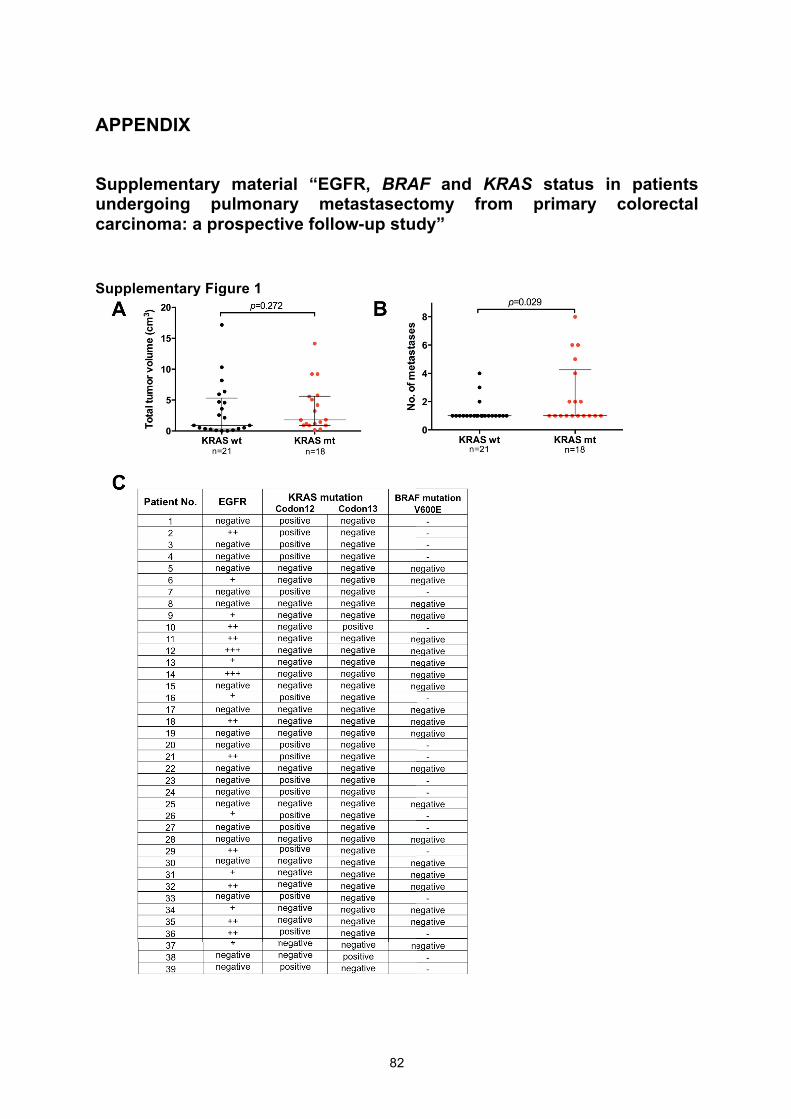
82
APPENDIX
Supplementary material “EGFR, BRAF and KRAS status in patients undergoing pulmonary metastasectomy from primary colorectal carcinoma: a prospective follow-up study”
Supplementary Figure 1

83
Supplementary material “Stromal expression of heat-shock protein 27 is associated with worse clinical outcome in patients with colorectal cancer lung metastases.”
Supplementary Table 1

84
Supplementary Table 2

85
Supplementary Table 3
MVD Vimentin
Total low high 2 low intermediate high 2 N=51 % N=25 % N=26 % p-value N=4 % N=21 % N=26 % p-value
Patients
Sex Male 29 56.9 16 31.4 13 25.5
0.313 2 3.9 13 25.5 14 27.5
0.913 Female 22 43.1 9 17.6 13 25.5 2 3.9 8 15.7 12 23.5
Age (years) Median 63 67 58.5
0.048 63.5 67 57.5
0.180a Range 33-83 33-83 45-77 50-68 44-83 33-78
Primary tumor
Location Colon 27 52.9 14 27.5 13 25.5
0.668 1 2.0 12 23.5 14 27.5
0.585 Rectum 24 47.1 11 21.5 13 25.5 3 5.9 9 17.6 12 23.5
T stage
pT1 1 2.1 0 0.0 1 2.1 0 0.0 1 2.1 0 0.0
pT2 7 14.6 3 6.2 4 8.3 0.848
0 0.0 3 6.2 4 8.3 0.186
pT3 34 70.8 19 39.6 15 31.2 4 8.3 10 20.8 20 41.7
pT4 6 12.5 3 6.2 3 6.2 0 0.0 5 10.4 1 2.1
N/A 3 -
N stage
pN0 21 43.8 12 25.0 9 18.8
0.709
1 2.1 9 18.8 11 22.9
0.456 pN1 11 22.9 6 12.5 5 10.4 0 0.0 5 10.4 6 12.5
pN2 16 33.3 7 14.6 9 18.8 3 8.3 5 10.4 8 16.7
N/A 3 -
Grading G1 2 3.9 2 3.0 0 0.0 1 2.0 0 0.0 1 2.0
G2 42 82.4 19 37.3 23 45.1 0.319 2 2.9 18 35.3 22 43.1 0.218
G3 7 13.7 4 7.8 3 5.9 1 2.0 3 5.9 3 5.9
Pulmonary metastasis
Previous liver metastasis
Yes 16 31.4 8 15.7 8 15.7 0.925
0 0.0 7 13.7 9 17.6 0.442
No 35 68.6 17 33.3 18 35.3 4 7.8 14 27.5 17 33.3
No. of nodules 1 33 64.7 16 31.4 17 33.3
0.918 2 3.9 11 21.6 20 39.2
0.195 >1 18 35.3 9 17.6 9 17.6 2 3.9 10 19.6 6 11.8
Stromal HSP27
low 17 33.3 10 19.6 7 13.7 3 5.9 12 23.5 2 3.9
intermediate 17 33.3 8 15.7 9 17.6 0.577 1 2.0 8 15.7 8 15.7 <0.001
high 17 33.3 7 13.7 10 19.6 0 0.0 1 2.0 16 31.4
Stromal alpha-SMA
low 14 27.5 9 17.6 5 9.8 4 7.8 10 19.6 0 0.0
intermediate 23 45.1 11 21.6 12 23.5 0.315 0 0.0 11 21.6 12 23.5 <0.001
high 14 27.5 5 9.8 9 17.6 0 0.0 0 0.0 14 27.5 aKruskal-Wallis test

86
CV

87
Curriculum vitae
Thomas Sebastian Schweiger Personal background
Date of birth: May 25th, 1987
Nationality: German
Education
2015/05 – present Residency in Thoracic Surgery, Division of Thoracic Surgery (Head: Prof. Walter Klepetko), Medical University of Vienna, Austria
2014/09 – 2015/05 Transplant coordination, Division of Thoracic Surgery (Head: Prof. Walter Klepetko), Medical University of Vienna, Austria
2012/10 – present MDPhD excellence program (Vascular biology), Medical University of Vienna, Austria
2010/04 – present Research Fellow at the Christian Doppler Laboratory for Diagnosis and Regeneration of Cardiac and Thoracic Diseases, Medical University of Vienna, Austria
2008/10 – 2014/07 Human medicine (Degree: Dr.med.univ./MD), Medical University of Vienna, Austria
2006/07 - 2007/04 Military Service
2006/06 High School Graduation
1997 - 2006 Katharinen-Gymnasium Ingolstadt, Germany
1993 - 1997 Primary School, Manching, Germany
Clinical Training
2014/04 - 2014/05 Clinical Clerkship at the Division of Thoracic Surgery, Medical University Vienna, Austria
88
2012/08 - 2012/09 Clinical Clerkship at the Department of Internal Medicine, Evangelisches Krankenhaus, Vienna, Austria
2012/07 - 2012/08 Clinical Clerkship at the Department of Pathology, Medical University Vienna, Austria
2011/06 - 2011/07 Clinical Clerkship at the Department of Emergency Medicine, Klinikum Ingolstadt, Germany
2010/07 - 2010/08 Clinical Clerkship at the Department of General, Visceral, Vascular and Thoracic Surgery, Charité Berlin, Germany
Scholarships and Honors
2014/12 Stipend of the Medical University of Vienna for outstanding study performance
2014/10 Poster Prize of the German Society for Thoracic Surgery
2014/06 ACO-ASSO Prize 2014
2014/05 Research Scholarship of the Medical University of Vienna
2014/04 Stipend of the “Hans und Blanca Moser- Stiftung”
2014/01 Research Scholarship of the Medical University of Vienna
2013/12 Stipend of the Medical University of Vienna for outstanding study performance
2013/05 Research Scholarship of the Medical University of Vienna
2012/12 Stipend of the Medical University of Vienna for outstanding study performance
2012/04 Research Scholarship of the Medical University of Vienna
2011/10 Research Scholarship of the Medical University of Vienna
Workshops
2014/03 Nahtkurs Gefäßchirurgie (OA Dr. C Senekowitsch), Wilhelminenspital, Vienna, Austria
2011/08 Theodor-Billroth-Academy Surgical Summer School 2011 (Prof. Dr. B Brücher, FACS), University of Tuebingen, Germany
Congresses and Meetings
2015/10 Annual Meeting oft he Austrian Society for Pulmonology, Graz, Austria
2015/09 7th ÖGMBT Anuual Meeting, Salzburg, Austria
2015/05 23rd European Conference on General Thoracic Surgery, Lisbon, Portugal
2014/10 ACO/ASSO Annual meeting 2014, St. Wolfgang, Austria
2014 06 Annual Congress of the Austrian Society of Surgery, Vienna, Austria
2014/03 Management of Laryngotracheal Problems, Vienna, Austria
2014/01 EACTS Academic Thoracic Surgery Club, Bern, Switzerland

89
2013/10 Meeting of the Austrian, German and Swiss Society of Thoracic Surgery, Basel, Switzerland
2013/10 ACO/ASSO Annual meeting 2013, St. Wolfgang, Austria
2013/05 Annual Congress of the German Society of Surgery, Munich, Germany
2012/11 36th Seminar of the Austrian Society for Surgical Research, Vienna, Austria
2012/06 Annual Congress of the Austrian Society of Surgery, Vienna, Austria
2012/04 Annual Congress of the German Society of Surgery, Berlin, Germany
2011/10 ACO/ASSO Annual meeting 2011, St. Wolfgang, Austria
2011/06 Annual Congress of the Austrian Society of Surgery, Vienna, Austria
2011/04 SkillsLab Symposium, Würzburg, Germany
2010/12 EACTS Meeting on Cardiac and Pulmonary Regeneration, Vienna, Austria
2010/10 Meeting of the Austrian, German and Swiss Society of Thoracic Surgery, Vienna, Austria
2010/04 SkillsLab Symposium, Münster, Germany
Original articles (peer-reviewed)
Schweiger T, Schwarz S, Traxler D, Dodier P, Aigner C, Lang G, Klepetko W, Hoetzenecker K Bronchoscopic Indocyanine Green Fluorescence Imaging of the Anastomotic Perfusion After Tracheal Surgery. Ann Thorac Surg. 2016 Feb 22 [Epub ahead of print] IF: 3.849
Schweiger T, Starkl V, Glueck O, Glogner C, Traxler D, Jedamzik J, Liebmann-Reindl S, Birner P, Streubel B, Klepetko W, Hoetzenecker K. Clinical impact of c-MET expression and mutational status in patients with colorectal cancer lung metastases. Eur J Cardiothorac Surg. 2015 Oct 24. pii: ezv323. IF: 3.304
Schweiger T, Nikolowsky C, Graeter T, Seebacher G, Laufer J, Glueck O, Glogner C, Birner P, Lang G, Klepetko W, Ankersmit HJ, Hoetzenecker K. Increased lymphangiogenesis in lung metastases from colorectal cancer is associated with early lymph node recurrence and decreased overall survival. Clin Exp Metastasis. 2015 Oct 23. IF: 3.491
Hoetzenecker K, Schweiger T, Schwarz S, Roesner I, Leonhard M, Denk-Linnert DM, Schneider-Stickler B, Bigenzahn W, Klepetko W. Summarized institutional experience of paediatric airway surgery. Eur J Cardiothorac Surg. 2015 Aug 7. pii: ezv263. IF: 3.304
Ghanim B, Schweiger T, Jedamzik J, Glueck O, Glogner C, Lang G, Klepetko W, Hoetzenecker K. Elevated inflammatory parameters and inflammation scores are associated with poor prognosis in patients undergoing pulmonary metastasectomy for colorectal cancer. Interact Cardiovasc Thorac Surg. 2015 Aug 4. pii: ivv206. IF: 1.155
von Karstedt S, Conti A, Nobis M, Montinaro A, Hartwig T, Lemke J, Legler K, Annewanter F, Campbell AD, Taraborrelli L, Grosse-Wilde A, Coy JF, El-Bahrawy MA, Bergmann F, Koschny R, Werner J,

90
Ganten TM, Schweiger T, Hoetzenecker K, Kenessey I, Hegedüs B, Bergmann M, Hauser C, Egberts JH, Becker T, Röcken C, Kalthoff H, Trauzold A, Anderson KI, Sansom OJ, Walczak H
Cancer Cell-Autonomous TRAIL-R Signaling Promotes KRAS-Driven Cancer Progression, Invasion, and Metastasis. Cancer Cell. 2015 Apr 13;27(4):561-73. IF: 23.893
Schweiger T, Nikolowsky C, Starlinger P, Traxler D, Zimmermann M, Birner P, Hegedüs B, Dome B, Bergmann M, Mildner M, Klepetko W, Hoetzenecker K, Ankersmit HJ
Stromal expression of heat-shock protein 27 is associated with worse clinical outcome in patients with colorectal cancer lung metastases. PLoS One. 2015 Mar 20;10(3):e0120724. IF: 3.534
Hoetzenecker K, Hochdaninger M, Traxler D, Gschwandtner M, Kasiri MM, Mitterbauer A, Schweiger T, Hegedus B, Klepetko W, Tschachler E, Ankersmit HJ, Mildner M
Antimicrobial peptides are highly abundant and active in post-operative pleural drainage fluids. Ann Thorac Surg. 2014 Sep;98(3):1042-50. IF: 3.631
Schweiger T, Kollmann D, Nikolowsky C, Traxler D, Guenova E, Lang G, Birner P, Klepetko W, Ankersmit HJ, Hoetzenecker K
Carbonic anhydrase IX is associated with early pulmonary spreading of primary colorectal carcinoma and tobacco smoking. Eur J Cardiothorac Surg. 2014 Jul;46(1):92-9. IF: 3.048
Schweiger T, Hegedüs B, Nikolowsky C, Hegedüs Z, Szirtes I, Mair R, Birner P, Döme B, Lang G, Klepetko W, Ankersmit HJ, Hoetzenecker K
EGFR, BRAF and KRAS Status in Patients Undergoing Pulmonary Metastasectomy from Primary Colorectal Carcinoma: A Prospective Follow-Up Study. Ann Surg Oncol. 2014 Mar;21(3):946-54 IF: 4.120
Hoetzenecker K, Schweiger T, Nikolowsky C, Lehmann L, Gittler F, Ankersmit H.J., Klepetko W, Lang G
Impact of resection techniques on postoperative lung function parameters in pulmonary metastasectomy. European Surgery (Acta Chir Austriaca) 2013 Apr, Volume 45(2), 93-97. IF: 0.283
Hoetzenecker K, Mitterbauer A, Guenova E, Schweiger T, Altmann P, Zimmermann M, Hofbauer H, Beer L, Klepetko W, Ankersmit HJ.
High levels of lung resident CD4+CD28null cells in COPD: implications of autoimmunity. Wien Klin Wochenschr. 2013 Mar;125(5-6):150-155. IF: 0.809
Hoetzenecker K, Zimmermann M, Hoetzenecker W, Schweiger T, Kollmann D, Mildner M, Hegedus B, Mitterbauer A, Hacker S, Birner P, Gabriel C, Gyöngyösi M, Blyszczuk P, Eriksson U, Ankersmit HJ.
Mononuclear cell secretome protects from experimental autoimmune myocarditis. Eur Heart J. 2013 Jan 14. [Epub ahead of print] IF: 14.781
Hoetzenecker K*, Assinger A*, Lichtenauer M, Mildner M, Schweiger T, Starlinger P, Jakab A, Berényi E, Petrási Z, Plass C, Gyöngyösi M, Volf I*, Ankersmit HJ*
Secretome of apoptotic peripheral blood cells (APOSEC) attenuates microvascular obstruction in a porcine closed chest reperfused acute myocardial infarction model: role of platelet aggregation and vasodilation. Basic Res Cardiol. 2012 Sep;107(5):292 IF: 7.348

91
Reviews and Case reports
Selimovic A, Klepetko W, Schweiger T, Hoetzenecker K, Mujicic E, Dinarevic S, Guska S, Rekic A, Kacamakovic H, Moro M, Kudumpvoc A, Kudumovic M, Katica A Intubation with flexible bronchoscope and surgical resection of a post-tracheostomy stenosis HealthMED 2015 March;(9)3:127-130. IF: -
Schweiger T, Hoetzenecker K, Bacher A, Aigner C, Klepetko W Reversible compression of the left lower lobe vein after right pneumonectomy Ann Thorac Surg. 2015 Mar;99(3):1067-9. IF: 3.631
Hoetzenecker K, Schweiger T, Bigenzahn W, Klepetko W Tracheal resection for porst-tracheostomy stenosis New Arm Med Jounral 2014 Dec;(8)4:19-22. IF: -
Schweiger T, Hoetzenecker K, Taghavi S, Klepetko W Extended cervico-thoracic metastasectomy for testicular non-seminomatous germ cell tumour masses through an inverse T and combined collar incision Eur J Cardiothorac Surg. 2015 May;47(5):931-3. IF: 3.048
Schweiger T, Hoetzencker K, Bacher A, Aigner C, Klepetko W Reversible compression of the left lower lobe vein after right pneumonectomy Ann Thorac Surg. 2015 Mar;99(3):1067-9. IF: 3.741 Schweiger T, Lang G, Klepetko W, Hoetzenecker K Prognostic factors in pulmonary metastasectomy: spotlight on molecular and radiological markers Eur J Cardiothorac Surg. 2014 Mar;45(3):408-16. IF: 3.048
Hoetzenecker K, Kollmann D, Schweiger T, Ankersmit HJ, Aigner C, Lang G, Taghavi S, Klepetko W. Unsuspected finding of a relapsing perichondritis during lung explantation. Ann Thorac Surg. 2012 Oct;94(4):1353. IF: 3.741
Published articles (non peer-reviewed)
Hoetzenecker K, Schweiger T, Klepetko W Neues zur pulmonalen Metastasektomie Wiener klinische Wochenschrift, ERS Spezial, Dezember 2012
Hoetzenecker K, Schweiger T, Klepetko W Neues zur pulmonalen Metastasektomie ÄrzteWoche, Nr. 42, 26. Jahrgang, 2012

92
Schweiger T 4. Sommer-Schule der Theodor-Billroth-Akademie in Tübingen - Eine Initiative zur Förderung des internationalen Chirurgennachwuchses Chirurgie, Nr. 03/11, 2011
Abstracts
Schweiger T, Starkl V, Glogner C, Glück O, Jedamzik J, Birner P, Klepetko W, Hoetzenecker K Clinical impact of c-MET expression and mutational status in patients with CRC lung metastases 7th ÖGMBT Anuual Meeting, Salzburg, Austria Schweiger T, Starkl V, Glogner C, Glück O, Jedamzik J, Birner P, Klepetko W, Hoetzenecker K c-MET overexpression and mutational status in patients with pulmonary metastases from primary CRC 56. Österreichischer Chirurgenkongress, Linz, Austrial Hoetzenecker K, Schweiger T, Leonhard M, Roesner I, Denk-Linnert D, Schneider-Stickler B, Bigenzahn W, Klepetko W Summarized institutional experience of pediatric airway surgery 56. Österreichischer Chirurgenkongress, Linz, Austrial Schweiger T, Graeter T, Seebacher G, Laufer J, Glück O, Glogner C, Jedamzik J, Birner P, Lang G, Klepetko W, Ankersmit HJ, Hoetzenecker K Lymphatic invasion in colorectal cancer lung metastases predicts the outcome after pulmonary metastasectomy 56. Österreichischer Chirurgenkongress, Linz, Austria Hoetzenecker K, Schweiger T, Matilla J, Aigner C, Lang G, Taghavi S, Klepetko W A Case series of carinal resections with tracheobronchial end-to-end anastomosis and end-to-side reimplantation of the remaining airway into the contralateral bronchus 56. Österreichischer Chirurgenkongress, Linz, Austria Ghanim B, Schweiger T, Lang G, Klepetko W, Hoetzenecker K Elevated inflammatory parameters and inflammation scores are associated with poor prognosis in patients undergoing curative pulmonary metastasectomy for colorectal cancer 23rd European Conference on General Thoracic Surgery, Lisbon, Portugal
Hoetzenecker K, Schweiger T, Leonhard M, Roesner I, Denk-Linnert D, Schneider-Stickler B, Bigenzahn W, Klepetko W Summarized institutional experience of pediatric airway surgery 23rd European Conference on General Thoracic Surgery, Lisbon, Portugal Schweiger T, Starkl V, Glogner C, Glück O, Jedamzik J, Birner P, Klepetko W, Hoetzenecker K Clinical impact of c-MET expression and mutational status in patients with primary colroectal cancer lung metastases 23rd European Conference on General Thoracic Surgery, Lisbon, Portugal Hoetzenecker K, Schweiger T, Lang G, Klepetko W Surgical treatment of diffuse tracheomalacia in adults: posterior stabilization using a polypropylene mesh Annual Meeting of the Austrian Society for Pneumonology 2014, Salzburg, Austria Schweiger T, Nikolowsky C, Mair R, Traxler D, Birner P, Döme B, Lang G, Ankersmit HJ, Klepetko W Hoetzenecker K

93
Lymphangiogenesis in patients undergoing pulmonary metastasectomy from metastatic colorectal carcinoma with negative thoracic lymph node staging 23. Jahrestagung der Deutschen Gesellschaft für Thoraxchirurgie 2014, Osnabrück, Germany Hoetzenecker K, Marta G, Schweiger T, Denk D, Bigenzahn W, Klepetko W Eine Adaptation der Couraud-Technik ermöglicht laryngotracheale Rekonstruktionen bei Erwachsenen als einzeitige Eingriffe 23. Jahrestagung der Deutschen Gesellschaft für Thoraxchirurgie 2014, Osnabrück, Germany Hoetzenecker K, Leonhard M, Marta G, Schweiger T, Denk D, Monnier P, Bigenzahn W, Klepetko W Korrektur laryngotrachealer Stenosen bei Kindern – eine Fallserie 23. Jahrestagung der Deutschen Gesellschaft für Thoraxchirurgie 2014, Osnabrück, Germany Hoetzenecker K, Schweiger T, Lang G, Klepetko W Posterior stabilization using polypropylene mesh in a patient with severe diffuse tracheomalacia 55. Österreichischer Chirurgenkongress 2014, Graz, Austria Hoetzenecker K, Hochdanninger M, Traxler D, Mitterbauer A, Schweiger T, Hegedüs B, Klepetko W, Ankersmit HJ, Mildner M High concentrations of antimicrobial peptides in post-operative pleural drainage fluids 55. Österreichischer Chirurgenkongress 2014, Graz, Austria Schweiger T, Traxler D, Nikolowsky C, Lang G, Birner P, Klepetko W Hegedüs B, Dome B, Hoetzenecker K, Ankersmit HJ Heat-shock proteins 27 and 70 in primary colorectal cancer and corresponding pulmonary metastases 55. Österreichischer Chirurgenkongress 2014, Graz, Austria Schweiger T, Hegedüs B, Nikolowsky C, Hegedüs Z, Szirtes I, Mair R, Birner P, Döme B, Lang G, Klepetko W, Ankersmit HJ, Hoetzenecker K Mutation in KRAS prognosticates early recurrence in patients undergoing pulmonary metastasectomy from primary colorectal carcinoma 55. Österreichischer Chirurgenkongress 2014, Graz, Austria Hoetzenecker K, Hochdanninger M, Traxler D, Mitterbauer A, Schweiger T, Hegedüs B, Klepetko W, Ankersmit HJ, Mildner M Antimicrobial peptides are highly abundant and active in post-operative pleural drainage fluids Meeting of the Austrian, German and Swiss Society of Thoracic Surgery 2013, Basel, Switzerland Schweiger T, Traxler D, Nikolowsky C, Lang G, Birner P, Klepetko W Hegedüs B, Dome B, Hoetzenecker K, Ankersmit HJ Heat-shock proteins 27 and 70 in primary colorectal cancer and corresponding pulmonary metastases Meeting of the Austrian, German and Swiss Society of Thoracic Surgery 2013, Basel, Switzerland Schweiger T, Hegedüs B, Nikolowsky C, Hegedüs Z, Szirtes I, Mair R, Birner P, Döme B, Lang G, Klepetko W, Ankersmit HJ, Hoetzenecker K Mutation in KRAS prognosticates early recurrence in patients under- going pulmonary metastasectomy from primary colorectal carcinoma Meeting of the Austrian, German and Swiss Society of Thoracic Surgery 2013, Basel, Switzerland T Schweiger, D Kollmann, C Nikolowsky, Denise Traxler, E Guenova, G Lang, P Birner, W Klepetko, HJ Ankersmit, K Hoetzenecker Early pulmonary spreading of primary colorectal carcinoma is associated with carbonic anhydrase IX expression and tobacco smoking Meeting of the Austrian, German and Swiss Society of Thoracic Surgery 2013, Basel, Switzerland Schweiger T, Traxler D, Nikolowsky C, Lang G, Birner P, Klepetko W Hegedüs B, Dome B, Hoetzenecker k, Ankersmit HJ

94
Heat-shock proteins 27 and 70 in primary colorectal cancer and corresponding pulmonary metastases ACO/ASSO Annual meeting 2013, St. Wolfgang, Austria Hoetzenecker K, Schweiger T, Nikolowsky C, Traxler D, Lehmann L, Gittler F, Mair R, Lang G, Klepetko W Pulmonary Metastasectomy at the Department of Thoracic Surgery, MUV, 2009-2013 – A computer-based prospective database ACO/ASSO Annual meeting 2013, St. Wolfgang, Austria Schweiger T, Hegedüs B, Nikolowsky C, Hegedüs Z, Szirtes I, Mair R, Birner P, Döme B, Lang G, Klepetko W, Ankersmit HJ, Hoetzenecker K Mutation in KRAS prognosticates early recurrence in patients undergoing pulmonary metastasectomy from primary colorectal carcinoma ACO/ASSO Annual meeting 2013, St. Wolfgang, Austria T Schweiger, K Hoetzenecker, C Nikolowsky, L Lehmann, R Wiebringhaus, HJ Ankersmit, G Lang, W Klepetko Impact of pulmonary metastasectomy on lung function parameters ACO/ASSO Annual meeting 2013, St. Wolfgang, Austria T Schweiger, D Kollmann, C Nikolowsky, Denise Traxler, E Guenova, G Lang, P Birner, W Klepetko, HJ Ankersmit, K Hoetzenecker Early pulmonary spreading of primary colorectal carcinoma is associated with carbonic anhydrase IX expression and tobacco smoking ACO/ASSO Annual meeting 2013, St. Wolfgang, Austria S Hacker, R Mittermayr, M Mildner, T Haider, S Nickl M Zimmermann, L Beer, D Lebherz-Eichinger, T Schweiger, A Mitterbauer, C Keibl, G Werba, M Frey, HJ Ankersmit Regenerative effects of secreted factors derived from peripheral blood mononuclear cells in cutaneous wound healing after full-thickness skin defects, burn and skin grafting: results of animal studies 54. Österreichischer Chirurgenkongress 2013, Vienna, Austria Abstract published in: European Surgery, Acta Chirurgica Austriaca, Volume 45, Suppl 2, 2013 D Kollmann, K Hoetzenecker, T Schweiger, HJ Ankersmit, C Aigner, G Lang, S Taghavi, W Klepetko Unsuspected finding of a relapsing polychondritis during lung explantation 54. Österreichischer Chirurgenkongress 2013, Vienna, Austria Abstract published in: European Surgery, Acta Chirurgica Austriaca, Volume 45, Suppl 2, 2013 Hoetzenecker K*, Assinger A*, Lichtenauer M, Mildner M,, Schweiger T, Starlinger P, Jakab A, Berenyi E, Petrase Z, Plass C, Gyöngösi M, Vold I*, Ankersmit HJ*.
Mononuclear cell secretome protects from experimental autoimmune myocarditis.
European Society of Cardiologists Annual Meeting 2012, Munich, Germany
Hoetzenecker K, Assinger A, Lichtenauer M, Mildner M, Schweiger T, Petráse Z, Plass C, Gyöngyösi M, Volf I, Ankersmit HJ
Secretome of apoptotic peripheral blood cells (APOSEC) attenuates microvascular obstruction in a porcine closed chest reperfused acute myocardial infarction model: role of platelet aggregation and vasodilation. European Society of Cardiologists Annual Meeting 2012, Munich, Germany
Hoetzenecker K*, Assinger A*, Lichtenauer M, Mildner M,, Schweiger T, Starlinger P, Jakab A, Berenyi E, Petrase Z, Plass C, Gyöngösi M, Vold I*, Ankersmit HJ*.
Mononuclear cell secretome protects from experimental autoimmune myocarditis. 3rd TERMIS World Congress 2012, Vienna, Austria
Hoetzenecker K, Assinger A, Lichtenauer M, Mildner M, Schweiger T, Petráse Z, Plass C, Gyöngyösi M, Volf I, Ankersmit HJ
Secretome of apoptotic peripheral blood cells (APOSEC) attenuates microvascular obstruction

95
in a porcine closed chest reperfused acute myocardial infarction model: role of platelet aggregation and vasodilation. 3rd TERMIS World Congress 2012, Vienna, Austria
T Schweiger, D Kollmann, C Nikolowsky, Denise Traxler, E Guenova, G Lang, P Birner, W Klepetko, HJ Ankersmit, K Hoetzenecker Early pulmonary spreading of primary colorectal carcinoma is associated with carbonic anhydrase IX expression and tobacco smoking 130th Annual Congress of the German Society of Surgery, Munich, Germany Abstract published in: Langebeck’s Archives of Surgery, Volume 398(4), 2013 S Hacker, R Mittermayr, M Mildner, T Haider, S Nickl, M Zimmermann, L Beer, D Lebherz-Eichinger, T Schweiger, A Mitterbauer, C Keibl, G Werba, M Frey, HJ Ankersmit Regenerative effests of secreted factors derived from peripheral blood mononuclear cells in cutaneous wound healing after full-thickness skin defects, burn and skin grafting: results of animal studies 3rd EACTS Meeting on Cardiac and Pulmonary Regeneration, Berlin, Germany Abstract published in: Interactive CardioVascular and Thoracic Surgery, Volume 16, 2013 K Hoetzenecker, M Zimmermann, T Schweiger, D Kollmann, M Mildner, A Mitterbauer, P Birner, M Lichtenauer, HJ Ankersmit Secretome from mononuclear cells confers immunosuppression in a murine autoimmune myocarditis model 36th Seminar of the Austrian Society for Surgical Research, Vienna, Austria Abstract published in: European Surgery, Acta Chirurgica Austriaca, Volume 44, Suppl 248, 2012 T Schweiger, D Kollmann, C Nikolowsky, Denise Traxler, E Guenova, G Lang, P Birner, W Klepetko, HJ Ankersmit, K Hoetzenecker Early pulmonary spreading of primary colorectal carcinoma is associated with carbonic anhydrase IX expression and tobacco smoking 36th Seminar of the Austrian Society for Surgical Research, Vienna, Austria Abstract published in: European Surgery, Acta Chirurgica Austriaca, Volume 44, Suppl 248, 2012 T Schweiger, K Hoetzenecker, C Nikolowsky, L Lehmann, R Wiebringhaus, HJ Ankersmit, G Lang, W Klepetko Impact of pulmonary metastasectomy on lung function parameters European Respiratory Society Annual Meeting 2012, Vienna, Austria Abstract published in: European Respiratory Journal, Volume 40, Suppl 56, 2012 T Schweiger, K Hoetzenecker, C Nikolowsky, L Lehmann, R Wiebringhaus, G Lang, P Birner, HJ Ankersmit, W Klepetko Hypoxic phenotype in pulmonary metastases of different primary tumors European Respiratory Society Annual Meeting 2012, Vienna, Austria Abstract published in: European Respiratory Journal, Volume 40, Suppl 56, 2012 K Hoetzenecker, A Assinger, M Lichtenauer, M Mildner, T Schweiger, A Mitterbauer, P Starlinger, M.Ernstbrunner, B Steinlechner, M Gyöngyösi, I Volf, HJ Ankersmit Secretome of apoptotoc peripheral blood cells (APOSEC) attenuates microvascular obstruction in a porcine closed chest reperfused acute myocardial infarction model: role of platelet aggregation in vitro and in vivo 53. Österreichischer Chirurgenkongress 2012, Salzburg, Austria Abstract published in: European Surgery, Acta Chirurgica Austriaca, Volume 44, Suppl 245, 2012 K Hoetzenecker, T Schweiger, M Mildner, C Plass, D Traxler, M Lichtenauer, M Gyöngyösi, HJ Ankersmit Secretome of apoptotic peripheral blood cells (APOSEC) induces coronary vasodilation: Impact on microvascular obstruction during acute myocardial infarction 53. Österreichischer Chirurgenkongress 2012, Salzburg, Austria Abstract published in: European Surgery, Acta Chirurgica Austriaca, Volume 44, Suppl 245, 2012
96
K Hoetzenecker, M Zimmermann, T Schweiger, D Kollmann, M Mildner, A Mitterbauer, P Birner, M Lichtenauer, HJ Ankersmit Secretome from mononuclear cells confers immunosuppression in a murine autoimmune myocarditis model 53. Österreichischer Chirurgenkongress 2012, Salzburg, Austria Abstract published in: European Surgery, Acta Chirurgica Austriaca, Volume 44, Suppl 245, 2012 Hoetzenecker K, Assinger A, Lichtenauer M, Mildner M, Schweiger T, Mitterbauer A, Starlinger P, Ernstbrunner M, Steinlechner B, Gyöngyösi M, Volf I, Ankersmit HJ. Secretome of apoptotic peripheral blood cells attenuates microvascular obstruction in acute myocardial infarction: role of platelet aggregation Abstract published in: J Heart Lung Transplant. 2012 Apr;31(4S):140 K Hoetzenecker, P Altmann, T Schweiger, A Hoda, A Aliabadi, G Lang, C Aigner, S Taghavi, W Klepetko Impact of lymphnode downstaging on the prognosis of pancoast tumors 9. PneumoUpdate 2012, Innsbruck, Austria T Schweiger, K Hoetzenecker, C Nikolowsky, L Lehmann, R Wiebringhaus, HJ Ankersmit, G Lang, W Klepetko Impact of pulmonary metastasectomy on lung function parameters 53. Österreichischer Chirurgenkongress 2012, Salzburg, Austria Abstract published in: European Surgery, Acta Chirurgica Austriaca, Volume 44, Suppl 245, 2012 T Schweiger, K Hoetzenecker, C Nikolowsky, L Lehmann, R Wiebringhaus, G Lang, P Birner, HJ Ankersmit, W Klepetko Hypoxic phenotype in pulmonary metastases of different primary tumors 53. Österreichischer Chirurgenkongress 2012, Salzburg, Austria Abstract published in: European Surgery, Acta Chirurgica Austriaca, Volume 44, Suppl 245, 2012 K Hoetzenecker, T Schweiger, L Lehmann, F Gittler, HJ Ankersmit, G Lang, W Klepetko Chirurgische Therapie pulmonaler Metastasen beim Mammakarzinom Senologie-ACO 2011, St. Wolfgang, Austria P Angleitner*, T Schweiger*, Michael Schmidts und Martin Schindl Chirurgia Practica – Peer teaching chirurgischer Fertigkeiten Skills Lab Symposium 2011, Würzburg, Germany
Presentations
Schweiger T, Starkl V, Glogner C, Glück O, Jedamzik J, Birner P, Klepetko W, Hoetzenecker K Clinical impact of c-MET expression and mutational status in patients with CRC lung metastases 7th ÖGMBT Anuual Meeting, Salzburg, Austria Schweiger T, Starkl V, Glogner C, Glück O, Jedamzik J, Birner P, Klepetko W, Hoetzenecker K Clinical impact of c-MET expression and mutational status in patients with primary colroectal cancer lung metastases. (Oral presentation) 23rd European Conference on General Thoracic Surgery, Lisbon, Portugal Schweiger T, Traxler D, Nikolowsky C, Lang G, Birner P, Klepetko W Hegedüs B, Dome B, Hoetzenecker K, Ankersmit HJ Heat-shock proteins 27 and 70 in primary colorectal cancer and corresponding pulmonary metastases. (Oral presentation) 55. Österreichischer Chirurgenkongress 2014, Graz, Austria

97
Schweiger T, Hegedüs B, Nikolowsky C, Hegedüs Z, Szirtes I, Mair R, Birner P, Döme B, Lang G, Klepetko W, Ankersmit HJ, Hoetzenecker K Mutation in KRAS prognosticates early recurrence in patients undergoing pulmonary metastasectomy from primary colorectal carcinoma. (Oral presentation) 55. Österreichischer Chirurgenkongress 2014, Graz, Austria Schweiger T, HoetzeneckerK, Klepetko W Posterior stabilization in a patient with severe tracheomalacia. (Oral presentation) Management of laryngotracheal problems 2014, Vienna, Austria Hoetzenecker K, Hochdanninger M, Traxler D, Mitterbauer A, Schweiger T, Hegedüs B, Klepetko W, Ankersmit HJ, Mildner M Antimicrobial peptides are highly abundant and active in post-operative pleural drainage fluids (Oral presentation) Meeting of the Austrian, German and Swiss Society of Thoracic Surgery, Basel, Switzerland Schweiger T, Traxler D, Nikolowsky C, Lang G, Birner P, Klepetko W Hegedüs B, Dome B, Hoetzenecker K, Ankersmit HJ Heat-shock proteins 27 and 70 in primary colorectal cancer and corresponding pulmonary metastases (Oral presentation) Meeting of the Austrian, German and Swiss Society of Thoracic Surgery, Basel, Switzerland Schweiger T, Hegedüs B, Nikolowsky C, Hegedüs Z, Szirtes I, Mair R, Birner P, Döme B, Lang G, Klepetko W, Ankersmit HJ, Hoetzenecker K Mutation in KRAS prognosticates early recurrence in patients under- going pulmonary metastasectomy from primary colorectal carcinoma (Poster presentation) Meeting of the Austrian, German and Swiss Society of Thoracic Surgery, Basel, Switzerland T Schweiger, D Kollmann, C Nikolowsky, Denise Traxler, E Guenova, G Lang, P Birner, W Klepetko, HJ Ankersmit, K Hoetzenecker Early pulmonary spreading of primary colorectal carcinoma is associated with carbonic anhydrase IX expression and tobacco smoking (Oral presentation) Meeting of the Austrian, German and Swiss Society of Thoracic Surgery, Basel, Switzerland Schweiger T, Traxler D, Nikolowsky C, Lang G, Birner P, Klepetko W Hegedüs B, Dome B, Hoetzenecker k, Ankersmit HJ Heat-shock proteins 27 and 70 in primary colorectal cancer and corresponding pulmonary metastases (Poster presentation) ACO/ASSO Annual meeting 2013, St. Wolfgang, Austria Schweiger T, Hegedüs B, Nikolowsky C, Hegedüs Z, Szirtes I, Mair R, Birner P, Döme B, Lang G, Klepetko W, Ankersmit HJ, Hoetzenecker K Mutation in KRAS prognosticates early recurrence in patients under- going pulmonary metastasectomy from primary colorectal carcinoma (Poster presentation) ACO/ASSO Annual meeting 2013, St. Wolfgang, Austria T Schweiger, K Hoetzenecker, C Nikolowsky, L Lehmann, R Wiebringhaus, HJ Ankersmit, G Lang, W Klepetko Impact of pulmonary metastasectomy on lung function parameters (Poster presentation) ACO/ASSO Annual meeting 2013, St. Wolfgang, Austria T Schweiger, D Kollmann, C Nikolowsky, Denise Traxler, E Guenova, G Lang, P Birner, W Klepetko, HJ Ankersmit, K Hoetzenecker Early pulmonary spreading of primary colorectal carcinoma is associated with carbonic anhydrase IX expression and tobacco smoking (Poster presentation) ACO/ASSO Annual meeting 2013, St. Wolfgang, Austria T Schweiger, D Kollmann, C Nikolowsky, Denise Traxler, E Guenova, G Lang, P Birner, W Klepetko, HJ Ankersmit, K Hoetzenecker

98
Early pulmonary spreading of primary colorectal carcinoma is associated with carbonic anhydrase IX expression and tobacco smoking (Oral presentation) 130th Annual Congress of the German Society of Surgery, Munich, Germany Abstract published in: Langebeck’s Archives of Surgery, Volume 398(4), 2013 T Schweiger, D Kollmann, C Nikolowsky, Denise Traxler, E Guenova, G Lang, P Birner, W Klepetko, HJ Ankersmit, K Hoetzenecker Early pulmonary spreading of primary colorectal carcinoma is associated with carbonic anhydrase IX expression and tobacco smoking (Oral presentation) 36th Seminar of the Austrian Society for Surgical Research, Vienna, Austria T Schweiger, K Hoetzenecker, C Nikolowsky, L Lehmann, R Wiebringhaus, HJ Ankersmit, G Lang, W Klepetko Impact of pulmonary metastasectomy on lung function parameters (Poster presentation) European Respiratory Society Annual Meeting 2012, Vienna, Austria T Schweiger, K Hoetzenecker, C Nikolowsky, L Lehmann, R Wiebringhaus, G Lang, P Birner, HJ Ankersmit, W Klepetko Hypoxic phenotype in pulmonary metastases of different primary tumors (Poster presentation) European Respiratory Society Annual Meeting 2012, Vienna, Austria
T Schweiger, K Hoetzenecker, C Nikolowsky, L Lehmann, R Wiebringhaus, HJ Ankersmit, G Lang, W Klepetko Impact of pulmonary metastasectomy on lung function parameters (Oral presentation) 53. Österreichischer Chirurgenkongress 2012, Salzburg, Austria T Schweiger, K Hoetzenecker, C Nikolowsky, L Lehmann, R Wiebringhaus, G Lang, P Birner, HJ Ankersmit, W Klepetko Hypoxic phenotype in pulmonary metastases of different primary tumors (Oral presentation) 53. Österreichischer Chirurgenkongress 2012, Salzburg, Austria
K Hoetzenecker, T Schweiger, L Lehmann, F Gittler, HJ Ankersmit, G Lang, W Klepetko Chirurgische Therapie pulmonaler Metastasen beim Mammakarzinom (Poster presentation) Senologie-ACO 2011, St. Wolfgang, Austria P Angleitner*, T Schweiger*, Michael Schmidts und Martin Schindl Chirurgia Practica – Peer teaching chirurgischer Fertigkeiten (Poster presentation) Skills Lab Symposium 2011, Würzburg, Germany
99
Review activity
2015-present Transplant International
Clinical Trials
Schweiger T (Study author), Klepetko W, Hoetzenecker K, Lang G – EUDRACT: 2013-001725-10 Bronchsoscopic indocyanine green fluorescence imaging for the evaluation of tracheal perfusion after surgery – A monocenter, prospective open-label feasibility study
Teaching activity
2016/03 JC Current Topics in Applied Immunology (summer term 2016; 861.111), Medical University of Vienna
2016/03 TS Applied Immunology and Tissue Regeneration (summer term 2016; 861.119), Medical University of Vienna
2013/10 Student teaching assistant „Themenspezifische Untersuchungtechniken I/Chirurgische Grundfertigkeiten“ (winter term 2013/14; 805.008), Medical University of Vienna
2012/10 Student teaching assistant „Themenspezifische Untersuchungtechniken I/Chirurgische Grundfertigkeiten“ (winter term 2012/13; 805.008), Medical University of Vienna
2012/03 Hands-on Workshop „Introduction to surgical suturing techniques“, MedSuccess 2012, Medical University of Vienna
2011/10 Student teaching assistant „Themenspezifische Untersuchungtechniken I/Chirurgische Grundfertigkeiten“ (winter term 2011/12; 805.008), Medical University of Vienna
2011/10 Student teaching assistant „Themenspezifische Untersuchungtechniken I“ ( winter term 2011/12; 805.008), Medical University of Vienna
2011/03 Hands-on Workshop „Introduction to surgical suturing techniques“, MedSuccess 2011, Medical University of Vienna
2010/10 - ongoing Co-Founder of „Chirurgia Practica“ (peer-teaching of surgical techniques) together with Dr. Philipp Angleitner (Supervisor: Ao. Univ.-Prof. Dr. Martin Schindl), Medical University of Vienna
Memberships
2015 - present Membership at Austrian Society of Cardio-Thoracic Surgery (ÖGHTC)
2015 - present Membership at European Society of Thoracic Surgeons (ESTS)
2014 - present Membership at Austrian Society of Surgical Oncology (ACO-ASSO)
2011 - present Membership at Berufsverband Österreichischer Chirurgen (BÖC)