Identification of O-mannosylated Virulence Factors in...
15
Identification of O-mannosylated Virulence Factors in Ustilago maydis Alfonso Ferna ´ ndez-A ´ lvarez 1¤ , Miriam Marı ´n-Menguiano 1 , Daniel Lanver 2 , Alberto Jime ´nez-Martı´n 1 , Alberto Elı´as-Villalobos 1 , Antonio J. Pe ´ rez-Pulido 1 , Regine Kahmann 2 , Jose ´ I. Ibeas 1 * 1 Centro Andaluz de Biologı ´a del Desarrollo, Universidad Pablo de Olavide, Consejo Superior de Investigaciones Cientı ´ficas, Sevilla, Spain, 2 Department of Organismic Interactions, Max-Planck-Institute for Terrestrial Microbiology, Marburg, Germany Abstract The O-mannosyltransferase Pmt4 has emerged as crucial for fungal virulence in the animal pathogens Candida albicans or Cryptococcus neoformans as well as in the phytopathogenic fungus Ustilago maydis. Pmt4 O-mannosylates specific target proteins at the Endoplasmic Reticulum. Therefore a deficient O-mannosylation of these target proteins must be responsible for the loss of pathogenicity in pmt4 mutants. Taking advantage of the characteristics described for Pmt4 substrates in Saccharomyces cerevisiae, we performed a proteome-wide bioinformatic approach to identify putative Pmt4 targets in the corn smut fungus U. maydis and validated Pmt4-mediated glycosylation of candidate proteins by electrophoretic mobility shift assays. We found that the signalling mucin Msb2, which regulates appressorium differentiation upstream of the pathogenicity-related MAP kinase cascade, is O-mannosylated by Pmt4. The epistatic relationship of pmt4 and msb2 showed that both are likely to act in the same pathway. Furthermore, constitutive activation of the MAP kinase cascade restored appressorium development in pmt4 mutants, suggesting that during the initial phase of infection the failure to O- mannosylate Msb2 is responsible for the virulence defect of pmt4 mutants. On the other hand we demonstrate that during later stages of pathogenic development Pmt4 affects virulence independently of Msb2, probably by modifying secreted effector proteins. Pit1, a protein required for fungal spreading inside the infected leaf, was also identified as a Pmt4 target. Thus, O-mannosylation of different target proteins affects various stages of pathogenic development in U. maydis. Citation: Ferna ´ndez-A ´ lvarez A, Marı ´n-Menguiano M, Lanver D, Jime ´nez-Martı ´n A, Elı ´as-Villalobos A, et al. (2012) Identification of O-mannosylated Virulence Factors in Ustilago maydis. PLoS Pathog 8(3): e1002563. doi:10.1371/journal.ppat.1002563 Editor: Barbara J. Howlett, University of Melbourne, Australia Received September 16, 2011; Accepted January 17, 2012; Published March 1, 2012 Copyright: ß 2012 Ferna ´ndez-A ´ lvarez et al. This is an open-access article distributed under the terms of the Creative Commons Attribution License, which permits unrestricted use, distribution, and reproduction in any medium, provided the original author and source are credited. Funding: This work was supported by Ministerio de Ciencia e Innovacio ´ n Grant BIO2010-16787. AFA and AEV were supported by fellowships from Ministerio de Ciencia e Innovacio ´ n. DL was supported by the Deutsche Forschungsgemeinschaft (GK1216). CABD is institutionally supported by CSIC, Universidad Pablo de Olavide and Junta de Andalucı ´a. The funders had no role in study design, data collection and analysis, decision to publish, or preparation of the manuscript. Competing Interests: The authors have declared that no competing interests exist. * E-mail: [email protected] ¤ Current address: Telomere Biology Laboratory, Cancer Research UK, London Research Institute, London, United Kingdom. Introduction O-mannosylation is an essential posttranslational protein modification in fungal cells [1]. This type of protein O- glycosylation, which adds mannoses to the nascent glycoproteins at the Endoplasmic Reticulum (ER) and Golgi Apparatus (AG), is required for correct protein conformation and stabilization [2]. In pathogenic fungi, such as Candida albicans or Cryptococcus neoformans, O-mannosylation is crucial for virulence [3–5]. The first step of the O-mannosylation pathway is catalyzed by protein O-manosyltransferases (Pmts), which add the first mannose to hydroxyl groups of serine and threonine in ER resident target proteins [6]. In fungi, the Pmt family is grouped into three subfamilies: Pmt1, Pmt2 and Pmt4, which mannosylate specific target proteins [7,8]. The number of members in these subfamilies differs in each organism; for example, in Saccharomyces cerevisiae or C. albicans the Pmt1 subfamily contains two members [9,10], while in Schizosaccharomyces pombe, C. neoformans, Aspergillus nidulans or in Ustilago maydis, the Pmt1 family contains only one member [11– 14]. PMT orthologs have also been found in humans, rat and Drosophila melanogaster but not in nematodes (Caenorhabditis elegans) or plants (Arabidopsis thaliana and Oryza sativa) [6,15–18]. Interestingly, the Pmt4 subfamily contains only one member in all organisms analyzed [6]. The protein O-mannosyltransferase Pmt4 has been characterized in several pathogenic fungi and was shown to be essential for virulence of fungal animal pathogens such as C. albicans and C. neoformans [4,5,10,19]. In addition, Pmt4 is relevant for virulence in the phytopahogenic fungus U. maydis [11]. It has been proposed that the loss of Pmt4 in fungal pathogens causes an alteration in their ability to adhere to the host, which might be caused by affecting the glycosylation of secreted proteins and cell wall proteins [5,20]. Despite their proposed relevance, Pmt4 target proteins have only been described in S. cerevisiae and C. albicans [21–23]. The lack of a defined consensus sequence that is targeted by Pmt4, coupled with the transient interaction of this O-mannosyltrans- ferase with its target proteins, makes identification of Pmt4 substrates difficult [21]. The only knowledge regarding Pmt4 target proteins is that they should contain a membrane anchor to facilitate their glycosylation in the ER lumen. This characteristic feature has been used to perform a search for Pmt4 substrates in S. cerevisiae [8]. However this type of in silico screening has not been carried out in pathogenic fungi, which would allow the identification of fungal virulence factors. In this work, we have PLoS Pathogens | www.plospathogens.org 1 March 2012 | Volume 8 | Issue 3 | e1002563
-
Upload
dangnguyet -
Category
Documents
-
view
218 -
download
0
Transcript of Identification of O-mannosylated Virulence Factors in...
Identification of O-mannosylated Virulence Factors inUstilago maydisAlfonso Fernandez-Alvarez1¤, Miriam Marın-Menguiano1, Daniel Lanver2, Alberto Jimenez-Martın1,
Alberto Elıas-Villalobos1, Antonio J. Perez-Pulido1, Regine Kahmann2, Jose I. Ibeas1*
1 Centro Andaluz de Biologıa del Desarrollo, Universidad Pablo de Olavide, Consejo Superior de Investigaciones Cientıficas, Sevilla, Spain, 2 Department of Organismic
Interactions, Max-Planck-Institute for Terrestrial Microbiology, Marburg, Germany
Abstract
The O-mannosyltransferase Pmt4 has emerged as crucial for fungal virulence in the animal pathogens Candida albicans orCryptococcus neoformans as well as in the phytopathogenic fungus Ustilago maydis. Pmt4 O-mannosylates specific targetproteins at the Endoplasmic Reticulum. Therefore a deficient O-mannosylation of these target proteins must be responsiblefor the loss of pathogenicity in pmt4 mutants. Taking advantage of the characteristics described for Pmt4 substrates inSaccharomyces cerevisiae, we performed a proteome-wide bioinformatic approach to identify putative Pmt4 targets in thecorn smut fungus U. maydis and validated Pmt4-mediated glycosylation of candidate proteins by electrophoretic mobilityshift assays. We found that the signalling mucin Msb2, which regulates appressorium differentiation upstream of thepathogenicity-related MAP kinase cascade, is O-mannosylated by Pmt4. The epistatic relationship of pmt4 and msb2 showedthat both are likely to act in the same pathway. Furthermore, constitutive activation of the MAP kinase cascade restoredappressorium development in pmt4 mutants, suggesting that during the initial phase of infection the failure to O-mannosylate Msb2 is responsible for the virulence defect of pmt4 mutants. On the other hand we demonstrate that duringlater stages of pathogenic development Pmt4 affects virulence independently of Msb2, probably by modifying secretedeffector proteins. Pit1, a protein required for fungal spreading inside the infected leaf, was also identified as a Pmt4 target.Thus, O-mannosylation of different target proteins affects various stages of pathogenic development in U. maydis.
Citation: Fernandez-Alvarez A, Marın-Menguiano M, Lanver D, Jimenez-Martın A, Elıas-Villalobos A, et al. (2012) Identification of O-mannosylated VirulenceFactors in Ustilago maydis. PLoS Pathog 8(3): e1002563. doi:10.1371/journal.ppat.1002563
Editor: Barbara J. Howlett, University of Melbourne, Australia
Received September 16, 2011; Accepted January 17, 2012; Published March 1, 2012
Copyright: � 2012 Fernandez-Alvarez et al. This is an open-access article distributed under the terms of the Creative Commons Attribution License, whichpermits unrestricted use, distribution, and reproduction in any medium, provided the original author and source are credited.
Funding: This work was supported by Ministerio de Ciencia e Innovacion Grant BIO2010-16787. AFA and AEV were supported by fellowships from Ministerio deCiencia e Innovacion. DL was supported by the Deutsche Forschungsgemeinschaft (GK1216). CABD is institutionally supported by CSIC, Universidad Pablo deOlavide and Junta de Andalucıa. The funders had no role in study design, data collection and analysis, decision to publish, or preparation of the manuscript.
Competing Interests: The authors have declared that no competing interests exist.
* E-mail: [email protected]
¤ Current address: Telomere Biology Laboratory, Cancer Research UK, London Research Institute, London, United Kingdom.
Introduction
O-mannosylation is an essential posttranslational protein
modification in fungal cells [1]. This type of protein O-
glycosylation, which adds mannoses to the nascent glycoproteins
at the Endoplasmic Reticulum (ER) and Golgi Apparatus (AG), is
required for correct protein conformation and stabilization [2]. In
pathogenic fungi, such as Candida albicans or Cryptococcus neoformans,
O-mannosylation is crucial for virulence [3–5].
The first step of the O-mannosylation pathway is catalyzed by
protein O-manosyltransferases (Pmts), which add the first mannose
to hydroxyl groups of serine and threonine in ER resident target
proteins [6]. In fungi, the Pmt family is grouped into three
subfamilies: Pmt1, Pmt2 and Pmt4, which mannosylate specific
target proteins [7,8]. The number of members in these subfamilies
differs in each organism; for example, in Saccharomyces cerevisiae or
C. albicans the Pmt1 subfamily contains two members [9,10], while
in Schizosaccharomyces pombe, C. neoformans, Aspergillus nidulans or in
Ustilago maydis, the Pmt1 family contains only one member [11–
14]. PMT orthologs have also been found in humans, rat and
Drosophila melanogaster but not in nematodes (Caenorhabditis elegans) or
plants (Arabidopsis thaliana and Oryza sativa) [6,15–18].
Interestingly, the Pmt4 subfamily contains only one member in
all organisms analyzed [6]. The protein O-mannosyltransferase
Pmt4 has been characterized in several pathogenic fungi and was
shown to be essential for virulence of fungal animal pathogens
such as C. albicans and C. neoformans [4,5,10,19]. In addition, Pmt4
is relevant for virulence in the phytopahogenic fungus U. maydis
[11]. It has been proposed that the loss of Pmt4 in fungal
pathogens causes an alteration in their ability to adhere to the host,
which might be caused by affecting the glycosylation of secreted
proteins and cell wall proteins [5,20].
Despite their proposed relevance, Pmt4 target proteins have
only been described in S. cerevisiae and C. albicans [21–23]. The
lack of a defined consensus sequence that is targeted by Pmt4,
coupled with the transient interaction of this O-mannosyltrans-
ferase with its target proteins, makes identification of Pmt4
substrates difficult [21]. The only knowledge regarding Pmt4
target proteins is that they should contain a membrane anchor to
facilitate their glycosylation in the ER lumen. This characteristic
feature has been used to perform a search for Pmt4 substrates in
S. cerevisiae [8]. However this type of in silico screening has not
been carried out in pathogenic fungi, which would allow the
identification of fungal virulence factors. In this work, we have
PLoS Pathogens | www.plospathogens.org 1 March 2012 | Volume 8 | Issue 3 | e1002563
performed an in silico screening of putative Pmt4 target proteins
in the plant pathogen U. maydis with a role during fungal
pathogenesis.
U. maydis is a basidiomycete fungus causing smut disease in
maize. For infection two yeast-like compatible haploid cells need
to mate and to form a filamentous dikaryon [24–26]. On the leaf
surface U. maydis senses a combination of physical-chemical plant-
derived signals and differentiates into an appressorium, a
morphogenetic structure which mediates fungal penetration into
the plant tissues [27]. Appressorium formation and penetration is
controlled by two MAP kinases, Kpp2/Ubc3 and Kpp6 [28–30].
Moreover, it is known that the perception of the physical surface
signal required for appressorium formation depends specifically on
the Kpp2/Ubc3 MAP kinase [27], and that its activation is likely
to involve the signalling mucin Msb2 [31]. Msb2 has been placed
upstream of the MAP kinase pathway, consisting of the MAPKK
kinase Kpp4/Ubc4, the MAPK kinase Fuz7/Ubc5 and the MAP
kinase Kpp2/Ubc3 [28,30,32–34]. After penetration of the plant
epidermis, U. maydis proliferates inside the plant as a mycelium.
During this process the fungus produces high amounts of putative
secreted proteins which are only found in related smut fungi and
are important for virulence [25,35]. Many of the respective
effector genes are organized in gene clusters [25]. At late stages of
the fungal invasion process, U. maydis induces the formation of
prominent tumors in the plant which will finally contain the
diploid spores [24,36,37].
The Pmt4 protein is the only O-mannosyltransferase required
for pathogenesis in U. maydis. The deletion of pmt4 causes a
drastic reduction in appressorium formation and, more impor-
tantly, a total loss of fungal capacity for plant penetration [11]
(Figure S1). In this work, we present novel Pmt4 target proteins,
identified by an in silico screening, with an essential role during U.
maydis pathogenic development. Among them, we detect the
signalling mucin Msb2 as a Pmt4 substrate, suggesting that an
altered glycosylation pattern of this sensor membrane protein
could be the cause for the defects in appressoria production in
Dpmt4 cells.
Results
Pmt4 substrate screening on the U. maydis proteomeSince the role of Pmt4 in virulence must be mediated via its
target proteins, we performed a search for such substrates in order
to explain the role of Pmt4 in appressorium formation and
penetration. Thus, we devised a bioinformatic approach to identify
putative Pmt4 targets from the 6787 proteins contained in the U.
maydis proteome. As Pmt4 mediates O-mannosylation of Ser/Thr-
rich membrane-associated proteins [8], we carried out three
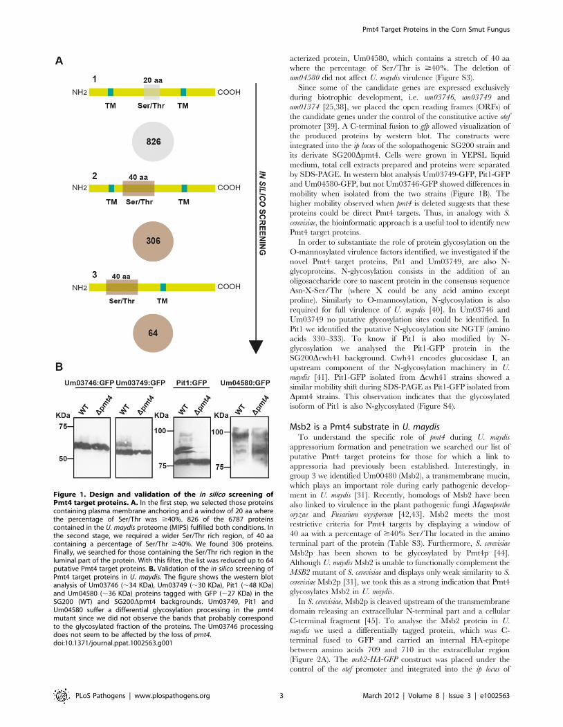
sequential in silico searches. First, we identified membrane proteins
containing a region of at least 20 aa in which the percentage of
Ser/Thr was $40%. With these parameters we found 826
proteins (12.17% of the U. maydis proteome) (Table S1). Then, we
performed a more restrictive search on the U. maydis proteome for
membrane attached proteins containing a wider region of 40 aa,
where the percentage of Ser/Thr was $40%. With this
bioinformatic approach we identified 306 proteins from the U.
maydis database (4.51% of the U. maydis proteome) (Table S2).
Since it has been described that the presence of a Ser/Thr rich
region in the amino terminal part of single-pass transmembrane
proteins triggers the recognition by Pmt4 [8], this was the search
criterion for the third round. 64 out of the 306 previously
identified sequences met these criteria (0.94% of the U. maydis
genome) (Table S3) (see Figure 1A).
The obtained putative Pmt4 substrates were categorized based
on their probable functions using FunCatDB from the MIPS U.
maydis database (http://mips.helmholtz-muenchen.de/genre/
proj/ustilago/). We found that the initially identified group of
826 proteins was mainly enriched in proteins associated with
transport, i.e. ion transport, detoxification by export as well as
homeostasis of cations. The more restricted group of 306 proteins
contained mostly unclassified proteins (52.1% of the proteins
contained in the list). Other enriched functional categories were
associated with inorganic chemical agent resistance and detoxifi-
cation by export. Finally, the most restrictive group of 64 proteins
was also mainly enriched in unclassified proteins (Figure S2).
In the genome of U. maydis many of the genes encoding small
secreted proteins are organized in gene clusters, most of them
implicated in virulence [25,35]. Interestingly, we found in our
screening four proteins included in these clusters: Um01235,
Um03746 and Um03749 (identified in group 1, Table S1); and
Um01374 (belonging to group 2, Table S2). The Um01235
protein is part of the cluster 2A. The deletion of all eight genes
from cluster 2A causes a hypervirulent phenotype [25]. On the
other hand, Um03746 and Um03749 reside in cluster 10A, which
contains 10 genes for effector proteins in total. The deletion of
cluster 10A reduces virulence [25]. Finally, Um01374 (Pit1) is one
of four proteins encoded by the pit (proteins important for tumors)
cluster. The deletion of the transmembrane gene pit1 or the
effector gene pit2 strongly reduces pathogenic development at the
level of tumor formation [38].
In silico screening validationTo confirm our bioinformatic results, we performed a
biochemical approach to study the role of Pmt4 in posttransla-
tional modification of its putative target proteins by western blot.
For this purpose we isolated candidate proteins from wild-type and
Dpmt4 strains and compared the mobility of these proteins during
SDS polyacrylamide gel electrophoresis (SDS-PAGE). Given the
critical role of cluster 10A during U. maydis virulence, we decided
to analyse the role of Pmt4 in the posttranslational modification of
Um03746 and Um03749. In addition we included the protein
Pit1. Finally, we also randomly selected an until now unchar-
Author Summary
The O-mannosyltransferase Pmt4 is essential for virulenceof animal and plant pathogenic fungi. This proteinattaches one mannose at serine/threonine residues of cellwall and secreted proteins modulating their locationand function. Thus, the crucial role of Pmt4 in fungalpathogenic development is probably caused by a defec-tive glycosylation of its target proteins altering host-fungus interaction. In this paper, we performed a screenfor Pmt4 target proteins employing the fungus Ustilagomaydis, which causes smut disease in maize plants. Thisallowed identifying novel Pmt4 target proteins having acrucial role on its virulence. One of these targets is thesignalling mucin Msb2, a conserved protein which actsupstream of MAP kinase cascades in various fungi andregulates early pathogenic development in U. maydis. Wepropose that Pmt4-dependent glycosylation of the extra-cellular domain of Msb2 is required for Msb2 activity andhence pathogenic development of U. maydis. This isdivergent to the situation in S. cerevisiae where themannosylated extracellular region of Msb2p possesses anegative regulatory function. In addition, we demonstrateimportant roles of Pmt4 during later stages of plantinfection and identified Pmt4 target proteins which couldbe responsible for the virulence defect of pmt4 mutantsduring tumor formation.
Pmt4 Target Proteins in the Corn Smut Fungus
PLoS Pathogens | www.plospathogens.org 2 March 2012 | Volume 8 | Issue 3 | e1002563
acterized protein, Um04580, which contains a stretch of 40 aa
where the percentage of Ser/Thr is $40%. The deletion of
um04580 did not affect U. maydis virulence (Figure S3).
Since some of the candidate genes are expressed exclusively
during biotrophic development, i.e. um03746, um03749 and
um01374 [25,38], we placed the open reading frames (ORFs) of
the candidate genes under the control of the constitutive active otef
promoter [39]. A C-terminal fusion to gfp allowed visualization of
the produced proteins by western blot. The constructs were
integrated into the ip locus of the solopathogenic SG200 strain and
its derivate SG200Dpmt4. Cells were grown in YEPSL liquid
medium, total cell extracts prepared and proteins were separated
by SDS-PAGE. In western blot analysis Um03749-GFP, Pit1-GFP
and Um04580-GFP, but not Um03746-GFP showed differences in
mobility when isolated from the two strains (Figure 1B). The
higher mobility observed when pmt4 is deleted suggests that these
proteins could be direct Pmt4 targets. Thus, in analogy with S.
cerevisiae, the bioinformatic approach is a useful tool to identify new
Pmt4 target proteins.
In order to substantiate the role of protein glycosylation on the
O-mannosylated virulence factors identified, we investigated if the
novel Pmt4 target proteins, Pit1 and Um03749, are also N-
glycoproteins. N-glycosylation consists in the addition of an
oligosaccharide core to nascent protein in the consensus sequence
Asn-X-Ser/Thr (where X could be any acid amino except
proline). Similarly to O-mannosylation, N-glycosylation is also
required for full virulence of U. maydis [40]. In Um03746 and
Um03749 no putative glycosylation sites could be identified. In
Pit1 we identified the putative N-glycosylation site NGTF (amino
acids 330–333). To know if Pit1 is also modified by N-
glycosylation we analysed the Pit1-GFP protein in the
SG200Dcwh41 background. Cwh41 encodes glucosidase I, an
upstream component of the N-glycosylation machinery in U.
maydis [41]. Pit1-GFP isolated from Dcwh41 strains showed a
similar mobility shift during SDS-PAGE as Pit1-GFP isolated from
Dpmt4 strains. This observation indicates that the glycosylated
isoform of Pit1 is also N-glycosylated (Figure S4).
Msb2 is a Pmt4 substrate in U. maydisTo understand the specific role of pmt4 during U. maydis
appressorium formation and penetration we searched our list of
putative Pmt4 target proteins for those for which a link to
appressoria had previously been established. Interestingly, in
group 3 we identified Um00480 (Msb2), a transmembrane mucin,
which plays an important role during early pathogenic develop-
ment in U. maydis [31]. Recently, homologs of Msb2 have been
also linked to virulence in the plant pathogenic fungi Magnaporthe
oryzae and Fusarium oxysporum [42,43]. Msb2 meets the most
restrictive criteria for Pmt4 targets by displaying a window of
40 aa with a percentage of $40% Ser/Thr located in the amino
terminal part of the protein (Table S3). Furthermore, S. cerevisiae
Msb2p has been shown to be glycosylated by Pmt4p [44].
Although U. maydis Msb2 is unable to functionally complement the
MSB2 mutant of S. cerevisiae and displays only weak similarity to S.
cerevisiae Msb2p [31], we took this as a strong indication that Pmt4
glycosylates Msb2 in U. maydis.
In S. cerevisiae, Msb2p is cleaved upstream of the transmembrane
domain releasing an extracellular N-terminal part and a cellular
C-terminal fragment [45]. To analyse the Msb2 protein in U.
maydis we used a differentially tagged protein, which was C-
terminal fused to GFP and carried an internal HA-epitope
between amino acids 709 and 710 in the extracellular region
(Figure 2A). The msb2-HA-GFP construct was placed under the
control of the otef promoter and integrated into the ip locus of
Figure 1. Design and validation of the in silico screening ofPmt4 target proteins. A. In the first step, we selected those proteinscontaining plasma membrane anchoring and a window of 20 aa wherethe percentage of Ser/Thr was $40%. 826 of the 6787 proteinscontained in the U. maydis proteome (MIPS) fulfilled both conditions. Inthe second stage, we required a wider Ser/Thr rich region, of 40 aacontaining a percentage of Ser/Thr $40%. We found 306 proteins.Finally, we searched for those containing the Ser/Thr rich region in theluminal part of the protein. With this filter, the list was reduced up to 64putative Pmt4 target proteins. B. Validation of the in silico screening ofPmt4 target proteins in U. maydis. The figure shows the western blotanalysis of Um03746 (,34 KDa), Um03749 (,30 KDa), Pit1 (,48 KDa)and Um04580 (,36 KDa) proteins tagged with GFP (,27 KDa) in theSG200 (WT) and SG200Dpmt4 backgrounds. Um03749, Pit1 andUm04580 suffer a differential glycosylation processing in the pmt4mutant since we did not observe the bands that probably correspondto the glycosylated fraction of the proteins. The Um03746 processingdoes not seem to be affected by the loss of pmt4.doi:10.1371/journal.ppat.1002563.g001
Pmt4 Target Proteins in the Corn Smut Fungus
PLoS Pathogens | www.plospathogens.org 3 March 2012 | Volume 8 | Issue 3 | e1002563
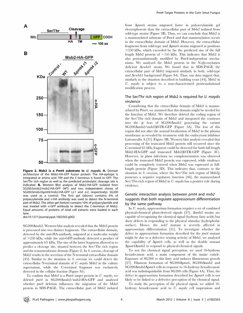
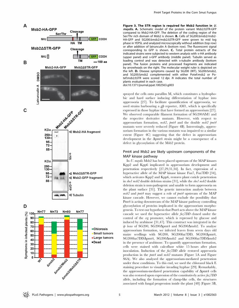
SG200Dmsb2. Western blot analysis revealed that the Msb2 protein
is processed into two distinct fragments. The extracellular domain,
detected by the anti-HA-antibody, migrated at a molecular weight
of .250 kDa, while the anti-GFP-antibody detected a product of
approximately 65 kDa. The size of the latter fragment allowed us to
predict a cleavage site, situated between the Ser/Thr rich region
and the transmembrane domain (Figure 2). In S. cerevisae, cleavage of
Msb2 results in the secretion of the N-terminal extracellular domain
[45]. Similar to the situation in S. cerevisiae we could detect the
extracellular N-terminal domain of U. maydis Msb2 in the culture
supernatant, while the C-terminal fragment was exclusively
detected in the cellular fraction (Figure S5).
To confirm that Msb2 is a Pmt4 target protein in U. maydis, we
deleted pmt4 in SG200Dmsb2/msb2-HA-GFP and analysed
whether pmt4 deletion influences the migration of the Msb2
protein in SDS-PAGE. The extracellular part of Msb2 isolated
from Dpmt4 strains migrated faster in polyacrylamide gel
electrophoresis than the extracellular part of Msb2 isolated from
wild-type strains (Figure 2B). Thus, we can conclude that Msb2 is
a mannosylated substrate of Pmt4 and that mannosylation occurs
in the extracellular domain of Msb2. However, the extracellular
fragments from wild-type and Dpmt4 strains migrated at positions
.250 kDa, which exceeded by far the predicted size of the full
length Msb2 protein of ,145 kDa. This indicates that Msb2 is
also postranslationally modified by Pmt4-independent mecha-
nisms. We analysed the Msb2 protein in the N-glycosylation
deficient Dcwh41 strain. We found that in SDS-PAGE the
extracellular part of Msb2 migrated similarly in both, wild-type
and Dcwh41 background (Figure S4). Thus, our data suggest that,
similarly to the situation described in budding yeast [44], Msb2 in
U. maydis is subject to a non-characterized posttranslational
modification process.
The Ser/Thr rich region of Msb2 is required for U. maydisvirulence
Considering that the extracellular domain of Msb2 is manno-
sylated by Pmt4, we assumed that this domain might be needed for
the function of Msb2. We therefore deleted the coding region of
the Ser/Thr rich domain of Msb2 and integrated the construct
into the ip locus of SG200Dmsb2 generating the variant
SG200Dmsb2/msb2DSTR-GFP (Figure 3A). The loss of this
region did not alter the normal localization of Msb2 in the plasma
membrane as revealed by treatment with the endocytosis inhibitor
Latrunculin A [31] (Figure 3B). Western blot analysis revealed that
processing of the truncated Msb2 protein still occurred since the
C-terminal 65 kDa fragment could be detected for both full length
Msb2-HA-GFP and truncated Msb2DSTR-GFP (Figure 3C).
However, in plant infections no complementation was observed
when the truncated Msb2 protein was expressed, while virulence
could be completely restored when Msb2 was expressed as full-
length protein (Figure 3D). This indicates that, contrary to the
situation in S. cerevisiae, where the Ser/Thr rich region of Msb2p
possesses a negative regulatory function [46], the mannosylated
Ser/Thr-rich region of Msb2 in U. maydis has a positive role during
virulence.
Genetic interaction analysis between pmt4 and msb2suggests that both regulate appressorium differentiationby the same pathway
In U. maydis, appressorium formation requires a set of combined
physical-chemical plant-derived signals [27]. Dmsb2 strains are
capable of recognizing the chemical signal (hydroxy fatty acids) but
show defects in responding to the physical stimulus (hydrophobic
surface). Hence, the msb2 mutant is severely affected in
appressorium differentiation [31]. To investigate whether the
defect in appressorium formation described for the pmt4 mutant
might be due to a defective sensing activity of Msb2, we analysed
the capability of Dpmt4 cells, as well as the double mutant
Dpmt4Dmsb2 to respond to physical-chemical signals.
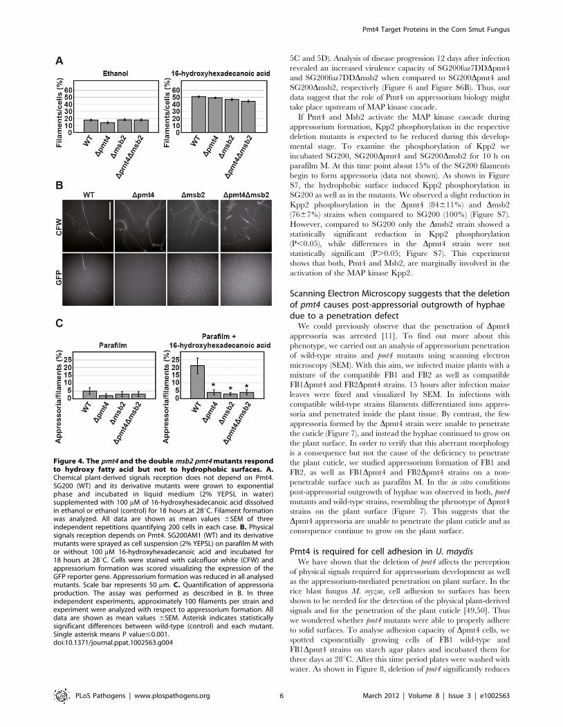
To test the chemical signal perception, we used 16-hydroxy
hexadecanoic acid, a main component of the maize cuticle.
Exposure of SG200 to this fatty acid induces filamentous growth
[27]. Filament formation of SG200Dpmt4, SG200Dmsb2 and
SG200Dmsb2Dpmt4 cells in response to 16-hydroxy hexadecanoic
acid was indistinguishable from SG200 cells (Figure 4A). Thus, the
defect in appressorium formation described for Dpmt4 cells is not
likely to be linked to a defective perception of the chemical signal.
To study the perception of the physical signal, we added 16-
hydroxy hexadecanoic acid to U. maydis cell suspensions and
Figure 2. Msb2 is a Pmt4 substrate in U. maydis. A. Domainarchitecture of the Msb2-HA-GFP fusion protein. The HA-epitope isintegrated at amino acid 709 and the C-terminus is fused to GFP. TheSer/Thr rich region as well as the predicted proteolytic cleavage site isindicated. B. Western Blot analysis of Msb2-HA-GFP isolated fromSG200Dmsb2/msb2-HA-GFP (WT) and two independent clones ofSG200Dmsb2Dpmt4/msb2-HA-GFP (#1 and #2, respectively). SG200was used as a control. The first gel (above) contains 6% ofpolyacrylamide and a-HA antibody was used to detect the N-terminalpart of Msb2. The other gel (below) contains 10% of polyacrylamide andwas treated with a-GFP antibody to detect the C-terminus of Msb2.Equal amounts of proteins of total cell extracts were loaded in eachlane.doi:10.1371/journal.ppat.1002563.g002
Pmt4 Target Proteins in the Corn Smut Fungus
PLoS Pathogens | www.plospathogens.org 4 March 2012 | Volume 8 | Issue 3 | e1002563
sprayed the cells onto parafilm M, which constitutes a hydropho-
bic and hard surface inducing differentiation of hyphae into
appressoria [27]. To facilitate quantification of appressoria, we
used strains harbouring a gfp reporter, AM1, which is specifically
expressed in those hyphae that have formed an appressorium [27].
We observed comparable filament formation of SG200AM1 and
the respective derivative mutants. However, with respect to
appressorium formation, msb2, pmt4 and the double msb2 pmt4
mutants were severely reduced (Figure 4B). Interestingly, appres-
sorium formation in the various mutants was impaired to a similar
extent (Figure 4C) suggesting that the defect in appressorium
development in the Dpmt4 strain might be a consequence of a
defect in glycosylation of the Msb2 protein.
Pmt4 and Msb2 are likely upstream components of theMAP kinase pathway
In U. maydis Msb2 has been placed upstream of the MAP kinases
Kpp2 and Kpp6 implicated in appressorium development and
penetration respectively [27,29,31,34]. In fact, expression of a
hyperactive allele of the MAP kinase kinase Fuz7, Fuz7DD [34],
which activates Kpp2 and Kpp6, restores plant cuticle penetration
in sho1 msb2 double deletion strains [31], while the sho1 msb2 double
deletion strain is non-pathogenic and unable to form appressoria on
the plant surface [31]. The genetic interaction analysis between
msb2 and pmt4 may suggest a role of pmt4 upstream of the MAP
kinase cascade. However, we cannot exclude the possibility that
Pmt4 is acting downstream of the MAP kinase pathway controlling
glycosylation of proteins implicated in the appressorium morpho-
genesis. To test our hypothesis that Pmt4 acts above the MAP kinase
cascade we used the hyperactive allele fuz7DD cloned under the
control of the crg promoter, which is repressed by glucose and
induced by arabinose [31,47]. This construct was integrated in the
ip locus of SG200, SG200Dpmt4 and SG200Dmsb2. To analyse
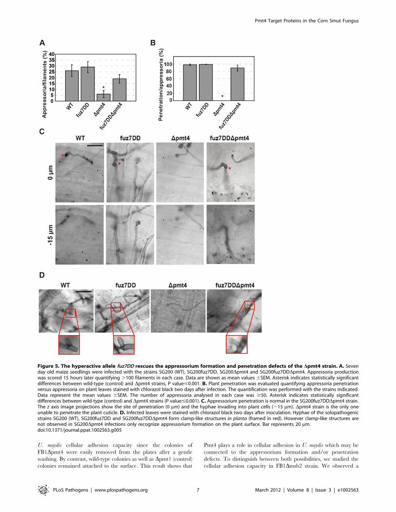
appressorium formation, we infected leaves from seven days old
maize seedlings with SG200, SG200fuz7DD, SG200Dpmt4,
SG200fuz7DDDpmt4, SG200Dmsb2 and SG200fuz7DDDmsb2
in the presence of arabinose. To quantify appressorium formation,
cells were stained with calcofluor white 15 hours after plant
inoculation. Induction of the fuz7DD allele restored appressoria
production in the pmt4 and msb2 mutants (Figure 5A and Figure
S6A). We also analysed the appressorium-mediated penetration
under these conditions. To this end, we used the chlorazol black E
staining procedure to visualize invading hyphae [29]. Remarkably,
the appressorium-mediated penetration capability of Dpmt4 cells
was also restored upon expression of the constitutively active fuz7DD
allele, including the formation of clamp-like cells, the structures
associated with fungal progression inside the plant [48] (Figure 5B,
Figure 3. The STR region is required for Msb2 function in U.maydis. A. Schematic model of the protein variant Msb2DSTR-GFPcompared to Msb2-HA-GFP. The deletion of the coding region of theSer/Thr rich domain of Msb2 is shown. B. Cells of SG200Dmsb2/msb2-HA-GFP and SG200Dmsb2/msb2DSTR-GFP were grown to mid logphase in YEPSL and analyzed microscopically without addition (top row)or after addition of latrunculin A (bottom row). The fluorescent signalcorresponding to GFP is shown. C. Total protein extracts of theindicated strains were subjected to western analysis with a-HA antibody(upper panel) and a-GFP antibody (middle panel). Tubulin served asloading control and was detected with a-tubulin antibody (bottompanel). The fusion proteins and processed fragments are indicatedby arrowheads on the right. The molecular weight ruler is depicted onthe left. D. Disease symptoms caused by SG200 (WT), SG200Dmsb2,and SG200Dmsb2 complemented with either Potef:msb2 or Po-tef:msb2DSTR were scored 12 dpi. N indicates the total number ofplants evaluated in each case.doi:10.1371/journal.ppat.1002563.g003
Pmt4 Target Proteins in the Corn Smut Fungus
PLoS Pathogens | www.plospathogens.org 5 March 2012 | Volume 8 | Issue 3 | e1002563
5C and 5D). Analysis of disease progression 12 days after infection
revealed an increased virulence capacity of SG200fuz7DDDpmt4
and SG200fuz7DDDmsb2 when compared to SG200Dpmt4 and
SG200Dmsb2, respectively (Figure 6 and Figure S6B). Thus, our
data suggest that the role of Pmt4 on appressorium biology might
take place upstream of MAP kinase cascade.
If Pmt4 and Msb2 activate the MAP kinase cascade during
appressorium formation, Kpp2 phosphorylation in the respective
deletion mutants is expected to be reduced during this develop-
mental stage. To examine the phosphorylation of Kpp2 we
incubated SG200, SG200Dpmt4 and SG200Dmsb2 for 10 h on
parafilm M. At this time point about 15% of the SG200 filaments
begin to form appressoria (data not shown). As shown in Figure
S7, the hydrophobic surface induced Kpp2 phosphorylation in
SG200 as well as in the mutants. We observed a slight reduction in
Kpp2 phosphorylation in the Dpmt4 (84611%) and Dmsb2
(7667%) strains when compared to SG200 (100%) (Figure S7).
However, compared to SG200 only the Dmsb2 strain showed a
statistically significant reduction in Kpp2 phosphorylation
(P,0.05), while differences in the Dpmt4 strain were not
statistically significant (P.0.05; Figure S7). This experiment
shows that both, Pmt4 and Msb2, are marginally involved in the
activation of the MAP kinase Kpp2.
Scanning Electron Microscopy suggests that the deletionof pmt4 causes post-appressorial outgrowth of hyphaedue to a penetration defect
We could previously observe that the penetration of Dpmt4
appressoria was arrested [11]. To find out more about this
phenotype, we carried out an analysis of appressorium penetration
of wild-type strains and pmt4 mutants using scanning electron
microscopy (SEM). With this aim, we infected maize plants with a
mixture of the compatible FB1 and FB2 as well as compatible
FB1Dpmt4 and FB2Dpmt4 strains. 15 hours after infection maize
leaves were fixed and visualized by SEM. In infections with
compatible wild-type strains filaments differentiated into appres-
soria and penetrated inside the plant tissue. By contrast, the few
appressoria formed by the Dpmt4 strain were unable to penetrate
the cuticle (Figure 7), and instead the hyphae continued to grow on
the plant surface. In order to verify that this aberrant morphology
is a consequence but not the cause of the deficiency to penetrate
the plant cuticle, we studied appressorium formation of FB1 and
FB2, as well as FB1Dpmt4 and FB2Dpmt4 strains on a non-
penetrable surface such as parafilm M. In the in vitro conditions
post-appressorial outgrowth of hyphae was observed in both, pmt4
mutants and wild-type strains, resembling the phenotype of Dpmt4
strains on the plant surface (Figure 7). This suggests that the
Dpmt4 appressoria are unable to penetrate the plant cuticle and as
consequence continue to grow on the plant surface.
Pmt4 is required for cell adhesion in U. maydisWe have shown that the deletion of pmt4 affects the perception
of physical signals required for appressorium development as well
as the appressorium-mediated penetration on plant surface. In the
rice blast fungus M. oryzae, cell adhesion to surfaces has been
shown to be needed for the detection of the physical plant-derived
signals and for the penetration of the plant cuticle [49,50]. Thus
we wondered whether pmt4 mutants were able to properly adhere
to solid surfaces. To analyse adhesion capacity of Dpmt4 cells, we
spotted exponentially growing cells of FB1 wild-type and
FB1Dpmt4 strains on starch agar plates and incubated them for
three days at 28uC. After this time period plates were washed with
water. As shown in Figure 8, deletion of pmt4 significantly reduces
Figure 4. The pmt4 and the double msb2 pmt4 mutants respondto hydroxy fatty acid but not to hydrophobic surfaces. A.Chemical plant-derived signals reception does not depend on Pmt4.SG200 (WT) and its derivative mutants were grown to exponentialphase and incubated in liquid medium (2% YEPSL in water)supplemented with 100 mM of 16-hydroxyhexadecanoic acid dissolvedin ethanol or ethanol (control) for 18 hours at 28uC. Filament formationwas analyzed. All data are shown as mean values 6SEM of threeindependent repetitions quantifying 200 cells in each case. B. Physicalsignals reception depends on Pmt4. SG200AM1 (WT) and its derivativemutants were sprayed as cell suspension (2% YEPSL) on parafilm M withor without 100 mM 16-hydroxyhexadecanoic acid and incubated for18 hours at 28uC. Cells were stained with calcofluor white (CFW) andappressorium formation was scored visualizing the expression of theGFP reporter gene. Appressorium formation was reduced in all analysedmutants. Scale bar represents 50 mm. C. Quantification of appressoriaproduction. The assay was performed as described in B. In threeindependent experiments, approximately 100 filaments per strain andexperiment were analyzed with respect to appressorium formation. Alldata are shown as mean values 6SEM. Asterisk indicates statisticallysignificant differences between wild-type (control) and each mutant.Single asterisk means P value#0.001.doi:10.1371/journal.ppat.1002563.g004
Pmt4 Target Proteins in the Corn Smut Fungus
PLoS Pathogens | www.plospathogens.org 6 March 2012 | Volume 8 | Issue 3 | e1002563
U. maydis cellular adhesion capacity since the colonies of
FB1Dpmt4 were easily removed from the plates after a gentle
washing. By contrast, wild-type colonies as well as Dpmt1 (control)
colonies remained attached to the surface. This result shows that
Pmt4 plays a role in cellular adhesion in U. maydis which may be
connected to the appressorium formation and/or penetration
defects. To distinguish between both possibilities, we studied the
cellular adhesion capacity in FB1Dmsb2 strain. We observed a
Figure 5. The hyperactive allele fuz7DD rescues the appressorium formation and penetration defects of the Dpmt4 strain. A. Sevenday old maize seedlings were infected with the strains SG200 (WT), SG200fuz7DD, SG200Dpmt4 and SG200fuz7DDDpmt4. Appressoria productionwas scored 15 hours later quantifying .100 filaments in each case. Data are shown as mean values 6SEM. Asterisk indicates statistically significantdifferences between wild-type (control) and Dpmt4 strains, P value#0.001. B. Plant penetration was evaluated quantifying appressoria penetrationversus appressoria on plant leaves stained with chlorazol black two days after infection. The quantification was performed with the strains indicated.Data represent the mean values 6SEM. The number of appressoria analysed in each case was $50. Asterisk indicates statistically significantdifferences between wild-type (control) and Dpmt4 strains (P value#0.001). C. Appressorium penetration is normal in the SG200fuz7DDDpmt4 strain.The z axis image projections show the site of penetration (0 mm) and the hyphae invading into plant cells (215 mm). Dpmt4 strain is the only oneunable to penetrate the plant cuticle. D. Infected leaves were stained with chlorazol black two days after inoculation. Hyphae of the solopathogenicstrains SG200 (WT), SG200fuz7DD and SG200fuz7DDDpmt4 form clamp-like structures in planta (framed in red). However clamp-like structures arenot observed in SG200Dpmt4 infections only recognize appressorium formation on the plant surface. Bar represents 20 mm.doi:10.1371/journal.ppat.1002563.g005
Pmt4 Target Proteins in the Corn Smut Fungus
PLoS Pathogens | www.plospathogens.org 7 March 2012 | Volume 8 | Issue 3 | e1002563

similar behaviour to wild-type cells (Figure S8). Because the loss of
Msb2 affects to appressorium formation but not penetration, our
data suggest a link between cellular adhesion capacity and
appressorium penetration in pmt4 mutant cells.
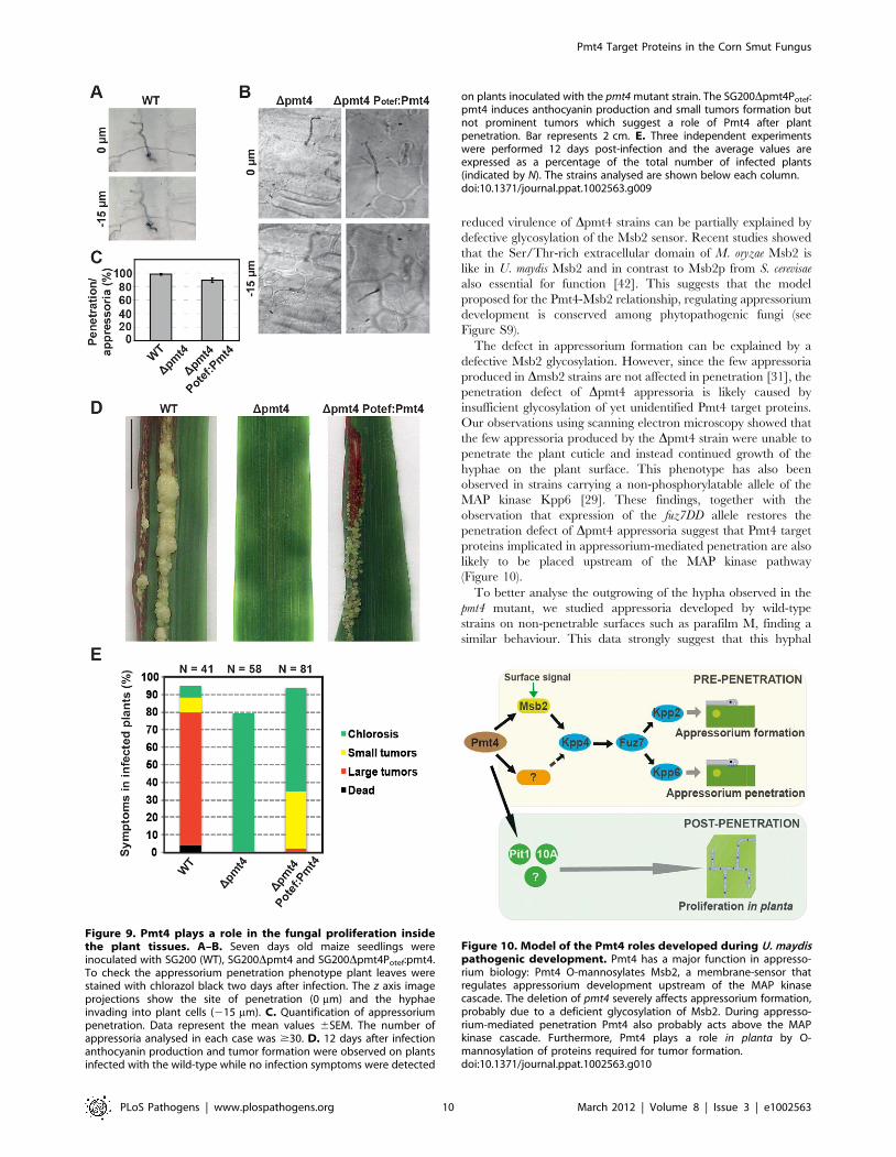
Pmt4 plays a role after appressorium penetrationWe have observed that although the plant infections with the
SG200fuz7DDDpmt4 strain were able to induce small tumors, the
virulence of this strain was severely reduced when compared to
SG200fuz7DD suggesting a role for Pmt4 during fungal
progression inside the plant tissues. To confirm this result, we
cloned the pmt4 ORF under the control of the otef promoter which
is active during early pathogenic development, but turned off
during the late biotrophic stage [51]. This construct was integrated
in the ip locus of the SG200Dpmt4 strain. To address if Pmt4 plays
a role after plant penetration, we inoculated maize plants with
SG200, SG200Dpmt4 and SG200Dpmt4Potef:pmt4. Disease
progression was scored 12 days after infection. Although
appressorium penetration was completely restored in plants
infected with SG200Dpmt4Potef:pmt4, tumor formation was
severely reduced compared to infections with SG200 (Figure 9).
This demonstrates, that Pmt4 is not only needed for early
pathogenic development, but has additional roles during the in
planta development, most likely by posttranslational modification of
fungal effectors required for tumor formation.
Discussion
In this paper we have performed in silico screening for putative
Pmt4 target proteins in the phytopathogen U. maydis. We have
found a high percentage of proteins containing Ser/Thr rich
regions with most of them having unknown functions. A screening
approach searching for transmembrane anchored proteins,
containing a stretch of 20 aa in which the percentage of Ser/
Thr was $40%, identified 826 proteins (12.17% of the U. maydis
proteome). By contrast, using similar parameters in S. cerevisiae 51
candidates (0.87% of the proteome) could be identified [8]. This
difference may be due to the high number of putative secreted
proteins present in the U. maydis proteome, which may be linked to
the biotrophic relationship with the host plant [25]. In order to
obtain a more restrictive group of Pmt4 target proteins we
screened the proteome of U. maydis for single-pass transmembrane
proteins containing a Ser/Thr-rich region facing the ER lumen.
Under these conditions, we found 64 proteins. The validation of
our screening retrieved novel Pmt4 substrates such as Um04580,
Figure 6. Expression of fuz7DD in the Dpmt4 strain partiallyrestores the tumor induction in maize. A. Disease progression inplant infections with the strains indicated. No infection symptoms weredetected on plants inoculated with the SG200Dpmt4 strain. TheSG200fuz7DDDpmt4 induces anthocyanin production and small tumorsformation. Bar represents 2 cm. B. Plants were infected with theindicated strains and symptoms were scored 12 days post-infection. Nindicates the total number of plants evaluated in each case.doi:10.1371/journal.ppat.1002563.g006
Figure 7. The pmt4 mutant appressorium shows outgrowing asa consequence of its incapability to penetrate the plant cuticle.Crosses of the sexually compatible strains FB1 and FB2, and FB1Dpmt4and FB2Dpmt4 were inoculated into maize seedlings. Leaves were fixedand analysed by Scanning Electron Microscopy (SEM) one day postinfection. In these images (left), we can observe appressoriumformation over a plant stomata (pointed with a red arrow) penetratingthe plant surface in the wild-type. However, in a similar scenario, withthe appressorium developed over plant stomata, the pmt4 mutant isunable to penetrate the plant cuticle, producing hyphal outgrowing.This morphologic structure seems to be a consequence of itsincapability to penetrate this surface as we can deduce from wild-type phenotype of the in vitro appressorium formation on the non-penetrable surface of parafilm M (right). The in vitro analysis wasperformed by fluorescence microscopy staining the cells with calcofluorwhite 15 hours after being sprayed on parafilm M (see Methods foradditional details). Scale bar on the in vitro conditions represents20 mm.doi:10.1371/journal.ppat.1002563.g007
Pmt4 Target Proteins in the Corn Smut Fungus
PLoS Pathogens | www.plospathogens.org 8 March 2012 | Volume 8 | Issue 3 | e1002563

or Pit1, which is essential for pathogenic development of U. maydis
[38]. Moreover, we have found that Um03749, which belongs to
the cluster 10A, required for tumor formation [25], is also a novel
Pmt4 target protein.
One of the most intriguing phenotypes associated with the pmt4
deletion in U. maydis is the strong reduction in appressorium
formation and appressorium-mediated penetration of the plant
cuticle [11]. Both processes are controlled by a MAP kinase
pathway, consisting of the MAPKK kinase Kpp4/Ubc4, the
MAPK kinase Fuz7/Ubc5 and the MAP kinases Kpp2/Ubc3 and
Kpp6 [28–30,32–34]. While Kpp2 is needed for appressorium
formation [30], Kpp6 has a specific role during the penetration
process [29]. Our results suggest that the role of Pmt4 during
appressorium formation consists probably not in the glycosylation
of downstream components of this pathway rather Pmt4 modifies
proteins placed upstream of the MAP kinase pathway. In U. maydis
the signalling mucin Msb2 is likely an activator of the MAP kinase
cascade and the deletion of msb2 leads to a strong reduction in
appressorium formation due to defects in perception of plant
surface signals [31]. We have identified Msb2 in our in silico
screening and confirmed that it is substrate of Pmt4 in U. maydis, as
it has been described for S. cerevisiae [44]. Our epistatic studies
comprising msb2 and pmt4 suggest that during appressorium
development both proteins act in the same pathway. A critical
question was if Pmt4 and Msb2 are needed for the activation of the
MAP kinase Kpp2, which is essential for filamentation and
appressorium formation in U. maydis [30]. Here we could show for
the first time that Kpp2 is phosphorylated during appressorium
formation in U. maydis. However, in pmt4 and msb2 mutants
phosphorylation of Kpp2 was only slightly reduced. In view of the
fact that under our experimental conditions only a minor fraction
of U. maydis filaments differentiate appressoria and that pmt4 and
msb2 mutants exhibit normal filament formation, a process that
depends also on Kpp2, we did not expect major differences in
Kpp2 phosphorylation. We do currently not know in which spatial
and temporal context Kpp2 phosphorylation induces either
filament formation or appressorium formation. It is conceivable
that appressorium formation requires a temporal coordinated
threshold of MAP kinase activity and that this process involves
Msb2 and its glycosylation status. A strong indication that Msb2
and Pmt4 act above the MAP kinase cascade is inferred from our
observation that expression of the constitutive active fuz7DD allele
restored appressorium formation in the Dmsb2 as well as Dpmt4
strains. Overall our data suggest that Msb2 and Pmt4 are needed
for the activation of the MAP kinase pathway, although formal
proof for this needs further experimentation.
In gel mobility shift analysis we found that Pmt4 specifically
glycosylates the extracellular domain of Msb2. In S. cerevisae the
extracellular domain of Msb2p has a negative regulatory function,
since deletion of this domain leads to the activation of the FG
(filamentous growth)-pathway specific MAP kinase Kss1p [46]. It
is therefore assumed, that in yeast the release of the extracellular
glycosylated domain of Msb2p activates the FG-pathway [45,46].
This release can be mimicked by the deletion of pmt4, which leads
to underglycosylated Msb2p and to the activation of the FG-
pathway in an Msb2p-dependent manner [44]. Thus, in S. cerevisiae
underglycosylated Msb2p activates the MAP kinase pathway. Our
data from U. maydis demonstrates that the extracellular domain of
Msb2 has a positive function, since deletion of the Ser/Thr-rich
region causes a decrease in pathogenicity similar to the deletion of
the entire msb2 gene. This observation is consistent with the
reduced appressorium formation in pmt4 deletion mutants.
Accordingly, Pmt4-mediated mannosylation of Msb2 seems to
be necessary for activation of the MAP kinase cascade and the
Figure 8. Pmt4 is required for cellular adhesion to solid surfacesin U. maydis. The strains indicated FB1 and FB1Dpmt4 were grown toA600 = 0.5 in YEPSL liquid medium and then were spotted on starchmedium plates, according to the random distribution scheme shown,and incubated for three days at 28uC. Later, the plate surface was gentlewashed. The FB1Dpmt1 strain was used as control of the effect of the lossof other protein O-mannosyltransferase. The deletion of pmt4 reducessignificantly the fungal cell adhesion to solid surfaces in U. maydis.doi:10.1371/journal.ppat.1002563.g008
Pmt4 Target Proteins in the Corn Smut Fungus
PLoS Pathogens | www.plospathogens.org 9 March 2012 | Volume 8 | Issue 3 | e1002563
reduced virulence of Dpmt4 strains can be partially explained by
defective glycosylation of the Msb2 sensor. Recent studies showed
that the Ser/Thr-rich extracellular domain of M. oryzae Msb2 is
like in U. maydis Msb2 and in contrast to Msb2p from S. cerevisae
also essential for function [42]. This suggests that the model
proposed for the Pmt4-Msb2 relationship, regulating appressorium
development is conserved among phytopathogenic fungi (see
Figure S9).
The defect in appressorium formation can be explained by a
defective Msb2 glycosylation. However, since the few appressoria
produced in Dmsb2 strains are not affected in penetration [31], the
penetration defect of Dpmt4 appressoria is likely caused by
insufficient glycosylation of yet unidentified Pmt4 target proteins.
Our observations using scanning electron microscopy showed that
the few appressoria produced by the Dpmt4 strain were unable to
penetrate the plant cuticle and instead continued growth of the
hyphae on the plant surface. This phenotype has also been
observed in strains carrying a non-phosphorylatable allele of the
MAP kinase Kpp6 [29]. These findings, together with the
observation that expression of the fuz7DD allele restores the
penetration defect of Dpmt4 appressoria suggest that Pmt4 target
proteins implicated in appressorium-mediated penetration are also
likely to be placed upstream of the MAP kinase pathway
(Figure 10).
To better analyse the outgrowing of the hypha observed in the
pmt4 mutant, we studied appressoria developed by wild-type
strains on non-penetrable surfaces such as parafilm M, finding a
similar behaviour. This data strongly suggest that this hyphal
Figure 9. Pmt4 plays a role in the fungal proliferation insidethe plant tissues. A–B. Seven days old maize seedlings wereinoculated with SG200 (WT), SG200Dpmt4 and SG200Dpmt4Potef:pmt4.To check the appressorium penetration phenotype plant leaves werestained with chlorazol black two days after infection. The z axis imageprojections show the site of penetration (0 mm) and the hyphaeinvading into plant cells (215 mm). C. Quantification of appressoriumpenetration. Data represent the mean values 6SEM. The number ofappressoria analysed in each case was $30. D. 12 days after infectionanthocyanin production and tumor formation were observed on plantsinfected with the wild-type while no infection symptoms were detected
on plants inoculated with the pmt4 mutant strain. The SG200Dpmt4Potef:pmt4 induces anthocyanin production and small tumors formation butnot prominent tumors which suggest a role of Pmt4 after plantpenetration. Bar represents 2 cm. E. Three independent experimentswere performed 12 days post-infection and the average values areexpressed as a percentage of the total number of infected plants(indicated by N). The strains analysed are shown below each column.doi:10.1371/journal.ppat.1002563.g009
Figure 10. Model of the Pmt4 roles developed during U. maydispathogenic development. Pmt4 has a major function in appresso-rium biology: Pmt4 O-mannosylates Msb2, a membrane-sensor thatregulates appressorium development upstream of the MAP kinasecascade. The deletion of pmt4 severely affects appressorium formation,probably due to a deficient glycosylation of Msb2. During appresso-rium-mediated penetration Pmt4 also probably acts above the MAPkinase cascade. Furthermore, Pmt4 plays a role in planta by O-mannosylation of proteins required for tumor formation.doi:10.1371/journal.ppat.1002563.g010
Pmt4 Target Proteins in the Corn Smut Fungus
PLoS Pathogens | www.plospathogens.org 10 March 2012 | Volume 8 | Issue 3 | e1002563
outgrowth is the consequence but not the cause of its incapability
to penetrate the plant cuticle. The penetration failure of Dpmt4
appressoria could be caused by an insufficient adhesion of the
fungus to the plant surface. For the rice blast fungus M. oryzae it has
been demonstrated that proper adhesion is critical for appressoria
to penetrate [50]. Interestingly, we have shown that Pmt4 plays an
important role in cellular adhesion to surfaces which might be
related to the appressorium penetration defect.
Our previous studies as well as this work demonstrated a
prominent role of Pmt4 in the appressorium biology. In addition,
we have now shown that Pmt4 is also necessary for tumor
induction in planta. We observed that pmt4 expression under the
control of the otef promoter, which is turned off during the
infection process [51], is not sufficient to restore normal tumor
formation in the SG200Dpmt4 strain. Likewise, expression of
fuz7DD in Dpmt4 strains did not rescue the virulence defect,
although initial appressorium formation and penetration were
restored. This suggests that the Pmt4 protein is also required
during fungal proliferation inside the plant tissue. For C. albicans it
has been demonstrated that the deletion of pmt genes is associated
with defects in cell wall integrity and secretion of fungal effectors,
probably disturbing the interaction with the host [5,10,52,53]. In
U. maydis, the biotrophic interaction between the plant and the
fungus is also mediated by fungal effectors [24]. The role of Pmt4
during this developmental stage could be related to the O-
mannosylation of Pit1 and Um03749 (see Figure 10), since both
are important for the biotrophic development [25,38]. Um03749
is a putative secreted protein which is directly modified by Pmt4.
The underglycosylation of Um03749 in Dpmt4 strains might affect
the function of this effector. Pit1 is a plasma membrane protein
that is genetically linked to the secreted effector Pit2. Both proteins
are specifically needed for tumor formation [38]. Although the
connection between these two proteins is currently unclear, it is
assumed, that they act in the same pathway [38]. Therefore,
mannosylation of Pit1 by Pmt4 could indirectly affect the function
of the secreted effector Pit2. This points to a role of Pmt4 in the
regulation of secreted effector proteins, needed for the biotrophic
development. In this context, considering the large number of
putative Pmt4 targets obtained in this study, it is tempting to
speculate that more Pmt4 targets exist, which contribute to the
establishment of biotrophic interaction between plant and fungus.
The bioinformatic search for Pmt4 target proteins in other
pathogenic fungi might highlight the role of Pmt4 during fungal
pathogenic development and allow the identification of novel
Pmt4 substrates required for fungus-host interactions.
Materials and Methods
Strains, growth conditions and plasmidsEscherichia coli DH5a and pGEM-T easy (Promega) were used
for cloning purposes. pING, a derivative of pONG [31] where
mcherry is exchanged by gfp, was used for cloning of ORFs from
pmt4, um03746, um03749 and um04580. To clone these ORFs, U.
maydis genomic DNA was used as template amplifying with the
primers Pmt4ORF-5 and Pmt4ORF-3, 03746ORF-5 and
03746ORF-3, 03749ORF-5 and 03749ORF-3, 04580ORF-5
and 04580ORF-3, respectively (sequences in Table S4). The
fragments were cut with SfiI and ligated with the 6.3 kb SfiI
fragment of pING. Expression of ORFs from pING derivatives is
controlled by the otef promoter.
To generate the msb2-HA-GFP construct, the primer combina-
tions oDL81/oDL125 and oDL82/oDL124 (Table S4) using U.
maydis genomic DNA as template generated two PCR products,
1.4 kb and 2.1 kb in length, respectively. Both fragments were cut
with SfiI and ligated with the 6.3 kb SfiI fragment of pING,
resulting in pPotef-msb2-HA-GFP. In this plasmid the msb2 gene
with an internal HA tag (corresponding to amino acid 709) is C-
terminally fused to GFP. Expression of the fusion gene is driven by
the otef promoter.
To generate the truncated msb2DSTR-GFP allele, the primer
combinations oDL124/oDL171 and oDL204/oDL125 using
pPotef-msb2-HA-GFP as template generated two PCR products,
0.1 kb and 1.3 kb in length, respectively. Both fragments were cut
with SfiI and XmaI and ligated with the 6.3 kb SfiI fragment of
pING resulting in pPotef-msb2DSTR-GFP. In this plasmid the
entire Ser/Thr-rich region of msb2, corresponding to amino acids
33 to 713 is deleted. All plasmids were linearized with SspI and
integrated into the ip locus.
U. maydis strains used in this study are listed in Table 1. Deletion
constructs were generated as described previously [25]. To generate
single deletion mutants of um04580 and pmt4 genes, fragments of the
59 and 39 flank of their open reading frames were generated by PCR
on U. maydis FB1 genomic DNA with the following primer com-
binations: Um04580KO5-1/Um04580KO5-2 and Um04580KO3-
1/Um4580KO3-2; UmPMT4KO5-1/UmPMT4KO5-2 and Um
PMT4KO3-1/UmPMT4KO3-2; (Sequences in Table S4). These
fragments were digested with SfiI and ligated with the 1.4 kb SfiI
nourseothricin (ClonNAT) resistance cassette [54].
Wild-type U. maydis strains FB1 (a1 b1) and FB2 (a2 b2) [55],
solopathogenic strain SG200 (a1:mfa2 bE1 bW2) [56] cells were
grown at 28uC in liquid YEPSL (0.4% bactopeptone, 1% yeast
extract and 0.4% saccharose) medium.
Pathogenicity assays were performed as described [25]. U.
maydis cultures were grown to exponential phase and concentrated
to an OD600 of 3, washed two times in water and injected into one
week old maize (Zea mays) seedlings (Golden Bantam). Disease
symptoms were quantified 7 to 25 days post infection. Virulence
tests were repeated at least three times using the number of plants
indicated in each figure.
DNA proceduresMolecular biology techniques were used as described by [57]. U.
maydis DNA was isolated following the protocol of [58]. Standard
U. maydis transformation procedure was used [59].
In silico screening analysisThe U. maydis proteome was downloaded from the MIPS FTP
server at ftp://ftpmips.gsf.de/ustilago/Umaydis_valid/Umaydis_
valid_orf_prot_121009, and the identifiers of proteins annotated
as transmembrane were downloaded from the MIPS web server. A
program to filter the protein sequences was written using the Perl
programming language. The program searches for proteins with a
fixed frequency of Ser/Thr within a window of selected length.
The most restrictive screening was carried out selecting only
transmembrane proteins, avoiding GPI-anchored and multipass
membrane proteins.
Western blot analysisProtein extracts were prepared from exponentially growing
cells, collected by centrifugation, washed with stop buffer (0.9%
NaCl, 1 mM NaN3, 10 mM EDTA, 50 mM NaF), and frozen on
dry ice. All manipulations were done on ice or in the cold room
(4u). For isolation of total protein extracts from surface-attached
hyphae, cell suspensions were incubated on parafilm M as
described below. Cells not attached to the surface were removed
in a water bath. Attached hyphae were harvested in Thorner
buffer (8 M urea, 5% SDS, 0.1 mM EDTA, 0.01% bromophenol
blue, 100 mM DTT, and 100 mM Tris-HCl, pH 6.8) using a cell
Pmt4 Target Proteins in the Corn Smut Fungus
PLoS Pathogens | www.plospathogens.org 11 March 2012 | Volume 8 | Issue 3 | e1002563
scraper (Greiner, Frickenhausen/Germany). Total protein extracts
were prepared by Fast-prep vortexing with glass beads (Sigma)
[60]. For isolation of secreted proteins, U. maydis cells were grown
in 50 mL of CM medium supplemented with 1% glucose to an
OD 600 of 0.4. Free-cell culture supernatant was obtained by
centrifugation at 3.000 g for ten minutes at 4uC. Secreted proteins
were isolated by incubation for 30 minutes on ice with 0.02%
sodium deoxycholate and further addition of 10% Trichloroacetic
acid. Samples were incubated overnight at 4uC and precipitated
proteins were collected by centrifugation at 12.000 g for
15 minutes at 4uC. The pellet was air dried and resuspended in
200 ml of loading buffer 2X (0.1 mM Tris-HCl pH 6.8, 4% SDS,
20% glycerol, 2.98 mM bromophenol blue, 0.2 M DTT). Purified
proteins were separated on SDS–PAGE (6–12% of polyacryl-
amide). Blots were probed with anti-HA antibodies (Roche) or
anti-GFP mouse IgG1 k antibodies (Roche). To detect phosphor-
ylated Kpp2 anti-phospho-p42/44 antibody (cell signaling,
Danvers/USA) was used. To detect total Kpp2 a polyclonal
anti-Kpp2 antibody was generated in rabbits using the Kpp2
antigen sequence N-CLTFSPRKRITVEEAL-C (Eurogentec,
Cologne/Germany). Anti-alpha tubulin was routinely used as
loading control. As secondary antibodies horseradish peroxidase–
conjugated anti-mouse IgG (Sigma) or anti-rabbit IgG (Cell
Signaling) were used. Supersignal (Pierce) was used to detect the
proteins analysed. Quantification of western blots was performed
using a chemocam imager (INTAS, Gottingen/Germany) and
ImageJ software.
Filament and appressorium induction on artificialsurfaces
The in vitro system for inducing filaments and appressoria in U.
maydis was applied as describes previously [27] with minor
modifications [31]. Cell cultures (2% YEPSL) were sprayed
(EcoSpray Labo Chimie, France) on Parafilm M (Pechiney Plastic
Packaging, Chicago, USA) and, if applicable, treated with 100 mM
(f.c.) 16-hydroxyhexadecanoic acid (Sigma). To quantify filaments
relative to yeast-like cells samples were directly analysed by light
microscopy. To quantify appressoria, surfaces were rinsed with
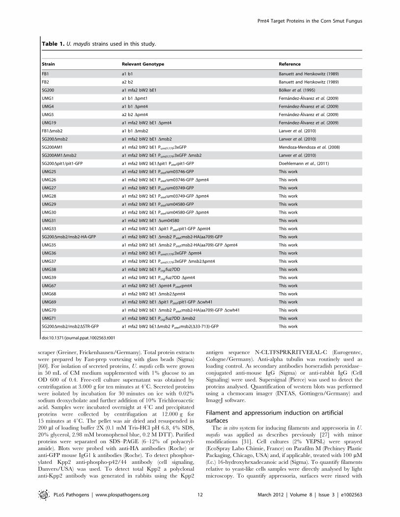
Table 1. U. maydis strains used in this study.
Strain Relevant Genotype Reference
FB1 a1 b1 Banuett and Herskowitz (1989)
FB2 a2 b2 Banuett and Herskowitz (1989)
SG200 a1 mfa2 bW2 bE1 Bolker et al. (1995)
UMG1 a1 b1 Dpmt1 Fernandez-Alvarez et al. (2009)
UMG4 a1 b1 Dpmt4 Fernandez-Alvarez et al. (2009)
UMG5 a2 b2 Dpmt4 Fernandez-Alvarez et al. (2009)
UMG19 a1 mfa2 bW2 bE1 Dpmt4 Fernandez-Alvarez et al. (2009)
FB1Dmsb2 a1 b1 Dmsb2 Lanver et al. (2010)
SG200Dmsb2 a1 mfa2 bW2 bE1 Dmsb2 Lanver et al. (2010)
SG200AM1 a1 mfa2 bW2 bE1 Pum01779:3xGFP Mendoza-Mendoza et al. (2008)
SG200AM1Dmsb2 a1 mfa2 bW2 bE1 Pum01779:3xGFP Dmsb2 Lanver et al. (2010)
SG200Dpit1/pit1-GFP a1 mfa2 bW2 bE1Dpit1 Potef:pit1-GFP Doehlemann et al., (2011)
UMG25 a1 mfa2 bW2 bE1 Potef:um03746-GFP This work
UMG26 a1 mfa2 bW2 bE1 Potef:um03746-GFP Dpmt4 This work
UMG27 a1 mfa2 bW2 bE1 Potef:um03749-GFP This work
UMG28 a1 mfa2 bW2 bE1 Potef:um03749-GFP Dpmt4 This work
UMG29 a1 mfa2 bW2 bE1 Potef:um04580-GFP This work
UMG30 a1 mfa2 bW2 bE1 Potef:um04580-GFP Dpmt4 This work
UMG31 a1 mfa2 bW2 bE1 Dum04580 This work
UMG33 a1 mfa2 bW2 bE1 Dpit1 Potef:pit1-GFP Dpmt4 This work
SG200Dmsb2/msb2-HA-GFP a1 mfa2 bW2 bE1 Dmsb2 Potef:msb2-HA(aa709)-GFP This work
UMG35 a1 mfa2 bW2 bE1 Dmsb2 Potef:msb2-HA(aa709)-GFP Dpmt4 This work
UMG36 a1 mfa2 bW2 bE1 Pum01779:3xGFP Dpmt4 This work
UMG37 a1 mfa2 bW2 bE1 Pum01779:3xGFP Dmsb2Dpmt4 This work
UMG38 a1 mfa2 bW2 bE1 Pcrg:fuz7DD This work
UMG39 a1 mfa2 bW2 bE1 Pcrg:fuz7DD Dpmt4 This work
UMG67 a1 mfa2 bW2 bE1 Dpmt4 Potef:pmt4 This work
UMG68 a1 mfa2 bW2 bE1 Dmsb2Dpmt4 This work
UMG69 a1 mfa2 bW2 bE1 Dpit1 Potef:pit1-GFP Dcwh41 This work
UMG70 a1 mfa2 bW2 bE1 Dmsb2 Potef:msb2-HA(aa709)-GFP Dcwh41 This work
UMG71 a1 mfa2 bW2 bE1 Pcrg:fuz7DD Dmsb2 This work
SG200Dmsb2/msb2DSTR-GFP a1 mfa2 bW2 bE1Dmsb2 Potef:msb2(D33-713)-GFP This work
doi:10.1371/journal.ppat.1002563.t001
Pmt4 Target Proteins in the Corn Smut Fungus
PLoS Pathogens | www.plospathogens.org 12 March 2012 | Volume 8 | Issue 3 | e1002563
water and later, stained with calcofluor white (1 mg ml21) (Sigma).
To quantify the production of appressoria, filaments stained with
calcofluor white were counted relative to filaments showing eGFP
fluorescence. Data are expressed as means 6SEM of triplicate
samples. Statistical significance was assessed using Statistical
Calculators (http://www.graphpad.com/quickcalcs/index.cfm)
and considered significant if P values were ,0.05.
Scanning Electron MicroscopyThe SEM analyses were performed on PHILIPS XL30
microscope. Infected leaves from maize two days post-infection
were fixed with 4% glutaraldehyde in cacodylate (0.1 M pH 7.4) at
4uC. The samples were washed several times in cacodylate.
Afterwards, a second fixation in 1% tetraoxide osmium was
carried out for two hours at 4uC. This was washed in cacodylate
containing 7.5% of sucrose. Dehydration with acetone at 4uC:
30 minutes with 70% acetone, 30 minutes with 80% acetone,
30 minutes 90% acetone and finally, one hour with 100% acetone.
The samples were then introduced into an SPI-Dry Critical Point
Drying apparatus and coated in gold.
Cellular adhesion assaysTo ascertain the effects of the deletion of pmt4 and msb2 on
cellular adhesion, we grew the strains to exponential phase in
YEPSL at 28uC and the cells were spotted on starch agar plates.
Starch medium contains, 0.25% starch from potato, 0.1%
ammonium sulphate, 0.1% sucrose and phosphate buffer
25 mM pH 7. Plates were incubated for three days at 28uC and
cellular colonies were rinsed with water to analyse their adhesion
properties.
Calcofluor white and chlorazol black E stainingTo analyze the pre-penetration stages of U. maydis using
fluorescence microscopy, cells were stained with calcofluor white.
Post-penetration stages were studied by optical microscopy of
chlorazol black E stained leaf samples as previously described [29].
MicroscopyCells were examined using a Leica fluorescence microscope,
equipped with a PlanApo 6100 lens. Analysis of the pre-
penetration stages was done using a Deltavision widefield
microscope (Applied Precision, Issaquah, WA). Image deconvolu-
tion was performed using z-series of between 7 and 23 focal planes,
acquired at 0.5 mm intervals. Image processing was carried out
using Adobe Photoshop CS5 and ImageJ.
Accession numbersU. maydis sequence data can be found in the UniProt data
library under accession numbers UniProt:Q4P380 for Pmt4,
UniProt:Q4P140 for Pmt1, UniProt:Q4PAX9 for Cwh41, Uni-
Prot:Q4PHD3 for Msb2, UniProt:Q9UQY5 for Kpp2/Ubc3,
UniProt:Q4PC32 for Kpp6, UniProt:Q8J230 for Kpp4/Ubc4,
UniProt:Q99078 for Fuz7/Ubc5, UniProt:Q4PET9 for Pit1,
UniProt:Q4PF78 for Um01235, UniProt:Q4P817 for Um03746,
UniProt:Q4P814 for Um03749 and UniProt:Q4P5N3 for
Um04580.
Supporting Information
Figure S1 The U. maydis life’s pathogenic cycle. The
sexual pathogenic cycle of U. maydis starts with the mating between
two sexually compatible strains on the plant surface to form a
dikaryon filament. A set of combined physical-chemical plant-
derived signals leads to hyphae differentiation into appressoria
which mediate plant penetration (1). Once inside the plant, the
fungus proliferates as mycelium developing the clamp-like cells
which ensure the maintenance of the dikaryotic state (2). During
this infection process, U. maydis induces the tumor formation in the
plant maize (3). Pmt4 is required for appressorium formation and
penetration, and thus the Dpmt4 strain is unable to proliferate
inside the plant tissues neither induce tumors.
(TIFF)
Figure S2 Enrichment analysis for FunCatDB (MUMDBMIPS) of Pmt4 putative target proteins.
(TIFF)
Figure S3 Um04580 is not required for U. maydispathogenic development. Plants were infected with the strains
indicated and symptoms were scored 12 days post-infection. N
indicates the total number of plants evaluated in each case.
(TIFF)
Figure S4 Pit1 and Msb2 processing in the cwh41mutant. A. Western blot analysis of Pit1 tagged with GFP in
the SG200 (WT), SG200Dpmt4 and SG200Dcwh41 backgrounds.
a-GFP antibody was used to detect the Pit1-GFP protein. We did
not observe the bands that correspond to the glycosylated fraction
of the protein in the cwh41 mutant. Thus, Pit1 processing depends
on protein N- and O-glycosylation pathways. B. Western Blot
analysis of Msb2-HA-GFP isolated from SG200Dmsb2/
msb2-HA-GFP (WT), SG200Dmsb2Dpmt4/msb2-HA-GFP and
SG200Dmsb2Dcwh41/msb2-HA-GFP. a-HA antibody was used
to detect the N-terminal part of Msb2. Equal amounts of proteins
of total cell extracts were loaded in each lane. The deletion of
cwh41 does not affect significantly the Msb2 mobility.
(TIFF)
Figure S5 N-terminal domain of U. maydis Msb2 issecreted. Western Blot analysis of Msb2-HA-GFP isolated from
SG200Dmsb2/msb2-HA-GFP culture supernatant (S) and total
extract (TE). SG200 was used as a negative control. The first gel
(above) was used to detect the N-terminal part of Msb2 with a-HA
antibody. The other gel (below) was treated with a-GFP antibody
to detect the C-terminus of Msb2. The extracellular N-terminal
domain of U. maydis Msb2 was identified in the culture
supernatant, while the C-terminal fragment was exclusively
detected in the cellular fraction.
(TIFF)
Figure S6 Expression of fuz7DD partially restores themsb2 mutant phenotypes. A. Seven days old maize seedlings
were infected with the strains SG200 (WT), SG200fuz7DD,
SG200Dmsb2 and SG200fuz7DDDmsb2 scoring appressoria
production 15 hours later (.100 filaments in each case). Data
are shown as mean values 6SEM. Asterisk indicates statistically
significant differences between wild-type (control) and Dmsb2
strains, P value#0.001. B. Symptoms in infected plants with the
strains indicated were scored 12 days post-infection. N indicates
the total number of plants evaluated in each case.
(TIFF)
Figure S7 Kpp2 phosphorylation in Dpmt4 and Dmsb2strains. A. The WT (SG200), Dpmt4 and Dmsb2 strains were
incubated on parafilm M with 100 mM 16-hydroxyhexadecanoic
acid for 10 h. Total proteins isolated before (left) and after
incubation on parafilm M (right) were subjected to western blot
analysis. The phosphorylated form of Kpp2 (P-Kpp2) and total
Kpp2 were detected using a-phospho-p44/42 antibody and a-
Kpp2 antibody, respectively. Asterisk denotes an unspecific
background signal. B. The relative Kpp2 phosphorylation from
Pmt4 Target Proteins in the Corn Smut Fungus
PLoS Pathogens | www.plospathogens.org 13 March 2012 | Volume 8 | Issue 3 | e1002563
three independent experiments. Kpp2 phosphorylation in WT
(SG200) was set to 1. The datasets from Dpmt4 and Dmsb2 strains
were compared with the wild-type dataset to calculate p-values (t-
test) given above column. Error bars indicate standard deviation.
(TIFF)
Figure S8 Msb2 is not required for cellular adhesion tosolid surfaces. The strains indicated were grown to A600 = 0.5
in YEPSL liquid medium and then were spotted on starch medium
plates and incubated for three days at 28uC. The deletion of msb2
does not affect the fungal cell adhesion to solid surfaces in U.
maydis.
(TIFF)
Figure S9 Working model of the possible divergentfunction of the Ser/Thr rich region of Msb2 in S.cerevisiae and phytopathogenic fungi such as U. maydis.The conserved plasma membrane protein Msb2 acts upstream of
MAP kinase cascades in fungi which regulates pseudohyphal
growth in S. cerevisiae and appressorium development in U. maydis.
In wild-type conditions (left) the extracellular domain of Msb2 is
O-mannosylated by Pmt4. The absence of Pmt4 could have a
divergent effect on the activation of the pathway in S. cerevisiae and
U. maydis (see discussion).
(TIFF)
Table S1 List of U. maydis proteins containing, at least,one plasma membrane anchoring and a window of 20 aawhere the percentage of Ser/Thr is $40%. The position of
the window and the percentage on 1 is shown.
(XLS)
Table S2 List of U. maydis proteins containing, at least,plasma membrane anchoring and a window of 40 aawhere the percentage of Ser/Thr is $40%. The position of
the window and the percentage on 1 is shown.
(XLS)
Table S3 List of U. maydis one trasmembrane proteinscontaining the window of 40 aa where the percentage ofSer/Thr is $40% in the luminal part. The position of the
window and the percentage on 1 is shown.
(XLS)
Table S4 Primers used in this study.
(DOC)
Acknowledgments
We would like to thank the Genetics department for their useful discussions
and comments, Vıctor Manuel Carranco Fabre for the graphic design in
the figures and Sandra Romero for the technical assistant. We also thank
Philip Muller for his help to generate the anti-Kpp2-antibody and Stefanie
Reissmann for fruitful discussions and for critical reading of the
manuscript. We thank all Ustilago community for providing strains and
plasmids.
Author Contributions
Conceived and designed the experiments: AFA DL AEV RK JII.
Performed the experiments: AFA MMM DL AJM AEV AJPP. Analyzed
the data: AFA DL AEV RK JII. Contributed reagents/materials/analysis
tools: AFA DL AEV RK JII. Wrote the paper: AFA DL AEV RK JII.
References
1. Lehle L, Strahl S, Tanner W (2006) Protein glycosylation, conserved from yeast
to man: A model organism helps elucidate congenital human diseases. Angew
Chem Int Ed Engl 45: 6802–6818.
2. Helenius A, Aebi M (2001) Intracellular functions of N-linked glycans. Science
291: 2364–2369.
3. Lengeler KB, Tielker D, Ernst JF (2008) Protein-O-mannosyltransferases in
virulence and development. Cell Mol Life Sci 65: 528–544.
4. Olson GM, Fox DS, Wang P, Alspaugh JA, Buchanan KL (2007) Role of
protein O-mannosyltransferase Pmt4 in the morphogenesis and virulence of
cryptococcus neoformans. Eukaryot Cell 6: 222–234.
5. Rouabhia M, Schaller M, Corbucci C, Vecchiarelli A, Prill SK, et al. (2005)
Virulence of the fungal pathogen candida albicans requires the five isoforms of
protein mannosyltransferases. Infect Immun 73: 4571–4580.
6. Willer T, Valero MC, Tanner W, Cruces J, Strahl S (2003) O-mannosyl glycans:
From yeast to novel associations with human disease. Curr Opin Struct Biol 13:
621–630.
7. Girrbach V, Strahl S (2003) Members of the evolutionarily conserved PMT
family of protein O-mannosyltransferases form distinct protein complexes
among themselves. J Biol Chem 278: 12554–12562.
8. Hutzler J, Schmid M, Bernard T, Henrissat B, Strahl S (2007) Membrane
association is a determinant for substrate recognition by PMT4 protein O-
mannosyltransferases. Proc Natl Acad Sci U S A 104: 7827–7832.
9. Gentzsch M, Tanner W (1996) The PMT gene family: Protein O-glycosylation
in saccharomyces cerevisiae is vital. EMBO J 15: 5752–5759.
10. Prill SK, Klinkert B, Timpel C, Gale CA, Schroppel K, et al. (2005) PMT family
of candida albicans: Five protein mannosyltransferase isoforms affect growth,
morphogenesis and antifungal resistance. Mol Microbiol 55: 546–560.
11. Fernandez-Alvarez A, Elias-Villalobos A, Ibeas JI (2009) The O-mannosyl-
transferase PMT4 is essential for normal appressorium formation and
penetration in ustilago maydis. Plant Cell 21: 3397–3412.
12. Goto M, Harada Y, Oka T, Matsumoto S, Takegawa K, et al. (2009) Protein O-
mannosyltransferases B and C support hyphal development and differentiation
in aspergillus nidulans. Eukaryot Cell 8: 1465–1474.
13. Willer T, Brandl M, Sipiczki M, Strahl S (2005) Protein O-mannosylation is
crucial for cell wall integrity, septation and viability in fission yeast. Mol
Microbiol 57: 156–170.
14. Willger SD, Ernst JF, Alspaugh JA, Lengeler KB (2009) Characterization of the
PMT gene family in cryptococcus neoformans. PLoS One 4: e6321.
15. Akasaka-Manya K, Manya H, Nakajima A, Kawakita M, Endo T (2006)
Physical and functional association of human protein O-mannosyltransferases 1
and 2. J Biol Chem 281: 19339–19345.
16. Grewal PK, Hewitt JE (2003) Glycosylation defects: A new mechanism for
muscular dystrophy? Hum Mol Genet 12 Spec No 2: R259–64.
17. Manya H, Chiba A, Margolis RU, Endo T (2006) Molecular cloning and
characterization of rat Pomt1 and Pomt2. Glycobiology 16: 863–873.
18. Martin-Blanco E, Garcia-Bellido A (1996) Mutations in the rotated abdomen
locus affect muscle development and reveal an intrinsic asymmetry in
drosophila. Proc Natl Acad Sci U S A 93: 6048–6052.
19. Mouyna I, Kniemeyer O, Jank T, Loussert C, Mellado E, et al. (2010) Members
of protein O-mannosyltransferase family in aspergillus fumigatus differentially
affect growth, morphogenesis and viability. Mol Microbiol 76: 1205–1221.
20. Bowman SM, Free SJ (2006) The structure and synthesis of the fungal cell wall.
Bioessays 28: 799–808.
21. Gentzsch M, Tanner W (1997) Protein-O-glycosylation in yeast: Protein-specific
mannosyltransferases. Glycobiology 7: 481–486.
22. Proszynski TJ, Simons K, Bagnat M (2004) O-glycosylation as a sorting
determinant for cell surface delivery in yeast. Mol Biol Cell 15: 1533–1543.
23. Weber Y, Prill SK, Ernst JF (2004) Pmt-mediated O mannosylation stabilizes an
essential component of the secretory apparatus, Sec20p, in candida albicans.
Eukaryot Cell 3: 1164–1168.
24. Brefort T, Doehlemann G, Mendoza-Mendoza A, Reissmann S, Djamei A,
et al. (2009) Ustilago maydis as a pathogen. Annu Rev Phytopathol 47: 423–445.
25. Kamper J, Kahmann R, Bolker M, Ma LJ, Brefort T, et al. (2006) Insights from
the genome of the biotrophic fungal plant pathogen ustilago maydis. Nature 444:
97–101.
26. Banuett F (1995) Genetics of ustilago maydis, a fungal pathogen that induces
tumors in maize. Annu Rev Genet 29: 179–208.
27. Mendoza-Mendoza A, Berndt P, Djamei A, Weise C, Linne U, et al. (2009)
Physical-chemical plant-derived signals induce differentiation in ustilago maydis.
Mol Microbiol 71: 895–911.
28. Mayorga ME, Gold SE (1999) A MAP kinase encoded by the ubc3 gene of
ustilago maydis is required for filamentous growth and full virulence. Mol
Microbiol 34: 485–497.
29. Brachmann A, Schirawski J, Muller P, Kahmann R (2003) An unusual MAP
kinase is required for efficient penetration of the plant surface by ustilago maydis.
EMBO J 22: 2199–2210.
30. Muller P, Aichinger C, Feldbrugge M, Kahmann R (1999) The MAP kinase
kpp2 regulates mating and pathogenic development in ustilago maydis. Mol
Microbiol 34: 1007–1017.
31. Lanver D, Mendoza-Mendoza A, Brachmann A, Kahmann R (2010) Sho1 and
Msb2-related proteins regulate appressorium development in the smut fungus
ustilago maydis. Plant Cell 22: 2085–2101.
Pmt4 Target Proteins in the Corn Smut Fungus
PLoS Pathogens | www.plospathogens.org 14 March 2012 | Volume 8 | Issue 3 | e1002563

32. Andrews DL, Egan JD, Mayorga ME, Gold SE (2000) The ustilago maydis ubc4
and ubc5 genes encode members of a MAP kinase cascade required forfilamentous growth. Mol Plant Microbe Interact 13: 781–786.
33. Banuett F, Herskowitz I (1994) Identification of fuz7, a ustilago maydis MEK/
MAPKK homolog required for a-locus-dependent and -independent steps in thefungal life cycle. Genes Dev 8: 1367–1378.
34. Muller P, Weinzierl G, Brachmann A, Feldbrugge M, Kahmann R (2003)Mating and pathogenic development of the smut fungus ustilago maydis are
regulated by one mitogen-activated protein kinase cascade. Eukaryot Cell 2:
1187–1199.35. Schirawski J, Mannhaupt G, Munch K, Brefort T, Schipper K, et al. (2010)
Pathogenicity determinants in smut fungi revealed by genome comparison.Science 330: 1546–1548.
36. Banuett F, Herskowitz I (1996) Discrete developmental stages during teliosporeformation in the corn smut fungus, ustilago maydis. Development 122:
2965–2976.
37. Doehlemann G, Wahl R, Vranes M, de Vries RP, Kamper J, et al. (2008)Establishment of compatibility in the ustilago maydis/maize pathosystem. J Plant
Physiol 165: 29–40.38. Doehlemann G, Reissmann S, Assmann D, Fleckenstein M, Kahmann R (2011)
Two linked genes encoding a secreted effector and a membrane protein are
essential for ustilago maydis-induced tumour formation. Mol Microbiol 81:751–66.
39. Spellig T, Bottin A, Kahmann R (1996) Green fluorescent protein (GFP) as anew vital marker in the phytopathogenic fungus ustilago maydis. Mol Gen Genet
252: 503–509.40. Schirawski J, Bohnert HU, Steinberg G, Snetselaar K, Adamikowa L, et al.
(2005) Endoplasmic reticulum glucosidase II is required for pathogenicity of
ustilago maydis. Plant Cell 17: 3532–3543.41. Fernandez-Alvarez A, Elias-Villalobos A, Ibeas JI (2010) Protein glycosylation in
the phytopathogen ustilago maydis: From core oligosaccharide synthesis to theER glycoprotein quality control system, a genomic analysis. Fungal Genet Biol
47: 727–735.
42. Liu W, Zhou X, Li G, Li L, Kong L, et al. (2011) Multiple plant surface signalsare sensed by different mechanisms in the rice blast fungus for appressorium
formation. PLoS Pathog 7: e1001261.43. Perez-Nadales E, Di Pietro A (2011) The membrane mucin Msb2 regulates
invasive growth and plant infection in fusarium oxysporum. Plant Cell 23:1171–1185.
44. Yang HY, Tatebayashi K, Yamamoto K, Saito H (2009) Glycosylation defects
activate filamentous growth Kss1 MAPK and inhibit osmoregulatory Hog1MAPK. EMBO J 28: 1380–1391.
45. Vadaie N, Dionne H, Akajagbor DS, Nickerson SR, Krysan DJ, et al. (2008)Cleavage of the signaling mucin Msb2 by the aspartyl protease Yps1 is required
for MAPK activation in yeast. J Cell Biol 181: 1073–1081.
46. Cullen PJ, Sabbagh W, Jr., Graham E, Irick MM, van Olden EK, et al. (2004) A
signaling mucin at the head of the Cdc42- and MAPK-dependent filamentous
growth pathway in yeast. Genes Dev 18: 1695–1708.
47. Bottin A, Kamper J, Kahmann R (1996) Isolation of a carbon source-regulated
gene from ustilago maydis. Mol Gen Genet 253: 342–352.
48. Scherer M, Heimel K, Starke V, Kamper J (2006) The Clp1 protein is required
for clamp formation and pathogenic development of ustilago maydis. Plant Cell
18: 2388–2401.
49. Talbot NJ (2003) On the trail of a cereal killer: Exploring the biology of
magnaporthe grisea. Annu Rev Microbiol 57: 177–202.
50. Tucker SL, Talbot NJ (2001) Surface attachment and pre-penetration stage
development by plant pathogenic fungi. Annu Rev Phytopathol 39: 385–417.
51. Doehlemann G, van der Linde K, Assmann D, Schwammbach D, Hof A, et al.
(2009) Pep1, a secreted effector protein of ustilago maydis, is required for
successful invasion of plant cells. PLoS Pathog 5: e1000290.
52. Timpel C, Zink S, Strahl-Bolsinger S, Schroppel K, Ernst J (2000)
Morphogenesis, adhesive properties, and antifungal resistance depend on the
Pmt6 protein mannosyltransferase in the fungal pathogen candida albicans.
J Bacteriol 182: 3063–3071.
53. Timpel C, Strahl-Bolsinger S, Ziegelbauer K, Ernst JF (1998) Multiple functions
of Pmt1p-mediated protein O-mannosylation in the fungal pathogen candida
albicans. J Biol Chem 273: 20837–20846.
54. Brachmann A, Konig J, Julius C, Feldbrugge M (2004) A reverse genetic
approach for generating gene replacement mutants in ustilago maydis. Mol
Genet Genomics 272: 216–226.
55. Banuett F, Herskowitz I (1989) Different a alleles of ustilago maydis are
necessary for maintenance of filamentous growth but not for meiosis. Proc Natl
Acad Sci U S A 86: 5878–5882.
56. Bolker M, Genin S, Lehmler C, Kahmann R (1995) Genetic regulation of
mating, and dimorphism in ustilago maydis. Can J Bot 73: 320–325.
57. Sambrook J, Frisch EF, Maniatis T (1989) Molecular cloning: A laboratory
manual. Cold Spring HarbourNew York: Cold Spring Harbour Laboratory
Press.
58. Hoffman CS, Winston F (1987) A ten-minute DNA preparation from yeast
efficiently releases autonomous plasmids for transformation of escherichia coli.
Gene 57: 267–272.
59. Schulz B, Banuett F, Dahl M, Schlesinger R, Schafer W, et al. (1990) The b
alleles of U. maydis, whose combinations program pathogenic development,
code for polypeptides containing a homeodomain-related motif. Cell 60:
295–306.
60. Moreno S, Klar A, Nurse P (1991) Molecular genetic analysis of fission yeast
schizosaccharomyces pombe. Methods Enzymol 194: 795–823.
Pmt4 Target Proteins in the Corn Smut Fungus
PLoS Pathogens | www.plospathogens.org 15 March 2012 | Volume 8 | Issue 3 | e1002563