Identification of Internal Parasites of Sheep and Goats
29
The University of Maine The University of Maine DigitalCommons@UMaine DigitalCommons@UMaine Honors College 5-2012 Identification of Internal Parasites of Sheep and Goats Identification of Internal Parasites of Sheep and Goats Amanda Chaney [email protected] Follow this and additional works at: https://digitalcommons.library.umaine.edu/honors Part of the Animal Sciences Commons, and the Veterinary Medicine Commons Recommended Citation Recommended Citation Chaney, Amanda, "Identification of Internal Parasites of Sheep and Goats" (2012). Honors College. 26. https://digitalcommons.library.umaine.edu/honors/26 This Honors Thesis is brought to you for free and open access by DigitalCommons@UMaine. It has been accepted for inclusion in Honors College by an authorized administrator of DigitalCommons@UMaine. For more information, please contact [email protected].
Transcript of Identification of Internal Parasites of Sheep and Goats
The University of Maine The University of Maine
DigitalCommons@UMaine DigitalCommons@UMaine
Honors College
5-2012
Identification of Internal Parasites of Sheep and Goats Identification of Internal Parasites of Sheep and Goats
Amanda Chaney [email protected]
Follow this and additional works at: https://digitalcommons.library.umaine.edu/honors
Part of the Animal Sciences Commons, and the Veterinary Medicine Commons
Recommended Citation Recommended Citation Chaney, Amanda, "Identification of Internal Parasites of Sheep and Goats" (2012). Honors College. 26. https://digitalcommons.library.umaine.edu/honors/26
This Honors Thesis is brought to you for free and open access by DigitalCommons@UMaine. It has been accepted for inclusion in Honors College by an authorized administrator of DigitalCommons@UMaine. For more information, please contact [email protected].
IDENTIFICATION OF INTERNAL PARASITES OF SHEEP AND GOATS
by
Amanda Chaney
A Thesis Submitted in Partial Fulfillment
of the Requirements for a Degree with Honors
(Animal and Veterinary Science)
The Honors College
University of Maine
May 2012
Advisory Committee:
James Weber, Associate Professor and Chair and Program Leader of Veterinary
Sciences, Advisor
Anne Lichtenwalner, Assistant Professor and Extension Veterinarian and
Director, University of Maine Animal Health Laboratory, Advisor
Robert Causey, Associate Professor
David Marcinkowski, Extension Dairy Specialist & Associate Professor
Mimi Killinger, Rezendes Preceptor for the Arts
Abstract
Abomasal worms are a major cause of small ruminant disease. Differentiation of
the most pathogenic nematode, H. contortus, from the other common species can be
difficult using standard diagnostic fecal floatation techniques because the ova are similar
in size and morphology. Known pure culture H. contortus fecal samples from West
Virginia University were used to develop morphologic assays using FITC-labeled lectin
agglutination and immunocytochemistry to identify species of abomasal worms. These
assays were applied to assess disease due to abomasal worms on selected small ruminant
farms in Maine. The diagnostic tests were used to test the hypothesis that H. contortus is
the most common internal parasite found on sheep and goat farms in Maine.
iii
Acknowledgements
The author would like to acknowledge Dr. James Weber for his mentorship
throughout the project. Completion of this research would not have been possible
without his guidance and support. The author would also like to thank Anne
Lichtenwalner for her aid in organization of the research and for her assistance in the
presentation of scientific information.
The author would like to acknowledge Mimi Killinger for her assistance with the
reading list and preparation for the defense. The author would also like to thank Robert
Causey and David Markinkowski for their input on research design and for offering their
time to serve on the thesis committee.
Finally, the author would like to thank Martin Stokes and Clare Thomas for their
guidance in writing a scientific paper.
iv
Table of Contents
Tables and Figures.................................................................................................... v
Introduction .............................................................................................................. 1
Materials and Methods ............................................................................................. 8
Results and Discussion ........................................................................................... 14
Conclusion ............................................................................................................. 19
Implications............................................................................................................ 20
References .............................................................................................................. 21
Appendices ............................................................................................................. 22
Author’s Biography ................................................................................................ 23
v
List of Tables, Figures, and Definitions
Fig. 1. Nematode ova 1
Fig. 2. The life cycle of a nematode 2
Fig. 3. Head region of H. contortus,
showing teeth located in buccal cavity
3
Fig. 4. Mucus membrane of anemic sheep 4
Fig. 5. Bottle jaw in sheep 4
Fig. 6. Ostertagia L4 in gastric gland of
abomasum
5
Fig. 7. Modified McMasters Chamber 9
Fig. 8. Baermann larval collection
technique
10
Table 1. Results of total fecal egg count
for Old Olk Farm in Maxfield, Maine.
14
Fig. 9. Fresh FITC-labeled H. contortus
ova without (A) and with (B) UV light.
16
Fig. 10. Formalin FITC-labeled H.
contortus ova without (A) and with (B)
UV light
17
Table 2. FITC-labeled lectin agglutination
test results for WVU ova
17
Fig. 11. H. contortus ova treated with
PNA-HRP 0, 10, and 20 minutes after
TMB was added
19
1
Introduction
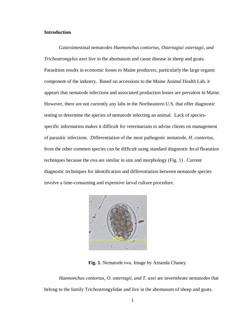
Gastrointestinal nematodes Haemonchus contortus, Ostertagiai ostertagii, and
Trichostrongylus axei live in the abomasum and cause disease in sheep and goats.
Parasitism results in economic losses to Maine producers, particularly the large organic
component of the industry. Based on accessions to the Maine Animal Health Lab, it
appears that nematode infections and associated production losses are prevalent in Maine.
However, there are not currently any labs in the Northeastern U.S. that offer diagnostic
testing to determine the species of nematode infecting an animal. Lack of species-
specific information makes it difficult for veterinarians to advise clients on management
of parasitic infections. Differentiation of the most pathogenic nematode, H. contortus,
from the other common species can be difficult using standard diagnostic fecal floatation
techniques because the ova are similar in size and morphology (Fig. 1) . Current
diagnostic techniques for identification and differentiation between nematode species
involve a time-consuming and expensive larval culture procedure.
Fig. 1. Nematode ova. Image by Amanda Chaney.
Haemonchus contortus, O. ostertagii, and T. axei are invertebrate nematodes that
belong to the family Trichostrongylidae and live in the abomasum of sheep and goats.
2
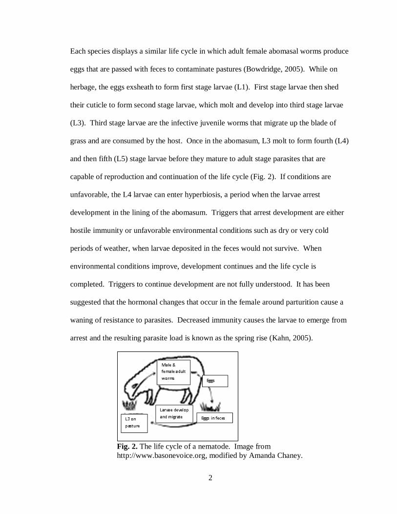
Each species displays a similar life cycle in which adult female abomasal worms produce
eggs that are passed with feces to contaminate pastures (Bowdridge, 2005). While on
herbage, the eggs exsheath to form first stage larvae (L1). First stage larvae then shed
their cuticle to form second stage larvae, which molt and develop into third stage larvae
(L3). Third stage larvae are the infective juvenile worms that migrate up the blade of
grass and are consumed by the host. Once in the abomasum, L3 molt to form fourth (L4)
and then fifth (L5) stage larvae before they mature to adult stage parasites that are
capable of reproduction and continuation of the life cycle (Fig. 2). If conditions are
unfavorable, the L4 larvae can enter hyperbiosis, a period when the larvae arrest
development in the lining of the abomasum. Triggers that arrest development are either
hostile immunity or unfavorable environmental conditions such as dry or very cold
periods of weather, when larvae deposited in the feces would not survive. When
environmental conditions improve, development continues and the life cycle is
completed. Triggers to continue development are not fully understood. It has been
suggested that the hormonal changes that occur in the female around parturition cause a
waning of resistance to parasites. Decreased immunity causes the larvae to emerge from
arrest and the resulting parasite load is known as the spring rise (Kahn, 2005).
Fig. 2. The life cycle of a nematode. Image from
http://www.basonevoice.org, modified by Amanda Chaney.
3
While the life cycles of H. contortus, O.ostertagii, and T. axei are very similar,
there are physiologically important differences in the life cycle which leads to differences
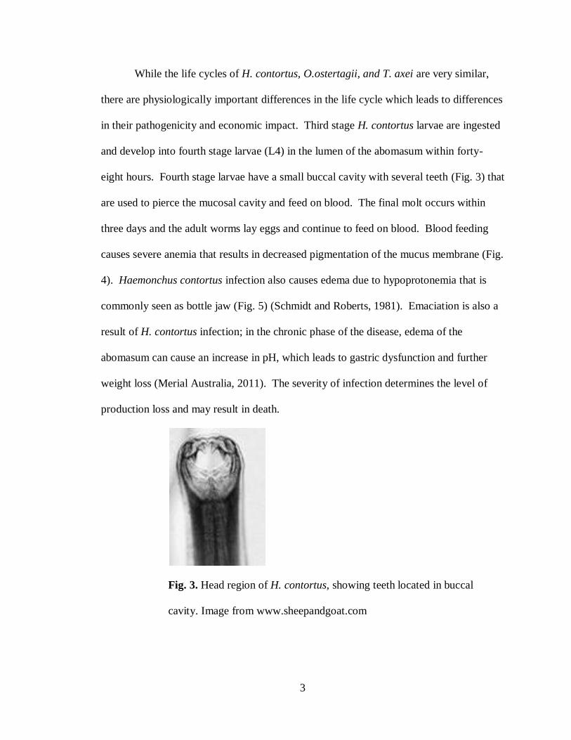
in their pathogenicity and economic impact. Third stage H. contortus larvae are ingested
and develop into fourth stage larvae (L4) in the lumen of the abomasum within forty-
eight hours. Fourth stage larvae have a small buccal cavity with several teeth (Fig. 3) that
are used to pierce the mucosal cavity and feed on blood. The final molt occurs within
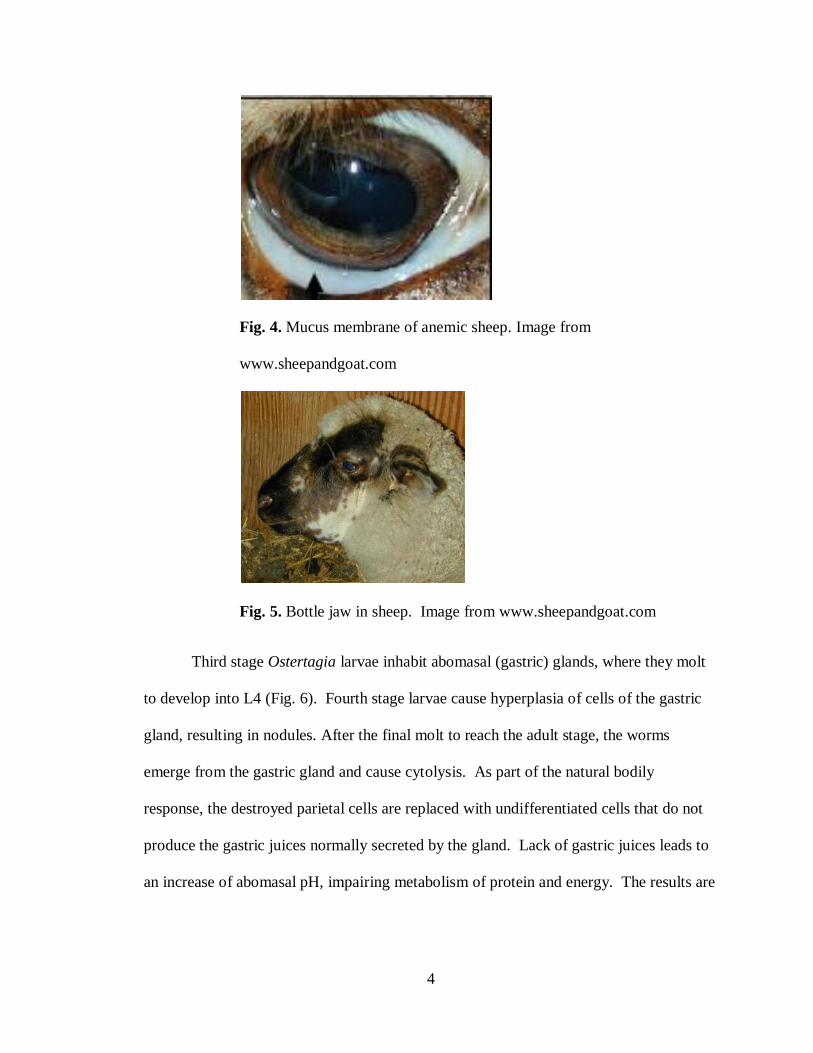
three days and the adult worms lay eggs and continue to feed on blood. Blood feeding
causes severe anemia that results in decreased pigmentation of the mucus membrane (Fig.
4). Haemonchus contortus infection also causes edema due to hypoprotonemia that is
commonly seen as bottle jaw (Fig. 5) (Schmidt and Roberts, 1981). Emaciation is also a
result of H. contortus infection; in the chronic phase of the disease, edema of the
abomasum can cause an increase in pH, which leads to gastric dysfunction and further
weight loss (Merial Australia, 2011). The severity of infection determines the level of
production loss and may result in death.
Fig. 3. Head region of H. contortus, showing teeth located in buccal
cavity. Image from www.sheepandgoat.com
4
Fig. 4. Mucus membrane of anemic sheep. Image from
www.sheepandgoat.com
Fig. 5. Bottle jaw in sheep. Image from www.sheepandgoat.com
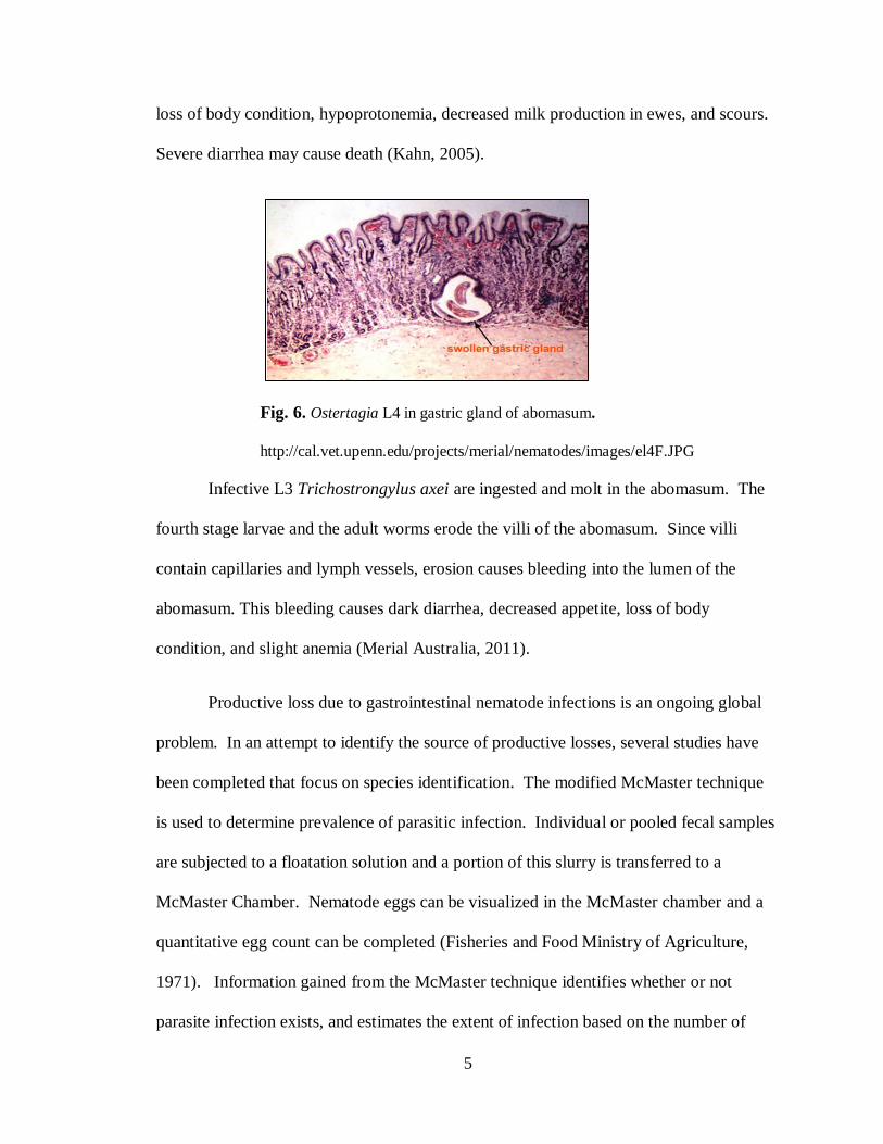
Third stage Ostertagia larvae inhabit abomasal (gastric) glands, where they molt
to develop into L4 (Fig. 6). Fourth stage larvae cause hyperplasia of cells of the gastric
gland, resulting in nodules. After the final molt to reach the adult stage, the worms
emerge from the gastric gland and cause cytolysis. As part of the natural bodily
response, the destroyed parietal cells are replaced with undifferentiated cells that do not
produce the gastric juices normally secreted by the gland. Lack of gastric juices leads to
an increase of abomasal pH, impairing metabolism of protein and energy. The results are
5
loss of body condition, hypoprotonemia, decreased milk production in ewes, and scours.
Severe diarrhea may cause death (Kahn, 2005).
Fig. 6. Ostertagia L4 in gastric gland of abomasum.
http://cal.vet.upenn.edu/projects/merial/nematodes/images/el4F.JPG
Infective L3 Trichostrongylus axei are ingested and molt in the abomasum. The
fourth stage larvae and the adult worms erode the villi of the abomasum. Since villi
contain capillaries and lymph vessels, erosion causes bleeding into the lumen of the
abomasum. This bleeding causes dark diarrhea, decreased appetite, loss of body
condition, and slight anemia (Merial Australia, 2011).
Productive loss due to gastrointestinal nematode infections is an ongoing global
problem. In an attempt to identify the source of productive losses, several studies have
been completed that focus on species identification. The modified McMaster technique
is used to determine prevalence of parasitic infection. Individual or pooled fecal samples
are subjected to a floatation solution and a portion of this slurry is transferred to a
McMaster Chamber. Nematode eggs can be visualized in the McMaster chamber and a
quantitative egg count can be completed (Fisheries and Food Ministry of Agriculture,
1971). Information gained from the McMaster technique identifies whether or not
parasite infection exists, and estimates the extent of infection based on the number of
6
eggs counted. Eggs of Haemonchus contortus, Ostertagia, and Trichostrongylus are
morphologically similar (Fig. 2), so it is not possible to identify the infective species
based on microscopic evaluation of Nematode eggs (Jurasek et al., 2010). In addition,
new research completed in the Kaplan lab at the University of Georgia has found that L3
morphology may not be a consistent and accurate method to differentiate between
Ostertagia and Trichostrongylys.
Since species identification cannot be determined from evaluation of Nematode
eggs, researchers in the 1970’s developed a technique in which they hatched the fecal
eggs and identified the species based on morphological differences of L3 larvae. Eggs
were incubated until L3 developmental stage was achieved. The L3 were then stained
with Grams Iodine and visualized under a microscope; species were identified based on
shape, internal structures of the head region, and tail morphology (Fisheries and Food
Ministry of Agriculture, 1971). Third stage larval isolation and identification is currently
the standard method used for identification and differentiation between Haemonchus,
Ostertagia, and Trichostrongylus. However, there are several downfalls to this
identification technique. The person completing the procedure must be trained to identify
morphological differences in larvae, and the incubation and identification is a time-
consuming and expensive process (Jurasek et al., 2010).
Recent studies have been conducted that aim to overcome the shortfalls of larval
identification. Researchers have identified three flourescein isothiocyanate (FITC)-
labeled lectins that bind to genus-specific carbohydrates on the surface of Haemonchus,
Ostertagia, or Trichostrongylus eggs. Osage orange seed agglutinin binds to the eggs of
H. contortus, O. circumcincta, and T. colubriformis. Jack bean agglutinin binds to the
7
eggs of H. contortus and O. circumcinus. Most importantly, Peanut agglutinin binds
selectively to H. contortus. Since the lectins have a fluorescent tag which can be
visualized under a fluorescent microscope, selective binding of Peanut agglutinin (PNA)
provides a rapid, simple, and less expensive identification technique for H. contortus
(Palmer and McCombe, 1996). Other studies have been completed to improve certain
aspects of the identification method. For example, Jurasek et al.(2010) conducted a study
that made adjustments to the PNA-FITC procedure to further reduce the time required for
identification of H. contortus.
Another recent study used an alternate technique for identifying anemic
individuals that may be infected with clinically relevant levels of H. contortus. Kaplan
et al. (2004) used the FAMACHA score system developed in South Africa to clinically
identify anemic sheep. In this system, the color of the ocular conjunctiva of sheep and
goats is compared with the images on the FAMACHA scoring card, which ranks ocular
conjunctiva color on a scale of one to five, with one being a red color of a healthy animal
and five being an almost white color of a severely anemic animal. Kaplan et al. (2004)
tested the reliability of the FAMACHA system by comparing FAMACHA evaluation of
animals with packed cell volume (PCV) and fecal egg counts (FEC) of the same animals.
The study concluded that there was a correlation between eye score, PCV, and FEC.
Anemic sheep display decreased PCV, increased FEC, and increased eye score. Since H.
contortus is the main parasite which causes severe anemia in sheep and goats, the
FAMACHA system can be used to identify animals infected with this nematode.
Identification of infected animals without extensive lab testing reduces costs while still
8
providing evidence of H. contortus infection; this information is useful in developing
treatment plans to reduce parasite prevalence.
Although recent research has developed assays for species-specific identification
of abomasal worms, these techniques are not available in labs in Maine. Gastrointestinal
nematode infections negatively impact the Maine sheep and goat industry. The objective
of this project was to streamline a diagnostic procedure for species-specific identification
that could be used by the University of Maine diagnostic lab to provide Maine famers
with an affordable service for parasite identification. This would benefit Maine sheep
and goat producers by allowing them to reduce parasitic disease on their farms, thus
increasing production.
Materials and Methods
Sheep fecal samples were obtained from Dr. Scott Bowdridge from the research
farm at West Virginia University (WVU) and from several commercial sheep and goat
farms in Maine. The WVU fecal samples were known positive for H. contortus. Each of
the following techniques was applied to each sample.
Total fecal egg count
The Modified McMasters technique was used to obtain a total fecal egg count.
To complete this technique, 2 grams of feces were weighed out and placed it in a 50 ml
centrifuge tube. A small amount of saturated sodium chloride solution (~15 ml) was
added and the feces were allowed to soak for one minute. The saturated sodium chloride
solution works to dissolve the feces and allow the ova to go into solution. A scoopula
was used to break up the fecal pellets, and the fecal solution was run through a mesh
9
strainer to further break up the feces if needed. Once the feces were well broken up,
more sodium chloride was added to bring the total volume to 30 ml. The McMasters
chamber was prepared by wetting it then gently tapping it on paper towel to remove
excess water. The centrifuge tube containing the fecal slurry was rocked back and forth
10 times and a pipette was used to draw up enough suspension to fill one chamber of the
McMasters slide. The rocking and pipetting procedure was repeated to fill the other side
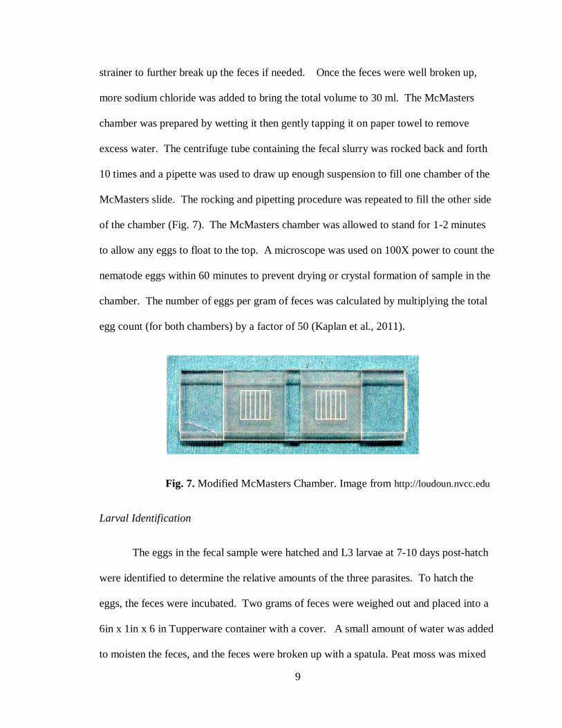
of the chamber (Fig. 7). The McMasters chamber was allowed to stand for 1-2 minutes
to allow any eggs to float to the top. A microscope was used on 100X power to count the
nematode eggs within 60 minutes to prevent drying or crystal formation of sample in the
chamber. The number of eggs per gram of feces was calculated by multiplying the total
egg count (for both chambers) by a factor of 50 (Kaplan et al., 2011).
Fig. 7. Modified McMasters Chamber. Image from http://loudoun.nvcc.edu
Larval Identification
The eggs in the fecal sample were hatched and L3 larvae at 7-10 days post-hatch
were identified to determine the relative amounts of the three parasites. To hatch the
eggs, the feces were incubated. Two grams of feces were weighed out and placed into a
6in x 1in x 6 in Tupperware container with a cover. A small amount of water was added
to moisten the feces, and the feces were broken up with a spatula. Peat moss was mixed

10
in to form a thin layer covering the bottom of the container. Water was sprinkled on top
until the mixture became moist. The covered container was incubated at 29oC for 7 days.
The Baermann larval collection technique (Fig. 8) was used to isolate the L3
larva from the feces. Motile L3 larvae were collected from the cultured fecal mix by
suspending 2 g of feces in a Kimwipe paper towel. The edges of the Kimwipe were
folded together, and the package was placed in a glass funnel filled w/warm water (the
funnel’s outlet was plugged to retain water). Larvae were collected from the bottom of
the funnel after 4-12 hours.
Fig. 8. Baermann larval collection technique. Image from
www.sheepandgoat.com
Grams iodine was added in a 1:1 ratio to the slide to stain free living nematodes
and to enhance visualization the internal structure of the L3 larvae. The sample was
viewed under the microscope to identify L3 of H. contortus, O. ostertagii, and T. axei.
Larval counts were used to determine the relative amount of each species causing parasite
infection.
11
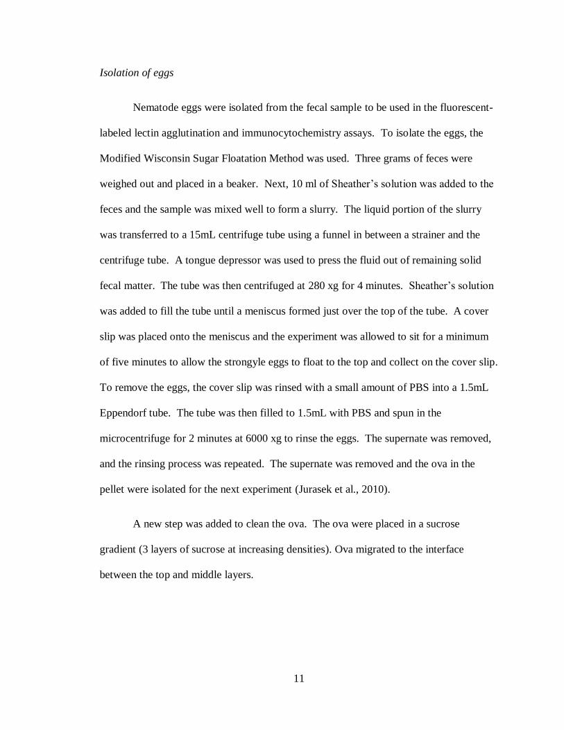
Isolation of eggs
Nematode eggs were isolated from the fecal sample to be used in the fluorescent-
labeled lectin agglutination and immunocytochemistry assays. To isolate the eggs, the
Modified Wisconsin Sugar Floatation Method was used. Three grams of feces were
weighed out and placed in a beaker. Next, 10 ml of Sheather’s solution was added to the
feces and the sample was mixed well to form a slurry. The liquid portion of the slurry
was transferred to a 15mL centrifuge tube using a funnel in between a strainer and the
centrifuge tube. A tongue depressor was used to press the fluid out of remaining solid
fecal matter. The tube was then centrifuged at 280 xg for 4 minutes. Sheather’s solution
was added to fill the tube until a meniscus formed just over the top of the tube. A cover
slip was placed onto the meniscus and the experiment was allowed to sit for a minimum
of five minutes to allow the strongyle eggs to float to the top and collect on the cover slip.
To remove the eggs, the cover slip was rinsed with a small amount of PBS into a 1.5mL
Eppendorf tube. The tube was then filled to 1.5mL with PBS and spun in the
microcentrifuge for 2 minutes at 6000 xg to rinse the eggs. The supernate was removed,
and the rinsing process was repeated. The supernate was removed and the ova in the
pellet were isolated for the next experiment (Jurasek et al., 2010).
A new step was added to clean the ova. The ova were placed in a sucrose
gradient (3 layers of sucrose at increasing densities). Ova migrated to the interface
between the top and middle layers.
12
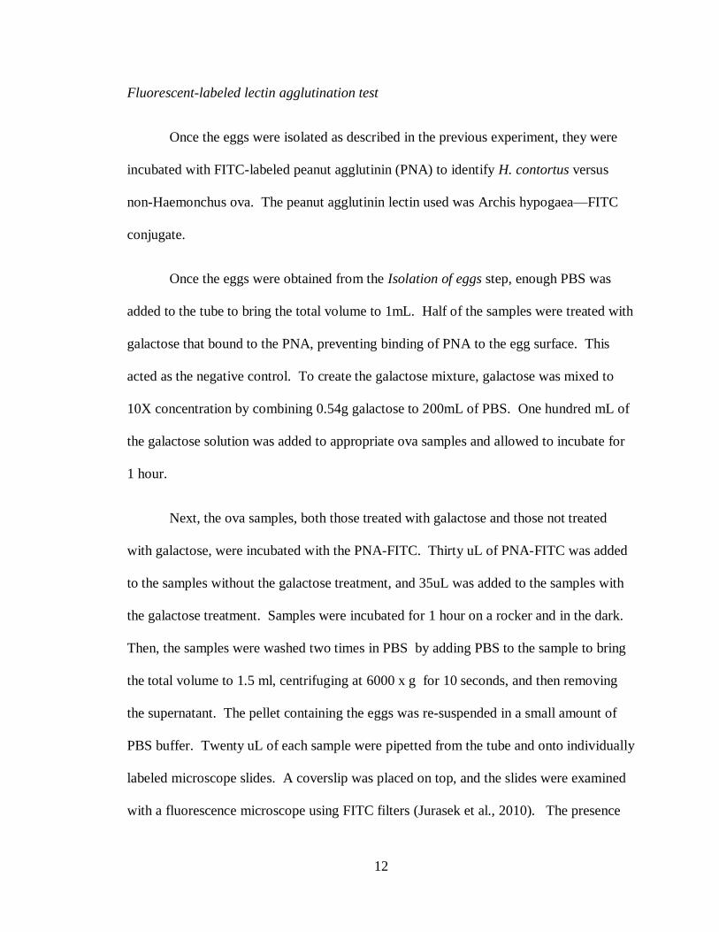
Fluorescent-labeled lectin agglutination test
Once the eggs were isolated as described in the previous experiment, they were
incubated with FITC-labeled peanut agglutinin (PNA) to identify H. contortus versus
non-Haemonchus ova. The peanut agglutinin lectin used was Archis hypogaea—FITC
conjugate.
Once the eggs were obtained from the Isolation of eggs step, enough PBS was
added to the tube to bring the total volume to 1mL. Half of the samples were treated with
galactose that bound to the PNA, preventing binding of PNA to the egg surface. This
acted as the negative control. To create the galactose mixture, galactose was mixed to
10X concentration by combining 0.54g galactose to 200mL of PBS. One hundred mL of
the galactose solution was added to appropriate ova samples and allowed to incubate for
1 hour.
Next, the ova samples, both those treated with galactose and those not treated
with galactose, were incubated with the PNA-FITC. Thirty uL of PNA-FITC was added
to the samples without the galactose treatment, and 35uL was added to the samples with
the galactose treatment. Samples were incubated for 1 hour on a rocker and in the dark.
Then, the samples were washed two times in PBS by adding PBS to the sample to bring
the total volume to 1.5 ml, centrifuging at 6000 x g for 10 seconds, and then removing
the supernatant. The pellet containing the eggs was re-suspended in a small amount of
PBS buffer. Twenty uL of each sample were pipetted from the tube and onto individually
labeled microscope slides. A coverslip was placed on top, and the slides were examined
with a fluorescence microscope using FITC filters (Jurasek et al., 2010). The presence
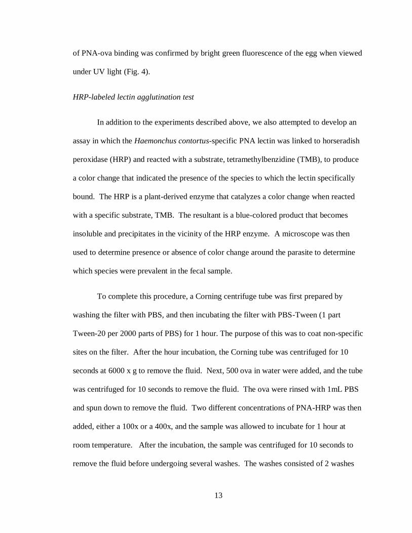
13
of PNA-ova binding was confirmed by bright green fluorescence of the egg when viewed
under UV light (Fig. 4).
HRP-labeled lectin agglutination test
In addition to the experiments described above, we also attempted to develop an
assay in which the Haemonchus contortus-specific PNA lectin was linked to horseradish
peroxidase (HRP) and reacted with a substrate, tetramethylbenzidine (TMB), to produce
a color change that indicated the presence of the species to which the lectin specifically
bound. The HRP is a plant-derived enzyme that catalyzes a color change when reacted
with a specific substrate, TMB. The resultant is a blue-colored product that becomes
insoluble and precipitates in the vicinity of the HRP enzyme. A microscope was then
used to determine presence or absence of color change around the parasite to determine
which species were prevalent in the fecal sample.
To complete this procedure, a Corning centrifuge tube was first prepared by
washing the filter with PBS, and then incubating the filter with PBS-Tween (1 part
Tween-20 per 2000 parts of PBS) for 1 hour. The purpose of this was to coat non-specific
sites on the filter. After the hour incubation, the Corning tube was centrifuged for 10
seconds at 6000 x g to remove the fluid. Next, 500 ova in water were added, and the tube
was centrifuged for 10 seconds to remove the fluid. The ova were rinsed with 1mL PBS
and spun down to remove the fluid. Two different concentrations of PNA-HRP was then
added, either a 100x or a 400x, and the sample was allowed to incubate for 1 hour at
room temperature. After the incubation, the sample was centrifuged for 10 seconds to
remove the fluid before undergoing several washes. The washes consisted of 2 washes
14
with PBS-Tween and 1 wash with PBS. To wash, 1mL of solution was added to the
sample, and the sample was centrifuged to remove the fluid. Once the washes were
completed, 0.5mL of the substrate, TMB, added. At time zero, and every five minutes
after, a drop of sample was viewed under the microscope. Once a blue shell formed
around the ova, the reaction was stopped by spinning down the tube, then rinsing once
with PBS-Tween and once with PBS. The ova were then resuspended in ~200uL of PBS
to obtain a concentrated sample for microscopic evaluation. Before transferring to a
slide, the ova were displaced from the membrane by gently pipetting up and down several
times before removing all fluid from the Corning centrifuge tube into a fresh tube.
Results and Discussion
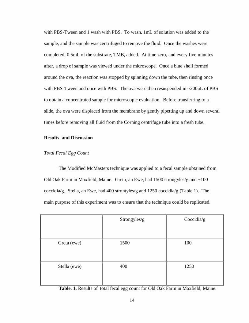
Total Fecal Egg Count
The Modified McMasters technique was applied to a fecal sample obtained from
Old Oak Farm in Maxfield, Maine. Greta, an Ewe, had 1500 strongyles/g and ~100
coccidia/g. Stella, an Ewe, had 400 strontyles/g and 1250 coccidia/g (Table 1). The
main purpose of this experiment was to ensure that the technique could be replicated.
Strongyles/g Coccidia/g
Greta (ewe) 1500 100
Stella (ewe) 400 1250
Table. 1. Results of total fecal egg count for Old Oak Farm in Maxfield, Maine.
15
Larval Identification
Feces from Old Oak Farm in Maxfield, Maine were placed in larval culture to
obtain L3 larvae for larval identification. For the first trial, 10g of feces were combined
with 10g of peat moss. When the sample was viewed under a microscope 7 days later,
there were many free-living nematodes. An explanation could be that the peat moss
contained many free-living nematodes that contaminated the sample.
For the second trial, the peat moss was first autoclaved to destroy any microbes
before being mixed with the feces. This resulted in a clean L3 sample, which was
beneficial for larval identification purposes.
The L3 larvae were combined with grams iodine and viewed under the
microscope at 40X to identify Haemonchus versus non-Haemonchus nematodes. It was
learned from the University of Georgia Parasitology Laboratory that larval identification
cannot be used to determine the difference between Trichostrongylus and Ostertagia as
was previously believed.
Fluorescent-labeled lectin agglutination test results: West Virginia University (WVU)
samples
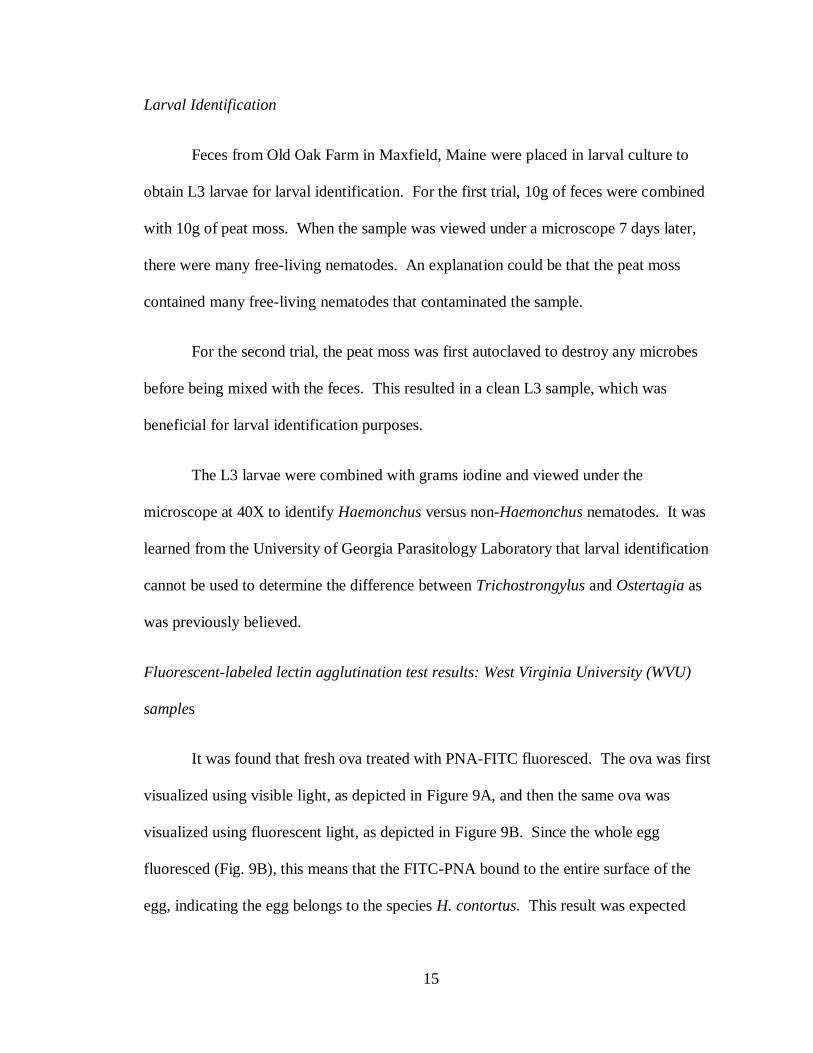
It was found that fresh ova treated with PNA-FITC fluoresced. The ova was first
visualized using visible light, as depicted in Figure 9A, and then the same ova was
visualized using fluorescent light, as depicted in Figure 9B. Since the whole egg
fluoresced (Fig. 9B), this means that the FITC-PNA bound to the entire surface of the
egg, indicating the egg belongs to the species H. contortus. This result was expected
16
since the sample was known H. contortus positive, and PNA binds specifically to H.
contortus.
Fig. 9. Fresh FITC-labeled H. contortus ova without (A) and with (B) UV light.
Image by Amanda Chaney
The fresh ova sample that was treated with galactose did not display fluorescence.
No fluorescence indicated that the galactose saturated the PNA, meaning the negative
control worked and that the PNA-FITC binding was specific for galactose residues on the
surface of the Haemonchus ova’s “egg shell.”
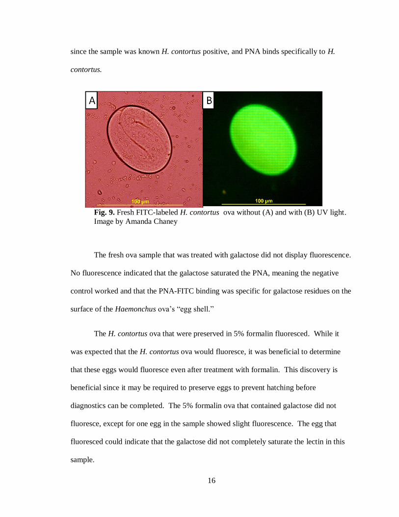
The H. contortus ova that were preserved in 5% formalin fluoresced. While it
was expected that the H. contortus ova would fluoresce, it was beneficial to determine
that these eggs would fluoresce even after treatment with formalin. This discovery is
beneficial since it may be required to preserve eggs to prevent hatching before
diagnostics can be completed. The 5% formalin ova that contained galactose did not
fluoresce, except for one egg in the sample showed slight fluorescence. The egg that
fluoresced could indicate that the galactose did not completely saturate the lectin in this
sample.
17
Fig. 10. Formalin FITC-labeled H. contortus ova without (A) and with (B) UV
light. Image by Amanda Chaney
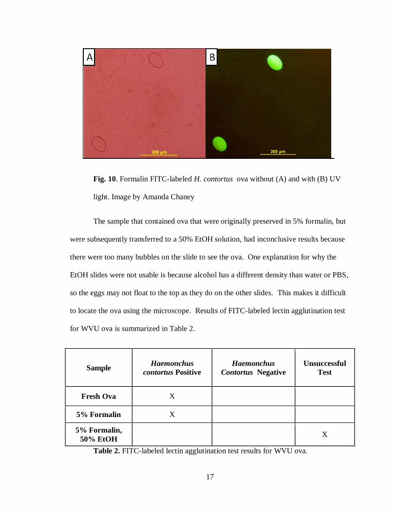
The sample that contained ova that were originally preserved in 5% formalin, but
were subsequently transferred to a 50% EtOH solution, had inconclusive results because
there were too many bubbles on the slide to see the ova. One explanation for why the
EtOH slides were not usable is because alcohol has a different density than water or PBS,
so the eggs may not float to the top as they do on the other slides. This makes it difficult
to locate the ova using the microscope. Results of FITC-labeled lectin agglutination test
for WVU ova is summarized in Table 2.
Sample Haemonchus
contortus Positive
Haemonchus
Contortus Negative
Unsuccessful
Test
Fresh Ova X
5% Formalin X
5% Formalin,
50% EtOH X
Table 2. FITC-labeled lectin agglutination test results for WVU ova.
18
The final sample was the L3 larva, which did not fluoresce. This result was
unexpected because the larvae should contain the same sugars on the surface as ova, and
thus, the PNA-FITC should be able to bind to the larvae. Lack of fluorescence could be
due to the inability of the lectins to penetrate the larvae cuticle, so no binding could
occur. Aside from the PNA-FITC binding, the non-fluorescent result was also not
expected because the L3 larvae actually auto-fluoresce using the fluorescent system at the
University of Georgia (personal communication).
Fluorescent-labeled lectin agglutination test results: Maine sheep and goat farm samples
No fluorescence of the samples occurred, indicating that the sheep was infected
with non-Haemonchus nematodes, either T. axei or O. ostertagii. However, it is possible
that the sheep was infected with H. contortus based on clinical signs, but no eggs were
being shed due to the season (hyperbiosis of H. contortus in the cold winter months).
HRP-labeled lectin agglutination test results:
The HRP-labeled lectin agglutination test was completed on fecal samples from
WVU that had been preserved in 5% formalin for five weeks. To date, only one trial has
been completed, and no negative control was used. For known H. contortus positive ova,
at time zero after TMB was added, no, or patchy, precipitation occurred (Fig 7). At a
time of 5 minutes, patchy precipitation occurred on the surface of the ova (Fig 7). At a
time of 10 minutes, blue precipitate covered the surface of the egg. At a time of 20
minutes, the precipitate started to fan out from the ova surface (Fig 7). Similar results
occurred for both concentrations of PNA-HRP, but the 400x concentration seemed to
have more non-specific binding of enzyme and resulting precipitate. The reaction was
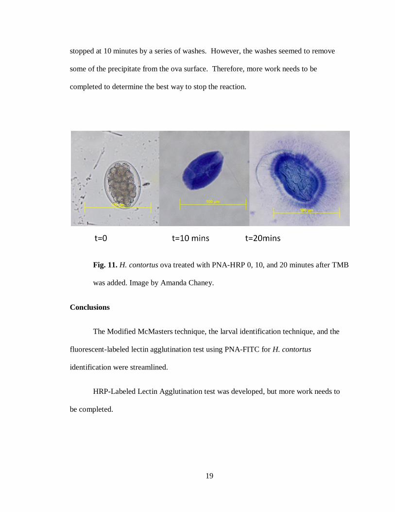
19
stopped at 10 minutes by a series of washes. However, the washes seemed to remove
some of the precipitate from the ova surface. Therefore, more work needs to be
completed to determine the best way to stop the reaction.
Fig. 11. H. contortus ova treated with PNA-HRP 0, 10, and 20 minutes after TMB
was added. Image by Amanda Chaney.
Conclusions
The Modified McMasters technique, the larval identification technique, and the
fluorescent-labeled lectin agglutination test using PNA-FITC for H. contortus
identification were streamlined.
HRP-Labeled Lectin Agglutination test was developed, but more work needs to
be completed.
20
Implications
Identification of the most pathogenic nematode, H. contortus, is important in
treating animals with a parasitic infection. The fluorescent-labeled PNA agglutination
test is ready for use in the University of Maine diagnostic lab alongside the larval
identification technique. In-house analysis must be completed on the sensitivity and
specificity of the procedure before it can be used alone. As for the HRP-labeled lectin
agglutination test, once the assay is fully developed, it will eliminate the need for an
expensive fluorescent microscope. This will make the identification of H. contortus more
available to Maine farmers and veterinarians. Overall, this research will benefit Maine
sheep and goat producers by providing more available identification methods for internal
parasites. Producers will then be better able to reduce parasitic disease and the economic
losses associated with it.
21
References
Bowdridge, S., 2005. The association between microsatellite marker CSRD 2138 and
parasite resistance in Katahdin Hair Sheep, pp 3-4.
Cerba, K.C., Stang, B.V., 2008. Comparison of methods to detect gastrointestinal
parasites in llamas and alpacas, JAMA, 232, 733-741.
Fisheries and Food Ministry of Agriculture, 1971. Manual of Veterinary Parasitological
Laboratory Techniques. H.M. Stationery Office, 1-40pp.
Jurasek, M.E., Bishop-Stewart, J.K., Storey, B.E., Kaplan, R.M., Kent, M.L., 2010.
Modification and further evaluation of a flourescein-labeled peanut agglutinin test
for identification of Haemonchus contortus eggs, Vet. Parasitol, 169, 209-213.
Kaplan, R.M., Burke, J.M., Terrill, T.H., Miller, J.E., Getz, W.R., Mobono, S., Valencia,
E., Williams, M.J., Williamson, L.H., Larsen, M., Vatta, A.F., 2004. Validation of
the FAMACHA © eye color chart for detecting clinical anemia in sheep and goats
on farms in the southern United States, Vet Parasitol, 123, 105-120.
Kahn, C. (Ed.), 2005. Merck Veterinary manual. Whitehouse Station, N.J., 262-265 pp.
Merial Australia. Available http://au.merial.com/disease_information/sheep/haemo.asp,
September 18, 2011.
Palmer, D.G., McCombe, I.L., 1996. Lectin staining of trichostrongylid nematode eggs of
sheep: rapid identification of Haemonchus contortus eggs with peanut agglutinin.
Int. J. Parisitol. 46, 147-150.
Schmidt, G.D., Roberts, L.S., 1981. Second edition foundations of parasitology. St.
Louis: C.V. Mosby
22
Appendicies
Appendix 1: Original budget
I am requesting funding to purchase the materials for lectin staining, to cover expenses of
survey mailing, and to assist in travel expenses to attend the Sheep and Goat conference.
I need several fluorescent-labeled lectins and galactose to complete the lectin-staining
method of nematode egg identification. The lectins are needed to tag and therefore
identify certain species of nematodes. Galactose is used to bind the lectins, preventing
their attachment to the nematode eggs; galactose is an important aspect of the
experimental control.
I hope to attend the UMaine Sheep and Goat Internal Parasite Seminar hosted at
Kennebec Valley Community College. Kennebec Valley Community College is 70
miles from my house and will cost me $18.27 to drive the 140 miles round trip with gas
prices being $3.55 and my Subaru averaging 27mpg.
I hope to receive funding that would enable me to mail a survey to 35 sheep and goat
producers throughout the state of Maine. Such a survey would provide me with
information such as flock or herd size, current parasite prevention and treatment
protocols, current or previous parasite infections, and other information relevant to my
project. Distributing a survey is also a good way both to inform sheep and goat
producers of our project and to communicate which farms are interested in participating
in our study.
Supplies— $
Lectin from Arachis hypogaea (peanut) –FITC conjugate (1mg) 56
Lectin from Concanavalin A—FITC conjugate (1mg) 48
Lectin from Methyl alpha-D-mannopyranosidase 30
Galactose 30
Travel to attend Sheep and Goat Internal Parasite Seminar 18.72
Postage to mail 35 surveys 15.40
Total Costs $198.12
23
Author’s Biography
Amanda Lynn Chaney of Searsport, Maine, majored in animal and veterinary
science with a concentration in pre-veterinary studies. She is also a student in the Honors
College. Amanda has enjoyed her time at UMaine, particularly her involvement in the
Witter Teaching and Research Center dairy and horse barns.
After graduation, Amanda plans to attend veterinary school. She then plans to
return to the state of Maine to practice large animal veterinary medicine.