Identification of Fasciola hepatica …Fasciola hepatica worms mature and lay eggs after 8 weeks...
6
Vol. 26, No. 10 JOURNAL OF CLINICAL MICROBIOLOGY, OCt. 1988, p. 2048-2053 0095-1137/88/102048-06$02.00/0 Copyright © 1988, American Society for Microbiology Identification of a 17-Kilodalton Fasciola hepatica Immunodiagnostic Antigen by the Enzyme-Linked Immunoelectrotransfer Blot Technique GEORGE V. HILLYER* AND MARICELIS SOLER DE GALANES Laboratory of Parasite Immunology, Department of Pathology, University of Puerto Rico School of Medicine, San Juan, Puerto Rico 00936-5067 Received 28 March 1988/Accepted 28 June 1988 Sera obtained from human patients, calves, sheep, and rabbits infected with Fasciola hepatica were tested by the Falcon assay screening test enzyme-linked immunosorbent assay (FAST-ELISA) and the enzyme-linked immunoelectrotransfer blot (EITB) techniques with Fasciola hepatica excretory-secretory antigens in order to evaluate their immunodiagnostic potential. The study included sera from 13 patients infected with F. hepatica or a history suggesting fascioliasis, 5 patients infected and treated with bithionol or praziquantel (3 were cured with bithionol), 10 patients infected with Schistosoma mansoni, 6 infected with Trichinella spiralis, and 13 controls and sera from calves, sheep, and rabbits with a primary F. hepatica infection. By FAST-ELISA with F. hepatica excretory-secretory antigens, the serum samples from fascioliasis patients gave the highest absorbance values, and the schistosomiasis patient sera gave intermediate values compared with a normal human serum control. Also by FAST-ELISA, the values for serum from patients with fascioliasis decreased steadily after cure, reaching normal levels 20 to 47 weeks postcure. In contrast, the serum from two patients who had been treated but were not yet cured had high levels of antibodies for up to 3 years of infection. By EITB, the serum samples from humans, rabbits, cattle, and sheep with fascioliasis recognized two antigenic polypeptides of 17 and 63 kilodaltons (kDa) in the form of sharp bands. For humans, this recognition lasted for at least 3 years of infection. Sera from individuals with schistosomiasis mansoni or trichinosis or from normal controls did not recognize the 17-kDa F. hepatica antigenic polypeptide. However, serum from one human with S. mansoni and one with T. spiralis infection had slight bands in the 63-kDa region, suggesting cross-reactivity. Reactivity to the 17-kDa polypeptide was absent in fascioliasis patients at 1 year postcure. Reactivity to the 63-kDa polypeptide was significantly diminished in fascioliasis patients at 1 year postcure. The sera from rabbits with a primary F. hepatica infection also recognized both the 17- and 63-kDa antigenic polypeptides by week 4 of infection. Reactivity to both antigens diminished significantly 6 weeks posture and disappeared by 8 weeks postcure. The sera from infected cattle and sheep recognized these two antigenic polypeptides by week 8 of infection. These studies suggest that the 17-kDa F. hepatica excretory secretory antigen is an excellent candidate for the immunodiagnosis of acute and chronic fascioliasis. Purification of this antigen and its application to quantitative serologic tests will permit further analysis of its predictive value to evaluate cure. Fasciola hepatica worms mature and lay eggs after 8 weeks of infection. However, symptoms often occur much earlier, at which time eggs would not be found in stool examination, making definitive parasitological diagnosis by this means ineffective. In addition, in humans the parasite eggs are often not found even after repeated stool examina- tions. Thus, serodiagnosis of fascioliasis is a useful alterna- tive to parasitologie diagnosis (4). Numerous antigens and test systems have been used for the serodiagnosis of fascioliasis, and numerous attempts have been made with conventional chromatographic tech- niques to isolate and characterize potential specific somatic or excretory-secretory (ES) antigens to define infections (4, 10). The enzyme-linked immunoelectrotransfer blot (EITB) technique described by Tsang et al. (13, 14) is a precise tool for identifying the molecular weights of potential sero- diagnostic antigens. This method was used to identify an F. hepatica ES antigen of 17 kilodaltons (kDa) which is recog- nized by the serum of humans, calves, sheep, and rabbits with fascioliasis, does not cross-react with the sera from humans with schistosomiasis mansoni or trichinosis, and * Corresponding author. disappears after successful chemotherapy, making it a po- tentially immunodiagnostic antigen. MATERIALS AND METHODS Antigens. F. hepatica ES antigens were isolated from live, adult worms essentially as described in Santiago and Hillyer (11). Live, intact adult F. hepatica worms were obtained from bovine livers at a local abbatoir and washed three or four times at room temperature during a 1-h period with 0.01 M phosphate-buffered saline, pH 7.2. The key element in the wash process is to remove all traces of blood and bile prior to the 3-h incubation in 0.01 M phosphate-buffered saline, pH 7.1, at 37°C. After incubation, the medium was collected and concentrated by vacuum dialysis. The antigen prepara- tion was centrifuged at 5,000 x g at 4°C, and the supernatant was collected. Protein concentration was determined by the Bradford method (1), and the antigens were stored at -80°C. Sodium dodecyl sulfate-polyacrylamide gel electrophoresis of ES obtained in this manner showed that it was composed primarily of a large cluster of proteins of 19 to 26 kDa and isolated bands of 17 and ca. 63 kDa (11). Sera. The following human sera were used in this study. (i) Sera from 13 patients infected with F. hepatica (5 defined by coprology, 8 by serology 14]). Of these patients, three were 2048 on June 29, 2020 by guest http://jcm.asm.org/ Downloaded from
Transcript of Identification of Fasciola hepatica …Fasciola hepatica worms mature and lay eggs after 8 weeks...
Vol. 26, No. 10JOURNAL OF CLINICAL MICROBIOLOGY, OCt. 1988, p. 2048-20530095-1137/88/102048-06$02.00/0Copyright © 1988, American Society for Microbiology
Identification of a 17-Kilodalton Fasciola hepaticaImmunodiagnostic Antigen by the Enzyme-Linked
Immunoelectrotransfer Blot TechniqueGEORGE V. HILLYER* AND MARICELIS SOLER DE GALANES
Laboratory of Parasite Immunology, Department ofPathology, University of Puerto Rico School of Medicine, San Juan,Puerto Rico 00936-5067
Received 28 March 1988/Accepted 28 June 1988
Sera obtained from human patients, calves, sheep, and rabbits infected with Fasciola hepatica were tested bythe Falcon assay screening test enzyme-linked immunosorbent assay (FAST-ELISA) and the enzyme-linkedimmunoelectrotransfer blot (EITB) techniques with Fasciola hepatica excretory-secretory antigens in order toevaluate their immunodiagnostic potential. The study included sera from 13 patients infected with F. hepaticaor a history suggesting fascioliasis, 5 patients infected and treated with bithionol or praziquantel (3 were curedwith bithionol), 10 patients infected with Schistosoma mansoni, 6 infected with Trichinella spiralis, and 13controls and sera from calves, sheep, and rabbits with a primary F. hepatica infection. By FAST-ELISA withF. hepatica excretory-secretory antigens, the serum samples from fascioliasis patients gave the highestabsorbance values, and the schistosomiasis patient sera gave intermediate values compared with a normalhuman serum control. Also by FAST-ELISA, the values for serum from patients with fascioliasis decreasedsteadily after cure, reaching normal levels 20 to 47 weeks postcure. In contrast, the serum from two patientswho had been treated but were not yet cured had high levels of antibodies for up to 3 years of infection. ByEITB, the serum samples from humans, rabbits, cattle, and sheep with fascioliasis recognized two antigenicpolypeptides of 17 and 63 kilodaltons (kDa) in the form of sharp bands. For humans, this recognition lasted forat least 3 years of infection. Sera from individuals with schistosomiasis mansoni or trichinosis or from normalcontrols did not recognize the 17-kDa F. hepatica antigenic polypeptide. However, serum from one human withS. mansoni and one with T. spiralis infection had slight bands in the 63-kDa region, suggesting cross-reactivity.Reactivity to the 17-kDa polypeptide was absent in fascioliasis patients at 1 year postcure. Reactivity to the63-kDa polypeptide was significantly diminished in fascioliasis patients at 1 year postcure. The sera fromrabbits with a primary F. hepatica infection also recognized both the 17- and 63-kDa antigenic polypeptides byweek 4 of infection. Reactivity to both antigens diminished significantly 6 weeks posture and disappeared by8 weeks postcure. The sera from infected cattle and sheep recognized these two antigenic polypeptides by week8 of infection. These studies suggest that the 17-kDa F. hepatica excretory secretory antigen is an excellentcandidate for the immunodiagnosis of acute and chronic fascioliasis. Purification of this antigen and itsapplication to quantitative serologic tests will permit further analysis of its predictive value to evaluate cure.
Fasciola hepatica worms mature and lay eggs after 8weeks of infection. However, symptoms often occur muchearlier, at which time eggs would not be found in stoolexamination, making definitive parasitological diagnosis bythis means ineffective. In addition, in humans the parasiteeggs are often not found even after repeated stool examina-tions. Thus, serodiagnosis of fascioliasis is a useful alterna-tive to parasitologie diagnosis (4).Numerous antigens and test systems have been used for
the serodiagnosis of fascioliasis, and numerous attemptshave been made with conventional chromatographic tech-niques to isolate and characterize potential specific somaticor excretory-secretory (ES) antigens to define infections (4,10).The enzyme-linked immunoelectrotransfer blot (EITB)
technique described by Tsang et al. (13, 14) is a precise toolfor identifying the molecular weights of potential sero-diagnostic antigens. This method was used to identify an F.hepatica ES antigen of 17 kilodaltons (kDa) which is recog-nized by the serum of humans, calves, sheep, and rabbitswith fascioliasis, does not cross-react with the sera fromhumans with schistosomiasis mansoni or trichinosis, and
* Corresponding author.
disappears after successful chemotherapy, making it a po-tentially immunodiagnostic antigen.
MATERIALS AND METHODS
Antigens. F. hepatica ES antigens were isolated from live,adult worms essentially as described in Santiago and Hillyer(11). Live, intact adult F. hepatica worms were obtainedfrom bovine livers at a local abbatoir and washed three orfour times at room temperature during a 1-h period with 0.01M phosphate-buffered saline, pH 7.2. The key element in thewash process is to remove all traces of blood and bile priorto the 3-h incubation in 0.01 M phosphate-buffered saline,pH 7.1, at 37°C. After incubation, the medium was collectedand concentrated by vacuum dialysis. The antigen prepara-tion was centrifuged at 5,000 x g at 4°C, and the supernatantwas collected. Protein concentration was determined by theBradford method (1), and the antigens were stored at -80°C.Sodium dodecyl sulfate-polyacrylamide gel electrophoresisof ES obtained in this manner showed that it was composedprimarily of a large cluster of proteins of 19 to 26 kDa andisolated bands of 17 and ca. 63 kDa (11).
Sera. The following human sera were used in this study. (i)Sera from 13 patients infected with F. hepatica (5 defined bycoprology, 8 by serology 14]). Of these patients, three were
2048
on June 29, 2020 by guesthttp://jcm
.asm.org/
Dow
nloaded from

F. HEPATICA IMMUNODIAGNOSTIC ANTIGENS 2049
treated with bithionol (50 mg/kg) and two with praziquantel(25 mg/kg) and followed parasitologically by the modifiedRitchie formol-ether concentration method (9) for up to 3years.
(ii) Sera were obtained from six individuals infected withSchistosoma mansoni, as determined by the presence ofparasite eggs in their stools (9), and four individuals positiveserologically by the circumoval precipitin (COP) reaction(7).
(iii) Sera from six individuals with confirmed trichinosiswere kindly donated by Shirley Maddison, Centers forDisease Control, Atlanta, Ga.Normal human serum samples were obtained from 13
individuals with no history of eating watercress and werenegative by enzyme-linked immunosorbent assay (ELISA)against S. mansoni soluble egg antigen (SEA) and F. hepat-ica ES antigens.The following sera from animals experimentally infected
with F. hepatica were used in this study. (i) A 7-week-oldNew Zealand White rabbit was prebled, infected with 30 F.hepatica metacercariae, bled periodically for 20 weeks,treated with a curative dose of rafoxanide (Merck) (6), andbled for an additional 30 weeks. (ii) Five 6-month-old calveswere prebled, infected with 500 F. hepatica metacercariae,and bled at 2-week intervals for 10 weeks. Each of the five2-week serum samples obtained from each bleeding waspooled for use in this study. (iii) Five sheep were prebled,infected orally with 400 F. hepatica metacercariae, and bledat 2-week intervals for 10 weeks. Each of the five 2-weekserum samples obtained from each bleeding was pooled foruse in this study.Immunologic tests. The Falcon assay screening test
ELISA (FAST-ELISA) was done as described by Hancockand Tsang (3). The antigen concentration for the assay was 4,ug/ml. All sera were tested at twofold dilutions from 1:32 to1:256. Goat anti-human immunoglobulin G (IgG) plus IgMplus IgA (heavy- and light-chain specific [H+L]) (Kirke-gaard & Perry Laboratories, Inc. [KPL], Gaithersburg, Md.)affinity-purified peroxidase-labeled conjugates were used ata 1:300 dilution in 0.3% phosphate-buffered saline-Tween.The reactions were read on a Bio-Rad Laboratories 2550ELISA reader at 600 nm.The EITB was done as described by Tsang et al. (13, 14)
and Santiago and Hillyer (11). All sera were tested at a 1:200dilution. The following affinity-purified peroxidase-labeledconjugates were used (at a 1:2,000 dilution): goat anti-humanIgG+IgM+IgA (H+L) (KPL), goat anti-bovine IgG (H+L)(KPL), goat anti-rabbit IgG (H+L) (Bio-Rad), and rabbitanti-sheep IgG (H+L) (KPL).
RESULTS
The first question was whether F. hepatica ES antigenscould identify antibodies in the serum of patients withconfirmed fascioliasis (n = 3) or suspected of having fascio-liasis due to history (n = 7) by FAST-ELISA, and this wasindeed the case (Fig. 1). The second question was whetherF. hepatica ES antigens cross-reacted with the serum ofindividuals infected with S. mansoni (confirmed by stoolexamination, n = 6; confirmed by positive COP reaction,n = 4). As can also be seen in Fig. 1, the serum samples fromindividuals infected with F. hepatica had the highest absorb-ance values against F. hepatica ES antigens, and the serafrom the individuals infected with S. mansoni had interme-diate but clearly reactive levels compared with a normalcontrol pool.
1.4
1.2`
1.0
EooCDn
:0
0.8
0.6
0.4
0.2
ses
ie
e
NHS Fh Sm
FIG. 1. FAST-ELISA with F. hepatica ES antigens reactedagainst normal human serum (NHS) or the serum of humansinfected with F. hepatica (Fh) of S. mansoni (Sm). Abs, Absorb-ance.
The third question was whether FAST-ELISA with F.hepatica ES antigens could be used to monitor the success ofchemotherapy in patients with fascioliasis. Figure 2 showsthe absorbance values for samples from three patientstreated and cured with bithionol and followed up for up to 50weeks. Before treatment, all three patients with fascioliasishad serum absorbance values similar to the positive controlserum. In one patient, antibody levels increased slightly 2weeks posttreatment (Fig. 2a) and then decreased by 5weeks posttreatment, steadily decreasing thereafter towardnormal values through the 23 weeks posttreatment tested. Inthe second patient, antibody levels decreased within 3 weeksposttreatment, steadily decreasing thereafter toward normalvalues through the 47 weeks posttreatment tested. In thethird patient, antibody levels dropped near to normal valuesby 16 weeks posttreatment, remaining at these low levelsthrough 50 weeks tested.The antibody absorbance patterns of samples from two
patients treated with praziquantel and not cured of fascio-liasis were followed for 83 and 147 weeks posttreatment. Asseen in Fig. 3, the antibody levels of these two infectedpatients remained high during the follow-up period, which inone case was almost 3 years.
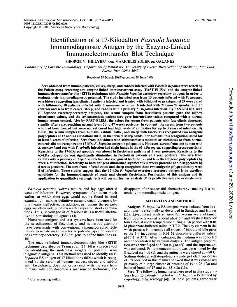
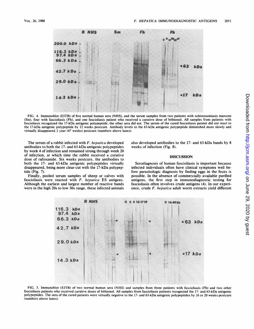
In order to best define the F. hepatica ES antigenicpolypeptides seen in infected patients, the EITB was per-formed. Sera from the five patients infected with F. hepaticaas defined parasitologically all reacted in EITB, producingmultiple bands. An apparently nonspecific cluster of 2+bands was seen at ca. 29 kDa (Fig. 4). One remarkableobservation was that the serum from all F. hepatica-infectedindividuals had antibodies which reacted with the 17- and63-kDa bands. Moreover, all eight serum samples frompatients having antibodies to F. hepatica ES antigens alsohad antibodies which reacted with the 17- and 63-kDa bands.In addition, the three patients who received curative dosesof bithionol had their antibody levels to the 17-kDa antigenicpolypeptide diminish rapidly (12 to 16 weeks postcure). Inthe same patients, antibody levels to the 63-kDa antigenicpolypeptide diminished more slowly (16 to 26 weeks), requir-ing almost 1 year for total disappearance. RepresentativeEITBs showing these patterns are shown in Fig. 4 and 5. The
VOL. 26, 1988
on June 29, 2020 by guesthttp://jcm
.asm.org/
Dow
nloaded from

2050 HILLYER AND SOLER DE GALANES
EC0.8oCD
0.6
.0
a
32 64 128 256
Reciprocal Serum DilutionFIG. 2. FAST-ELISA of serum from three fascioliasis patients reacted with F. hepatica ES antigens. These patients were treated with
bithionol and cured of infection as determined by multiple parasitological examinations (formol-ether concentration method). Abs,Absorbance; NHS, normal human serum; POS, positive control serum. Numbers beside symbols indicate weeks posttreatment.
serum from patients treated with praziquantel but not cureddid not have diminished antibody to the 17- and 63-kDaantigenic polypeptides up to 147 or more weeks posttreat-ment (and postinfection) (Fig. 6).As can be seen in Fig. 4 and 6, the serum from humans
infected with either S. mansoni or Trichinella spiralis re-acted with F. hepatica ES antigens, primarily in the 20- to30-kDa region, as did normal human serum, with one or two
bands at ca. 29 kDa. Additional bands cross-reactive withthe serum of S. mansoni-infected individuals were seen inthe 15- and 40-kDa regions. In Fig. 6, one of the two humanS. mansoni serum samples (but not the two in Fig. 4) and oneof the three T. spiralis sera also appeared to react with the63-kDa antigenic polypeptide. Moreover, the serum from T.spiralis-infected individuals also recognized several anti-genic polypeptides of less than 14 kDa.
a1.4
-0- O
154-} 27-0- 55_ 83-à- NHS> PO6
64 128 256
1.2
1.0
0.8
0.6
0.4
0.2
0.0
b
<> o+.. 4- 12- 24_ 136{} 147-NHS,-P06
32 64 128 256
Reciprocal Serum DilutionFIG. 3. FAST-ELISA of the serum from two fascioliasis patients reacted with F. hepatica ES antigens. These patients were treated with
praziquantel and not cured of infection as determined by multiple parasitological examinations (formol-ether concentration method). Abs,Absorbance; NHS, normal human serum; POS, positive control serum. Numbers beside symbols indicate weeks posttreatment.
1.4
1.2
E 1.0.
O0.8
0.6
0.4
0.2
0.0
J. CLIN. MICROBIOL.
on June 29, 2020 by guesthttp://jcm
.asm.org/
Dow
nloaded from
F. HEPATICA IMMUNODIAGNOSTIC ANTIGENS 2051
8 NUS
200.0 kDa -
116.3 kDa - |97.4 kOa -
Sm Fb
I66.3 kDa -
42.7 kDa -
29.0 kba -
Fb
0 O 47
Ilût 463 kDa
417 kDa14.3 kDa '-iI
FIG. 4. Immunoblot (EITB) of five normal human sera (NHS), and the serum samples from two patients with schistosomiasis mansoni(Sm), four with fascioliasis (Fh), and one fascioliasis patient who received a curative dose of bithionol. All samples from patients withfascioliasis recognized the 17-kDa antigenic polypeptide; the other sera did not. The serum of the cured fascioliasis patient did not react tothe 17-kDa antigenic polypeptide by 12 weeks postcure. Antibody levels to the 63-kDa antigenic polypeptide diminished more slowly andvirtually disappeared 1 year (47 weeks) postcure (numbers above lanes).
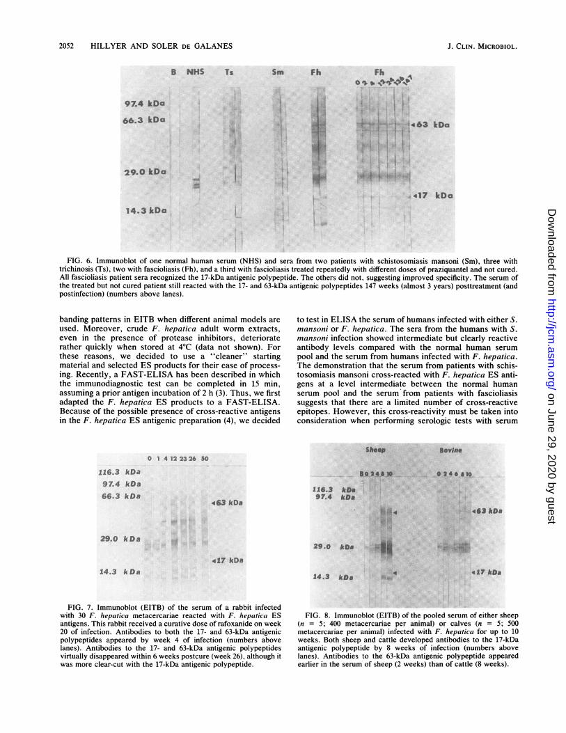
The serum of a rabbit infected with F. hepatica developedantibodies to both the 17- and 63-kDa antigenic polypeptidesby week 4 of infection and remained strong through week 20of infection, at which time the rabbit received a curativedose of rafoxanide. Six weeks postcure, the antibodies toboth the 17- and 63-kDa antigenic polypeptides virtuallydisappeared, being more clear-cut with the 17-kDa polypep-tide (Fig. 7).
Finally, pooled serum samples of sheep or calves withfascioliasis were reacted with F. hepatica ES antigens.Although the earliest and largest number of reactive bands*were in the high 20s to low 30s range, these infected animals
B NHS
116.3 kDa97.4 kDa66.3 kDa
42.7 kDa
29.0 kDa
14.3 kDa
also developed antibodies to the 17- and 63-kDa bands by 8weeks of infection (Fig. 8).
DISCUSSION
Serodiagnosis of human fascioliasis is important becauseinfected individuals often have clinical symptoms well be-fore parasitologic diagnosis by finding eggs in the feces ispossible. In the absence of commercially available purifiedantigens, the first step in immunodiagnostic testing forfascioliasis often involves crude antigens (4). In our experi-ence, crude F. hepatica adult worm extracts yield different
0 24161719
.~:~ O 164950
4 63 kDa
417 kDa
FIG. 5. Immunoblot (EITB) of two normal human sera (NHS) and samples from three patients with fascioliasis (Fh) and two otherfascioliasis patients who received curative doses of bithionol. All samples from fascioliasis patients recognized the 17- and 63-kDa antigenicpolypeptides. The sera of the cured patients were virtually negative to the 17- and 63-kDa antigenic polypeptides by 16 or 20 weeks postcure(numbers above lanes).
VOL. 26, 1988
I
i
'l'"1
>-*
L---.
m i: 1
1! ;
on June 29, 2020 by guesthttp://jcm
.asm.org/
Dow
nloaded from
2052 HILLYER AND SOLER DE GALANES
B NHS Ts Sm Fh
97.4 kDa
66.3 kDa
29.0 kDa
FhO 'I. s u
s R . I
417 kDa
14.3kDa5
FIG. 6. Immunoblot of one normal human serum (NHS) and sera from two patients with schistosomiasis mansoni (Sm), three withtrichinosis (Ts), two with fascioliasis (Fh), and a third with fascioliasis treated repeatedly with different doses of praziquantel and not cured.All fascioliasis patient sera recognized the 17-kDa antigenic polypeptide. The others did not, suggesting improved specificity. The serum ofthe treated but not cured patient still reacted with the 17- and 63-kDa antigenic polypeptides 147 weeks (almost 3 years) posttreatment (andpostinfection) (numbers above lanes).
banding patterns in EITB when different animal models areused. Moreover, crude F. hepatica adult worm extracts,even in the presence of protease inhibitors, deterioraterather quickly when stored at 4°C (data not shown). Forthese reasons, we decided to use a "cleaner" startingmaterial and selected ES products for their ease of process-ing. Recently, a FAST-ELISA has been described in whichthe immunodiagnostic test can be completed in 15 min,assuming a prior antigen incubation of 2 h (3). Thus, we firstadapted the F. hepatica ES products to a FAST-ELISA.Because of the possible presence of cross-reactive antigensin the F. hepatica ES antigenic preparation (4), we decided
116.3 kDa
97.4 kDa
66.3 kDa
to test in ELISA the serum of humans infected with either S.mansoni or F. hepatica. The sera from the humans with S.mansoni infection showed intermediate but clearly reactiveantibody levels compared with the normal human serumpool and the serum from humans infected with F. hepatica.The demonstration that the serum from patients with schis-tosomiasis mansoni cross-reacted with F. hepatica ES anti-gens at a level intermediate between the normal humanserum pool and the serum from patients with fascioliasissuggests that there are a limited number of cross-reactiveepitopes. However, this cross-reactivity must be taken intoconsideration when performing serologic tests with serum
Dovine
0 24601I02.48,10
463 kDa
116.3 kDi97.4 k<Da ,~
29.0 kDa
t6à kDa
29.0 kDa
417 kDa
14.3 kDa14.3 kt<0 4 .17 kle
FIG. 7. Immunoblot (EITB) of the serum of a rabbit infectedwith 30 F. hepatica metacercariae reacted with F. hepatica ESantigens. This rabbit received a curative dose of rafoxanide on week20 of infection. Antibodies to both the 17- and 63-kDa antigenicpolypeptides appeared by week 4 of infection (numbers abovelanes). Antibodies to the 17- and 63-kDa antigenic polypeptidesvirtually disappeared within 6 weeks postcure (week 26), although itwas more clear-cut with the 17-kDa antigenic polypeptide.
FIG. 8. Immunoblot (EITB) of the pooled serum of either sheep(n = 5; 400 metacercariae per animal) or calves (n = 5; 500metacercariae per animal) infected with F. hepatica for up to 10weeks. Both sheep and cattle developed antibodies to the 17-kDaantigenic polypeptide by 8 weeks of infection (numbers abovelanes). Antibodies to the 63-kDa antigenic polypeptide appearedearlier in the serum of sheep (2 weeks) than of cattle (8 weeks).
ShoopO 1 4 12 23 26 50
J. CLIN. MICROBIOL.
1
-E
e1
on June 29, 2020 by guesthttp://jcm
.asm.org/
Dow
nloaded from

F. HEPATICA IMMUNODIAGNOSTIC ANTIGENS 2053
from patients who have been exposed to schistosomes.Recently, Espino et al. (2) did not find any antigenic cross-reactivity when testing serum from humans with a widevariety of parasitic infections, including schistosomiasis(although the species was not defined, it was presumably S.mansoni or S. haematobium or both). However, this may bedue to differences in assay conditions or in the preparation ofantigen. Espino et al. (2) obtained their F. hepatica ESantigens after a 24-h incubation; on the other hand, ourswere obtained after a 3-h incubation.The decrease in antibody levels seen by FAST-ELISA in
the cured fascioliasis patients confirms a previous study ofours reporting antibody changes after chemotherapy in onecured patient in which crude F. hepatica worm antigen wasused in the ELISA (5).
Irving and Howell (8) isolated F. hepatica ES antigens byincubating live worms obtained from infected mice for 5 to 7days in serum-free medium. Three major antigenic polypep-tides incorporated radiolabeled amino acids of 23, 24, and 26kDa. We have also found this to be one region of majorantigenic polypeptide activity in rabbits with fascioliasis(12). In the present report, we found that this region alsocontained some antigens cross-reactive with S. mansoni andT. spiralis and nonspecific cross-reactivity with normalhuman serum.Our observation herein that humans, rabbits, cattle, and
sheep infected with F. hepatica all develop antibodies to the17-kDa antigenic polypeptide seen by EITB suggests thatthis may be a useful marker to detect infections by thismethod.The uniformity of finding the 17-kDa band when the sera
of patients with F. hepatica were tested by EITB with F.hepatica ES antigen preparations, and not finding thesereactions with an antigenically closely related trematode,such as S. mansoni, or one with cross-reacting antibodies,such as T. spiralis, suggests that the 17-kDa band may bespecific for fascioliasis. The observation that it disappearedin treated and cured patients but not in treated but not curedpatients suggests that it may also serve as a marker to definethe success of chemotherapy. This is reinforced by the studyon the rabbit that was infected and then treated and cured, inwhich antibodies to the 17-kDa antigenic polypeptide ap-peared by week 4 of infection and virtually disappearedwithin 6 weeks postcure. The observation that infectedhumans still have similarly high levels of antibodies as atpretreatment 3 years later suggests that this fluke is long-lived in its human host.
ACKNOWLEDGMENTS
These studies were supported by Public Health Service grant AI22906 from the National Institutes of Health.The human serum samples were collected by Guillermo Vazquez,
UPR Medical Sciences Campus Infectious Diseases Unit, Ramôn
Bermùdez, Veterans Administration Center, San Juan, and BernardChristensen, Bayamôn. The sera from sheep and cattle were col-lected by El Tahir M. Haroun, and the gift is warmly acknowledged.
LITERATURE CITED
1. Bradford, M. M. 1976. A rapid and sensitive method for thequantitation of microgram quantities of protein utilizing theprinciple of protein-dye binding. Anal. Biochem. 72:248-254.
2. Espino, A. M., B. E. Duménigo, R. Fernàndez, and C. M. Finlay.1987. Immunodiagnosis of human fasciliasis by enzyme-linkedimmunosorbent assay using excretory-secretory products. Am.J. Trop. Med. Hyg. 37:605-608.
3. Hancock, K., and V. C. W. Tsang. 1986. Development andoptimization of the FAST-ELISA for detecting antibodies toSchistosoma mansoni. J. Immunol. Methods 92:167-186.
4. Hillyer, G. V. 1986. Fascioliasis, paragonimiasis, clonorchiasis,and opisthorchiasis, p. 39-68. In K. W. Walls, and P. M.Schantz (ed.), Immunodiagnosis of parasitic diseases, vol. 1:helminthic diseases. Academic Press, Inc., Boca Raton, Fla.
5. Hillyer, G. V., R. H. Bermudez, and A. G. Ramirez de Arellano.1984. Use of immunologic techniques to predict success oftherapy in human fascioliasis: a case report. Bol. Asoc. Med.P. R. 76:116-119.
6. Hillyer, G. V., and A. del Llano de Diaz. 1976. Use of immuno-logic techniques to detect chemotherapeutic success in infec-tions with Fasciola hepatica. I. Rabbit infections. Am. J. Trop.Med. Hyg. 25:307-311.
7. Hillyer, G. V., E. Ruiz Tiben, W. B. Knight, I. G6mez de Rios,and R. P. Pelley. 1979. Immunodiagnosis of infection withSchistosoma mansoni: comparison of ELISA, radioimmunoas-say, and precipitation tests performed with antigens from eggs.Am. J. Trop. Med. Hyg. 28:661-669.
8. Irving, D. O., and M. J. Howell. 1982. Characterization ofexcretory-secretory antigens of Fasciola hepatica. Parasitology85:179-188.
9. Knight, W. B., R. A. Hiatt, B. L. Cline, and L. S. Ritchie. 1976.A modification of the formol-ether concentration technique forincreased sensitivity in detecting Schistosoma mansoni eggs.Am. J. Trop. Med. Hyg. 25:818-823.
10. Reddington, J. J., R. W. Leid, and R. B. Wescott. 1984. Areview of the antigens of Fasciola hepatica. Vet. Parasitol. 14:209-229.
11. Santiago, N., and G. V. Hillyer. 1986. Isolation of potentialserodiagnostic Fasciola hepatica antigens by electroelutionfrom polyacrylamide gels. Am. J. Trop. Med. Hyg. 35:1210-1217.
12. Santiago, N., G. V. Hillyer, M. Garcia Rosa, and M. H. Morales.1986. Identification of functional Fasciola hepatica antigens inexperimental infections in rabbits. Am. J. Trop. Med. Hyg. 35:135-140.
13. Tsang, V. C. W., G. E. Bers, and K. Hancock. 1985. Enzyme-linked immunoelectrotransfer blot (EITB), p. 389-414. In T. T.Ngo and H. M. Lenhoff (ed.), Enzyme-mediated immunoassay.Plenum Press, New York.
14. Tsang, V. C. W., J. M. Peralta, and A. R. Simons. 1983.Enzyme-linked immunoelectrotransfer blot techniques (EITB)for studying the specificities of antigens and antibodies sepa-rated by gel electrophoresis. Methods Enzymol. 92:377-391.
VOL. 26, 1988
on June 29, 2020 by guesthttp://jcm
.asm.org/
Dow
nloaded from


![Serum [3H]-fucose labelled glycoproteins in Fasciola hepatica](https://static.fdocuments.in/doc/165x107/623f6350b395777077658644/serum-3h-fucose-labelled-glycoproteins-in-fasciola-hepatica.jpg)