Identification of ‘Campylobacter upsaliensis’ and other catalase-negative campylobacters from...
6
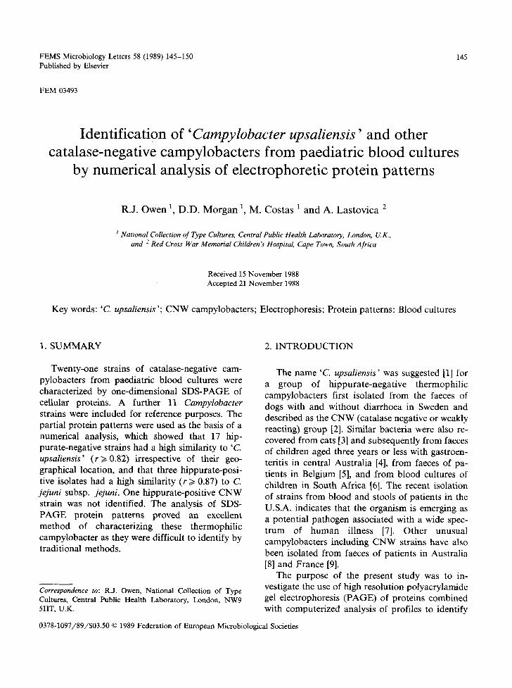
FEMS Microbiology Letters 58 (1989) 145-150 145 Published by Elsevier FEM 03493 Identification of 'Campylobacter upsaliensis' and other catalase-negative campylobacters from paediatric blood cultures by numerical analysis of electrophoretic protein patterns R.J. Owen a, D.D. Morgan 1, M. Costas 1 and A. Lastovica 2 I National Collection of Type Cultures, Central Public Health Laboratory, London, U.K., and 2 Red Cross War Memorial Children's Hospital, Cape Town, South Africa Received 15 November 1988 Accepted 21 November 1988 Key words: 'C. upsaliensis '; CNW campylobacters; Electrophoresis; Protein patterns: Blood cultures 1. SUMMARY 2. INTRODUCTION Twenty-one strains of catalase-negative cam- pylobacters from paediatric blood cultures were characterized by one-dimensional SDS-PAGE of cellular proteins. A further 11 Campylobacter strains were included for reference purposes. The partial protein patterns were used as the basis of a numerical analysis, which showed that 17 hip- purate-negative strains had a high similarity to 'C. upsaliensis' (r>~0.82) irrespective of their geo- graphical location, and that three hippurate-posi- tive isolates had a high similarity (r >~0.87) to C. jejuni subsp, jejuni. One hippurate-positive CNW strain was not identified. The analysis of SDS- PAGE protein patterns proved an excellent method of characterizing these thermophilic campylobacter as they were difficult to identify by traditional methods. Correspondence to: R.J. Owen, National Collection of Type Cultures, Central Public Health Laboratory, London, NW9 5HT, U.K. The name 'C. upsaliensis' was suggested [1] for a group of hippurate-negative thermophilic campylobacters first isolated from the faeces of dogs with and without diarrhoea in Sweden and described as the CNW (catalase negative or weakly reacting) group [2]. Similar bacteria were also re- covered from cats [3] and subsequently from faeces of children aged three years or less with gastroen- teritis in central Australia [4], from faeces of pa- tients in Belgium [5], and from blood cultures of children in South Africa [6]. The recent isolation of strains from blood and stools of patients in the U.S.A. indicates that the organism is emerging as a potential pathogen associated with a wide spec- trum of human illness [7]. Other unusual campylobacters including CNW strains have also been isolated from faeces of patients in Australia [8] and France [9]. The purpose of the present study was to in- vestigate the use of high resolution polyacrylamide gel electrophoresis (PAGE) of proteins combined with computerized analysis of profiles to identify 0378-1097/89/$03.50 © 1989 Federation of European Microbiological Societies
Transcript of Identification of ‘Campylobacter upsaliensis’ and other catalase-negative campylobacters from...
FEMS Microbiology Letters 58 (1989) 145-150 145 Published by Elsevier
FEM 03493
Identification of 'Campylobacter upsaliensis' and other catalase-negative campylobacters from paediatric blood cultures
by numerical analysis of electrophoretic protein patterns
R.J. Owen a, D.D. Morgan 1, M. Costas 1 and A. Lastovica 2
I National Collection of Type Cultures, Central Public Health Laboratory, London, U.K., and 2 Red Cross War Memorial Children's Hospital, Cape Town, South Africa
Received 15 November 1988 Accepted 21 November 1988
Key words: 'C. upsaliensis '; CNW campylobacters; Electrophoresis; Protein patterns: Blood cultures
1. SUMMARY 2. INTRODUCTION
Twenty-one strains of catalase-negative cam- pylobacters from paediatric blood cultures were characterized by one-dimensional SDS-PAGE of cellular proteins. A further 11 Campylobacter strains were included for reference purposes. The partial protein patterns were used as the basis of a numerical analysis, which showed that 17 hip- purate-negative strains had a high similarity to 'C. upsaliensis' (r>~0.82) irrespective of their geo- graphical location, and that three hippurate-posi- tive isolates had a high similarity (r >~ 0.87) to C. jejuni subsp, jejuni. One hippurate-positive CNW strain was not identified. The analysis of SDS- PAGE protein pat terns proved an excellent method of characterizing these thermophilic campylobacter as they were difficult to identify by traditional methods.
Correspondence to: R.J. Owen, National Collection of Type Cultures, Central Public Health Laboratory, London, NW9 5HT, U.K.
The name 'C. upsaliensis' was suggested [1] for a group of hippurate-negative thermophilic campylobacters first isolated from the faeces of dogs with and without diarrhoea in Sweden and described as the CNW (catalase negative or weakly reacting) group [2]. Similar bacteria were also re- covered from cats [3] and subsequently from faeces of children aged three years or less with gastroen- teritis in central Australia [4], from faeces of pa- tients in Belgium [5], and from blood cultures of children in South Africa [6]. The recent isolation of strains from blood and stools of patients in the U.S.A. indicates that the organism is emerging as a potential pathogen associated with a wide spec- trum of human illness [7]. Other unusual campylobacters including CNW strains have also been isolated from faeces of patients in Australia [8] and France [9].
The purpose of the present study was to in- vestigate the use of high resolution polyacrylamide gel electrophoresis (PAGE) of proteins combined with computerized analysis of profiles to identify
0378-1097/89/$03.50 © 1989 Federation of European Microbiological Societies

146
the var ious C N W strains i so la ted f rom paed ia t r i c b l o o d cul tures in South Africa.
3. M A T E R I A L S A N D M E T H O D S
3.1. Bacteria used The 21 cl inical isolates of C N W campylo -
bac te rs and 11 reference s t ra ins ob t a ined f rom the N a t i o n a l Col lec t ion of Type Cul tures ( N C T C ) are l is ted in Table 1, wi th their a l te rna t ive s t ra in numbers and sources. Conven t iona l test resul ts on the strains and cl inical deta i ls of the pa t ien t s f rom whom they were i so la ted are descr ibed separa te ly [10]. 'C. upsaliensis' is in quo ta t ions th roughou t to indica te it is not a val id name, a l though it has come into c o m m o n usage. Al l s t ra ins were grown on 5% ( v / v ) de f ib r ina t ed horse b l o o d agar. Cul- tures were incuba ted for 48 h at 3 7 ° C unde r microaerophi l i c cond i t ions ( abou t 5% oxygen) at- t a ined by incuba t ion in anae rob ic j a r s (wi thout cata lys t ) f rom which 75% of the air had been wi thdrawn (580 m m Hg be low a tmospher i c pres- sure) and rep laced with an anae rob ic gas mix ture con ta in ing 10% hydrogen , 10% ca rbon d iox ide and 80% ni t rogen.
3.2. Preparation o f protein samples and electro- phoresis
F o r each p r o t e i n sample , a p p r o x i m a t e l y 0 .05-0.1 g wet weight of the bac te r i a were harves ted d i rec t ly f rom b lood agar p la tes and suspended in abou t 60 t~l of doub le s t rength lysis buf fe r (20% v / v glycerol , 2% v / v 2 -mercap to - e thanol , 4% w / v sod ium dodecy l su lpha te (SDS) and 70% v / v s tacking gel buffer) . The p ro t e in samples were then ex t rac ted as descr ibed previ- ous ly [11] and were run on d i scon t inuous SDS- po lyac ry l amide gels, which were cast to al low for a 10 m m stacking gel. The f inal p o l y a c r y l a m i d e concen t ra t ions were 10% w / v for the separa t ion gel and 5% w / v for the s tacking gel. Ful l de ta i l s of the me thods used in gel p r e p a r a t i o n and e lect ro- phores is were descr ibed prev ious ly [12].
3.3. Scanning o f gels and computations The s ta ined p ro te in pa t t e rns in the d r i ed gels
were scanned (LKB Ul t ro scan X L laser dens i to -
Table 1
Campylobacter strains studied
Species/group Strain number a Reference no. in Figs. 1 and 2
Reference strains "C. upsaliensis" NCTC 11540 1 'C. upsaliensis" NCTC 11926 2 C. laridis NCTC 11844 3 C. laridis NCTC 11458 16 C. laridis NCTC 11352 T 24 C. jejuni ssp. jejuni NCTC 11168 12 C. jejuni ssp. jejuni NCTC 11251 x 25 C. jejuni ssp. doylei NCTC 11951 -r 23 C. coli NCTC 11366 T 13 C. coli NCTC 11353 15 C. fetus ssp. fetus NCTC 10842 v 14
Hippurate-negative b isolates CNW A614/87; 5.86 5 CNW A620/87; 211.86 10, CNW A618/87; 158.86 7, CNW A617/87; 129,86 6 CNW A622/87; 10.87 17 CNW A621/87; 3.87 18, CNW A686/88; 144.85 - CNW A687/88; 147.85 - CNW A688/88; 148.85 - CNW A689/88; 66.86 - CNW A690/88; 182.86 - CNW A691/88; 77.86 - CNW A692/88; 34.88 - CNW A693/88; 19.88 - CNW A694/88; 46.88 - CNW A695/88; 74.88 -
Hippurate-positwe b isolates CNW A615/87; 28.85 4 CNW A613/87; 1.86 8 CNW A603/87; 34.85 9 CNW A619/87; 159.86 22
11 20, 21
19
a 'A' number denotes number used for NCTC field strains. Other numbers are original (Cape Town) isolate numbers. NCTC, National Collection of Type Cultures, London. T, type strain.
b Hippurate hydrolysis results were from Lastovica et al. [10] and NCTC unpublished results.
meter ) at a scan speed of 20 c m / m i n and an a bso rba nc e range of 0 to 1, and the abso rbance values were r ecorded on magne t ic disk. The reten- t ion t imes for mo lecu la r weight es t imat ions and to ta l areas of each p ro t e in b a n d were ca lcu la ted us ing an L K B 2220 record ing in tegrator . The ini- t ia l ( s tacking g e l / s e p a r a t i o n gel interface) and

final (bromophenol blue marker) bands were de- leted and protein patterns were corrected for gel- to-gel variation by segmented linear correction as described previously [12]. The length-corrected traces on the reference gel (the gel containing standard strain reference pattern) were composed of 590 absorbance values after removal of the initial and final bands. The principal band region (39-50 kDa) and a general background trend of 0.4 (fraction of absorbance) in each trace were removed to increase discrimination among pat- terns. Similarities between all pairs of processed traces were expressed as the Pearson product mo- ment correlation coefficient (converted for con- venience to a percent value). The best fit between each pair was obtained by laterally shifting one corrected trace with respect to the other in single point steps of 160/~m up to 5 points either side of the initial alignment. Strains were then clustered by the method of unweighted pair-group average linkage (UPGMA). Computat ions were carried out on a Compaq 386 microcomputer using a program package written in Turbo Pascal and based on previously described programs [13].
147
4. RESULTS
4.1. General features of PAGE protein patterns One-dimensional SDS-PAGE of whole cell pro-
tein extracts of the 21 CNW campylobacters and 11 reference strains produced patterns containing at least 40 discrete bands with molecular weights of 17-100 kDa. Proteins with molecular weights outside this range were not resolved under the electrophoresis conditions used. PAGE patterns are illustrated in Fig. 1. Duplicate and triplicate samples of three strains were included as a mea- sure of reproducibility which was estimated to be at least 95% (Fig. 2).
4.2. Numerical analysis Numerical analysis of partial PAGE protein
profiles based on determination of the Pearson correlation coefficient and U P G M A clustering re- vealed four clusters of two or more strains at the 73% similarity level as shown in Fig. 2. The phe- nons were as follows (from the top of the pheno- gram in Fig. 2):
Phenon 1. This cluster comprised eight strains: N C T C 11540, which was a reference strain of 'C.
13 12 11 10 9 8 X 7 6 5 4 3 2 1 25 24 23 22 21 20 X 19 18 17 16 15 14
Fig. 1. Electrophoretic protein patterns of CNW isolates and reference strains. The numbers refer to those used in Table 1 and Fig. 2. Molecular weight marker (track labelled x) are (from top to bottom: ovotransferrin, 76-78 kDa; albumin, 66.25 kDa; ovalbumin, 42.7 kDa; carbonic anhydrase, 30 kDa; myoglobin, 17.2 kDa.

148
STRAIN PERCENTAGE SIMILARITY
P H E N O N 100 90 80 70 60 50 40 30 20
NCTC 1154O A 6 1 4 / 8 7 A 6 2 0 / 8 7 A 6 2 0 / 8 7 A 6 1 8 / 8 7 A 6 1 8 / 8 7
1 A 6 1 8 / 8 7
A 6 1 7 / 8 7 A 6 2 2 / 8 7 A 6 2 1 / 8 7 A 6 2 1 / 8 7 NCTC 1 1 9 2 6
I NCTC 1 1 8 4 4
2 NCTC 1 1 4 5 8 NCTC 1 1 3 5 2 A 6 1 5 / 8 7 A 6 1 3 / 8 7
3 A 6 0 3 / 8 7 NCTC 1 1 1 6 8 NCTC 11351 NCTC 11951
4 [ N C T C 1 1 3 6 6 NCTC 1 1 3 5 3
5 - N C T C 1 0 8 4 2 MW STD MW STD
6 - A 6 1 9 / 8 7
1 5 - -
10 11
7 20 21
6 - - 17 18 19
2 3
16 24
4 8 9
12 25 23 13 15 14
1 I J t
22
Fig. 2. Phenogram of the cluster analysis based on partial protein patterns of strains (vertical axis) listed in Table 1. The numbers on the horizontal axis indicate the percentage similarities as determined by the Pearson product moment correlation coefficient ( × 100) and unweighted pair group average linkage clustering. The following strains (see Phenon 1) were used to check within- and between-gel reproducibility: triplicate cultures of A618/87; duplicate cultures of A620 /87 and A621/87.
upsaliensis ', representing the Swedish dog isolates [2]; NCTC 11926, which was a representative of the Australian human faecal isolates [3]; and six CNW strains from paediatric blood cultures in South Africa.
Phenon 2. This cluster contained three strains of C. laridis, which included the type strain NCTC 11352 T, and NCTC 11844 which was a urease- positive variant of C. laridis [14].
Phenon 3. This cluster contained three reference
Table 2
Mean intra- and inter-phenon percentage similarities (_+ SD) as determined by the Pearson product moment correlation coefficient ( r ) and U P G M A clustering. All phenons were formed at the 73% S level
Species/group Phenon 1 2 3 4 5 6 (n =12) ( n = 3 ) ( n = 6 ) ( n = 2 ) ( n = l ) ( n = l )
'C. upsal iensis ' /CNW 1 86.6 4- 5,5 C. laridis 2 64.3+_5,0 85.3+_0.9 C. jejuni 3 67.3 _+ 5.0 69.2 -+ 4.9 77,4 4- 5.9 C. coil 4 60.6-+3,2 67.0-+4.7 65.2_+5.9 83 C. fetus 5 51.3 4- 2.9 48.7 -+ 2.5 60.5 _+ 4.7 44.0 ± 4.0 A619/87 6 23.9-+4.3 15.3-+1.2 16.2-+4.6 5.5-+5.5
100 *
21 100 *
n = number of strains in each phenon. * = only one strain included in phenon.

strains of C. jejuni including NCTC 11351 T and three hippurate-positive CNW isolates from paediatric blood cultures in South Africa. The latter isolates were linked to C. jejuni subsp, jejuni at r = 0.87.
Phenon 4. This cluster contained two C. coli strains, one of which was NCTC 11366 "r. Two further strains did not fall in the above clusters. C. fetus subsp, fetus NCTC 10842 T (phenon 5) and CNW isolate A619/87 (phenon 6), which had a low level of similarity to all the other campylo- bacters in the study. The mean intra- and inter- phenon percentage similarities are shown in Table 2. The protein patterns of a further 11 hippurate- negative CNW strains were compared in a sep- arate analysis in which A618/87 was included as a reference. The strains had an average similarity to A618/87 of 90% and could be identified as mem- bers of phenon 1 (i.e. 'C. upsaliensis ').
5. DISCUSSION
The results demonstrate that CNW campylo- bacters from paediatric blood cultures are com- prised of diverse micro-organisms. The hippurate- negative CNW strains had protein patterns that were consistent with their identification as 'C. upsaliensis '. There were no obvious protein profile differences betwen the South African blood iso- lates and representatives of the dog and human faecal isolates from Sweden [2] and Australia [4]. Lastovica et al. [10] showed that the 'C. upsalien- sis' strains examined here were homogeneous in biochemical characteristics except for some be- tween-strain variations in alkaline phosphatase production and sensitivity to triphenyltetrazolium chloride (TTC) production, and in their serologi- cal reactions with C. jejuni and C. coil antisera (some strains were serotyped as PEN 28 [10]). We found no correlation between these test dif- ferences and the protein patterns of the strains.
The numerical analysis results agreed with DNA-DNA homology data in showing that 'C. upsaliensis' is a distinct taxon within the thermo- philic campylobacters [2,4,15] and support its in- dependent species status. However, it is important to note that these CNW strains can be confused
149
with atypical (hippurate-negative) strains of C. jejuni, which have been isolated from humans with diarrhoea and from poultry in the U.S.A. [16], although the latter strains were catalase-positive.
The hippurate-positive CNW strains, except for A619/87, were identified as C. jejuni subsp, jejuni on their protein patterns, but they were atypical of subsp, jejuni in their inability to produce catalase. Such strains are rarely reported although a strain was described recently from an enteritis case in Canada [17]. The protein patterns also showed that the strains were distinct from C. jejuni subsp. doylei, even though members of that subspecies were typically weak catalase producers. The iden- tity of A619/87 is unknown, but the strain is of interest as it was hippurate-positive but was not a member of C. jejuni.
We conclude that the majority of these CNW strains from paediatric blood cultures belonged to 'C. upsaliensis '. However, we found that weak or no catalase activity is not unique to this species, and can be a feature of some strains resembling C. jejuni, subsp, jejuni.
REFERENCES
[1] Sandstedt, K. and Ursing, J. (1986) in Abstracts of XIV Int. Congr. Microbiol., abstr, no. p.138-17, p. 61. IUMS, Manchester.
[2] Sandstedt, K., Ursing, J. and Walder, M. (1983) Curr. Microbiol. 8, 209-213.
[3] Gebhart, C.J., Ward, G.E. and Finsmith, L.A. (1984) in Abstracts of Annu. Meet. Am. Soc. Microbiol., abstr, no. C55, p. 245, ASM, Washington.
[4] Steele, T.W., Sangster, N. and Lanser, J.A. (1985) J. Clin. Microbiol. 22, 71-74.
[5] Goossens, H., Van den Borre, C., Vlaes, L., Van Naelten, C. and Butzler J.P. (1988) in Campylobacter IV (Kaijser, B. and Falsen, E., eds.), Abstract No. 241, p. 170, Goterna, Kung~ilv.
[6] Lastovica, A.J., Kirby, R. and Ambrosio, R.E. (1985) in Campylobacter III (Pearson, A.D., Skirrow, M.B., Lior, H. and Rowe, B., eds.) p. 201, Public Health Laboratory Service, London.
[7] Pattton, C.M., Shaffer, N., Edmonds, P., Barrett, T.J., Lambert M.A., Baker, D. and Brenner, D.J. (1988) in Campylobacter IV (Kaijser, B. and Falsen, E., eds.), pp. 158-160, Goterna, Kung~ilv.
[8] Tee, W., Anderson, B.N., Ross, B.C. and Dwyer, B. (1987) J. Clin. Microbiol. 25, 1248-1252.
[9] Megraud, F. and Bonnet, F. (1986) J. Infect. 12, 275-276.

150
[10] Lastovica, A.J., Le Roux, E. and Penner, J.L. (1988) J. Clin. Microbiol. In press.
[11] Costas, M., Holmes, B., and Sloss, L.L. (1987) J. Appl. Bacteriol. 63, 319-328.
[12] Owen, R.J., Costas, M., Morgan, D.D., On, S.L.W., Hill, L.R., Pearson, A.D. and Morgan, D.R. (1988) Antonie van Leeuwenhoek J. Microbiol. 54, In press.
[13] Jackman, P.J.H., Feltham, R.K.A. and Sneath, P.H.A. (1983) Microbios Lett. 23, 87-98.
[14] Owen, R.J., Costas, M., Sloss, L., and Bolton, F.J. (1988). J. Appl. Bacteriol. 65, 69-78.
[15] Roop, R.M., II, Smibert, R.M., Johnson, J.L. and Krieg, N.R. (1985) Canad. J. Microbiol. 31,823-831.
[16] Torten, P.A., Patton, C.M., Tenover, F.C., Barrett, T.J.. Stamm, W.E., Steigerwalt, A.G., Lin, J.Y., Holmes, K.K and Brenner, D.J. (1987) J. Clin Microbiol. 25, 1747-1752.
[17] Borczyk, A. and Lior, H. (1985) in Campylobacter lII (Pearson, A.D., Skirrow. M.B., Lior, H. and Rowe, B., eds.), p. 235, Public Health Laboratory Service, London.