Identification of biomarkers in the hair of dogs: new diagnostic possibilities in the study and...
10
RESEARCH PAPER Identification of biomarkers in the hair of dogs: new diagnostic possibilities in the study and control of visceral leishmaniasis Jairo Torres Magalhães-Junior & Paulo Roberto Ribeiro Mesquita & Wyllian Franz dos Santos Oliveira & Fábio Santos Oliveira & Carlos Roberto Franke & Frederico de Medeiros Rodrigues & Jailson Bittencourt de Andrade & Stella Maria Barrouin-Melo Received: 15 June 2014 /Revised: 10 August 2014 /Accepted: 11 August 2014 /Published online: 30 August 2014 # Springer-Verlag Berlin Heidelberg 2014 Abstract Visceral leishmaniasis (VL) is a zoonosis whose etiologic agent in the Americas is Leishmania infantum, and dogs are the main host. Research and innovation in diagnostic techniques are essential to improve the surveillance and con- trol of VL in endemic areas. The present study investigates the profile of the volatile organic compounds (VOCs) emitted by healthy dogs and by dogs infected by L. infantum to detect variations in the VOCs that may be used as biomarkers in the diagnosis of VL. In total, 36 dogs were selected from an endemic area and divided into three groups: G1, not infected with L. infantum; G2, infected without clinical signs of VL; and G3, infected with clinical signs of VL. To analyze the profiles of the VOCs emitted by dogs from the three groups, solid-phase microextraction (SPME) combined with gas chromatography-mass spectrometry (GC-MS) was used. Var- iations were observed between the profiles of the VOCs emitted in the three groups studied, and they also differentiat- ed infected animals with or without clinical signs. Six VOCs were identified as potential biomarkers of infection, with significant variations between healthy dogs (G1) and infected dogs (G2+G3). The detection of variations between groups G2 and G3 suggested that the profiles of some VOCs may be related to the type of immune response and the parasite load of the infected dogs. This study demonstrated the possibility of analysis of VOCs as biomarkers of VL in diagnostic, clinical, and epidemiological work. Keywords Metabolomics . VOCs . Canine visceral leishmaniasis . Leishmania infantum . Biomarkers . Disease diagnostics Introduction Considered by the World Health Organization a neglected disease [1], visceral leishmaniasis (VL) is a protozoan zoono- sis that occurs in 62 countries and mainly affects children, the elderly, and immunocompromised individuals [2]. In the Americas, the etiologic agent is Leishmania infantum, which is transmitted between mammalian hosts by the phlebotomine vector of the Lutzomyia longipalpis species [3]. Dogs are considered the main domestic animal hosts of the disease due to their proximity to humans and their Anal Bioanal Chem (2014) 406:6691–6700 DOI 10.1007/s00216-014-8103-2 Electronic supplementary material The online version of this article (doi:10.1007/s00216-014-8103-2) contains supplementary material, which is available to authorized users. J. T. Magalhães-Junior (*) : C. R. Franke : S. M. Barrouin-Melo Laboratório de Infectologia Veterinária, Hospital de Medicina Veterinária, Universidade Federal da Bahia - UFBA, Avenida Adhemar de Barros, 500. Campus Universitário de Ondina, Salvador, BA 40170-110, Brazil e-mail: [email protected] P. R. R. Mesquita : W. F. S. Oliveira : F. M. Rodrigues Empresa Baiana de Desenvolvimento Agrícola S.A. - EBDA, Avenida Adhemar de Barros, Salvador, BA 40170-110, Brazil P. R. R. Mesquita : W. F. S. Oliveira : J. B. de Andrade Instituto de Química, UFBA, Campus Universitário de Ondina, Salvador, BA 40170-290, Brazil F. S. Oliveira Centro de Ciências da Saúde, Universidade Federal do Recôncavo da Bahia, Cajueiro, Santo Antônio de Jesus, BA 44570-000, Brazil J. B. de Andrade INCT de Energia e Ambiente, UFBA, Salvador, BA 40170-290, Brazil S. M. Barrouin-Melo Departamento de Anatomia, Patologia e Clínicas, Escola de Medicina Veterinária e Zootecnia, UFBA, Salvador, BA 40170-110, Brazil
-
Upload
stella-maria -
Category
Documents
-
view
212 -
download
0
Transcript of Identification of biomarkers in the hair of dogs: new diagnostic possibilities in the study and...
RESEARCH PAPER
Identification of biomarkers in the hair of dogs: new diagnosticpossibilities in the study and control of visceral leishmaniasis
Jairo Torres Magalhães-Junior & Paulo Roberto Ribeiro Mesquita &
Wyllian Franz dos Santos Oliveira & Fábio Santos Oliveira &
Carlos Roberto Franke & Frederico de Medeiros Rodrigues &
Jailson Bittencourt de Andrade & Stella Maria Barrouin-Melo
Received: 15 June 2014 /Revised: 10 August 2014 /Accepted: 11 August 2014 /Published online: 30 August 2014# Springer-Verlag Berlin Heidelberg 2014
Abstract Visceral leishmaniasis (VL) is a zoonosis whoseetiologic agent in the Americas is Leishmania infantum, anddogs are the main host. Research and innovation in diagnostictechniques are essential to improve the surveillance and con-trol of VL in endemic areas. The present study investigates theprofile of the volatile organic compounds (VOCs) emitted byhealthy dogs and by dogs infected by L. infantum to detectvariations in the VOCs that may be used as biomarkers in the
diagnosis of VL. In total, 36 dogs were selected from anendemic area and divided into three groups: G1, not infectedwith L. infantum; G2, infected without clinical signs of VL;and G3, infected with clinical signs of VL. To analyze theprofiles of the VOCs emitted by dogs from the three groups,solid-phase microextraction (SPME) combined with gaschromatography-mass spectrometry (GC-MS) was used. Var-iations were observed between the profiles of the VOCsemitted in the three groups studied, and they also differentiat-ed infected animals with or without clinical signs. Six VOCswere identified as potential biomarkers of infection, withsignificant variations between healthy dogs (G1) and infecteddogs (G2+G3). The detection of variations between groupsG2 and G3 suggested that the profiles of some VOCs may berelated to the type of immune response and the parasite load ofthe infected dogs. This study demonstrated the possibility ofanalysis of VOCs as biomarkers of VL in diagnostic, clinical,and epidemiological work.
Keywords Metabolomics . VOCs . Canine visceralleishmaniasis . Leishmania infantum . Biomarkers . Diseasediagnostics
Introduction
Considered by the World Health Organization a neglecteddisease [1], visceral leishmaniasis (VL) is a protozoan zoono-sis that occurs in 62 countries and mainly affects children, theelderly, and immunocompromised individuals [2]. In theAmericas, the etiologic agent is Leishmania infantum, whichis transmitted between mammalian hosts by the phlebotominevector of the Lutzomyia longipalpis species [3].
Dogs are considered the main domestic animal hosts of thedisease due to their proximity to humans and their
Anal Bioanal Chem (2014) 406:6691–6700DOI 10.1007/s00216-014-8103-2
Electronic supplementary material The online version of this article(doi:10.1007/s00216-014-8103-2) contains supplementary material,which is available to authorized users.
J. T. Magalhães-Junior (*) : C. R. Franke : S. M. Barrouin-MeloLaboratório de Infectologia Veterinária, Hospital de MedicinaVeterinária, Universidade Federal da Bahia - UFBA, AvenidaAdhemar de Barros, 500. Campus Universitário de Ondina,Salvador, BA 40170-110, Brazile-mail: [email protected]
P. R. R. Mesquita :W. F. S. Oliveira : F. M. RodriguesEmpresa Baiana de Desenvolvimento Agrícola S.A. - EBDA,Avenida Adhemar de Barros, Salvador, BA 40170-110, Brazil
P. R. R. Mesquita :W. F. S. Oliveira : J. B. de AndradeInstituto de Química, UFBA, Campus Universitário de Ondina,Salvador, BA 40170-290, Brazil
F. S. OliveiraCentro de Ciências da Saúde, Universidade Federal do Recôncavo daBahia, Cajueiro, Santo Antônio de Jesus, BA 44570-000, Brazil
J. B. de AndradeINCT de Energia e Ambiente, UFBA, Salvador, BA 40170-290,Brazil
S. M. Barrouin-MeloDepartamento de Anatomia, Patologia e Clínicas, Escola deMedicina Veterinária e Zootecnia, UFBA, Salvador, BA 40170-110,Brazil
susceptibility to acquiring and developing strong infectionsassociated with severe disease. Therefore, dogs are objects ofstudy and a focus of public actions to monitor and control thedisease [4]. The development of clinical signs in dogs dependson the type of immune response triggered by the infection in agiven individual and the parasite load [5, 6].
One of the main limitations to the effective control of thedisease is still the lack of an effective and routine feasiblediagnostic tool to identify dogs that actually transmit theparasite to the vector. Dogs that have greater parasite loadsare effective transmitters of parasites to the phlebotomine [7],whereas animals with subclinical infection, which have areduced and oscillating parasite load, seem to play a lesserrole in the infection of the vectors [8]. The presence ofresistant animals in endemic areas would represent an obstacleto the expansion of the infection because these animals wouldcompete with the susceptible dogs as a food source for theinsect vectors. Thus, the development of accurate diagnostictechniques for early diagnosis of infectiousness in dogs isessential for the success of actions to monitor and control VL.
For this purpose, metabolomics, which is a science appliedto the study of chemical compounds resulting from cellularprocesses [9], may contribute in innovative ways to improvethe knowledge and the diagnosis of VL. The idea that chem-ical compounds resulting from cellular activities can reflectthe health status of an individual is not new: in China (1500–2000 BC), doctors used ants to detect the presence of glucosein the urine of patients with suspected diabetes [10]. Severalbiological samples can be used as analytical matrices inmetabolomic studies, including animal hair.
Hair is a skin appendage with functions in the body thatinclude the secretion and excretion of substances produced byendogenous metabolism [11]. Studies that analyze animal haircomponents as indicators of the internal metabolic processingof medicines [12] or in forensic toxicological research [13] areexamples of the potential applications of these samples inmetabolomics. Hair samples are easily collected, and the hairnaturally becomes impregnated with secreted and excretedsubstances produced endogenously by several metabolic pro-cesses [11]. In a previous study, our group demonstrated thathair samples from seropositive and seronegative dogs for anti-L. infantum antibodies showed clear differences in their vol-atile organic compound (VOC) emission profiles [14]. Thepossibility of using hair samples from dogs and other animalsin the diagnosis of VL provides a new perspective to advancethe knowledge of the pathophysiology of this infection and ofthe interactions between parasite, host, and vector.
Among the techniques used to extract VOCs, headspacesolid-phase microextraction (HS-SPME) stands out because itis simple, fast, solvent-free, inexpensive, and easy to associatewith the gas chromatography equipment coupled with massspectrometers (GC-MS) used in the identification of the com-pounds [15]. These techniques have been applied to analyze
VOC profiles and identify biomarkers of plant diseases [16],breast cancer [17], lung cancer [18], and diabetes [19].
The present study investigates the feasibility of usingVOCs in dog hair as biomarkers associated withL. infantum infection and severity of VL. Our hypothe-sis was that dogs naturally infected by L. infantum andwith active clinical disease would be different frominfected clinically healthy dogs, and that both categorieswould be different from noninfected healthy dogs withrespect to the VOC profile composition of the hair. Thismethod encourages the development of new ways toindicate the propensity of a canine host to developsevere disease associated with high parasite loads andthe possible state of active infectiousness, thus contrib-uting to the study of parasite transmissibility in VL.
Methods
Animals and study area
The VL-endemic area studied comprised the MetropolitanRegion of Salvador (MRS), located in the northeast of thestate of Bahia, Brazil (12° 58′ 16″ S 38° 30′ 39″W), where 74dogs naturally exposed to the infection were selected. All dogswere pets and lived with their owners. The animals werescreened by indirect ELISA for the presence of anti-Leishmania sp. antibodies in serum samples, followed byparasitological diagnosis of the infection by culture, and byPCR tests, both using fine-needle spleen aspirates. The inclu-sion criteria to select the 36 dogs with distinct profiles evalu-ated in the study and divided into three groups were asfollows: (G1), 12 healthy dogs with negative results on thethree diagnostic tests; (G2), 12 dogs with no clinical signs butwith positive ELISA test and at least one of the parasitologicaltests; and (G3), 12 dogs with clinical signs of VL and positiveon ELISA and on at least one of the parasitological tests. Theexclusion criteria were as follows: borderline or positiveresults on any of the laboratory tests; the presence ofany clinical change for group G1; the presence of anyclinical change and negative parasitological tests ingroup G2; negative serological or parasitological testsin group G3; and history or the presence of infectionwith other pathogens and/or degenerative chronic dis-eases for all groups. No distinction of gender, age, orbreed was made among the animals examined, all ofwhich were evaluated with the consent of their guardians asrecorded on informed consent forms.
All procedures were approved by the ethics committee foranimal research at the School of Veterinary Medicine andAnimal Science of the Federal University of Bahia (UFBA)under Protocol No. 19/2011.
6692 J. T. Magalhães-Junior et al.
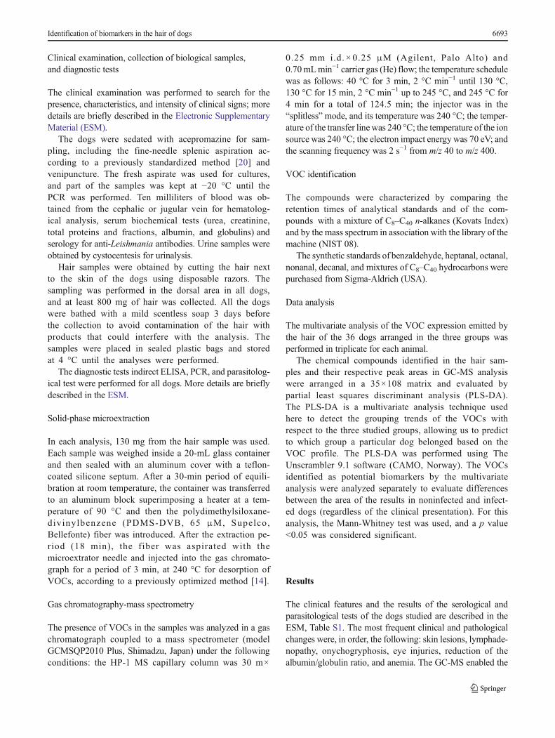
Clinical examination, collection of biological samples,and diagnostic tests
The clinical examination was performed to search for thepresence, characteristics, and intensity of clinical signs; moredetails are briefly described in the Electronic SupplementaryMaterial (ESM).
The dogs were sedated with acepromazine for sam-pling, including the fine-needle splenic aspiration ac-cording to a previously standardized method [20] andvenipuncture. The fresh aspirate was used for cultures,and part of the samples was kept at −20 °C until thePCR was performed. Ten milliliters of blood was ob-tained from the cephalic or jugular vein for hematolog-ical analysis, serum biochemical tests (urea, creatinine,total proteins and fractions, albumin, and globulins) andserology for anti-Leishmania antibodies. Urine samples wereobtained by cystocentesis for urinalysis.
Hair samples were obtained by cutting the hair nextto the skin of the dogs using disposable razors. Thesampling was performed in the dorsal area in all dogs,and at least 800 mg of hair was collected. All the dogswere bathed with a mild scentless soap 3 days beforethe collection to avoid contamination of the hair withproducts that could interfere with the analysis. Thesamples were placed in sealed plastic bags and storedat 4 °C until the analyses were performed.
The diagnostic tests indirect ELISA, PCR, and parasitolog-ical test were performed for all dogs. More details are brieflydescribed in the ESM.
Solid-phase microextraction
In each analysis, 130 mg from the hair sample was used.Each sample was weighed inside a 20-mL glass containerand then sealed with an aluminum cover with a teflon-coated silicone septum. After a 30-min period of equili-bration at room temperature, the container was transferredto an aluminum block superimposing a heater at a tem-perature of 90 °C and then the polydimethylsiloxane-divinylbenzene (PDMS-DVB, 65 μM, Supelco,Bellefonte) fiber was introduced. After the extraction pe-riod (18 min), the fiber was aspirated with themicroextrator needle and injected into the gas chromato-graph for a period of 3 min, at 240 °C for desorption ofVOCs, according to a previously optimized method [14].
Gas chromatography-mass spectrometry
The presence of VOCs in the samples was analyzed in a gaschromatograph coupled to a mass spectrometer (modelGCMSQP2010 Plus, Shimadzu, Japan) under the followingconditions: the HP-1 MS capillary column was 30 m×
0.25 mm i.d. × 0.25 μM (Agilent, Palo Alto) and0.70mLmin−1 carrier gas (He) flow; the temperature schedulewas as follows: 40 °C for 3 min, 2 °C min−1 until 130 °C,130 °C for 15 min, 2 °C min−1 up to 245 °C, and 245 °C for4 min for a total of 124.5 min; the injector was in the“splitless”mode, and its temperature was 240 °C; the temper-ature of the transfer line was 240 °C; the temperature of the ionsource was 240 °C; the electron impact energy was 70 eV; andthe scanning frequency was 2 s−1 from m/z 40 to m/z 400.
VOC identification
The compounds were characterized by comparing theretention times of analytical standards and of the com-pounds with a mixture of C8–C40 n-alkanes (Kovats Index)and by the mass spectrum in association with the library of themachine (NIST 08).
The synthetic standards of benzaldehyde, heptanal, octanal,nonanal, decanal, and mixtures of C8–C40 hydrocarbons werepurchased from Sigma-Aldrich (USA).
Data analysis
The multivariate analysis of the VOC expression emitted bythe hair of the 36 dogs arranged in the three groups wasperformed in triplicate for each animal.
The chemical compounds identified in the hair sam-ples and their respective peak areas in GC-MS analysiswere arranged in a 35×108 matrix and evaluated bypartial least squares discriminant analysis (PLS-DA).The PLS-DA is a multivariate analysis technique usedhere to detect the grouping trends of the VOCs withrespect to the three studied groups, allowing us to predictto which group a particular dog belonged based on theVOC profile. The PLS-DA was performed using TheUnscrambler 9.1 software (CAMO, Norway). The VOCsidentified as potential biomarkers by the multivariateanalysis were analyzed separately to evaluate differencesbetween the area of the results in noninfected and infect-ed dogs (regardless of the clinical presentation). For thisanalysis, the Mann-Whitney test was used, and a p value<0.05 was considered significant.
Results
The clinical features and the results of the serological andparasitological tests of the dogs studied are described in theESM, Table S1. The most frequent clinical and pathologicalchanges were, in order, the following: skin lesions, lymphade-nopathy, onychogryphosis, eye injuries, reduction of thealbumin/globulin ratio, and anemia. The GC-MS enabled the
Identification of biomarkers in the hair of dogs 6693
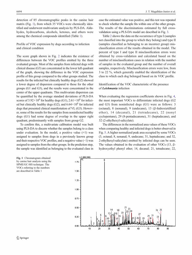
detection of 85 chromatographic peaks in the canine hairmatrix (Fig. 1), from which 35 VOCs were chemically iden-tified and underwent multivariate analysis by PLS-DA. Alde-hydes, hydrocarbons, alcohols, ketones, and ethers wereamong the chemical compounds identified (Table 1).
Profile of VOC expression by dogs according to infectionand clinical condition
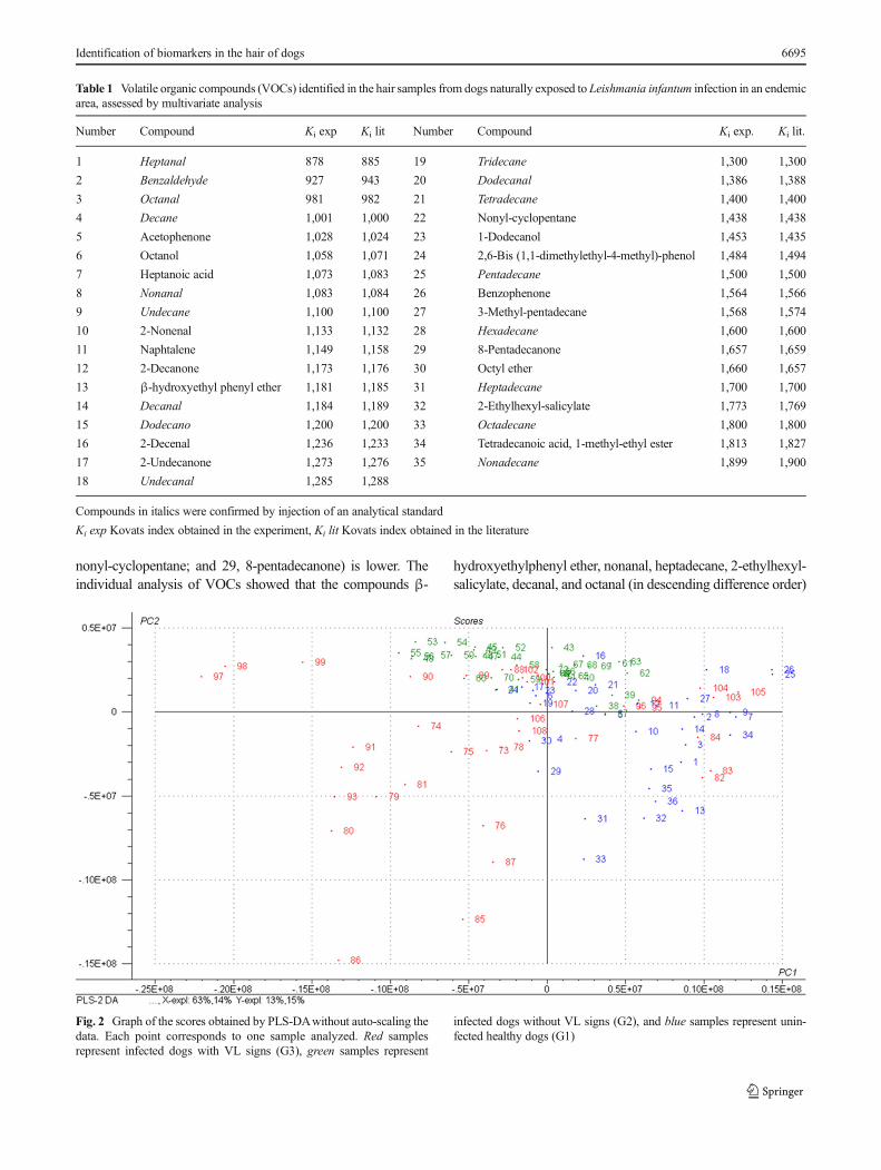
The score graph shown in Fig. 2 indicates the existence ofdifferences between the VOC profiles emitted by the threeevaluated groups. Most of the samples from infected dogs withclinical disease (G3) are concentrated in the lower left quadrantof the graph, showing the difference in the VOC expressionprofile of this group compared to the other groups studied. Theresults for the infected but clinically healthy dogs (G2) showeda lower degree of dispersion compared to those for the othergroups (G1 and G3), and the results were concentrated in thecenter of the upper quadrants. This multivariate dispersion canbe quantified by the average standard deviations of PLS-DAscores of 3.92×10⁶ for healthy dogs (G1), 2.61×10⁶ for infect-ed but clinically healthy dogs (G2), and 6.64×10⁶ for infecteddogs that presented clinical manifestation of VL (G3). Howev-er, some of the results for the samples from noninfected healthydogs (G1) had some degree of overlap in the upper rightquadrant, predominantly with samples from group G2.
To confirm this, a multivariate calibration model was builtusing PLS-DA to discern whether the samples belong to a classunder evaluation. In the model, a positive value (+1) wasassigned to samples from dogs in a previously known groupand their respective VOC profiles, and a negative value (−1) wasassigned to samples from the other groups. In the prediction step,the sample was identified as belonging to the evaluated class in
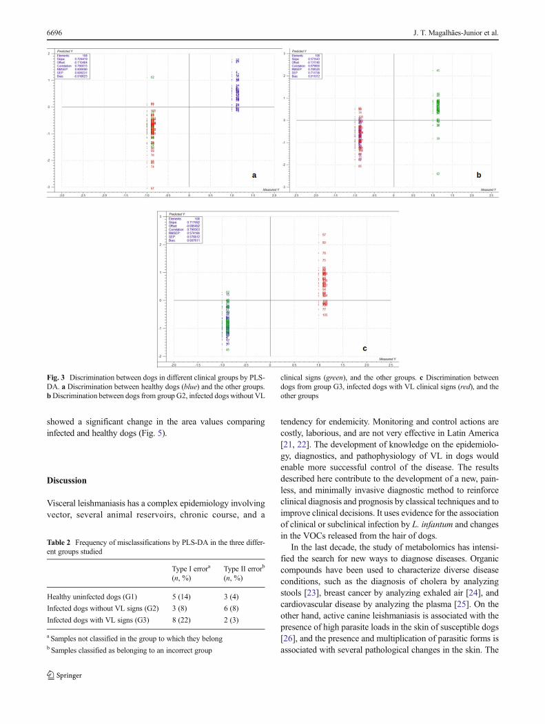
case the estimated value was positive, and this test was repeatedto check whether the sample fits within one of the other groups.The results of the discriminations estimated by full cross-validation using a PLS-DA model are described in Fig. 3.
Table 2 shows the data on the occurrence of type I (samplesnot classified into the group to which they belong) and type II(samples classified as belonging to an incorrect group) mis-classification errors of the results obtained in the model. Thepercent of type I and type II misclassification errors wereobtained by cross-validation and calculated based on thenumber of misclassification cases in relation with the numberof samples in the evaluated group and the number of overallsamples, respectively. Misclassification errors were low, from3 to 22 %, which generally enabled the identification of theclass to which each dog belonged based on its VOC profile.
Identification of the VOC characteristic of the presenceof Leishmania infection
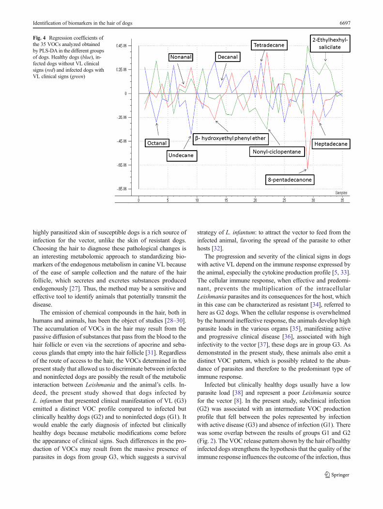
When evaluating the regression coefficients shown in Fig. 4,the most important VOCs to differentiate infected dogs (G2and G3) from noninfected dogs (G1) were as follows: 3(octanal), 8 (nonanal), 9 (undecane), 13 (β-hidroxietilfenilether), 14 (decanal), 21 (tetradecane), 22 (nonylcyclopentane), 29 (8-pentadecanone), 31 (heptadecane), and32 (2-ethylhexyl-salicylate).
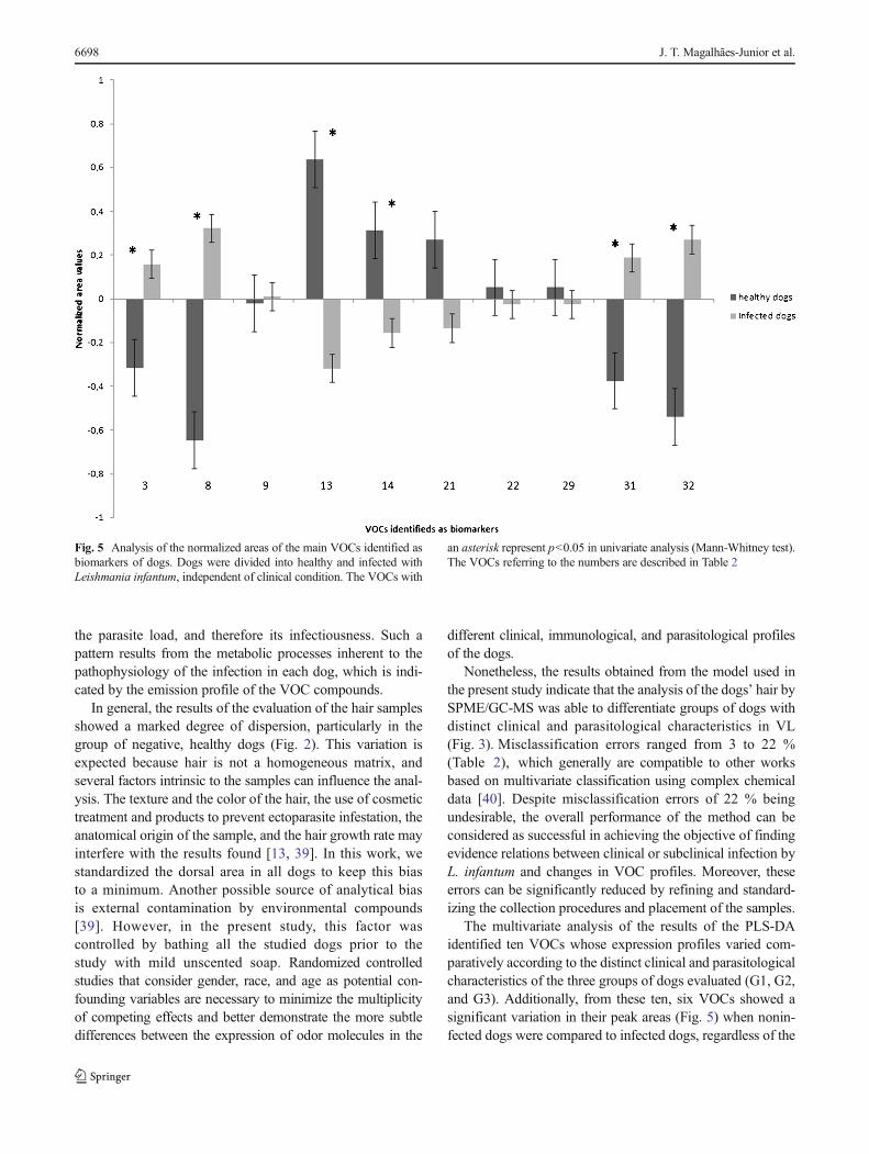
The differences in the normalized area values of these VOCswhen comparing healthy and infected dogs is better observed inFig. 5. A higher normalized peak area occupied by some VOCs(3, octanal; 8, nonanal; 9, undecane; 31, heptadecane; and 32,2-ethylhexyl-salicylate) emitted by infected dogs can be seen.The values obtained in the evaluation of other VOCs (13, β-hydroxyethyl phenyl ether; 14, decanal; 21, tetradecane; 22,
Fig. 1 Chromatogram obtainedby canine hair analysis using theSPME/GC-MS technique. TheVOCs referring to the numbersare described in Table 1
6694 J. T. Magalhães-Junior et al.
nonyl-cyclopentane; and 29, 8-pentadecanone) is lower. Theindividual analysis of VOCs showed that the compounds β-
hydroxyethylphenyl ether, nonanal, heptadecane, 2-ethylhexyl-salicylate, decanal, and octanal (in descending difference order)
Fig. 2 Graph of the scores obtained by PLS-DAwithout auto-scaling thedata. Each point corresponds to one sample analyzed. Red samplesrepresent infected dogs with VL signs (G3), green samples represent
infected dogs without VL signs (G2), and blue samples represent unin-fected healthy dogs (G1)
Table 1 Volatile organic compounds (VOCs) identified in the hair samples from dogs naturally exposed to Leishmania infantum infection in an endemicarea, assessed by multivariate analysis
Number Compound Ki exp Ki lit Number Compound Ki exp. Ki lit.
1 Heptanal 878 885 19 Tridecane 1,300 1,300
2 Benzaldehyde 927 943 20 Dodecanal 1,386 1,388
3 Octanal 981 982 21 Tetradecane 1,400 1,400
4 Decane 1,001 1,000 22 Nonyl-cyclopentane 1,438 1,438
5 Acetophenone 1,028 1,024 23 1-Dodecanol 1,453 1,435
6 Octanol 1,058 1,071 24 2,6-Bis (1,1-dimethylethyl-4-methyl)-phenol 1,484 1,494
7 Heptanoic acid 1,073 1,083 25 Pentadecane 1,500 1,500
8 Nonanal 1,083 1,084 26 Benzophenone 1,564 1,566
9 Undecane 1,100 1,100 27 3-Methyl-pentadecane 1,568 1,574
10 2-Nonenal 1,133 1,132 28 Hexadecane 1,600 1,600
11 Naphtalene 1,149 1,158 29 8-Pentadecanone 1,657 1,659
12 2-Decanone 1,173 1,176 30 Octyl ether 1,660 1,657
13 β-hydroxyethyl phenyl ether 1,181 1,185 31 Heptadecane 1,700 1,700
14 Decanal 1,184 1,189 32 2-Ethylhexyl-salicylate 1,773 1,769
15 Dodecano 1,200 1,200 33 Octadecane 1,800 1,800
16 2-Decenal 1,236 1,233 34 Tetradecanoic acid, 1-methyl-ethyl ester 1,813 1,827
17 2-Undecanone 1,273 1,276 35 Nonadecane 1,899 1,900
18 Undecanal 1,285 1,288
Compounds in italics were confirmed by injection of an analytical standard
Ki exp Kovats index obtained in the experiment, Ki lit Kovats index obtained in the literature
Identification of biomarkers in the hair of dogs 6695
showed a significant change in the area values comparinginfected and healthy dogs (Fig. 5).
Discussion
Visceral leishmaniasis has a complex epidemiology involvingvector, several animal reservoirs, chronic course, and a
tendency for endemicity. Monitoring and control actions arecostly, laborious, and are not very effective in Latin America[21, 22]. The development of knowledge on the epidemiolo-gy, diagnostics, and pathophysiology of VL in dogs wouldenable more successful control of the disease. The resultsdescribed here contribute to the development of a new, pain-less, and minimally invasive diagnostic method to reinforceclinical diagnosis and prognosis by classical techniques and toimprove clinical decisions. It uses evidence for the associationof clinical or subclinical infection by L. infantum and changesin the VOCs released from the hair of dogs.
In the last decade, the study of metabolomics has intensi-fied the search for new ways to diagnose diseases. Organiccompounds have been used to characterize diverse diseaseconditions, such as the diagnosis of cholera by analyzingstools [23], breast cancer by analyzing exhaled air [24], andcardiovascular disease by analyzing the plasma [25]. On theother hand, active canine leishmaniasis is associated with thepresence of high parasite loads in the skin of susceptible dogs[26], and the presence and multiplication of parasitic forms isassociated with several pathological changes in the skin. The
Fig. 3 Discrimination between dogs in different clinical groups by PLS-DA. a Discrimination between healthy dogs (blue) and the other groups.bDiscrimination between dogs from group G2, infected dogs without VL
clinical signs (green), and the other groups. c Discrimination betweendogs from group G3, infected dogs with VL clinical signs (red), and theother groups
Table 2 Frequency of misclassifications by PLS-DA in the three differ-ent groups studied
Type I errora
(n, %)Type II errorb
(n, %)
Healthy uninfected dogs (G1) 5 (14) 3 (4)
Infected dogs without VL signs (G2) 3 (8) 6 (8)
Infected dogs with VL signs (G3) 8 (22) 2 (3)
a Samples not classified in the group to which they belongb Samples classified as belonging to an incorrect group
6696 J. T. Magalhães-Junior et al.
highly parasitized skin of susceptible dogs is a rich source ofinfection for the vector, unlike the skin of resistant dogs.Choosing the hair to diagnose these pathological changes isan interesting metabolomic approach to standardizing bio-markers of the endogenous metabolism in canine VL becauseof the ease of sample collection and the nature of the hairfollicle, which secretes and excretes substances producedendogenously [27]. Thus, the method may be a sensitive andeffective tool to identify animals that potentially transmit thedisease.
The emission of chemical compounds in the hair, both inhumans and animals, has been the object of studies [28–30].The accumulation of VOCs in the hair may result from thepassive diffusion of substances that pass from the blood to thehair follicle or even via the secretions of apocrine and seba-ceous glands that empty into the hair follicle [31]. Regardlessof the route of access to the hair, the VOCs determined in thepresent study that allowed us to discriminate between infectedand noninfected dogs are possibly the result of the metabolicinteraction between Leishmania and the animal’s cells. In-deed, the present study showed that dogs infected byL. infantum that presented clinical manifestation of VL (G3)emitted a distinct VOC profile compared to infected butclinically healthy dogs (G2) and to noninfected dogs (G1). Itwould enable the early diagnosis of infected but clinicallyhealthy dogs because metabolic modifications come beforethe appearance of clinical signs. Such differences in the pro-duction of VOCs may result from the massive presence ofparasites in dogs from group G3, which suggests a survival
strategy of L. infantum: to attract the vector to feed from theinfected animal, favoring the spread of the parasite to otherhosts [32].
The progression and severity of the clinical signs in dogswith active VL depend on the immune response expressed bythe animal, especially the cytokine production profile [5, 33].The cellular immune response, when effective and predomi-nant, prevents the multiplication of the intracellularLeishmania parasites and its consequences for the host, whichin this case can be characterized as resistant [34], referred tohere as G2 dogs. When the cellular response is overwhelmedby the humoral ineffective response, the animals develop highparasite loads in the various organs [35], manifesting activeand progressive clinical disease [36], associated with highinfectivity to the vector [37], these dogs are in group G3. Asdemonstrated in the present study, these animals also emit adistinct VOC pattern, which is possibly related to the abun-dance of parasites and therefore to the predominant type ofimmune response.
Infected but clinically healthy dogs usually have a lowparasite load [38] and represent a poor Leishmania sourcefor the vector [8]. In the present study, subclinical infection(G2) was associated with an intermediate VOC productionprofile that fell between the poles represented by infectionwith active disease (G3) and absence of infection (G1). Therewas some overlap between the results of groups G1 and G2(Fig. 2). The VOC release pattern shown by the hair of healthyinfected dogs strengthens the hypothesis that the quality of theimmune response influences the outcome of the infection, thus
Fig. 4 Regression coefficients ofthe 35 VOCs analyzed obtainedby PLS-DA in the different groupsof dogs. Healthy dogs (blue), in-fected dogs without VL clinicalsigns (red) and infected dogs withVL clinical signs (green)
Identification of biomarkers in the hair of dogs 6697
the parasite load, and therefore its infectiousness. Such apattern results from the metabolic processes inherent to thepathophysiology of the infection in each dog, which is indi-cated by the emission profile of the VOC compounds.
In general, the results of the evaluation of the hair samplesshowed a marked degree of dispersion, particularly in thegroup of negative, healthy dogs (Fig. 2). This variation isexpected because hair is not a homogeneous matrix, andseveral factors intrinsic to the samples can influence the anal-ysis. The texture and the color of the hair, the use of cosmetictreatment and products to prevent ectoparasite infestation, theanatomical origin of the sample, and the hair growth rate mayinterfere with the results found [13, 39]. In this work, westandardized the dorsal area in all dogs to keep this biasto a minimum. Another possible source of analytical biasis external contamination by environmental compounds[39]. However, in the present study, this factor wascontrolled by bathing all the studied dogs prior to thestudy with mild unscented soap. Randomized controlledstudies that consider gender, race, and age as potential con-founding variables are necessary to minimize the multiplicityof competing effects and better demonstrate the more subtledifferences between the expression of odor molecules in the
different clinical, immunological, and parasitological profilesof the dogs.
Nonetheless, the results obtained from the model used inthe present study indicate that the analysis of the dogs’ hair bySPME/GC-MS was able to differentiate groups of dogs withdistinct clinical and parasitological characteristics in VL(Fig. 3). Misclassification errors ranged from 3 to 22 %(Table 2), which generally are compatible to other worksbased on multivariate classification using complex chemicaldata [40]. Despite misclassification errors of 22 % beingundesirable, the overall performance of the method can beconsidered as successful in achieving the objective of findingevidence relations between clinical or subclinical infection byL. infantum and changes in VOC profiles. Moreover, theseerrors can be significantly reduced by refining and standard-izing the collection procedures and placement of the samples.
The multivariate analysis of the results of the PLS-DAidentified ten VOCs whose expression profiles varied com-paratively according to the distinct clinical and parasitologicalcharacteristics of the three groups of dogs evaluated (G1, G2,and G3). Additionally, from these ten, six VOCs showed asignificant variation in their peak areas (Fig. 5) when nonin-fected dogs were compared to infected dogs, regardless of the
Fig. 5 Analysis of the normalized areas of the main VOCs identified asbiomarkers of dogs. Dogs were divided into healthy and infected withLeishmania infantum, independent of clinical condition. The VOCs with
an asterisk represent p<0.05 in univariate analysis (Mann-Whitney test).The VOCs referring to the numbers are described in Table 2
6698 J. T. Magalhães-Junior et al.
clinical presentation, which defines them as potential bio-markers of L. infantum infection in dogs.
In conclusion, the VOCs emitted in the hair of dogscan be used both as biomarkers of the presence orabsence of L. infantum infection and as indicators ofthe intensity of disease, thus parasitism, which variesaccording to the clinical condition. Indication of theprobable degree of infectiousness, as indicated by theprofile found in the analysis of the dog hair samples bySPME/GC-MS, may be an important diagnostic tool toadvance the study and control of visceral leishmaniasisand should be thoroughly investigated, including itsdirect comparison with the current methods of screen-ing. The characteristics inherent to the nature of thesamples also suggest a benefit in the field of animalwelfare and in the knowledge of the interactions betweenparasite and host.
Acknowledgments This study was performed with the support of theNational Council for Scientific and Technological Development(Conselho Nacional de Desenvolvimento Científico e Tecnológico -CNPq – 479753/2009-1) and the Bahia State Research Foundation(Fundação deAmparo à Pesquisa do Estado da Bahia - FAPESB – PedidoNo. 498/ 2011 and No. 1799/2012). We thank the Brazilian FederalAgency for the Support and Evaluation of Graduate Education(Coordenação de Aperfeiçoamento de Pessoal de Nível Superior -CAPES) for granting a master’s degree scholarship (J.T. Magalhães-Junior). The authors thank Clauceane de Jesus (LIVE-UFBA) for helpingwith serological and parasitological tests and Estéfane da C. Nunes(EBDA) for helping with the chemical identification of the volatileorganic compounds.
References
1. WHO (2011) Leishmaniasis: worldwide epidemiological and drugaccess update. WHO, Geneva
2. Alvar J, Vélez ID, Bern C, Herrero M, Desjeux P, Cano J, Jannin J,den Boer M (2012) Leishmaniasis worldwide and global estimates ofits incidence. PLoS One 7:e35671
3. Lainson R, Rangel EF (2005) Lutzomyia longipalpis and the eco-epidemiology of American visceral leishmaniasis, with particularreference to Brazil: a review. Mem Inst Oswaldo Cruz 100:811–827
4. Silva AVM, Paula AA, Cabrera MAA, Carreira JCA (2005)Leishmaniasis in domestic dogs: epidemiological aspects. CadSaude Publica 21:324–328
5. Kaye PM, Svensson M, Ato M, Maroof A, Polley R, Stager S,Zubairi S, Engwerda CR (2004) The immunopathology of experi-mental visceral leishmaniasis. Immunol Rev 201:239–253
6. SanchezMA, Diaz NL, ZerpaO, Negron E, Convit J, Tapia FJ (2004)Organ-specific immunity in canine visceral leishmaniasis: analysis ofsymptomatic and asymptomatic dogs naturally infected withLeishmania chagasi. Am J Trop Med Hyg 70:618–624
7. Da Costa-Val AP, Cavalcanti RR, de Figueiredo Gontijo N,Marques Michalick MS, Alexander B, Williams P, Melo MN(2007) Canine visceral leishmaniasis: relationships betweenclinical status, humoral immune response, haematology andLutzomyia (Lutzomyia) longipalpis infectivity. Vet J 174:636–643.doi:10.1016/j.tvjl.2006.11.006
8. Travi BL, Tabares CJ, Cadena H, Ferro C, Osorio Y (2001) Caninevisceral leishmaniasis in Colombia: relationship between clinical andparasitologic status and infectivity for sand flies. Am J TropMedHyg64:119–1249
9. Daviss B (2005) Growing pains for metabolomics. Scientist 19:25–2810. van der Greef J, Smilde AK (2005) Symbiosis of chemometrics and
metabolomics: past, present, and future. J Chemometr 19:376–38611. Stenn K, Paus R (2001) Controls of hair follicle cycling. Physiol Rev
81:449–49412. Gratacos-Cubarsi M, Castellari M, Valero A, Garcia-Regueiro J
(2006) Hair analysis for veterinary drug monitoring in livestockproduction. J Chromatogr B 834:14–25
13. Vincenti M, Salomone A, Gerace E, Pirro V (2013) Application ofmass spectrometry to hair analysis for forensic toxicological investi-gations. Mass Spectrom Rev 32:312–332
14. Oliveira LS, Rodrigues FM, de Oliveira FS, Mesquita PR, Leal DC,Alcântara AC, Souza BM, Franke CR, Pereira PAP, de Andrade JB(2008) Headspace solid phase microextraction/gas chromatography–mass spectrometry combined to chemometric analysis for volatileorganic compounds determination in canine hair: a new tool to detectdog contamination by visceral leishmaniasis. J Chromatogr B 875:392–398
15. Zhang Z, Li G (2010) A review of advances and new developmentsin the analysis of biological volatile organic compounds. MicrochemJ 95:127–139
16. Hantao LW, Aleme HG, Passador MM, Furtado EL, Ribeiro FAL,Poppi RJ, Augusto F (2013) Determination of disease biomarkers inEucalyptus by comprehensive two-dimensional gas chromatographyand multivariate data analysis. J Chromatogr A 1279:86–91. doi:10.1016/j.chroma.2013.01.013
17. Phillips M, Cataneo RN, Ditkoff BA, Fisher P, Greenberg J,Gunawardena R, Kwon CS, Rahbari-Oskoui F, Wong C (2003)Volatile markers of breast cancer in the breath. Breast J 9:184–191.doi:10.1046/j.1524-4741.2003.09309.x
18. Di Natale C, Macagnano A, Martinelli E, Paolesse R, D’ArcangeloG, Roscioni C, Finazzi-Agrò A, D’Amico A (2003) Lung canceridentification by the analysis of breath by means of an array of non-selective gas sensors. Biosens Bioelectron 18:1209–1218. doi:10.1016/S0956-5663(03)00086-1
19. Dalton P, Gelperin A, Preti G (2004) Volatile metabolic monitoring ofglycemic status in diabetes using electronic olfaction. DiabetesTechnol 6:534–544
20. Barrouin-Melo SM, Larangeira DF, de Andrade Filho FA, Trigo J,Juliao FS, Franke CR, Palis Aguiar PH, Conrado dos-Santos WL,Pontes-de-Carvalho L (2006) Can spleen aspirations be safely usedfor the parasitological diagnosis of canine visceral leishmaniosis? Astudy on assymptomatic and polysymptomatic animals. Vet J 171:331–339. doi:10.1016/j.tvjl.2004.11.010
21. Romero GA, Boelaert M (2010) Control of visceral leishmaniasis inLatin America—a systematic review. PLoS Negl Trop Dis 4:e584
22. Ribeiro VM, da Silva SM,Menz I, Tabanez P, dos Santos Nogueira F,Werkhaüser M, da Fonseca ALS, Dantas-Torres F (2013) Control ofvisceral leishmaniasis in Brazil: recommendations from Brasileish.Parasitol Vectors 6:8
23. Garner C, Smith S, Bardhan P, Ratcliffe N, Probert C (2009) A pilotstudy of faecal volatile organic compounds in faeces from cholerapatients in Bangladesh to determine their utility in disease diagnosis.Trans R Soc Trop Med Hyg 103:1171–1173
24. Mandy M, Cornelia F, Malgorzata L, Oliver S, Achim S, Dorothee S(2012) Volatile organic compounds (VOCs) in exhaled breath ofpatients with breast cancer in a clinical setting. Ginekol Pol 83:730–736
25. Rupérez FJ, Ramos-Mozo P, Teul J, Martinez-Pinna R, Garcia A,Malet-Martino M, Camafeita E, Lopez JA, Pastor-Vargas C, Egido J(2012) Metabolomic study of plasma of patients with abdominalaortic aneurysm. Anal Bioanal Chem 403:1651–1660
Identification of biomarkers in the hair of dogs 6699
26. Queiroz NM, da Silveira R, Fau - de Noronha ACF Jr, de Noronha AJr, Fau - Oliveira TMFS, Oliveira T, Fau - Machado RZ, Machado R,Fau - Starke-Buzetti WA, Starke-Buzetti WA (2011) Detection ofLeishmania (L.) chagasi in canine skin. Vet Parasitol 178(1873–2550(Electronic)):1–8. doi:10.1016/j.vetpar.2010.12.033
27. Harkey M (1993) Anatomy and physiology of hair. Forensic Sci Int63:9–18
28. Dorea J, Costa J, Holzbecher J, Ryan D,Marsden P (1987) Antimonyaccumulation in hair during treatment of leishmaniasis. Clin Chem33:2081–2082
29. Agusa T, Kunito T, Iwata H, Monirith I, Chamnan C, TanaTS, Subramanian A, Tanabe S (2007) Mercury in hair andblood from residents of Phnom Penh (Cambodia) and possi-ble effect on serum hormone levels. Chemosphere 68:590–596
30. Anielski P (2008) Hair analysis of anabolic steroids in connectionwith doping control—results from horse samples. J Mass Spectrom43:1001–1008
31. Henderson G (1993) Mechanisms of drug incorporation into hair.Forensic Sci Int 63:19–29
32. Knols BGJ, Meijerink J (1997) Odors influence mosquito behavior.Sci Med 4:56–63
33. Solano-Gallego L, Miró G, Koutinas A, Cardoso L, Pennisi MG,Ferrer L, Bourdeau P, Oliva G, Baneth G (2011) LeishVet guide-lines for the practical management of canine leishmaniosis.Parasitol Vectors 4:1–16
34. Grimaldi G, Tesh R (1993) Leishmaniases of the NewWorld: currentconcepts and implications for future research. Clin Microbiol Rev 6:230–250
35. Alves CF, de Amorim IF, Moura EP, Ribeiro RR, Alves CF, MichalickMS, Kalapothakis E, Bruna-RomeroO, TafuriWL, TeixeiraMM (2009)Expression of IFN-γ, TNF-α, IL-10 and TGF-β in lymph nodes asso-ciates with parasite load and clinical form of disease in dogs naturallyinfected with Leishmania (Leishmania) chagasi. Vet ImmunolImmunopathol 128(4):349–358
36. Pinelli E, Killick-Kendrick R,Wagenaar J, BernadinaW, Del Real G,Ruitenberg J (1994) Cellular and humoral immune responses in dogsexperimentally and naturally infected with Leishmania infantum.Infect Immun 62:229–235
37. Guarga JL, Moreno J, Lucientes J, Gracia MJ, Peribáñez MA, AlvarJ, Castillo JA (2000) Canine leishmaniasis transmission: higher in-fectivity amongst naturally infected dogs to sand flies is associatedwith lower proportions of T helper cells. Res Vet Sci 69:249–253.doi:10.1053/rvsc.2000.0419
38. Manna L, Reale S, Vitale F, Gravino AE (2009) Evidence for arelationship between Leishmania load and clinical manifestations.Res Vet Sci 87:76–78
39. Wennig R (2000) Potential problems with the interpretation of hairanalysis results. Forensic Sci Int 107:5–12
40. Brereton R (2009) Chemometrics for pattern recognition.http://www.wiley.com/WileyCDA/WileyTitle/productCd-0470987251.html
6700 J. T. Magalhães-Junior et al.