Identification of biofloc microscopic composition as the ...
10
ORIGINAL ARTICLE Identification of biofloc microscopic composition as the natural bioremediation in zero water exchange of Pacific white shrimp, Penaeus vannamei, culture in closed hatchery system Hidayah Manan 1 • Julia Hwei Zhong Moh 1 • Nor Azman Kasan 2 • Suhaimi Suratman 3 • Mhd Ikhwanuddin 1 Received: 13 December 2015 / Accepted: 9 May 2016 / Published online: 16 June 2016 Ó The Author(s) 2016. This article is published with open access at Springerlink.com Abstract Study on the microscopic composition of biofloc in closed hatchery culture system was carried out to determine the interaction between the aggregation flocs in the bioreme- diation process for the decomposition and degradation of organic matter loaded in the shrimp culture tanks. The study was done for 105 days of culture period in zero water exchange. All of the organic loaded in the culture tanks identified comes from the shrimp feces, uneaten fed, and the decomposed macro- and microorganisms died in the culture tanks. All of the microscopic organisms in the biofloc were identified using Advance microscopes Nikon 80i. From the present study, there were abundances and high varieties of phytoplankton, zooplankton, protozoa, nematodes and algae species identified as aggregates together in the flocs accu- mulation. All of these microscopic organisms identified implemented the symbiotic process together for food supply, become the algae grazer, act as natural water stabilizer in regulating the nutrients in culture tank and serve as decom- poser for dead organic matter in the water environment. Heterotrophic bacteria identified from Pseudomonas and Aeromonas family consumed the organic matter loaded at the bottom of culture tank and converted items through chemical process as useful protein food to be consumed back by the shrimp. Overall it can be concluded that the biofloc organisms identified really contributed as natural bioremediation agents in zero water exchange culture system to ensure the water quality in the optimal condition until the end of culture period. Keywords Biofloc compositions Organic matter Bioremediation Symbiotic process Introduction Bioremediation is a process where microorganisms were stimulated with nutrients and other chemicals to enable them to wipe out contaminants in the targeted area and break down the hazardous substances into less toxic or non-toxic sub- stances (Das 2014). Microorganisms are considered as the first living organisms to have evolved and are adaptive with the ecological changes. Nowadays, the use of microorgan- isms such as bacteria as biodegradation and bioremediation agent has come to attention because of its ability to reduce hazard, success in degrading natural and synthetic substances and accumulating toxic compound (Karigar and Rao 2011). According to Das et al. (2006), microorganisms are respon- sible for carbon fixation, nitrogen fixation, methane meta- bolism and sulfur metabolism, thus controlling the biogeochemical cycle. Microorganisms are able to produce diverse metabolic enzymes that can assist for safe removal of contaminants either by direct destruction or converting to safer or less toxic intermediate (Dash and Das 2012). Any microorganisms used as bioremediation has to possess resistant genotype for the particular pollutant and because of it, microorganisms possess certain unique characteristic which make them suitable for bioremediation processes (Stelting et al. 2010). & Mhd Ikhwanuddin [email protected] Hidayah Manan [email protected] 1 Institute of Tropical Aquaculture, Universiti Malaysia Terengganu, 21030 Kuala Nerus, Terengganu, Malaysia 2 School of Fisheries and Aquaculture Sciences, Universiti Malaysia Terengganu, 21030 Kuala Nerus, Terengganu, Malaysia 3 Institute of Oceanography and Environment, Universiti Malaysia Terengganu, 21030 Kuala Nerus, Terengganu, Malaysia 123 Appl Water Sci (2017) 7:2437–2446 DOI 10.1007/s13201-016-0421-4
Transcript of Identification of biofloc microscopic composition as the ...

ORIGINAL ARTICLE
Identification of biofloc microscopic composition as the naturalbioremediation in zero water exchange of Pacific white shrimp,Penaeus vannamei, culture in closed hatchery system
Hidayah Manan1 • Julia Hwei Zhong Moh1 • Nor Azman Kasan2 • Suhaimi Suratman3 •
Mhd Ikhwanuddin1
Received: 13 December 2015 / Accepted: 9 May 2016 / Published online: 16 June 2016
� The Author(s) 2016. This article is published with open access at Springerlink.com
Abstract Study on themicroscopic composition of biofloc in
closed hatchery culture system was carried out to determine
the interaction between the aggregation flocs in the bioreme-
diation process for the decomposition and degradation of
organic matter loaded in the shrimp culture tanks. The study
was done for 105 days of culture period in zero water
exchange. All of the organic loaded in the culture tanks
identified comes from the shrimp feces, uneaten fed, and the
decomposed macro- and microorganisms died in the culture
tanks. All of the microscopic organisms in the biofloc were
identified using Advance microscopes Nikon 80i. From the
present study, there were abundances and high varieties of
phytoplankton, zooplankton, protozoa, nematodes and algae
species identified as aggregates together in the flocs accu-
mulation. All of these microscopic organisms identified
implemented the symbiotic process together for food supply,
become the algae grazer, act as natural water stabilizer in
regulating the nutrients in culture tank and serve as decom-
poser for dead organic matter in the water environment.
Heterotrophic bacteria identified from Pseudomonas and
Aeromonas family consumed the organic matter loaded at the
bottom of culture tank and converted items through chemical
process as useful protein food to be consumed back by the
shrimp. Overall it can be concluded that the biofloc organisms
identified really contributed as natural bioremediation agents
in zero water exchange culture system to ensure the water
quality in the optimal condition until the end of culture period.
Keywords Biofloc compositions � Organic matter �Bioremediation � Symbiotic process
Introduction
Bioremediation is a process where microorganisms were
stimulated with nutrients and other chemicals to enable them
to wipe out contaminants in the targeted area and break down
the hazardous substances into less toxic or non-toxic sub-
stances (Das 2014). Microorganisms are considered as the
first living organisms to have evolved and are adaptive with
the ecological changes. Nowadays, the use of microorgan-
isms such as bacteria as biodegradation and bioremediation
agent has come to attention because of its ability to reduce
hazard, success in degrading natural and synthetic substances
and accumulating toxic compound (Karigar and Rao 2011).
According to Das et al. (2006), microorganisms are respon-
sible for carbon fixation, nitrogen fixation, methane meta-
bolism and sulfur metabolism, thus controlling the
biogeochemical cycle. Microorganisms are able to produce
diverse metabolic enzymes that can assist for safe removal of
contaminants either by direct destruction or converting to
safer or less toxic intermediate (Dash and Das 2012). Any
microorganisms used as bioremediation has to possess
resistant genotype for the particular pollutant and because of
it, microorganisms possess certain unique characteristic
which make them suitable for bioremediation processes
(Stelting et al. 2010).
& Mhd Ikhwanuddin
Hidayah Manan
1 Institute of Tropical Aquaculture, Universiti Malaysia
Terengganu, 21030 Kuala Nerus, Terengganu, Malaysia
2 School of Fisheries and Aquaculture Sciences, Universiti
Malaysia Terengganu, 21030 Kuala Nerus, Terengganu,
Malaysia
3 Institute of Oceanography and Environment, Universiti
Malaysia Terengganu, 21030 Kuala Nerus, Terengganu,
Malaysia
123
Appl Water Sci (2017) 7:2437–2446
DOI 10.1007/s13201-016-0421-4
Biofloc technology (BFT) is a promising technology
which promotes the retention of waste and its conversion to
biofloc as natural food for shrimp in the aquaculture system
(Panigrahi et al. 2014). Biofloc consists of microorganisms
such as heterotrophic bacteria, algae (dinoflagellates and
diatoms), fungi, ciliates, flagellates, rotifers, nematodes,
metazoans and detritus that conglomerate together and per-
form symbiotic processes to maintain the water quality,
maintain bio-security, support high density of shrimp culture
and reduce water exchange in the aquaculture system. In the
biofloc technology (BFT) application, protein is utilized as a
feed for the shrimp when the heterotrophic microbe in the
biofloc converts the nitrogenous waste in the culture tank
from the uneaten feed into protein. Development of dense
heterotrophic bacterial community rather than algae domi-
nated will overcome the waste generated in the aquaculture
system through in situ bioremediation (Panigrahi et al.
2014). By addition of cheap carbohydrate sources such as
molasses or tapioca usually in ration around C: N 12–15:1 in
the water column, biofloc will convert the toxic nutrients in
the water to beneficial food sources for shrimp consumption.
Avnimelech (2009) found out that in high stocking density
and zero or minimal water exchange the additional carbon
source encourage the development of heterotrophic bacteria
in the pond or tank. Schneider et al. (2005) discovered that
addition of organic nitrogenous waste, ammonium will be
converted into bacterial biomass if C:N ratio is balanced at
ratio 10–15:1. Usually in BFT, heterotrophic bacteria are
more dominant than nitrifying bacteria because of their
higher growth rate andmicrobial biomass yield per substrate,
thus making many fold increase of heterotrophic bacteria
(Hargreaves 2006). Identification of the microscopic biofloc
composition can help in better understanding the application
of biofloc. From the identification of each class of organisms’
function (phytoplankton as primary producer, zooplankton
as the algae grazer, bacteria and protozoa as organic matter
decomposer) that occurs in the zero water exchange culture
system, the interaction happening between the organisms in
the biofloc system can be understood. Because of the
potential of biofloc technology for bioremediation in the
aquaculture system, present study was conducted to identify
the biofloc microscopic composition and to determine the
biofloc performance as the natural bioremediation agent for
removal of organic matter loader in the culture tank in zero
water exchange system.
Methodology
Experimental design
Rounded tank with capacity 8 ton (height = 1.2 m and
diameter = 3.3 m) was stocked with Pacific White shrimp,
Penaeus vannamei, postlarvae at PL10 with density of 100
PL per cubic meter, m3. Six tanks were used in the
experiment for treatments (T1, T2 and T3 and for control
tanks, C1, C2 and C3). Molasses as carbohydrate or carbon
sources at ratio C:N 10:1 were transferred to the treatment
culture tank after being fermented for 24 h to boost the
breakdown process by the bacteria or the microorganisms
for biofloc formulation. Shrimp were cultured for 105 days
until reaching harvested size at PL115. During the culture
period, the microorganisms in the culture tank were sam-
pled every week to identify the microorganism’s compo-
sition in the biofloc aggregation in the closed and zero
water exchange system. All water parameters were checked
weekly for pH, salinity, DO, TDS, and temperature using
YSI multi-probe YSI 556 and nutrients (ammonia, nitrite,
nitrate) were analyzed with spectrophotometer by ammo-
nia–salicylate method (Standard Method 8155), nitrite
diazotization method (Standard Method 8507) and nitrate–
cadmium reduction method (Standard Method 8192) of
(DR/2400 Procedure manual 2002).
Sample collection
3 L of water sample from treatment tank was filtered using
plankton net 20 lm for microscopic plankton identifica-
tion. For bacterial identification, sample water was pipetted
out using micropipette and serial diluted until 10-5 for
bacteria analysis. Sample for plankton analysis was left
24 h for the substrate to settle at the bottom and concen-
trated to 10 ml of water sample and then preserved with
10 % formalin. All water samples were taken back to
laboratory for further analysis.
Microbial identification
Bacteria were isolated using trypticase soy agar (TSA) and
selective agar thiosulphate–citrate–bile salts–sucrose agar
(TCBS) for isolating Vibrio sp. The colony-forming unit
(CFU) from fifth time serial dilution (10-5) was selected
for colony counting. Gram staining also was done to
identify Gram-positive and -negative bacteria. Catalase test
was done to identify Gram-positive bacilli. API kit (Bio-
merieux) API20E and API 20NE were used to identify
Gram-negative bacteria. Incubation box, tray and lid were
prepared for the strip preparation. For the inoculum
preparation, an ampule of API NaCl 0.85 % (2 ml) was
selected and 1–4 colonies of bacteria were picked up using
inoculation loop from the agar plate and then suspension
was prepared with the turbidity equivalent to 0.5 McFar-
land. For the API 20NE strip inoculation, test nitrate
reduction (NO3) and p-nitrophenyl-b-D-galactopyranosidehydrolysis (PNPG) were inoculated by distributing the
saline suspension into the tubes using Pasteur pipette. API
2438 Appl Water Sci (2017) 7:2437–2446
123

AUX medium was added to approximately 200 ll of theremaining suspension into the ampule and was homoge-
nized well. Tubes and cupules of test glucose fermentation
(GLU) and Phenyl-acetate assimilation test (PAC) were
filled with the suspension. Mineral oil was added to the
cupules of 3 tests (GLU), arginine hydrolysis (ADH), and
urea hydrolysis (URE) until convex meniscuses formed.
The incubation box was closed and incubated at 29 ± 2 �Cfor 24 h. After the incubation period, the strips were read
by referring to the reading table. The reactions for (GLU,
ADH, URE, aesculin hydrolysis test (ESC)), gelatine
hydrolysis (GEL) and (PNPG) were recorded on result
sheet. For NO3 test, 1 drop of NIT 1 and 1 drop of NIT 2
reagents were added to NO3 cupule. For tryptophan
deaminase test (TRP), 1 drop of JAMES reagent was added
and immediate reaction took place. NIT 1, NIT 2 and
JAMES reagents were removed using pipette and test NO3
and TRP were covered with mineral oil. Kit was reincu-
bated at 29 ± 2 �C for 24 h and all tests were read again
except for NO3, TRP and underlined GLU, which were
only read once at 24 h. Identification is obtained with the
numerical profile. Database (V6.0) in the API web index
was used by entering the seven digit numerical profile in
the identification software for species identification.
For API20E strip inoculation, bacterial suspension were
distributed into the tubes with pipette for citrate assimila-
tion test (CIT), Voges–Proskauer (VP) test for acetyl
methyl carbinol detection, gelatine hydrolysis (GEL) test
by filled in both tube and cupules, and for test ADH, lysine
decarboxylase test (LDC), Ornithine decarboxylase test
(ODC) and production of hydrogen sulfide test (H2S) and
urea hydrolysis (URE) filled with mineral oil in the
cupules. The incubation box was closed and incubated at
36 ± 2 �C for 18–24 h. The strip was read by referring the
reading table after incubation period. For Tryptophan
deaminase test (TDA), 1 drop of TDA reagent was added,
for Indole production test (IND) 1 drop of JAMES reagent
was added and for VP test, VP 1 and VP 2 reagents were
added. Identification is obtained with numerical profile
nine digit using the database (v4.1) in the API web index
for species identification.
Plankton microscopic identification
Advance microscope Nikon 80i was used for biofloc
microscopic identification and for plankton length and size
measurements. Qualitative and quantitative analyses of
phytoplankton and zooplankton were done by Lackey’s
method. Compound microscope was used for phytoplankton
counting. The cover slip was placed over a drop of water in
the slide and whole of cover slip was examined by parallel
overlapping strips to count all the organisms in the drop.
About 22 strips were examined in each drop. Number of
subsamples to be taken depended on examining 2–3 suc-
cessive subsamples without addition of an encounter species
when compared to the examined subsamples in the same
sample (American Public Health Association APHA 1989).
Calculation formula:
Density cells l�1� �
¼ C � At
As � S� V
� volume of concentrated sample mlð Þvolume of actual water filtered
whereC Number of organisms counted, At Area of cover slip
(22 mm922 mm),SNumber of strip counted,AsAreaof strip
(22 mm91 mm), V Volume of sample under the cover slip.
Results
There were various types of microscopic organisms identified
from bioflocculation. All of the microscopic organisms
identified come from different classes of phytoplankton algae
and also numerous of algae grazer such as rotifer and nema-
tode also the protozoa, Vorticella sp. (Fig. 1). From Gram
staining, rod-shaped Gram-positive bacteria were also iden-
tified, which are Bacillus sp., from the positive result of
catalase test. From the API kit analysis, species of bacteria
identified come from heterotrophic bacteria (Aeromonas
hydrophila, Pseudomonas aeruginosa) and also anaerobic
bacteria, Vibrio sp. (ex. V. fluvialis). For the water parameter
results,meandissolvedoxygen,DO,was 6.67 ± 0.97 mg l-1
(5.9–9.53 mg l-1; n = 12), mean temperature 28 ± 0.30 �C(26–28 �C; n = 12), mean pH 7.36 ± 0.49 (6.1–8.2;
n = 12), mean salinity 33.66 ± 1.45 ppt (31–36 ppt;
n = 12) and mean total dissolved solid, TDS 33.52 ± 1.33
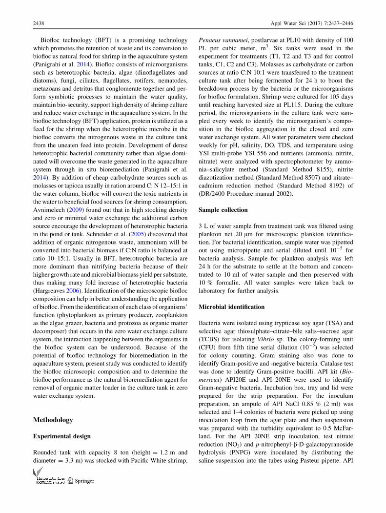
(31.5–35.5 mg l-1; n = 12). The bioremediation processwas
successfully carried out by themicroorganism in the biofloc as
the nutrients ammonia, NH3, drops from 8.0 to 0.3 mg l-1,
nitrite drops from 0.8 to 0.5, and nitrate drops from 15.3 to
5.7 mg l-1 during the culture period (Fig. 2). Species Pseu-
domonas sp. and Aeromonas sp. were identified to be domi-
nant from the colony-forming unit (CFU) counting.Vibrio sp.
was also identified as aggregates in the bioflocculation
(Table 1). The percentage of bacteria identified dominantly
come from heterotrophic bacterial species (ex. Aeromonas
hydrophyila) and also from Vibrio spp. (V. alginolyticus and
V. fluvialis) (Fig. 3). There were a lot of and varieties of
microscopic organism compositions identified from biofloc-
culation in treatment tank 1 (Table 2) treatment tank 2
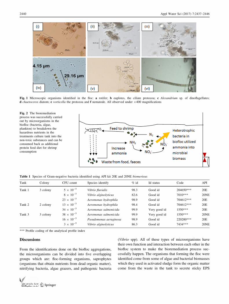
(Table 3) and treatment tank 3 (Table 4). The density of
microorganism was also identified based on the Day of Cul-
ture (DOC) of shrimp, density of microorganism in DOC11
shrimp (Fig. 4), DOC17 (Fig. 5), DOC30 (Fig. 6), DOC58
(Fig. 7), DOC65 (Fig. 8), DOC76 (Fig. 9), and for DOC93
(Fig. 10).
Appl Water Sci (2017) 7:2437–2446 2439
123
Discussions
From the identifications done on the biofloc aggregations,
the microorganisms can be divided into five overlapping
groups which are: floc-forming organisms, saprophytes
(organisms that obtain nutrients from dead organic matter),
nitrifying bacteria, algae grazers, and pathogenic bacteria
(Vibrio spp). All of these types of microorganisms have
their own function and interaction between each other in the
biofloc system to make the bioremediation process suc-
cessfully happen. The organisms that forming the floc were
identified come from some of algae and bacterial biomasses
which they used in activated sludge form the organic matter
come from the waste in the tank to secrete sticky EPS
Fig. 1 Microscopic organisms identified in the floc: a rotifer; b euplotes, the ciliate protozoa; c Alexandrium sp. of dinoflagellates;
d chaetoceros diatom; e vorticella the protozoa and f nematode. All observed under 9400 magnifications
Fig. 2 The bioremediation
process was successfully carried
out by microorganisms in the
biofloc (bacteria, algae,
plankton) to breakdown the
hazardous nutrients in the
treatments culture tank into the
non-toxic substances and can be
consumed back as additional
protein feed diet for shrimp
consumption
Table 1 Species of Gram-negative bacteria identified using API kit 20E and 20NE biomerieux
Tank Colony CFU count Species identify % id Id status Code API
Tank 1 3 colony 5 9 10-5 Vibrio fluvialis 98.3 Good id 204650*** 20E
8 9 10-5 Vibrio alginolyticus 82.6 Good id 7010*** 20NE
23 9 10-5 Aeromonas hydrophila 98.9 Good id 704612*** 20E
Tank 2 2 colony 13 9 10-5 Aeromonas hydrophila 98.4 Good id 704612*** 20E
34 9 10-5 Aeromonas salmonicida 99.9 Very good id 1550*** 20E
Tank 3 3 colony 38 9 10-5 Aeromonas salmonicida 99.9 Very good id 1550*** 20NE
16 9 10-5 Pseudomonas aeruginosa 98.9 Good id 220200*** 20E
3 9 10-5 Vibrio alginolyticus 86.3 Good id 7434*** 20NE
*** Profile coding of the analytical profile index
2440 Appl Water Sci (2017) 7:2437–2446
123

(Medina and Neis 2007). These EPS were known to have
significant effect on the physiochemical properties of the
microbial aggregates including structure, surface charge,
flocculation, settling properties, dewatering and absorptive
capacity Sheng et al. (2010). The floc itself also as the
bioremediation agent is able to stick the detritus from the
wastes together with other organisms such as protozoa and
zooplankton during bioflocculation and bacteria in the floc
take up ammonia in the water which mostly comes from the
metabolic waste of shrimp and convert it into microbial
protein. Hargreaves (2013) discovered that bioflocs are
accumulation of algae, bacteria, protozoan, and other kinds
Fig. 3 Percentage of bacterial
species identified accumulated
together in the biofloc in
treatment tank 1 (T1), tank 2
(T2) and tank 3 (T3)
Table 2 Type of microorganisms identified as aggregates in the biofloc in Tank 1
Plankton Tank Phylum Class Genera
Phytoplankton Tank 1 Ohcrophyta Bacillariophyceae Nitzschia
Navicula
Licmophora
Amphora
Cymbella
Radiolarian
Cyclotella
Leptocylindrus
Coscinodiscophyceae Cosinodiscus
Tank 1 Cynophyta Cynophyceae Gomphoperia
Oscillatoria
Tank 1 Chlorophyta Chlorophyceae Borodinellopsis
Chlorella
Chlamydomonas
Tetraselmis
Zooplankton Tank 1 Rotifera Brachionidae Brachionus
Protozoa Tank 1 Ciliophora Euplotidae Euplotes
Ciliophora Vorticellidae Vorticella
Ciliophora Parameciidae Paramecium
Euglenoidea Euglenaceae Euglena
Ciliophora Ciliatea Cilliate
Nematode Tank 1 Nematoda Nematode
Appl Water Sci (2017) 7:2437–2446 2441
123

of particulate organic matter such as feces and uneaten feed
which are held together in a loose matrix of mucus secreted
by bacteria and bound by filamentous algae or held by
electrostatic attraction. During siphoning process, it can
also be seen that the waste at the bottom of the tank was
aggregated in small rounded shape, which means the biofloc
microorganisms work in settling down the detritus and
acted as the bioremediation agent in neutralizing the pol-
lutant in the bottom of the tank and makes the condition of
water optimum. For the saprophytes group or the organisms
obtaining nutrients from the dead organic matter, hetero-
trophic bacteria is in this classification. Heterotrophic bac-
teria identified from the study are Aeromonas spp and
Pseudomonas spp. Heterotrophic bacteria used the organic
compound from the organic matter left in the tank as the
sources of energy and food which contrast with the auto-
trophic organisms such as phytoplankton and algae. Proto-
zoa identified in the biofloc treatment tank such as ciliate,
vorticella, euplotes and paramecium also classified as
saprophytic protozoa as absorb organic matter through their
cell wall for food and takes 40 % of the nutrients for the
production of protozoan biomass (Merriam Co 1913; Lal
2006). All of these saprophytic microorganisms acted as
bioremediation agents in neutralizing the nutrients (am-
monia, nitrite and nitrate) from the wastes of uneaten feed
and shrimp’s fecal secretion that produced ammonia prod-
uct. The denitrifying bacteria, Pseudomonas sp., was iden-
tified in the bioflocculation that worked to convert the
nitrates NO3- into gaseous nitrogen; N2 makes the water
condition less toxic and maintains the water quality
(Schramm et al. 1999). This bacteria also will convert the
nitrates in the water to the beneficial protein for shrimp
consumption besides getting the food from the pellet given.
These are proved through Hargreaves’s (2013) study in
biofloc system, whereas some of the nitrogen is incorpo-
rated into the bacterial cells that become the main compo-
nent of biofloc, shrimp consumption of this microbial
protein will effect for a second time and contribute to
shrimp growth. Zooplankton, protozoa and protozoa para-
site are classified under the algae grazer category. The
organisms identified in the bioflocculation were such as
nematode, gastrotrich, euplotes protozoa, vorticella
Table 3 Type of microorganisms identified as aggregates in the biofloc in Tank 2
Plankton Tank Phylum Class Genera
Phytoplankton Tank 2 Ohcrophyta Bacillariophyceae Nitzschia
Leptocylindrus
Navicula
Cyclotella
Melosira
Licmophora
Cymbella
Coscinodiscophyceae Cosinodiscus
Cynophyta Cynophyceae Oscillatoria
Gomphosperia
Gloeocapsa
Chlorophyta Chlorophyceae Chlorella
Borodinellopsis
Scenedesmus
Tetraselmis
Dinophyta Dinophyceae Protoperidinium
Alexandrium
Zooplankton Tank 2 Arthropoda Copepoda Copepod
Rotifera Brachionidae Branchius
Protozoa Tank 2 Sarcomastigophora Radiolaria Radiolarian
Ciliophora Euplotidae Euplotes
Ciliophora Parameciidae Paramecium
Ciliophora Ciliatea Cilliate
Euglenoidea Euglenaceae Euglena
Ciliophora Vorticellidae Vorticella
Nematode Tank 2 Nematoda Nematode
2442 Appl Water Sci (2017) 7:2437–2446
123

protozoa, ciliate, rotifer and copepod which in dense com-
position as higher food from algae and phytoplankton types
were available in the biofloc treatment tank (Hargreaves
2013). During culture starting from DOC11 to DOC93
(Figs. 4, 5, 6, 7, 8, 9, 10), algae from three different classes
indentified to dominate the biofloc culture treatment are
Chlorophyceae (Green algae), Bacillariophyceae (Diatoms),
Cyanophyceae (Blue green algae) and occasionally from
Dinophyceae (Dinoflagellates) class. This finding is same as
the studies by Galvez (2015) and Schrader et al. (2011)
which found out that the most abundant algae in this biofloc
study came from cyanobacteria class followed by Chloro-
phyta, Heterokontophyta, Euglenophyta and Dinophyta.
The organisms classified under pathogenic group identified
are protozoa that are harmful to the shrimp and the patho-
genic bacteria Vibrio sp. such as Vibrio alginolyticus that
Table 4 Type of microorganisms identified as aggregates in the biofloc in Tank3
Plankton Tank Phylum Class Genera
Phytoplankton Tank 3 Ohcrophyta Bacillariophyceae Nitzschia
Leptocylindrus
Cyclotella
Licmophora
Navicula
Coscinodiscophyceae Cosinodiscus
Cynophyta Cynophyceae Oscillatoria
Gomphosperia
Gloeocapsa
Chlorophyta Chlorophyceae Chlorella
Borodinellopsis
Dinophyta Dinophyceae Protoperidinium
Alexandrium
Zooplankton Tank 3 Arthropoda Copepoda Copepod
Rotifera Brachionidae Branchius
Gastrotricha Chaetonotida Gastrotrich
Protozoa Tank 3 Euglenoidea Euglenaceae Euglena
Ciliophora Vorticellidae Vorticella
Ciliophora Euplotidae Euplotes
Ciliophora Parameciidae Paramecium
Ciliophora Ciliatea Cilliate
Sarcomastigophora Radiolaria Radiolarian
Nematode Tank 3 Nematoda Nematode
Fig. 4 Density of
microorganisms composition
identified from the
bioflocculation in the water
column of treatment culture
tanks for Day of culture,
DOC11
Appl Water Sci (2017) 7:2437–2446 2443
123

can give infection to the shrimp as suggested by Wei and
Wendy (2012). The Vibrio sp. was also identified in the
biofloc aggregation but in less CFU number. These were not
lethal to the shrimps as it is being controlled by the biofloc
itself through higher diversity of phytoplankton and algae
and also can compete with dominant number of hetero-
trophic bacteria. Refer to study done by Emerenciano et al.
(2013) they discovered that the natural probiotic in the
biofloc could internally or externally against the Vibrio sp.
and ectoparasite from giving harmful to the shrimp. Com-
peting with the dominant heterotrophic bacteria and nitri-
fying bacteria for the essential nutrients such as nitrogen
Fig. 5 Density of
microorganisms composition
identified from bioflocculation
in the water column of treatment
culture tanks for Day of culture,
DOC17
Fig. 6 Density of
microorganism composition
identified from the
bioflocculation in the water
column of treatment culture
tanks for Day of culture,
DOC30
Fig. 7 Density of
microorganism composition
identified from the
bioflocculation in the water
column of treatment culture
tanks for Day of culture,
DOC58
2444 Appl Water Sci (2017) 7:2437–2446
123
also will limit the Vibrio sp. group from uncontrolled
growth (Emerenciano et al. 2013).
Conclusion
Bioflocculation is a promising technology towards friendly
aquaculture environment. In fact, it can supply additional
diet for shrimp’s consumption from the biofloculant of a
variety microorganisms identified in the floc; biofloc also
were recognized to be efficient and successful as a biore-
mediation and biodegradation agent for maintaining the
water quality in the close aquaculture system with the zero
water exchange.
Acknowledgments The author would like to thank private hatchery
(AB Hatchery Sdn. Bhd) for PL shrimp supply for this research
project. Authors also would like to thank Fisheries officer Mr Hanif,
Assistant Science officer Mr. Sabri Muda and Research Officer Mr.
Ikhwan Zakariah and all the hatchery officers and staffs of Institute of
Tropical Aquaculture, Universiti Malaysia Terengganu for all the
assistance and guidance throughout the research period.
0 1000 2000 3000 4000
Tank1
Tank 2
Tank 3
Density (cells/L)Tr
eatm
ents
nematode
protozoa
zooplankton
Dinophyceae
Chlorophyceae
cynaophyceae
Bacillariophyceae
Fig. 8 Density of
microorganism composition
identified from the
bioflocculation in the water
column of treatment culture
tanks for Day of culture,
DOC65
0 1000 2000 3000 4000
Tank1
Tank 2
Tank 3
Density (cells/L)
Trea
tmen
ts
nematode
protozoa
zooplankton
Dinophyceae
Chlorophyceae
cynaophyceae
Bacillariophyceae
Fig. 9 Density of
microorganism composition
identified from the
bioflocculation in the water
column of treatment culture
tanks for Day of culture,
DOC76
0 500 1000 1500 2000 2500 3000
Tank1
Tank 2
Tank 3
Density (cells/L)
Trea
tmen
ts
nematode
protozoa
zooplankton
Dinophyceae
Chlorophyceae
cynaophyceae
Bacillariophyceae
Fig. 10 Density of
microorganism composition
identified from the
bioflocculation in the water
column of treatment culture
tanks for Day of culture,
DOC93
Appl Water Sci (2017) 7:2437–2446 2445
123

Open Access This article is distributed under the terms of the
Creative Commons Attribution 4.0 International License (http://
creativecommons.org/licenses/by/4.0/), which permits unrestricted
use, distribution, and reproduction in any medium, provided you give
appropriate credit to the original author(s) and the source, provide a
link to the Creative Commons license, and indicate if changes were
made.
References
American Public Health Association. APHA (1989) Phytoplankton
counting technique. In: Standard methods for the examination of
water and wastewater, pp 10–23
Avnimelech Y (2009) Biofloc technology—a practical guide book.
The world Aquaculture Society, EUA, BatonRouge, Louisiana
Das S (2014) Microbial biodegradation and bioremediation. Labora-
tory of Environmental Microbiology and Ecology (LenME).
National Institute of Technology, Rourkela Odisha (ElsevierBook First Edition)
Das S, Lyla PS, Khan SA (2006) Marine microbial diversity and
ecology: present status and future perspectives. Curr Sci
90(10):1325–1335
Dash HR, Das S (2012) Bioremediation of mercury and importance of
bacterial mergenes. Int Biodeterior Biodegrad 75:207–213
Emerenciano M, Gaxiola G, Gerard C (2013) Biofloc Technology
(BFT): a review for aquaculture application and animal food
industry. Biomass Now-Cultiv Util. doi:10.57772/53902
Galvez AO (2015) Plankton communities in shrimp monoculture,
integrated biofloc system. Global Aquaculture Advocate MAY/
JUN 2015
Hargreaves JA (2006) Photosynthetic suspended-growth systems in
aquaculture. Aquac Eng 34:344–363
Hargreaves JA (2013) Biofloc production systems for aquaculture.
SRAC, Southern Regional Aquaculture Center Publication. No.
4503, pp 1–12
Karigar CS, Rao SS (2011) Role of microbial enzymes in the
bioremediation of pollutants: a review. Enzym Res. doi:10.4061/
2011/805187
Lal R (2006) Encyclopedia of soil science. In: Griffiths BS (ed)
Nature. Scottish Crop Research Institute, Invergowrie. doi:10.
1081/E-ESS-120042737
Medina M, Neis U (2007) Symbiotic algal bacterial wastewater
treatment: effect of food to microorganism ratio and hydraulic
retention time on the process performance. Water Sci Technol
55(11):165–171
Merriam Co G (1913) Protozoa, their role and impact on aquaculture.
Webster Revised Unabridged Dictionary, Encyclopedia Dic-
tionary (Aquaculture) published
Panigrahi A, Sundaram M, Ravichandran P, Gopal C, (2014)
Microbial soup-Eco based approach for shrimp culture and
management. CIBA (Indian Council of Agricultural Research)
ENVIS Centre Newsletter vol 12:2. 2 Apr–Jun 2014
Schrader KK, Green BW, Perschbacher PW (2011) Development of
phytoplankton communities and common off-flavors in a
biofloctechnology system used for the culture of channel catfish
(Ictaluruspunctatus). Aquac eng 45:118–126
Schramm A, de Beer DA, Van den Heuvel S, Ottengraf S (1999)
Microscale distribution of populations and activities of Nitro-
sospira and Nitrospira spp. along a macroscale100 sustainable
aquaculture technique gradient in a nitrifying bioreactor: quan-
tification by in situ hybridization and the useofmicrosensors.
Appl Environ Microbiol 65:3690–3696
Schneider O, Sereti V, Eding EH, Verreth JAJ (2005) Analysis of
nutrient flows in integrated intensive aquaculture systems. Aquac
Eng 32:379–401
Sheng G, Yu H, Li X (2010) Extracellular Polymeric Substances
(EPS) of microbial aggregates in biological wastewater treat-
ment systems: a review. Biotechnol Adv 28(6):882–894
Stelting S, Burns RG, Sunna A, Visnovsky G, Bunt C (2010)
Immobilization of Pseudomonas sp. strain ADP: a stable inocu-
lant for the bioremediation of atrazine. In: 19th World congress
of soil science, soil solutions for a changing world, Brisbane,
Australia
Wei LS, Wendy W (2012) Chracterization of vibrio alginolyticus
isolated from white leg shrimp (Litopenaeus vannamei) with
emphasis on its antibiogram and heavy metal resistance pattern.
Verterinarski Arh 82(2):221–227
2446 Appl Water Sci (2017) 7:2437–2446
123