Identification and Fragmentation of Sucralose Using ... · Identification and Fragmentation of...
8
Identification and Fragmentation of Sucralose Using Accurate-Mass Q-TOF LC/MS and Molecular Structure Correlator Software Authors Imma Ferrer and E. Michael Thurman Center for Environmental Mass Spectrometry Department of Environmental Engineering University of Colorado, Boulder, CO 80309 Jerry Zweigenbaum Agilent Technologies, Inc. 2850 Centerville Road Wilmington, DE 19808 Application Note Food and Environmental Abstract The use of Accurate-Mass Q-TOF LC/MS and MS/MS, in both positive and negative electrospray ionization (ESI) modes, was evaluated for the identification of sucralose in water. Response and fragmentation pathways were investigated. Sucralose responded well using Q-TOF LC/MS in either the positive or negative ion ESI modes. The overall signal intensity obtained in positive ion mode was approximately twice that of negative ion mode. In positive ion mode, sucralose was detected by its sodium adduct [M+Na] + at m/z 419.0038. Accurate mass MS/MS measurements provided structural confirma- tion of the sodiated fragments obtained (m/z 221.0187 and m/z 238.9848). In nega- tive ion mode, the deprotonated molecule was observed ([M-H] - at m/z 395.0073). Fragmentation by MS/MS yielded one characteristic fragment ion (m/z 359.0306). Agilent MassHunter Molecular Structure Correlator (MSC) software was used to draw and investigate the fragmentation pathways for the negative and positive ion MS/MS analyses. The MSC software proved to be a useful tool in assisting with the characterization of the fragment ion structures.
-
Upload
hoangduong -
Category
Documents
-
view
221 -
download
0
Transcript of Identification and Fragmentation of Sucralose Using ... · Identification and Fragmentation of...
Identification and Fragmentation ofSucralose Using Accurate-MassQ-TOF LC/MS and MolecularStructure Correlator Software
Authors
Imma Ferrer and E. Michael Thurman
Center for Environmental Mass
Spectrometry
Department of Environmental
Engineering
University of Colorado,
Boulder, CO 80309
Jerry Zweigenbaum
Agilent Technologies, Inc.
2850 Centerville Road
Wilmington, DE 19808
Application Note
Food and Environmental
Abstract
The use of Accurate-Mass Q-TOF LC/MS and MS/MS, in both positive and negative
electrospray ionization (ESI) modes, was evaluated for the identification of
sucralose in water. Response and fragmentation pathways were investigated.
Sucralose responded well using Q-TOF LC/MS in either the positive or negative ion
ESI modes. The overall signal intensity obtained in positive ion mode was
approximately twice that of negative ion mode.
In positive ion mode, sucralose was detected by its sodium adduct [M+Na]+ at
m/z 419.0038. Accurate mass MS/MS measurements provided structural confirma-
tion of the sodiated fragments obtained (m/z 221.0187 and m/z 238.9848). In nega-
tive ion mode, the deprotonated molecule was observed ([M−H]− at m/z 395.0073).
Fragmentation by MS/MS yielded one characteristic fragment ion (m/z 359.0306).
Agilent MassHunter Molecular Structure Correlator (MSC) software was used to
draw and investigate the fragmentation pathways for the negative and positive ion
MS/MS analyses. The MSC software proved to be a useful tool in assisting with the
characterization of the fragment ion structures.
2
Introduction
Due to its intense sweetness, noncaloric properties, lowbioaccumulation potential, low toxicity, and the dietaryrequirements of many consumers, sucralose has become oneof the most popular artificial sweeteners used worldwide.Because the human body does not metabolize sucralose, itends up in wastewater and surface water. Current waste-water treatment technologies do not address sucralose, so itis now ubiquitous in the environment. This is a point of signif-icant concern. A recent study revealed the biological effectsof sucralose in the aquatic environment, which may haveimportant toxicological consequences [1]. For these reasons,there is growing interest in measuring sucralose in drinkingwater, groundwater, surface water, wastewater, and aquatic environments.
Due to its solubility, sucralose is readily analyzed by LC/MS. Itcontains three chlorine atoms, which produce a distinctivechlorine signature when analyzed by MS. Based on manypapers published describing the analysis of sucralose,LC/MS/MS with multiple reaction monitoring (MRM) in neg-ative ion mode is the most popular method. However, theMRM transitions used are not selective enough to identifysucralose in water with the same confidence as with accuratemass. The transitions are not discriminatory because theyinvolve a chlorine loss, which can be present in many othercommon organic molecules.
This application note evaluates the use of an Agilent 6540Accurate-Mass Q-TOF LC/MS system in both positive andnegative ESI modes for the unequivocal identification ofsucralose in water. Response and the usefulness of molecularstructure correlation software were investigated. The comple-mentary study of Analytical Methodologies for the Detectionof Sucralose in Water in Analytical Chemistry [2] provides adetailed comparison of Q-TOF LC/MS and LC/MS/MS for thedetection of sucralose in environmental water samples.
Experimental
A detailed description of the experimental procedures can befound in the complementary journal article published inAnalytical Chemistry [2].
Standard preparationSucralose was purchased from Sigma-Aldrich (St. Louis, MO,USA). A stock solution of sucralose (1,000 µg/mL) was prepared in water and stored at −18 °C. From this solution,working standard solutions were prepared by dilution withmethanol and water.
InstrumentationThe standard was analyzed using an Agilent 1290 InfinityBinary LC System coupled to an Agilent 6540 Accurate-MassQ-TOF LC/MS system with Agilent Jet Stream technology forelectrospray ionization.
The HPLC was equipped with a binary pump with an inte-grated vacuum degasser (G4220A) and an autosampler(G4226A). The HPLC parameters are shown in Table 1.
Table 1. HPLC Parameters
Instrument Agilent 1290 Infinity Binary LC System
Mobile phases (A) acetonitrile(B) 0.1% formic acid in water
Gradient Linear: Initial mobile phase composition was 10% A,held constant for 1.7 minutes, followed by a lineargradient to 100% A, for a total run time of 10 minutes.
Flow rate 0.4 mL/min
Column Agilent ZORBAX Eclipse Plus reversed phase C18 analytical column, 50 × 2.1 mm, 1.8 µm particle size (p/n 959741-902)
Column temperature 25 °C
Injection volume 20 µL
3
Q-TOF MS accurate mass spectra were recorded across therange 30−1,000 m/z at 2 GHz. Polarity switching was notused; samples were injected twice, one under positive ionmode and the other under negative ion mode. MS/MS experi-ments were also carried out in both positive and negative ionmodes. The Q-TOF MS and MS/MS parameters are shown inTable 2.
Results and Discussion
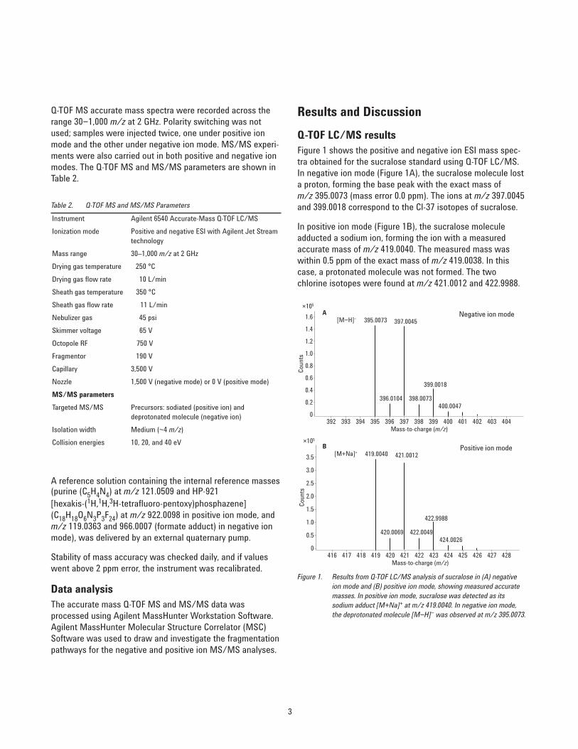
Q-TOF LC/MS results Figure 1 shows the positive and negative ion ESI mass spec-tra obtained for the sucralose standard using Q-TOF LC/MS.In negative ion mode (Figure 1A), the sucralose molecule losta proton, forming the base peak with the exact mass ofm/z 395.0073 (mass error 0.0 ppm). The ions at m/z 397.0045and 399.0018 correspond to the Cl-37 isotopes of sucralose.
In positive ion mode (Figure 1B), the sucralose moleculeadducted a sodium ion, forming the ion with a measuredaccurate mass of m/z 419.0040. The measured mass waswithin 0.5 ppm of the exact mass of m/z 419.0038. In thiscase, a protonated molecule was not formed. The two chlorine isotopes were found at m/z 421.0012 and 422.9988.
Figure 1. Results from Q-TOF LC/MS analysis of sucralose in (A) negativeion mode and (B) positive ion mode, showing measured accuratemasses. In positive ion mode, sucralose was detected as itssodium adduct [M+Na]+ at m/z 419.0040. In negative ion mode,the deprotonated molecule [M−H]− was observed at m/z 395.0073.
Table 2. Q-TOF MS and MS/MS Parameters
Instrument Agilent 6540 Accurate-Mass Q-TOF LC/MS
Ionization mode Positive and negative ESI with Agilent Jet Streamtechnology
Mass range 30–1,000 m/z at 2 GHz
Drying gas temperature 250 °C
Drying gas flow rate 10 L/min
Sheath gas temperature 350 °C
Sheath gas flow rate 11 L/min
Nebulizer gas 45 psi
Skimmer voltage 65 V
Octopole RF 750 V
Fragmentor 190 V
Capillary 3,500 V
Nozzle 1,500 V (negative mode) or 0 V (positive mode)
MS/MS parameters
Targeted MS/MS Precursors: sodiated (positive ion) and deprotonated molecule (negative ion)
Isolation width Medium (~4 m/z)
Collision energies 10, 20, and 40 eV
A reference solution containing the internal reference masses(purine (C5H4N4) at m/z 121.0509 and HP-921[hexakis-(1H,1H,3H-tetrafluoro-pentoxy)phosphazene](C18H18O6N3P3F24) at m/z 922.0098 in positive ion mode, andm/z 119.0363 and 966.0007 (formate adduct) in negative ionmode), was delivered by an external quaternary pump.
Stability of mass accuracy was checked daily, and if valueswent above 2 ppm error, the instrument was recalibrated.
Data analysisThe accurate mass Q-TOF MS and MS/MS data wasprocessed using Agilent MassHunter Workstation Software.Agilent MassHunter Molecular Structure Correlator (MSC)Software was used to draw and investigate the fragmentationpathways for the negative and positive ion MS/MS analyses.
1.6A
B
[M−H]−Negative ion mode
1.4
1.2
1.0
0.8
0.6
0.4
0.2
0392 393 394 395
395.0073 397.0045
396.0104 398.0073
399.0018
400.0047
396 397Mass-to-charge (m/z)
Counts
398 399 400 401 402 403 404
×105
[M+Na]+Positive ion mode
3.5
3.0
2.5
2.0
1.5
1.0
0.5
0416 417 418 419
419.0040 421.0012
420.0069 422.0049
422.9988
424.0026
420 421Mass-to-charge (m/z)
Counts
422 423 424 425 426 427 428
×105
4
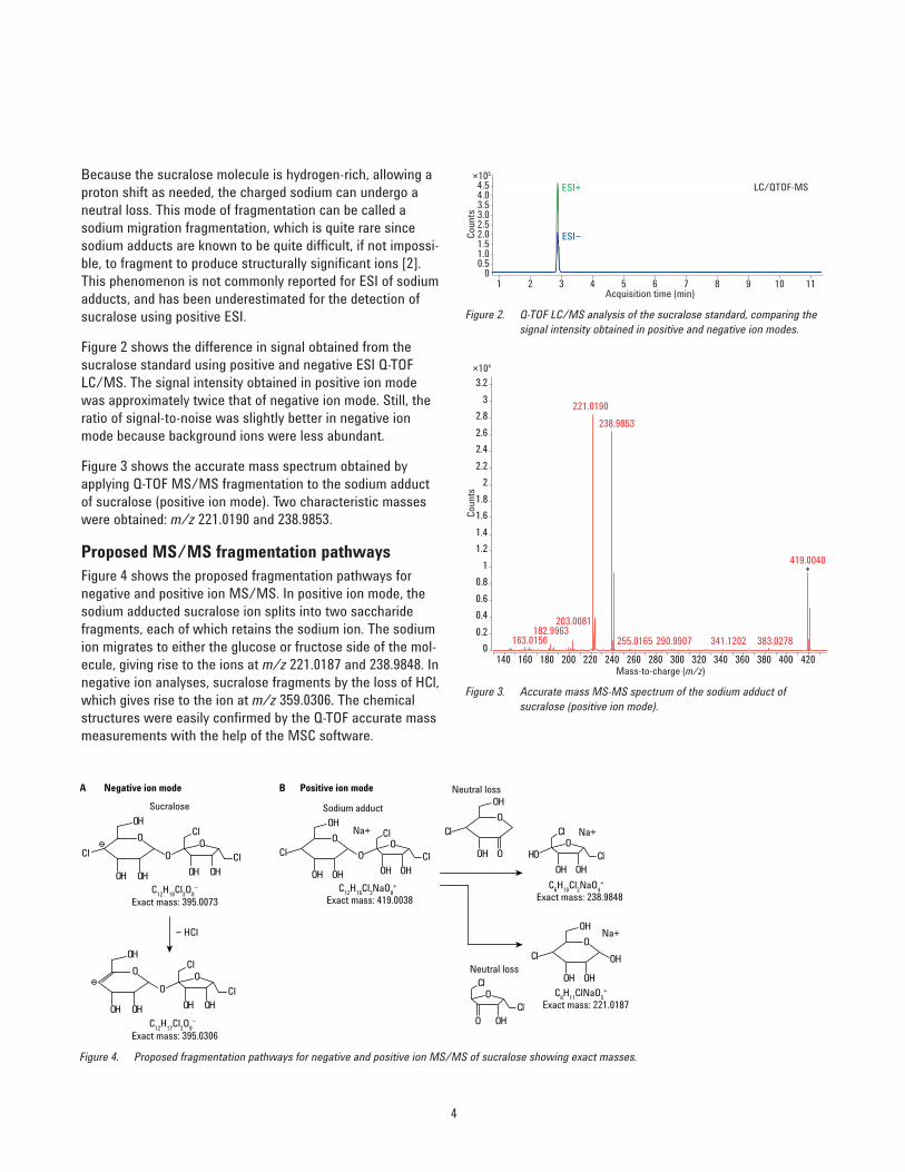
Because the sucralose molecule is hydrogen-rich, allowing aproton shift as needed, the charged sodium can undergo aneutral loss. This mode of fragmentation can be called asodium migration fragmentation, which is quite rare sincesodium adducts are known to be quite difficult, if not impossi-ble, to fragment to produce structurally significant ions [2].This phenomenon is not commonly reported for ESI of sodiumadducts, and has been underestimated for the detection ofsucralose using positive ESI.
Figure 2 shows the difference in signal obtained from thesucralose standard using positive and negative ESI Q-TOFLC/MS. The signal intensity obtained in positive ion modewas approximately twice that of negative ion mode. Still, theratio of signal-to-noise was slightly better in negative ionmode because background ions were less abundant.
Figure 3 shows the accurate mass spectrum obtained byapplying Q-TOF MS/MS fragmentation to the sodium adductof sucralose (positive ion mode). Two characteristic masseswere obtained: m/z 221.0190 and 238.9853.
Proposed MS/MS fragmentation pathwaysFigure 4 shows the proposed fragmentation pathways fornegative and positive ion MS/MS. In positive ion mode, thesodium adducted sucralose ion splits into two saccharidefragments, each of which retains the sodium ion. The sodiumion migrates to either the glucose or fructose side of the mol-ecule, giving rise to the ions at m/z 221.0187 and 238.9848. Innegative ion analyses, sucralose fragments by the loss of HCl,which gives rise to the ion at m/z 359.0306. The chemicalstructures were easily confirmed by the Q-TOF accurate massmeasurements with the help of the MSC software.
Figure 4. Proposed fragmentation pathways for negative and positive ion MS/MS of sucralose showing exact masses.
LC/QTOF-MS
00.51.01.52.02.53.03.54.04.5
1 2 3 4 5 6 7 8 9 10 11
ESI+
ESI−
Acquisition time (min)
Counts
×105
Figure 2. Q-TOF LC/MS analysis of the sucralose standard, comparing thesignal intensity obtained in positive and negative ion modes.
Figure 3. Accurate mass MS-MS spectrum of the sodium adduct ofsucralose (positive ion mode).
0
0.2
0.4
0.6
0.8
1
1.2
1.4
1.6
1.8
2
2.2
2.4
2.6
2.8
3
3.2
221.0190
238.9853
419.0040
203.0081182.9963
163.0156 383.0278255.0165 290.9907 341.1202
140 160 180 200 220 240 260 280 300 320 340 360 380 400 420
×104
Mass-to-charge (m/z)
Counts
A
OO
O Cl
Cl
Cl
Cl
Cl
OH OH OH OH
OH
OO
O
OH OH OH OH
OH
Negative ion mode
Sucralose
B Positive ion mode
Sodium adduct
Exact mass: 395.0073C
12H
18Cl
3O
8−
Exact mass: 395.0306C
12H
17Cl
2O
8−
Exact mass: 419.0038C
12H
19Cl
3NaO
8+
OO
O
OH OH OH OH
OH
Cl
Cl
Cl
Na+
H
Exact mass: 238.9848C
6H
10Cl
2NaO
4+
OO Cl
Cl
OH OH
Na+
Exact mass: 221.0187C
6H
11ClNaO
5+
Na+O
OH
OH OH
OH
ClNeutral loss
OCl
Cl
O OH
Neutral loss
O
OH O
OH
Cl
− HCl
5
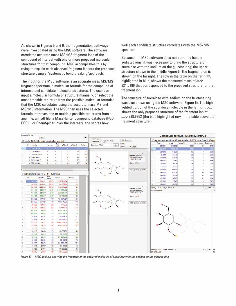
As shown in Figures 5 and 6, the fragmentation pathwayswere investigated using the MSC software. The software correlates accurate mass MS/MS fragment ions of the compound of interest with one or more proposed molecularstructures for that compound. MSC accomplishes this bytrying to explain each observed fragment ion into the proposedstructure using a “systematic bond-breaking”approach.
The input for the MSC software is an accurate mass MS/MSfragment spectrum, a molecular formula for the compound ofinterest, and candidate molecular structures. The user caninput a molecular formula or structure manually, or select themost probable structure from the possible molecular formulasthat the MSC calculates using the accurate mass MS andMS/MS information. The MSC then uses the selected formula, retrieves one or multiple possible structures from a.mol file, an .sdf file, a MassHunter compound database (PCD,PCDL), or ChemSpider (over the Internet), and scores how
well each candidate structure correlates with the MS/MSspectrum.
Because the MSC software does not currently handle sodiated ions, it was necessary to draw the structure ofsucralose with the sodium on the glucose ring; the upperstructure shown in the middle Figure 5. The fragment ion isshown on the far right. The row in the table on the far right,highlighted in blue, shows the measured mass of m/z221.0189 that corresponded to the proposed structure for thatfragment ion.
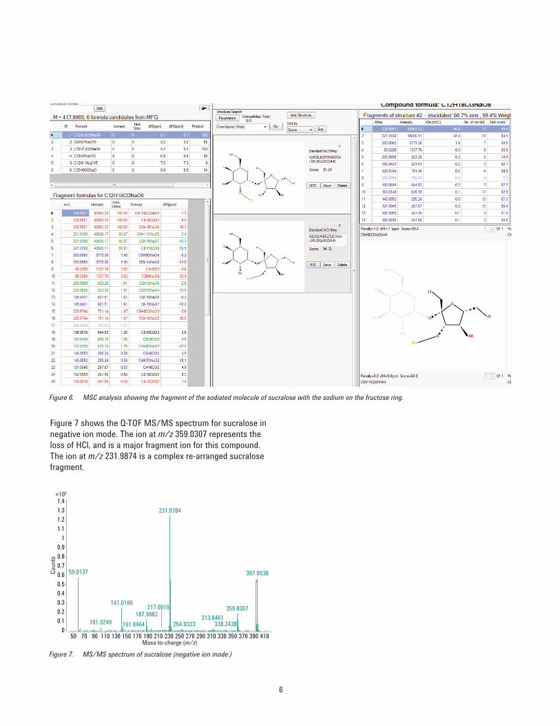
The structure of sucralose with sodium on the fructose ringwas also drawn using the MSC software (Figure 6). The high-lighted portion of the sucralose molecule in the far right boxshows the only proposed structure of the fragment ion atm/z 238.9852 (the blue highlighted row in the table above thefragment structure.)
Figure 5. MSC analysis showing the fragment of the sodiated molecule of sucralose with the sodium on the glucose ring.
6
Figure 7 shows the Q-TOF MS/MS spectrum for sucralose innegative ion mode. The ion at m/z 359.0307 represents theloss of HCl, and is a major fragment ion for this compound.The ion at m/z 231.9874 is a complex re-arranged sucralosefragment.
Figure 7. MS/MS spectrum of sucralose (negative ion mode.)
0
0.1
0.2
0.3
0.4
0.5
0.6
0.7
0.8
0.9
1
1.1
1.2
1.3
1.4
231.9784
59.0137
397.0038
141.0166 359.0307
187.9882 101.0249
313.9461
217.0019
254.9333
338.2438
161.0464
50 70 90 110 130 150 170 190 210 230 250 270 290 310 330 350 370 390 410
×105
Mass-to-charge (m/z)
Coun
ts
Figure 6. MSC analysis showing the fragment of the sodiated molecule of sucralose with the sodium on the fructose ring.
7
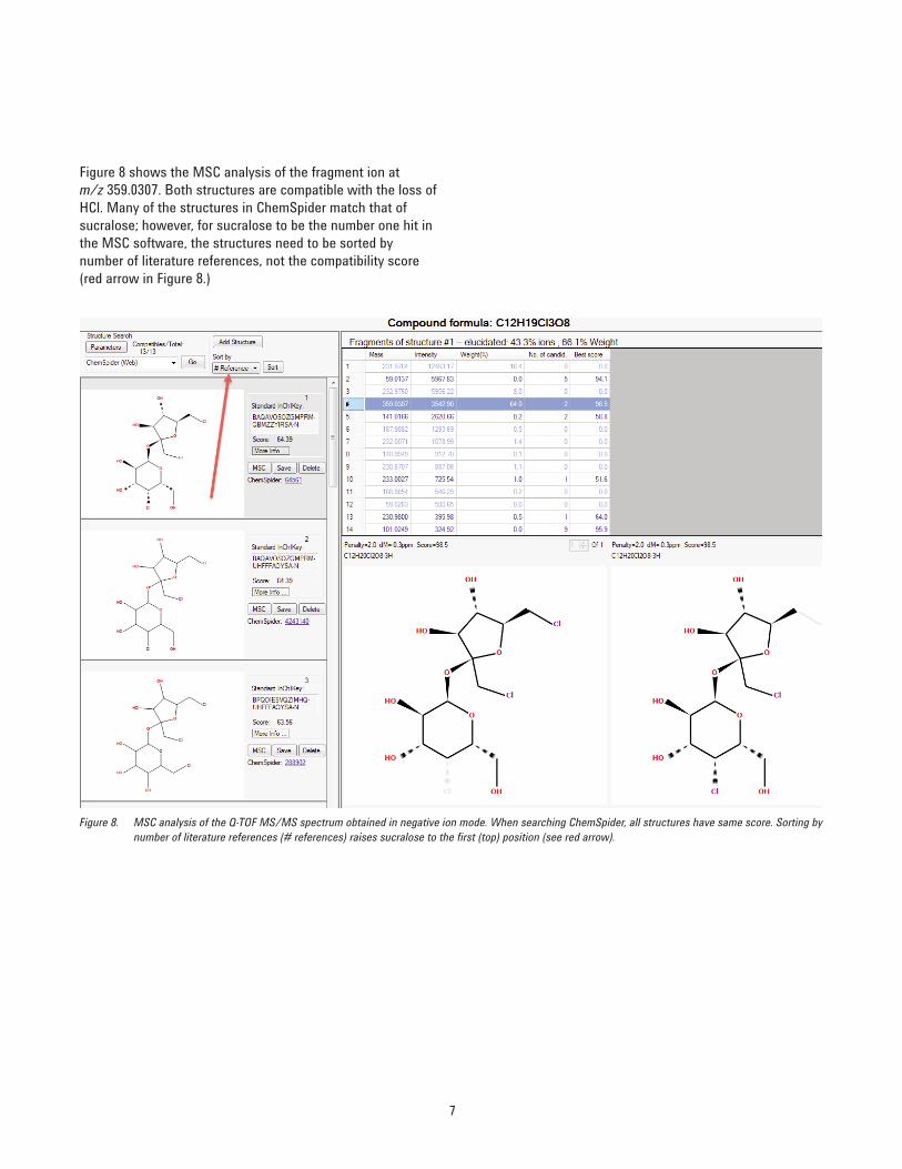
Figure 8 shows the MSC analysis of the fragment ion atm/z 359.0307. Both structures are compatible with the loss ofHCl. Many of the structures in ChemSpider match that ofsucralose; however, for sucralose to be the number one hit inthe MSC software, the structures need to be sorted bynumber of literature references, not the compatibility score(red arrow in Figure 8.)
Figure 8. MSC analysis of the Q-TOF MS/MS spectrum obtained in negative ion mode. When searching ChemSpider, all structures have same score. Sorting bynumber of literature references (# references) raises sucralose to the first (top) position (see red arrow).
www.agilent.com/chem
Agilent shall not be liable for errors contained herein or for incidental or consequentialdamages in connection with the furnishing, performance, or use of this material.
Information, descriptions, and specifications in this publication are subject to changewithout notice.
© Agilent Technologies, Inc., 2014Printed in the USAMarch 6, 20145991-4066EN
Conclusions
Sucralose responded well using Q-TOF LC/MS when oper-ated in either the positive or negative ESI mode. Sucraloseformed a strong sodium adduct in positive ion mode andreadily lost a proton in negative ion mode. The overall signalintensity obtained in positive ion mode was approximatelytwice that of negative ion mode. As demonstrated in thecomplementary study of detection methodologies forsucralose in water, for the triple quadrupole LC/MS MRMmethod, sensitivity was higher in the positive ion mode(using the two transitions shown in this application note),than in the negative ion mode [2].
Contrary to what is commonly reported for ESI analyses ofsodium adducts, the strong sodium adduct formed in the positive ion mode was easily fragmented by MS/MS. Thetwo characteristic accurate mass fragments produced can beused to identify sucralose unequivocally. The MSC softwareis a useful tool to assist with the characterization of fragment ion structures.
References
1. A.K.E Wiklund, M. Breitholtz, B.E. Bengtsson, andM. Adolfsson-Erici “Sucralose – An ecotoxicologicalchallenger?” Chemosphere 2012, 86, 50−55.
2. I. Ferrer, J.A. Zweigenbaum, and E.M. Thurman“Analytical Methodologies for the Detection ofSucralose in Water” Analytical Chemistry 2013, 85,9581-9587.
For More Information
These data represent typical results. For more informationon our products and services, visit our Web site atwww.agilent.com/chem.

![Bruker Product Overview - Mass Spectrometry · 2019. 12. 1. · maXis II resolution: 80,000 scan speed: 1-50 Hz (MS & MS/MS) fragmentation: CID, ETD size [mm]: 1320 x 800 x 2845 timsTOF](https://static.fdocuments.in/doc/165x107/60e2c47e70177f7a0e366140/bruker-product-overview-mass-spectrometry-2019-12-1-maxis-ii-resolution.jpg)