Hypochlorous acid/ N -chloramines are naturally produced DNA repair...
6
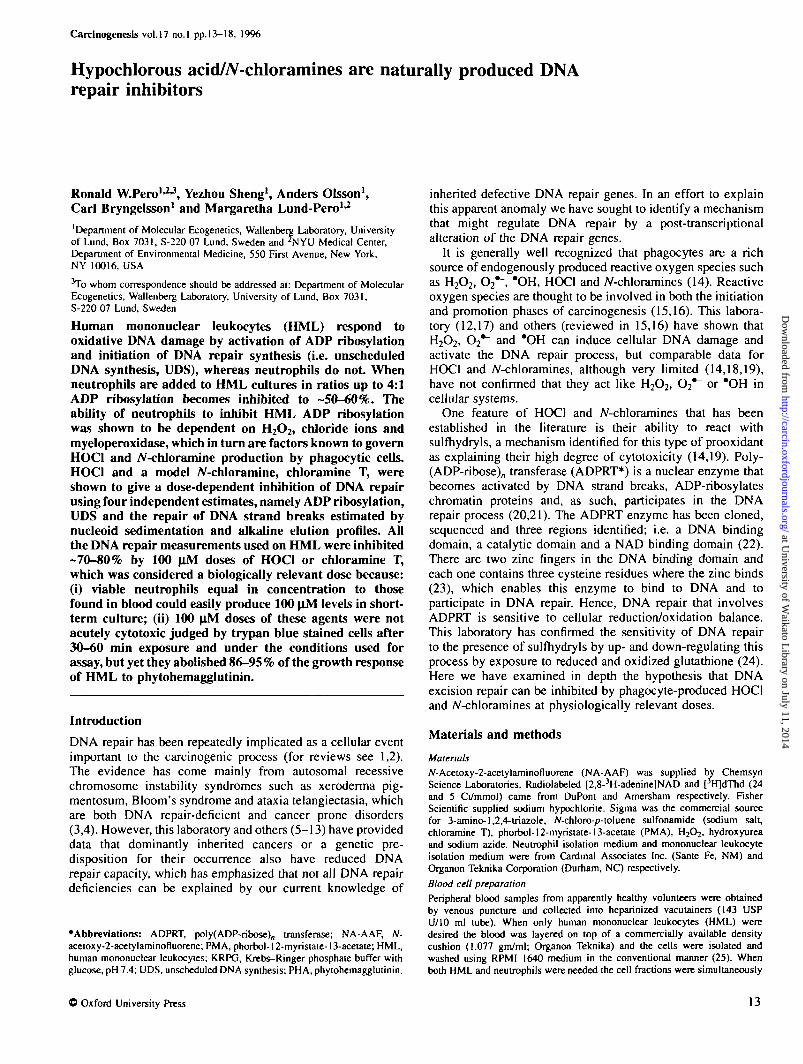
Carcinogeneds vol.17 no.l pp.13-18, 1996 Hypochlorous acid/iV-chloramines are naturally produced DNA repair inhibitors Ronald W.Pero 1 * 3 , Yezhou Sheng 1 , Anders Olsson 1 , Carl Bryngelsson 1 and Margaretha Lund-Pero 1 * 2 'Department of Molecular Ecogenetics, Wallenberg Laboratory, University of Lund, Box 7031, S-220 07 Lund, Sweden and W U Medical Center, Department of Environmental Medicine, 550 First Avenue, New York, NY 10016, USA 3 To whom correspondence should be addressed at: Department of Molecular Ecogenetics, Wallenberg Laboratory, University of Lund, Box 7031, S-220 07 Lund, Sweden Human mononuclear leukocytes (HML) respond to oxidative DNA damage by activation of ADP ribosylation and initiation of DNA repair synthesis (i.e. unscheduled DNA synthesis, UDS), whereas neutrophils do not When neutrophils are added to HML cultures in ratios up to 4:1 ADP ribosylation becomes inhibited to -50-60%. The ability of neutrophils to inhibit HML ADP ribosylation was shown to be dependent on H 2 O 2 , chloride ions and myeloperoxidase, which in turn are factors known to govern HOC1 and iV-chloramine production by phagocytic cells. HOC1 and a model A r -chloramine, chloramine T, were shown to give a dose-dependent inhibition of DNA repair using four independent estimates, namely ADP ribosylation, UDS and the repair of DNA strand breaks estimated by nucleoid sedimentation and alkaline elution profiles. All the DNA repair measurements used on HML were inhibited -70-80% by 100 uM doses of HOC1 or chloramine T, which was considered a biologically relevant dose because: (i) viable neutrophils equal in concentration to those found in blood could easily produce 100 uM levels in short- term culture; (ii) 100 |iM doses of these agents were not acutely cytotoxic judged by trypan blue stained cells after 30-60 min exposure and under the conditions used for assay, but yet they abolished 86-95% of the growth response of HML to phytohemagglutinin. Introduction DNA repair has been repeatedly implicated as a cellular event important to the carcinogenic process (for reviews see 1,2). The evidence has come mainly from autosomal recessive chromosome instability syndromes such as xeroderma pig- mentosum, Bloom's syndrome and ataxia telangiectasia, which are both DNA repair-deficient and cancer prone disorders (3,4). However, this laboratory and others (5—13) have provided data that dominantly inherited cancers or a genetic pre- disposition for their occurrence also have reduced DNA repair capacity, which has emphasized that not all DNA repair deficiencies can be explained by our current knowledge of •Abbreviations: ADPRT, poly(ADP-ribose) n transferase; NA-AAF, N- acetoxy-2-acetylaminofiuorene; PMA, phorbol-l2-myristate-13-acetate; HML, human mononuclear leukocytes; KRPG, Krebs-Ringer phosphate buffer with glucose, pH 7.4; UDS, unscheduled DNA synthesis; PHA, phytohemagglutinin. inherited defective DNA repair genes. In an effort to explain this apparent anomaly we have sought to identify a mechanism that might regulate DNA repair by a post-transcriptional alteration of the DNA repair genes. It is generally well recognized that phagocytes are a rich source of endogenously produced reactive oxygen species such as H 2 O 2 , O 2 *~, *OH, HOC1 and N-chloramines (14). Reactive oxygen species are thought to be involved in both the initiation and promotion phases of carcinogenesis (15,16). This labora- tory (12,17) and others (reviewed in 15,16) have shown that H 2 O 2 , O 2 *~ and *OH can induce cellular DNA damage and activate the DNA repair process, but comparable data for HOC1 and JV-chloramines, although very limited (14,18,19), have not confirmed that they act like H 2 O 2 , O 2 *~ or "OH in cellular systems. One feature of HOC1 and A'-chloramines that has been established in the literature is their ability to react with sulfhydryls, a mechanism identified for this type of prooxidant as explaining their high degree of cytotoxicity (14,19). Poly- (ADP-ribose) n transferase (ADPRT*) is a nuclear enzyme that becomes activated by DNA strand breaks, ADP-ribosylates chromatin proteins and, as such, participates in the DNA repair process (20,21). The ADPRT enzyme has been cloned, sequenced and three regions identified; i.e. a DNA binding domain, a catalytic domain and a NAD binding domain (22). There are two zinc fingers in the DNA binding domain and each one contains three cysteine residues where the zinc binds (23), which enables this enzyme to bind to DNA and to participate in DNA repair. Hence, DNA repair that involves ADPRT is sensitive to cellular reduction/oxidation balance. This laboratory has confirmed the sensitivity of DNA repair to the presence of sulfhydryls by up- and down-regulating this process by exposure to reduced and oxidized glutathione (24). Here we have examined in depth the hypothesis that DNA excision repair can be inhibited by phagocyte-produced HOC1 and A'-chloramines at physiologically relevant doses. Materials and methods Materials JV-Acetoxy-2-acetylaminofiuorene (NA-AAF) was supplied by Chemsyn Science Laboratories. Radiolabeled [2,8- 3 H-adenine]NAD and [%]dThd (24 and 5 Ci/mmol) came from DuPont and Amersham respectively. Fisher Scientific supplied sodium hypochlorite. Sigma was the commercial source for 3-amino-1,2,4-triazole, A'-chloro-p-toluene sulfonamide (sodium salt, chloramine T), phorbol-12-myristate-13-acelate (PMA), H 2 O2, hydroxyurea and sodium azide. Neutrophil isolation medium and mononuclear leukocyte isolation medium were from Cardinal Associates Inc. (Sante Fe, NM) and Organon Teknika Corporation (Durham, N Q respectively. Blood cell preparation Peripheral blood samples from apparently healthy volunteers were obtained by venous puncture and collected into heparinized vacutainers (143 USP U/10 ml tube). When only human mononuclear leukocytes (HML) were desired the blood was layered on top of a commercially available density cushion (1.077 gm/ml; Organon Teknika) and the cells were isolated and washed using RPMI 1640 medium in the conventional manner (25). When both HML and neutrophils were needed the cell fractions were simultaneously © Oxford University Press 13 at University of Waikato Library on July 11, 2014 http://carcin.oxfordjournals.org/ Downloaded from
-
Upload
margaretha -
Category
Documents
-
view
214 -
download
1
Transcript of Hypochlorous acid/ N -chloramines are naturally produced DNA repair...
Carcinogeneds vol.17 no.l pp.13-18, 1996
Hypochlorous acid/iV-chloramines are naturally produced DNArepair inhibitors
Ronald W.Pero1*3, Yezhou Sheng1, Anders Olsson1,Carl Bryngelsson1 and Margaretha Lund-Pero1*2
'Department of Molecular Ecogenetics, Wallenberg Laboratory, Universityof Lund, Box 7031, S-220 07 Lund, Sweden and W U Medical Center,Department of Environmental Medicine, 550 First Avenue, New York,NY 10016, USA3To whom correspondence should be addressed at: Department of MolecularEcogenetics, Wallenberg Laboratory, University of Lund, Box 7031,S-220 07 Lund, Sweden
Human mononuclear leukocytes (HML) respond tooxidative DNA damage by activation of ADP ribosylationand initiation of DNA repair synthesis (i.e. unscheduledDNA synthesis, UDS), whereas neutrophils do not Whenneutrophils are added to HML cultures in ratios up to 4:1ADP ribosylation becomes inhibited to -50-60%. Theability of neutrophils to inhibit HML ADP ribosylationwas shown to be dependent on H2O2, chloride ions andmyeloperoxidase, which in turn are factors known to governHOC1 and iV-chloramine production by phagocytic cells.HOC1 and a model Ar-chloramine, chloramine T, wereshown to give a dose-dependent inhibition of DNA repairusing four independent estimates, namely ADP ribosylation,UDS and the repair of DNA strand breaks estimated bynucleoid sedimentation and alkaline elution profiles. Allthe DNA repair measurements used on HML were inhibited-70-80% by 100 uM doses of HOC1 or chloramine T,which was considered a biologically relevant dose because:(i) viable neutrophils equal in concentration to thosefound in blood could easily produce 100 uM levels in short-term culture; (ii) 100 |iM doses of these agents were notacutely cytotoxic judged by trypan blue stained cells after30-60 min exposure and under the conditions used forassay, but yet they abolished 86-95% of the growth responseof HML to phytohemagglutinin.
Introduction
DNA repair has been repeatedly implicated as a cellular eventimportant to the carcinogenic process (for reviews see 1,2).The evidence has come mainly from autosomal recessivechromosome instability syndromes such as xeroderma pig-mentosum, Bloom's syndrome and ataxia telangiectasia, whichare both DNA repair-deficient and cancer prone disorders(3,4). However, this laboratory and others (5—13) have provideddata that dominantly inherited cancers or a genetic pre-disposition for their occurrence also have reduced DNArepair capacity, which has emphasized that not all DNA repairdeficiencies can be explained by our current knowledge of
•Abbreviations: ADPRT, poly(ADP-ribose)n transferase; NA-AAF, N-acetoxy-2-acetylaminofiuorene; PMA, phorbol-l2-myristate-13-acetate; HML,human mononuclear leukocytes; KRPG, Krebs-Ringer phosphate buffer withglucose, pH 7.4; UDS, unscheduled DNA synthesis; PHA, phytohemagglutinin.
inherited defective DNA repair genes. In an effort to explainthis apparent anomaly we have sought to identify a mechanismthat might regulate DNA repair by a post-transcriptionalalteration of the DNA repair genes.
It is generally well recognized that phagocytes are a richsource of endogenously produced reactive oxygen species suchas H2O2, O2*~, *OH, HOC1 and N-chloramines (14). Reactiveoxygen species are thought to be involved in both the initiationand promotion phases of carcinogenesis (15,16). This labora-tory (12,17) and others (reviewed in 15,16) have shown thatH2O2, O2*~ and *OH can induce cellular DNA damage andactivate the DNA repair process, but comparable data forHOC1 and JV-chloramines, although very limited (14,18,19),have not confirmed that they act like H2O2, O2*~ or "OH incellular systems.
One feature of HOC1 and A'-chloramines that has beenestablished in the literature is their ability to react withsulfhydryls, a mechanism identified for this type of prooxidantas explaining their high degree of cytotoxicity (14,19). Poly-(ADP-ribose)n transferase (ADPRT*) is a nuclear enzyme thatbecomes activated by DNA strand breaks, ADP-ribosylateschromatin proteins and, as such, participates in the DNArepair process (20,21). The ADPRT enzyme has been cloned,sequenced and three regions identified; i.e. a DNA bindingdomain, a catalytic domain and a NAD binding domain (22).There are two zinc fingers in the DNA binding domain andeach one contains three cysteine residues where the zinc binds(23), which enables this enzyme to bind to DNA and toparticipate in DNA repair. Hence, DNA repair that involvesADPRT is sensitive to cellular reduction/oxidation balance.This laboratory has confirmed the sensitivity of DNA repairto the presence of sulfhydryls by up- and down-regulating thisprocess by exposure to reduced and oxidized glutathione (24).Here we have examined in depth the hypothesis that DNAexcision repair can be inhibited by phagocyte-produced HOC1and A'-chloramines at physiologically relevant doses.
Materials and methods
MaterialsJV-Acetoxy-2-acetylaminofiuorene (NA-AAF) was supplied by ChemsynScience Laboratories. Radiolabeled [2,8-3H-adenine]NAD and [%]dThd (24and 5 Ci/mmol) came from DuPont and Amersham respectively. FisherScientific supplied sodium hypochlorite. Sigma was the commercial sourcefor 3-amino-1,2,4-triazole, A'-chloro-p-toluene sulfonamide (sodium salt,chloramine T), phorbol-12-myristate-13-acelate (PMA), H2O2, hydroxyureaand sodium azide. Neutrophil isolation medium and mononuclear leukocyteisolation medium were from Cardinal Associates Inc. (Sante Fe, NM) andOrganon Teknika Corporation (Durham, NQ respectively.Blood cell preparation
Peripheral blood samples from apparently healthy volunteers were obtainedby venous puncture and collected into heparinized vacutainers (143 USPU/10 ml tube). When only human mononuclear leukocytes (HML) weredesired the blood was layered on top of a commercially available densitycushion (1.077 gm/ml; Organon Teknika) and the cells were isolated andwashed using RPMI 1640 medium in the conventional manner (25). Whenboth HML and neutrophils were needed the cell fractions were simultaneously
© Oxford University Press 13
at University of W
aikato Library on July 11, 2014
http://carcin.oxfordjournals.org/D
ownloaded from

R-W.Pero et al
isolated by layering the blood sample on top of neutrophil isolation medium(CardinaJ Associates) and carrying out all steps in the density gradient isolationusing Krebs-Ringer phosphate buffer with glucose, pH 7.4 (KRPG; 26),according to the procedure of Nathan (27)
Estimation of viability by trypan blue exclusion
Regardless of the isolation method used for blood cell fractionation, HMLwere always resuspended in 10-20% serum- or plasma-supplemented RPMI1640 medium, pelleted and then resuspended again in either physiologicalsaline or KRPG buffer for treatment with either HOCI or chloramine T. HOGconcentration was determined from the £235 = 100/M/cm. HML viability wasmonitored by cellular exclusion of trypan blue (0.2% isotonic solution + 5%serum) after 15 min incubation with the dye at 37°C. The stained (non-viable)and unstained (viable) HML were recorded 30-60 min after each treatmentas per cent viability (i.e. unstained HML).
ADPRT assay
The procedure was adapted from the permeabilized cell technique of Berger(28) with modifications as previously described (11). Duplicate samples of1X106 HML in the presence of 0-4X10⁶ neutrophils were cultured in I mlKRPG buffer for 30 min at 37°C in the presence of PMA (25 ng/ml). Afterthis co-incubation the HML + neutrophil mixtures were harvested bycentrifugation, permeabilized and ADPRT activity determined by a radio-metric procedure as described in detail elsewhere (11). In other experimentsduplicate HML samples of 1 X10⁶ per ml KRPG buffer were directly treatedwith 0-100 |iM dose ranges of HOCI or chloramine T for 30 min at 37°C,which was then followed immediately by treatment with standardized dosesof either H2O2 (100 |iM) or PMA (25 ng/ml) for another 30 min beforeanalysis of ADPRT activity as already outlined above (II).
Unscheduled DNA synthesis (UDS) assay
Approximately 5X 10* HML were exposed to a standardized 10 \iM dose ofNA-AAF for 1 h at 37°C in 5 ml 20% autologous plasma-supplementedRPMI 1640 medium. The NA-AAF-treated HML were harvested, resuspendedin physiological saline and next immediately exposed to a dose range ofHOCI or chloramine T from 0 to 100 jiM for 30 min at 37CC. The NA-AAF + HOCI- or + chloramine T-treated HML were again harvested bycentrifugation and then incubated overnight at 37°C in fresh 20% autologousplasma-supplemented RPMI 1640 medium containing 10 |iM hydroxyureaand 10 aCi/ml [3H]dThd (24 Ci/mmol). Finally, the HML were harvested, theDNA extracted and immobilized onto nitrocellulose filters, the radioactivitydetermined by scintillation counting, the DNA quantified by the diphenylamineprocedure and UDS calculated as already described (15,16,20).
Nucleoid sedimentation assay
The repair of H2O2-induced DNA damage in the presence of chloramine Twas evaluated using nucleoid sedimentation. HML (5X IO*/ml) were culturedin physiological saline in the presence or absence of 10 uM H2O2 for30 min at 0°C. Next ± 100 uM chloramine T was added at 0°C, the incubationcontinued for another 120 min at 37°C and 50 ul aliquots (250 000 HML)taken at various time points during this period for the measurement ofDNA damage and repair. Nucleoid bodies were prepared and centrifuged at25 000 r.p.m. in a Beckman SW 50.1 rotor for 30 min at 4°C after layeringon top of 15-30% sucrose gradients as described (24). The sedimentation ofnucleoids was recorded as a percentage of the untreated control nucleoidsedimentation distance.
Alkaline elution assay for single-strand DNA breaks
HMLs were prepared and exposed to 100 u_M H2O2 ± 100 uM chloramineT as described in the method for nucleoid sedimentation. The alkaline elutionassay was carried out as described by Kohn and co-workers (29), withmodifications to measure the unlabeled DNA by microfluorometry (30).Briefly, 2X10* cells in ice-cold phosphate-buffered saline were layered onto2 pore size and 25 mm diameter polycarbonate filters (Millipore), lysed with4 ml 2 M NaCI, 0.04 M EDTA, 0.2% sarkosyl, 0.5 mg/ml proteinase K, pH10.0, washed with 2.5 ml 0.02 M EDTA, pH 10.0, and eluted in the darkwith 0.01 M sodium EDTA, pH 12.3, at a flow rate of 0.038 ml/min. Fractionswere collected every 90 min for 9 h and they represented the single-stranddamaged DNA that eluted under the above conditions. After this elution filterswere cut into pieces, dissolved in 8 ml eluting solution, vortexed, spun downand the DNA measured in the supernatant, which represented the undamagedfraction of DNA retained on filters. The results were calculated as a per centof total DNA by the formula: DNA retained on filter (%) = [|ig DNA onfilter/(ng DNA on filter + I ug DNA in fractions eluted)lX10O.
HOC! measurementMixed cultures of HML + neutrophils were assayed for the production ofHOCI in the extracellular conditioned medium by removal of the cells bycentrifugation following the incubation period and immediate trapping ofthe produced HOCI with taurine (20 mM). Taurine chloramine was then
14
quantified spectrophotometrically by using the conversion of I to I2 (e =2.29X10⁴/M/cm). Details of this procedure have been described by Weisset al. (31).
Measurement of lymphocyte response to phytohemagglutinin (PHA)HML initially suspended in 10% autologous plasma-supplemented RPMI1640 medium were first exposed to 0-100 uM doses of chloramineT or HOCIin physiological saline for 30 min at 37°C at a cell density of 1.2X IC^/ml.The cells were then harvested by centrifugation and placed in RPMI 1640medium fortified with 10% autologous plasma. Next the HML were subjectedto a standard 96-well microculture technique where 200 000 cells/200 ul/well were used. Highly purified PHA (Flow Laboratories, catalogue no. 16-935-60) was added at a final concentration of 6 Jig/ml and the cells incubatedfor 48 h al 37°C in a 5% CO2 atmosphere before pulsing for an additional48 h with [3H]dThd (5 nCi/ml, 2 Ci/mmol; Amersham).
Results
Neutrophil modulation of ADP ribosylation in HMLWhen H2O2, O2*~ or *OH generating systems are used todamage DNA in HML there is activation of ADP ribosylation(12,17). Contrarily, terminally differentiated neutrophils do notrespond to the induction of DNA damage by activation ofADP ribosylation (32). For example, in our laboratory when4X I06 human purified neutrophils were exposed to a standard-ized dose of 100 uM H2O2 in KRPG buffer for 30 min therewas no measurable activation of ADP ribosylation whendetermined as reported in Materials and methods. These earlierresults could be explained by the possibility that neutrophilsmay produce inhibitors of ADPRT which prevent their activa-tion. In order to test this possibility we have combined, underconditions that permit viable cell culturing, a standardizedamount of HML (1X10⁶) together with increasing amountsof neutrophils from 0—4X10⁶ cells per culture. Next thesecombined cultures were exposed to PMA to activate ADPribosylation in HML and to induce the production of oxyradicals by neutrophils. The data in Figure 1 show that whenHML + neutrophil ratios reached 1:2 (X106 cells/ml), whichis comparable with the proportion and concentration in blood,HML ADP ribosylation began to become severely inhibited.The respiratory burst induced by PMA exposure of neutrophilswas monitored by HOCI production using the taurine trappingtechnique (31). It was concluded that either the presence of2X106 neutrophils or the production of -80 uM HOCI or
200
HOCl/N-chloramineo
150
2 3 4Incubation with neutrophiU (X 106)
Fig. 1. Effect of the presence of neutrophils on PMA-activated ADPRTactivity in HML. A standardized I 000 000 purified HML were combinedwith purified neutrophils in the indicated proportions and then were treatedwith 25 ng/ml PMA to induce neutrophil oxy radical products and toactivate HML ADP ribosylaiion. HOCI production was measured in theculture media and ADPRT in the cell pellets as described in Materials andmethods. There is no ADPRT activity in neutrophils (32). Data points arethe average of duplicate samples.
at University of W
aikato Library on July 11, 2014
http://carcin.oxfordjournals.org/D
ownloaded from

Hypochlorous acld//V-chloramlnes are DNA repair inhibitors
A'-chloramine was sufficient to cause inhibition of HML ADPribosylation.
Dependence of neutrophil modulation of ADP ribosylation inHML on myeloperoxidase activityMyeloperoxidase activity is specific to phagocytes and thisenzyme is the only endogenous source of HOCI production(14,33). In an effort to distinguish between whether the HMLADPRT inhibition induced by neutrophils was due to HOCIproduction or due to the presence of some other inhibitingfactor(s) coming from neutrophils we have determined if HMLADPRT was dependent on myeloperoxidase activity. Thereare three factors known to govern HOCI production byneutrophils, namely H2O2, chloride ions and myeloperoxidase.The data presented in Table I show that: (i) addition to HML+ neutrophil cultures of H2O2, which is the substrate formyeloperoxidase and thus stimulates HOCI production, resultsin a dose-dependent inhibition of HML ADPRT activity; (ii)addition to HML + neutrophil cultures of sodium fluoride,which can compete for chloride and thus inhibit the formationof HOCI, results in a dose-dependent activation of HMLADPRT activity, due to the increased presence of H2O2 dueto inhibition of myeloperoxidase, which in turn stimulatesADPRT activity (11); (iii) addition to HML + neutrophilcultures of 3-aminotriazole or sodium azide, which are well-established inhibitors of meyloperoxidase, also activates HMLADPRT activity and thus blocks the ability of neutrophils toinhibit ADPRT. Taken together these data strongly suggestthat HOCI, and not some other neutrophil factor, is responsiblefor inhibition of HML ADPRT activity.
Effects ofHOCl and chloramine T on HML viability estimatedby trypan blue exclusionThe cytotoxicity of HOCI and A/-chloramines is well established(14,19). Hence, it is important to determine that any bio-Table I. Dependence of ADPRT activity in HML on H2O2, chloride ions
and myeloperoxidase activity
HML + neutrophils H2O2 NaF Myeloperoxidase ADPRT activity(X 106) (U.M) (mM) inhibitor (% control ± SD)
\+r1+2"1+2°1+2°1+2"1+2"l + l 9"l + l.9"l + l.9"1 + 1.9"1+2°1+2C
1+2°1+2*1+2°
Control10 uM20 uM30 uM40
100100100100100100100100100100
Control1050
100Control
10 mM 3-AT50 mM 3-AT10 |iM azide
100 uM azide
1OO±I477±841±536±626±330±4
l00±17114±12163±20174±23100±9139±I3220±3765O±577l0±7l
The cells were treated with 0-100 uJvl HjOz in KRPG buffer for 1 h at 37°C.The per cent of control determinations was calculated by dividing the ADPRTactivity present in HML + neutrophil cultures by the ADPRT activity present inHML cultures only. There is no ADPRT activity in neutrophils. The ADPRTactivities for HML + neutrophils (I + 2XI06) treated with 100 U.M fyOjranged from 414 to 616 c.p.m. (9.1-13.6 pmol) TCA-precipitable radioactiveNAD.•The cells were first washed in 0.1 M phosphate buffer, 1.5 mM MgSO4, pH 7.4,treated with 100 \xM fyO? and then incubated in the presence of increasingamounts of NaF for 1 h at 37°C.The cells were simultaneously treated with 100 |im H2O7 and with the indicatedconcentrations of 3-amino-1,2,4 triazole (3-AT) or sodium azide in KRPG bufferfor I h at 37°C.
chemical effects on DNA repair and cell function induced bythese prooxidants are not related to acute cytotoxicity (i.e.trypan blue staining within 30-60 min after exposure). Wehave observed that HML are very sensitive to HOC1 andthe model AZ-chloramine, chloramine T, after 1-2 washes inphysiological saline or KRPG buffer. For example, underthese conditions the per cent HML unstained cells forHOC1 were: 25 uM, 96 ± 10%; 50 uM, 86 ± 7%; 75 uM,67 ± 5%; 100 |iM, 55 ± 15%. The corresponding viabilityvalues for chloramine T were: 25 |iM, 98 ± 10%; 50 uM,69 ± 8%; 75 uM, 29 ± 5%; 100 uM, 16 ± 12%. However,if HML washed in either saline or KRPG buffer are firstsuspended in 10-20% serum/plasma-supplemented RPMI1640medium, pelleted and then suspended again in saline or KRPGbuffer for exposure to HOC1 or chloramine T then there is nosignificant acute cytotoxicity (i.e. estimated as trypan bluestained cells immediately after the exposure period of 30 min)within the dose range 0-100 u,M. Consequently, we havemonitored and used non-acute cytotoxicity conditions through-out this study.
Effects ofHOCl and chloramine T on HML ADP ribosylationHML ADP ribosylation was activated by treatment withstandardized doses of either 100 u,M H2O2 or 25 ng/ml PMAand then the ability of chloramine T or HOC1 to inhibit ADPRT
3
h120
100
SO
60
40
20
n.
/N/ \\\\\
i\
0 20 40 60 80 100Chlonmine T ((iM)
20 40 60 80 100HOCl(uM)
Fig. 2. Effect of chloramine T and HOC1 on H2O2-activated ADPRTactivity in HML. HML were first treated with 100 (iM H2O2 and thenimmediately exposed to the indicated doses of chloramine T or HOCI. Datapoints and bars represent mean ± SD.
0 20 40 60 80 100 0Chlonmine T (pM)
20 40 60 80 100H0Cl(pM)
Fig. 3. Effect of chloramine T and HOCI on PMA-activated ADPRTactivity in HML. HML were first treated with 25 ng/ml PMA and thenimmediately exposed to the indicated doses of chloramine T or HOCI. Datapoints and bars represent mean ± SD. Reproduced from Biochimie, TJ, 390(1995).
15
at University of W
aikato Library on July 11, 2014
http://carcin.oxfordjournals.org/D
ownloaded from

R.W.Pero et aL
was assessed by addition of these agents to HML cultures for30 min as described in Materials and methods. Both chloramineT and HOC1 gave a dose-dependent inhibition of ADP ribosyl-ation regardless of whether it was induced by H2O2 or PMA(Figures 2 and 3). About 70-80% inhibition of ADPRT activitywas observed at doses of 100 \iM H0C1 or chloramine T.These doses and the effects on HML ADPRT activity werecomparable with those produced physiologically by neutrophils(Figure 1).
Effects of HOCl and chloramine T on other estimates ofDNA repairDNA excision repair is a cellular process involving severalenzymatic events (1-3,20,21) and it is possible that althoughADPRT is implicated in DNA repair (20,21), the inhibitoryeffects of HOCl or chloramine T on ADPRT might not bereflected in other quantitative estimates of DNA repair. Wehave confirmed that this is not the case using three otherindependent estimates of DNA repair, namely UDS and therepair of DNA strand breaks estimated by nucleoid sedimenta-tion and alkaline elution profiles. NA-AAF-induced UDS wasdose dependency inhibited by 67 and 75% at doses of 100(iM chloramine T and HOCl respectively (calculated fromFigure 4). This inhibition was apparently irreversible, sincethe cells were washed free of these two prooxidants beforeestimating UDS over an 18 h incubation period. Similar resultswere obtained when the repair of H2O2-induced DNA strandbreaks measured up to 2 h after H2O2 treatment by eithernucleoid sedimentation (Figure 5) or alkaline elution (Figure6) was inhibited by exposure to 100 (iM chloramine T.Furthermore, the effects of HOCl and chloramine T on DNArepair appear to be quite independent of any direct effects onDNA damage induction, because neither agent could be shownto induce significant DNA damage in HML in short-termculture (Figures 5-7).
Effects of HOCl and chloramine Ton HML PHA responsivenessThe effect of DNA repair inhibition caused by HOCl orchloramine T exposure on the function of HML was examinedby quantifying the growth induced by the polyclonal mitogenicstimulus PHA. The data in Table II clearly demonstrate thatthere was a dose-dependent inhibition of the growth responseto PHA after exposure to 50-100 uM doses of HOCl orchloramine T and that the level of inhibition was nearly
0 20 40 60 80 100Chloramirte T (pM)
20 40 60 80 100HOCl(pM)
Fig. 4. Effect of chloramine T and HOCl on NA-AAF-induced UDS. HMLwere first treated with 10 \lM NA-AAF, washed, treated with the indicateddoses of chloramine T or HOCl in physiological saline and then UDSquantified over an 18 h incubation period in fresh medium as described inMaterials and methods. Data points represent mean ± SD.
complete, being 86-95% compared with the control levels atdoses of 100 \\M. These data support the hypothesis that DNArepair inhibition by HOCl/A'-chloramine type products resultsin a proportionate loss of immune cell (i.e. HML) respons-iveness without directly inducing DNA damage (Figure 7),but by permitting its accumulation (Figures 5 and 6).
x 100
it8 0
| § 60
1 * 40
1 20
Control level
10|iMH2<>2+ 100 uM chlonunine T J
30 60 90Repair time (min)
120
Fig. S. Effect of chloramine T on the repair of DNA strand breaks inducedin HML by H2O2 and quantified by nucleoid sedimentation Data points arethe average of duplicate samples or mean ± SD with bars; paired Mestcomparisons of 90 and 120 min ± chloramine T points were significant(/> < 0.05).
^ 100
11 80
£ 60
Ig 40
20
100 uM H2O2
100 \sM HJOJ + 100 (iM Chloramine T
30 60Rep&ir time (min)
90
Fig. 6. Effect of chloramine T on the repair of DNA single-strand breaksinduced in HML by H2O2 and quantified by the alkaline elution technique.Data points and bars are mean ± S.D. The 60 and 90 min points aresignificantly different (P < 0.05) when tested by the paired Mest.
0 20 40 60 80 100 0Chloramine T (nM)
20 40 60 80 100HOCl(uM)
Fig. 7. Lack of induction of DNA strand breaks in HML treated withchloramine T or HOCl when assessed by nucleoid sedimentation. Datapoints and bars represent mean ± SD; r-test comparisons yielded nostatistical differences.
16
at University of W
aikato Library on July 11, 2014
http://carcin.oxfordjournals.org/D
ownloaded from

Hypochlorous aridAV-chloramlnes are DNA repair inhibitors
Table II. The effect of pre-incubation with chloramine T or HOC1 on the PHAresponse of HML
Treatment
None50 uM chloramine T75 |iM chloramine T100 (iM chloramiDe T50 uM HOC175 nM HOC1100 nM HOC1
n
24121212121212
[3H]dThd c.p.m. incorporated/200 000 HML
11 770±6557525+29I1
6781 ±409*537 ±46'
7l09±415"357V3±217»1627±194"
There was no cytotoxicity 30 min after exposure to HOG or chloramine T.Details of this method and of the assessment of the PHA response after 96 h inmicroculrure are presented in Materials and methods."Mean ± SD, /-test comparison to no treatment, P < 0 001.
Discussion
This study presents evidence that neutrophil production ofHOG or iV-chloramines compromises DNA repair and immunecell (i.e. HML) responsiveness. Four independent measures ofDNA repair (i.e. ADPRT activity, UDS and the repair of DNAstrand breaks estimated by nucleoid sedimentation and alkalineelution profiles) all gave dose-response curves for DNA repairinhibition by HOG and the model A'-chloramine, chloramineT, where -70-80% inhibition occurred at 100 (iM doses(Figures 2-6). Moreover, 100-200 |iM doses of HOG orchloramine T also resulted in -60% inhibition of DNA repairwhen it was produced naturally by cultured neutrophils (Figure1) or -86-95% inhibition of growth response when it wasadded directly to HML cultures activated by the mitogen PHA(Table II). The strong correspondence of these data, combinedwith the potential for irreversibility (Figure 4) and selectivity(Figure 7) of the effects on DNA repair, have emphasized aconsistency and specificity in the data that is convincing of animportant physiological role for HOG and iV-chloramines asDNA repair inhibitors.
Moreover, there are two equally compelling explanationsfor the data reported in this study. One is that ROCVN-chloramines are very broad spectrum biologically reactiveoxidants affecting separate mechanisms in at least threebiochemical processes we have studied, namely ADPRT activ-ity, UDS and PHA mitogenic stimulation of HML. Thisinterpretation is supported by the fact that the dose-responsecurves for inhibition by HOGAV-chloramines were slightlydifferent for the ADPRT and UDS measures (Figures 2-4), aswell as that the observed effects on PHA HML responsiveness(Table II) could have been explained by interference in PHAbinding to receptors and not related to DNA damage andrepair. The other possibility is that these biochemical processeshave a common controlling mechanism that affects all threeassay procedures. There are data available to support thiscontention. Although it is true that the repair of single-strandbreaks is not ADPRT dependent (24), the repair of bulky NA-AAF DNA lesions that induce large patch UDS is ADPRTdependent and inhibited by H2O2 (24). Likewise, the PHAresponse of HML is inhibited by ADPRT inhibitors, whichdemonstrates the dependence on ADPRT (34,35).
In addition, our data are further supported by biochemicalconsiderations. HOG and yV-chloramines are prooxidantsexpected to be reactive with sulfhydryl amino acid-containingproteins and both ADPRT activity (24) and UDS (24), as wellas the responsiveness of HML (36,37), have been shown tobe reduction/oxidation-sensitive biochemical processes.
A role for DNA repair in cancer and ageing has previouslybeen based on the concept that there are inherited or acquireddefects in the genes that carry out DNA repair (1^4) or on theconcept that there is a regulation of the level of mutationsfound in oncogenes. Although there is ample evidence for theoccurrence of DNA repair gene defects (4), their associationwith rare genetic events such as chromosome instabilitysyndromes makes it difficult to postulate a more generalizedrole for DNA repair in cancer of the breast, colon and lung,even if heterozygote populations are considered (17). Thedata reported on here offer a new approach where post-transcriptional regulation of DNA repair is by production ofendogenous DNA repair inhibitors (HOG and M-chloramines),which occur as by-products of the inflammatory response vianormal phagocytic function (Figures 1-5). This concept ofDNA repair regulation would predict that modulation of theinflammatory response, either by inheritance or by acquisition,for example from dietary factors, would up- or down-regulateDNA repair and, as a consequence, affect immune respons-iveness, cancer susceptibility and ageing. Support for thishypothesis comes from: (i) overactive inflammatory responses,such as is the case with ulcerative colitis, have increasedcancer risk (15,33,38) and suppressed HML ADPRT activity(13); (ii) dietary factors, such as a reduced calorific intake,can enhance DNA repair (39), inhibit spontaneous cancerdevelopment (15) and extend longevity (15); (iii) immuno-suppressive corticosteroids also suppress ADPRT in vivo (40)and immunosuppression in turn has been linked with cancerand ageing (15,41); (iv) tamoxifen inhibits the production ofH2O2 and thus HOG by neutrophils (42), enhances ADPRTin vivo (43) and can prevent cancer (44); (v) ADPRT activityand UDS correlate with the lifespan of mammals (43,44, andreferences therein) and this correlation was not related to theamount of ADPRT present but rather to its activity (46). Theseavailable data are considered ample justification for otherinvestigators to further validate this hypothesis.
AcknowledgementsWe gratefully acknowledge the technical assistance of Juliana Powell andPaul Liebman. This work was supported by the Swedish Cancer Society(1728-B93-O5XBC), Lund University Medical Faculty, John and AugustaPersson Foundation, King Gustaf V Jubilee Fund and Oxigene Inc.
References1. Harris.C.C. (1991) Chemical and physicaJ carcinogenesis: advances and
perspectives for the 1990s. Cancer Res., 51, 5023S-5044S.2.Bohr,V.A. (1991) Gene specific DNA repair. Carcinogenesis, 12, 1983-
1992.3.Setlow,R.B. (1978) Repair deficient human disorders and cancer. Nature,
271, 713-717.4. Arlett.C.F. (1985) Do defects in DNA repair contribute to cancer? In
Miller.HJ., Weber.W. and Kuttapa.T. (eds), Familial Cancer. FirstInternational Research Conference on Familial Cancer. Karger, Basel.Switzerland, pp. 234-237.
5. Pero.R.W., Miller.D.G., Lipkin.M., Markowitz.M.M., Gupta,S.,Winawer.SJ., Enker.W. and Good.R. (1983) Reduced capacity for DNArepair synthesis in patients with or genetically predisposed to colorectalcancer. J. Natl Cancer Inst., 70, 867-875.
6.Pero,R.W., Ritchie.M, Winawer.SJ., Markowitz,M.M. and Miller.D.G.(1985) Unscheduled DNA synthesis in mononuclear leukocytes frompatients with colorectal polyps. Cancer Res., 45, 3388-3391.
7.Pero,R.W., Heim.S. and Bryngelsson.C. (1986) Lower rates of thymidineincorporation into DNA of skin fibroblasts from patients with adenomatosisof the colon and rectum. Carcinogenesis, 7, 541-545.
8.Kovacs,E., Stucki.D., Weber,W. and Miller.HJ. (1986) Impaired DNArepair synthesis in lymphocytes of breast cancer patients. Eur. J. CancerClin, Oncol, 22, 863-869.
17
at University of W
aikato Library on July 11, 2014
http://carcin.oxfordjournals.org/D
ownloaded from

R.W.Pero tt aL
9. Kovacs.E. and Almendral.A. (1987) Reduced DNA repair synthesis inhealthy women having first degree relatives with breast cancer. Eur. J.Cancer Clin. Oncol., 23, 1051-1057.
10. Pero.R.W., Johnson,D.B., Markowitz.M.M., Doyle.G.A., Lund-Pero,M.,SeidegardJ., Halper.M. and Miller.D.G. (1989) DNA repair synthesis inindividuals with and without a familial history of cancer. Carcinogenesis,10, 693-697.
11. Pero.R.W. et al. (1989) Adenosine diphosphate ribosyl transferase responsesto a standardized dose of hydrogen peroxide in the mononuclear leukocytesof patients with a diagnosis of cancer Carcinogenesis, 10, 1657-1664.
12. Markowitz.M.M., Johnson.D.B., Pero,R.W., Winawer.SJ. and Miller.D.G.(1988) Effects of cumene hydroperoxide on adenosine disphosphate nbosyltransferase in mononuclear leukocytes of patients with adenomatous polypsin the colon. Carcinogenesis, 9, 349-355.
l3.Markowitz,M.M., Rozen.P, Pero.R.W., Tobi.M. and Miller.D.G. (1988)Hydrogen peroxide induced adenosine diphosphate ribosyl transferase(ADPRT) response in patients with inflammatory bowel disease. Gut, 29,1680-1686.
14.Cochrane,C.G. (1992) Mechanisms of cell damage by oxidants. InJesaitis^A.J. and Dratz,E.A. (eds), The Molecular Basis of OxidativeDamage by Leukocytes. CRC Press, Boca Raton, FL, pp. 149-162.
15.Cross,C.E., Holliwell.B., Borish,E.T., Pryor.W.A., Ames.B.N., Saul.R L,McCordJ.M. and Harman.D. (1987) Oxygen radicals and human disease.Ann. Intern. Med., 107, 526-545.
16. Cerutti.P.A. (1985) Prooxidant states and tumor promotion. Science, 227,375-381.
17. Pero.R.W., Roush.G.C, Markowitz,M.M. and Miller.D.G. (1990) OxidaUvestress, DNA repair and cancer susceptibility. Cancer Detect. Prevent, 14,555-561.
18.Van Rensburg.C.EJ., Van Staden.A.M. and Anderson.R. (1991)Inactivation of poly(ADP-ribose) polymerase by hypochlorous acid. FreeRadical Biol. Med., 11, 285-291.
19.Schraufstatter,I.U., Browne.K., Harris.A., Hyslop.P.A., Jackson J.H.,Quehenberger.O. and Cochrane.C.G. (1990) Mechanisms of hypochloriteinjury of target cells. J. Clin. Invest., 85, 554-562.
20. Boulikas.T. (1991) Relation between carcinogenesis, chromatin structureand poly(ADP-ribosylation). AntiCancer Res., 11, 489-528.
21. CleaverJ.E., Borek.C, Milam.K. and Morgan.W.F. (1987) The role ofpoly(ADP-ribose) synthesis in toxicity and repair of DNA damage.Pharmacol. Ther, 31, 269-293.
22.Jacobson,M.K. and Jacobson.E.L. (eds) (1989) ADP-ribose TransferReactions. Mechanisms and Biological Significance. Springer-Verlag, NewYork, NY, pp. 1-527.
23.Mazen,A., De MurciaJ.M., Molinete,M., Simonin,F, Gradwohl.G.,Poirier.G. and De Murcia,G. (1989) Poly(ADP-ribose) polymerase: a novelfinger protein. Nucleic Acids Res., 17, 4689-4698
24. Pero.R.W., Anderson,M.W., Doyle.G.A., Anna,C.H., Romagna,E,Markowitz,M.M. and Bryngelsson.C. (1990) Oxidative stress inducesDNA damage and inhibits the repair of DNA lesions induced by N-acetoxy-2-acetylaminofluorene in human peripheral mononuclearleukocytes. Cancer Res., 50, 4619-4625.
25.Boyum,A. (1968) Separation of leucocytes from blood and bone marrow.Scand. J. Clin. Lab. Invest., 21 (suppl. 97), 7.
26. De la HarpeJ. and Nathan.C.F. (1985) A semi-automated micro-assayfor H2O2 release by human blood monocytes and mouse peritonealmacrophages. J. Immunol. Methods, 78, 323-336.
27. Nathan.C.F. (1987) Neutrophil activation on biological surfaces. J. Clin.Invest., 80, 1550-1560.
28. Berger.N.A. (1978) Nucleic acid synthesis in permeablilized eukaryoticcells. Methods Cell Biol., 20, 325-340.
29.Kohn,K.W., Erig.R.A.G., Erickson.L.C. and Zwelling,L.A. (1981) InFriedberg.E. and Hanawalt,P. (eds), DNA Repair: A Laboratory Manualof Research Procedures. Marcel Dekker, New York, NY, pp. 379-402.
3O.Cesarone,C.F., Bolognesi.C. and Santi.L. (1979) Improved micro-fluorometric DNA determination in biological material using 33258HoechsL Ann. Biochem., 100, 188-197.
31. Weiss.S.J., Klein.R., Slivka^i. and Wei, M. (1982) Chlorination of taurineby human neutrophils. J. Clin. Invest., 70, 598-607.
32.Ikai,K, Ueda.K., Fukushima.M., Nakamura,T. and Hayaishi.O. (1980)Poly(ADP-nbose) synthesis, a marker of granulocyte differentiation. Proc.Natl Acad. Sci. USA, 77, 3682-3685.
33.Clark,R.A. (1983) Extracellular effects of the myeloperoxidase-hydrogenperoxide-halide system. Adv. Inflammation Res., 5, 107-146.
34.Johnstone,A.P. and Williams.G.T. (1982) Role of DNA breaks and ADP-ribosyl transferase activity on eukaryotic differentiation demonstrated inhuman lymphocytes. Nature, 300, 368-370.
35.Scovassi,A.I., Stefanini.M., Lagomargini.P, Izzo.R. and Bertazzoni,U.(1987) Response of mammalian ADP-nbosyl transferase to lymphocytestimulation, mutagen treatment and cell cycling. Carcinogenesis, 8,1295-1300.
36.Carson,D.A., Selo.S. and Wasson.D.B. (1986) Lymphocyte dysfunctionafter DNA damage by toxic oxygen species: a model of immunodeficiency.J. Exp. Med., 163, 746-751.
37.Gougerot-Pocidalo,M.-A., Fay,M., Roche,Y. and Chollet-Martin.S. (1988)Mechanisms by which oxidative injury inhibits the proliferative responseof human lymphocytes to PHA. Effect of the thiol compound 2-mercaptoethanol. Immunology, 64, 281-288.
38.KirsnerJ.B. (1978) Inflammatory bowel disease. Am. J. Gastroenteroi,69, 253-271.
39.LipmanJ.M., Turturro,A. and Hart.R.W. (1989) The influence of dietaryrestriction on DNA repair in rodents. A preliminary study. Mech. AgeingDev., 48, 135-143
40. Pero.R.W., Salford.L.G., Stromblad.L.G. and Anderson.C. (1992)Mononuclear leukocyte ADP-ribosylation as an indicator of immunefunction in malignant-glioma patients treated with betamethasone forcerebral edema. J. Neurosurg., 77, 601-606.
41.Bradt,P. (1983) Tumor immunology—three decades in review Annu. Rev.Microbtol, 37, 447-476.
42.Troll,W. and LimJ.S. (1991) Tamoxifen suppresses tumor promoter-induced hydrogen peroxide in human neutrophils. Proc. Am. Assoc. CancerRes., 32, 149.
43. Pero.R.W., Olsson.H., Killander.F. and Troll.W. (1992) Elevation of ADPribosylation as an indicator of mononuclear leukocyte responsiveness inbreast cancer patients treated with Tamoxifen. Eur. J. Cancer, 28A,1803-1806.
44.Jordan,V.C., Fntz.M.F. and Gottardis.M.N. (1987) Strategies for breastcancer therapy with antiestrogens. J. Steroid Biochem., 27, 493-498.
45. Pero.R.W., Holmgren,K. and Persson.L. (1985) Gamma-radiation inducedADP-ribosyl transferase activity and mammalian longevity. Mutat. Res.,142, 69-73.
46.Grube,K. and Burkle.A. (1992) Poly(ADP-nbose) polymerase activity inmononuclear leukocytes of 13 mammalian species correlates with species-specific life span. Proc. Natl Acad. Sci. USA, 89, 11759-11763.
Received on March 24, 1995, revised on September 18, 1995; accepted onSeptember 26, 1995
18
at University of W
aikato Library on July 11, 2014
http://carcin.oxfordjournals.org/D
ownloaded from