HYPERSENSITIVITY REACTIONS. Innocous materials can cause hypersensitivity in certain individuals...
33
HYPERSENSITIVITY REACTIONS
-
Upload
stewart-wright -
Category
Documents
-
view
219 -
download
1
Transcript of HYPERSENSITIVITY REACTIONS. Innocous materials can cause hypersensitivity in certain individuals...

HYPERSENSITIVITY REACTIONS

HYPERSENSITIVITY REACTIONS
Innocous materials can cause hypersensitivity in certain
individuals
leading to unwanted inflammationdamaged cells and tissues
Non-proper reaction of the immune system to foreign substances
Mainly harmless substances – after second or multiple exposure

An overview of hypersensitivity reactions
Type I. „immediate”
Type II. Type III. Type IV.„late”
Antibody mediated T cell mediated

TYPES OF ANTIBODY MEDIATED HYPERSENSITIVITY REACTIONS
FcRIα)

TYPE I HYPERSENSITIVITY REACTION
ALLERGY

SENSITISATION PROCESS
IgE
Fc RIea
bg
foszfatidil-kolin
PGD2
PGD2
PAF
LTC4
PIP2
IP3
DAG
Ca2+Ca2+
Ca2+foszfolipid
PKC
LTC4
LTD4LTE4
LYSO-PC arachidonsav
szekretorosgranulum
mediátorokcitokinekIL-3, IL-4,IL-5, IL-6
TNFa
endoplazmásretikulum
PI-PLCg
Ca2+
Ca2+
szekréció
MAP-kináz
proteinek(miozin-
könnyűlánc)foszforilációja
PLA2
ciklooxigenáz
5-lipoxigenáz
ITAM
ITAM
ITAM
ITAM
ITA
M
ITA
M
citokin génektranszkripciója
SykLyn
NFATAP-1 NF- Bk
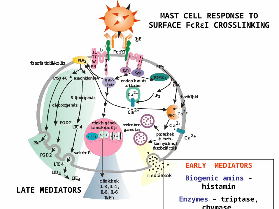
MAST CELL RESPONSE TO SURFACE FcRεI CROSSLINKING
EARLY MEDIATORS
Biogenic amins – histamin
Enzymes – triptase, chymase, carboxypeptidase
LATE MEDIATORS

The effect of mast cell degranulation varies with the tissue exposed to allergen

Systemic anaphylaxis is caused by allergens that reach the blood stream

Types of IgE-derived allergic responses
SYNDROME ALLERGENS ROUTE OF ENTRY RESPONSE
systemic anaphylaxis
drugsanti-serum
peanuts
intravenous(either directly or
after rapid absorption)
edema, increased vascular permeabilitytracheal occlusion
circulatory collapse, death
acuteurticaria
bug biteallergy test
subcutan local increase in blood flow and vascular
permeability
allergicrhinitis
pollendust mite
drops
inhaled irritation and edema of nasal mucosa
airway inflammation
asthma animal furpollen
dust mite drops
inhaled bronchial constriction, increased mucus
production
food allergy nut, peanuts,fish, shellfish
milk, eggs
oral vomiting, diarrheapruritis (itching)urticaria (hives)
anaphylaxia (rare)

Mast cell degranulation, allergic reaction in the skin of
a sensibilized individual
Prick test

ImmunoCAPSpecific IgE Blood Test
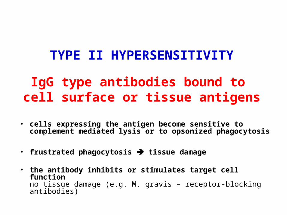
TYPE II HYPERSENSITIVITY
IgG type antibodies bound to cell surface or tissue antigens
• cells expressing the antigen become sensitive to complement mediated lysis or to opsonized phagocytosis
• frustrated phagocytosis tissue damage
• the antibody inhibits or stimulates target cell function no tissue damage (e.g. M. gravis – receptor-blocking antibodies)
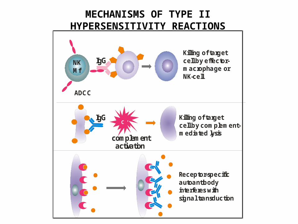
MECHANISMS OF TYPE II HYPERSENSITIVITY REACTIONS
Killing of target cell by effector-macrophage orNK-cell
Killing of targetcell by complement-mediated lysis
complement activation
IgG
IgG
Receptor-specific autoantibodyinterferes withsignal transduction
NKMf
C '
ADCC

DEVELOPMENT OF DRUG SENSITIVITY I.

DEVELOPMENT OF DRUG SENSITIVITY II.

The tissue, which can not be
phagocytosed, is damaged
Absorbed antigen (drug)
FcR
C3R
C3b C3b
C3b
C3b C3b C3b
FRUSTRATED PHAGOCYTOSIS MEDIATED BY IgG TYPE ANTIBODIES
Binding Opsonization Internalization Enzyme release
Opsonized surface Binding Frustrated Enzyme release phagocytosis

Examples - Type II hypersensitivity
Newborn haemolytic anaemiaTransfusion reaction
Drug-derived • Haemolitic anaemia• Thrombocytopenia• Agranulocytosis
• Penicillin-based antibiotics• Anti-arithmic quinidine

TYPE III HYPERSENSITIVITY
Antibodies binding to soluble antigensforming small circulating immune complexes
which are deposited in various tissues
Depends on:Size of immune complexes
Antigen-antibody ratio Affinity of antibodyIsotype of antibody

THE PROCESS OF TISSUE DAMAGE CAUSED BY IMMUNE COMPLEXES
Immune complexes activate the complement system, neutrophils, basophils and thrombocytes
Blood vessel wall
permeability
Frustrated phagocytosis
Antig e n
Antib o d y
Va so a c tivea m ine s
Ba so p hilg ra nulo c yte
Thro m b o c yte s
PM NC he m o ta xis
Im m une c o m p le x
C om p le m e nt-a c tiva tion(C 3a , C 5a )
De p ositio n
End othe liumBa sa l m e m b ra neVe sse l wa ll
C '
C '
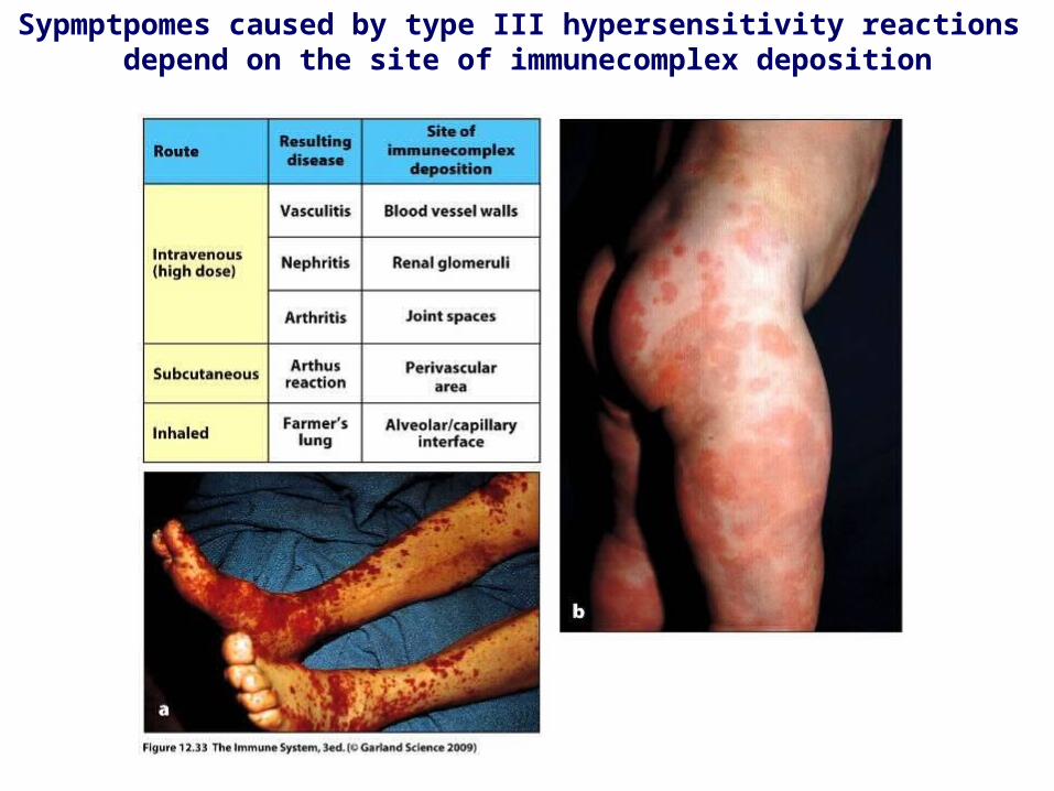
Sypmptpomes caused by type III hypersensitivity reactions depend on the site of immunecomplex deposition

Arthus-reaction• Localized Type III hypersensitivity • Local vasculitis develops as a result of immune complex deposition • Inhaled antigens (fungi, animal feces) may induce similar reaction in
the lung• IgG type antibody • ‘Farmer lung’ and ‘piegeon breeder lung’

TYPE IV HYPERSENSITIVITY REACTION
T CELL MEDIATED PROCESS
MACROPHAGES ARE INVOLVED

Type IV hypersensitivity reaction
Chemokines, cytokines,cytotoxins
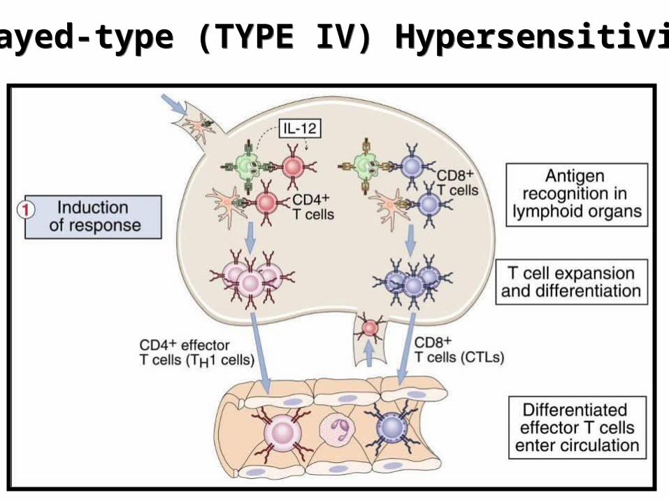
Delayed-type (TYPE IV) HypersensitivityDelayed-type (TYPE IV) Hypersensitivity

Delayed-type (Type IV) HypersensitivityDelayed-type (Type IV) Hypersensitivity

Delayed-type hypersensitivity (DTH) (e.g., tuberculin skin test)
TH1 from a previous immunization (memory)

Tuberculin skin test
Ag = antigen
Mycobacterium protein (PPD) Introduction of Ag

Chemical Mediators of DTH

*a contact-sensitizing agent is usually a small molecule that penetrates the skin then binds to self-proteins, making them “look” foreign
Contact Dermatitis
DTH as a result of a contact-sensitizing agent*

Poison ivy Anacardiaceae (family), Toxicodendron (genus)Toxicodendron radicans or Rhus toxicodendron