HYPERNATRAEMIC DEHYDRATION INFANTILE GASTRO-ENTERITIS
8
HYPERNATRAEMIC DEHYDRATION IN INFANTILE GASTRO-ENTERITIS BY DUNCAN MACAULAY and MARGARET I. BLACKHALL From the Duchess of York Hospital for Babies, Manchester (RECEIVED FOR PUBLICATION DECEMBER 19, 1960) Although it is possible to find occasional reports written before the last war of the occurrence of hyperelectrolytaemia in sick infants and children (for early references see Rapoport, 1947) the recog- nition of the condition as a fairly common one and the elucidation of the clinical features date from 1947 when Rapoport gave an account of 14 patients with hyperosmolarity of the plasma. Of the 14 patients, 10 were suffering from diarrhoea. Five of these were noted to have symptoms of central nervous system disturbance, from marked lethargy to con- vulsions. The first indication of the possible frequency of this state came in a report by Tarail, Bass and Runco (1953) who found that, of 191 infants with diarrhoea, 21 or 11% had serum sodium levels of 150 mEq/litre or higher. In 1955 Finberg and Harrison gave an account of seven years' experience of infantile diarrhoea involving 274 patients. They reported an incidence of hyper- natraemia (serum sodium level over 150 mEq/l.) of 25% and pointed out the great frequency of 'manifestations referable to the nervous system', nearly two-thirds of the hypernatraemic cases showing such signs. They reported, for the first time, the occurrence of raised protein concentrations in the cerebrospinal fluid, and drew attention to the fact that nearly half the cases did not show the customary signs of dehydration. They indicated the danger that rapid rehydration with dilute fluids might give rise to symptoms similar to those seen in water intoxication, especially to convulsions, and they suggested that hypernatraemia could be a cause of permanent brain damage. This paper seemed to define a distinct clinical entity of consider- able significance. It was followed by one by Weil and Wallace in 1956 which emphasized the points made by Finberg and Harrison, especially the neuro- logical disturbances. Several other papers from America have reported similar findings and opinion in that country is probably represented by the account of 'Hypertonic dehydration' given by Cooke (1959). According to this writer hypertonic dehydration occurs in approximately 20% of cases of diarrhoea in infancy; 'of particular importance are the cerebral changes which result from severe hyperosmolarity'; the development of oedema during treatment is referred to as 'characteristic'. From consideration of the available reports the 'clinical picture' of hypertonic dehydration emerges as follows: the patients are predominantly infants in the first year of life; as the result of fluid loss in the stools and by vomiting there is often loss of 10-15% of the body weight without the classical signs of such a severe degree of dehydration, e.g. the skin turgor may be deceptively normal; symptoms and signs of cerebral damage are common and include disturbances of consciousness ranging from lethargy to coma; hyperirritability to stimuli in spite of extreme lethargy; increased muscle tone amounting in many cases to generalized rigidity with very brisk tendon reflexes; convulsions, especially during rehydration; raised concentration of protein in the cerebrospinal fluid and rather exceptionally, the development of sub-dural effusions. Oedema is liable to appear during the administration of rehydrating fluids, sometimes before full rehydration has been effected. It has been suggested (Franz and Segar, 1959) that this state is becoming commoner and also (Finberg, 1958) that it may be due in many instances to the unwise administration of fluids with 'high-solute' content. We wish to describe the two cases which first drew our attention to this condition and then to give an account of an analysis of the records of 100 infants with diarrhoea admitted to the Duchess of York Hospital for Babies on whom serum- electrolyte estimations were performed. Case Reports Case 1. A baby girl, 3 months of age, was admitted to hospital with a history of diarrhoea and vomiting for five days. She had been perfectly well before then. On the day of admission, after a vomit, she became pale, 543 copyright. on March 21, 2022 by guest. Protected by http://adc.bmj.com/ Arch Dis Child: first published as 10.1136/adc.36.189.543 on 1 October 1961. Downloaded from
Transcript of HYPERNATRAEMIC DEHYDRATION INFANTILE GASTRO-ENTERITIS

HYPERNATRAEMIC DEHYDRATION IN INFANTILEGASTRO-ENTERITIS
BY
DUNCAN MACAULAY and MARGARET I. BLACKHALLFrom the Duchess of York Hospital for Babies, Manchester
(RECEIVED FOR PUBLICATION DECEMBER 19, 1960)
Although it is possible to find occasional reportswritten before the last war of the occurrence ofhyperelectrolytaemia in sick infants and children(for early references see Rapoport, 1947) the recog-nition of the condition as a fairly common one andthe elucidation of the clinical features date from 1947when Rapoport gave an account of 14 patients withhyperosmolarity of the plasma. Of the 14 patients,10 were suffering from diarrhoea. Five of thesewere noted to have symptoms of central nervoussystem disturbance, from marked lethargy to con-vulsions. The first indication of the possiblefrequency of this state came in a report by Tarail,Bass and Runco (1953) who found that, of 191infants with diarrhoea, 21 or 11% had serum sodiumlevels of 150 mEq/litre or higher. In 1955 Finbergand Harrison gave an account of seven years'experience of infantile diarrhoea involving 274patients. They reported an incidence of hyper-natraemia (serum sodium level over 150 mEq/l.)of 25% and pointed out the great frequency of'manifestations referable to the nervous system',nearly two-thirds of the hypernatraemic casesshowing such signs. They reported, for the firsttime, the occurrence of raised protein concentrationsin the cerebrospinal fluid, and drew attention tothe fact that nearly half the cases did not show thecustomary signs of dehydration. They indicatedthe danger that rapid rehydration with dilutefluids might give rise to symptoms similar to thoseseen in water intoxication, especially to convulsions,and they suggested that hypernatraemia could be acause of permanent brain damage. This paperseemed to define a distinct clinical entity of consider-able significance. It was followed by one by Weiland Wallace in 1956 which emphasized the pointsmade by Finberg and Harrison, especially the neuro-logical disturbances. Several other papers fromAmerica have reported similar findings and opinionin that country is probably represented by theaccount of 'Hypertonic dehydration' given byCooke (1959). According to this writer hypertonic
dehydration occurs in approximately 20% of casesof diarrhoea in infancy; 'of particular importanceare the cerebral changes which result from severehyperosmolarity'; the development of oedemaduring treatment is referred to as 'characteristic'.From consideration of the available reports the
'clinical picture' of hypertonic dehydration emergesas follows: the patients are predominantly infantsin the first year of life; as the result of fluid loss inthe stools and by vomiting there is often loss of10-15% of the body weight without the classicalsigns of such a severe degree of dehydration,e.g. the skin turgor may be deceptively normal;symptoms and signs of cerebral damage are commonand include disturbances of consciousness rangingfrom lethargy to coma; hyperirritability to stimuliin spite of extreme lethargy; increased muscle toneamounting in many cases to generalized rigiditywith very brisk tendon reflexes; convulsions,especially during rehydration; raised concentrationof protein in the cerebrospinal fluid and ratherexceptionally, the development of sub-duraleffusions. Oedema is liable to appear during theadministration of rehydrating fluids, sometimesbefore full rehydration has been effected. It hasbeen suggested (Franz and Segar, 1959) that thisstate is becoming commoner and also (Finberg,1958) that it may be due in many instances to theunwise administration of fluids with 'high-solute'content.We wish to describe the two cases which first
drew our attention to this condition and then togive an account of an analysis of the records of100 infants with diarrhoea admitted to the Duchessof York Hospital for Babies on whom serum-electrolyte estimations were performed.
Case ReportsCase 1. A baby girl, 3 months of age, was admitted
to hospital with a history of diarrhoea and vomitingfor five days. She had been perfectly well before then.On the day of admission, after a vomit, she became pale,
543
copyright. on M
arch 21, 2022 by guest. Protected by
http://adc.bmj.com
/A
rch Dis C
hild: first published as 10.1136/adc.36.189.543 on 1 October 1961. D
ownloaded from
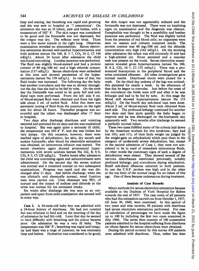
ARCHIVES OF DISEASE IN CHILDHOODlimp and staring; her breathing was rapid and gruntingand she was sent to hospital as '? pneumonia'. Onadmission she was an ill infant, pale and listless, with atemperature of 1020 F. The skin turgor was consideredto be good and the fontanelle was not depressed, butthe tongue was dry. The reflexes were brisk. Therewere a few moist sounds in the right lung. Otherwiseexamination revealed no abnormalities. Serum electro-lyte estimation showed well-marked hypernatraemia andmild acidosis (serum Na 168, K 5 7, CO2 16-2, Cl 144mEq/l.). About 24 hours after admission the childwas found convulsing. Lumbar puncture was performed.The fluid was slightly blood-stained and had a proteincontent of 80 mg./100 ml. and a high chloride concen-tration (137 mEq/l.). Serum electrolytes were recheckedat this time and showed persistence of the hyper-natraemia (serum Na 159 mEq/l.). In view of this, thefluid intake was increased. The following day the babyhad another convulsion and she was so drowsy through-out the day that she had to be fed by tube. On the sixthday the fontanelle was noted to be quite full and sub-dural taps were performed. From the left side 6 ml.of blood-stained fluid were obtained and from the rightside about 2 ml. of turbid fluid. After this there waspersistent oozing of fluid from the puncture on the rightside for about 24 hours. Thereafter progress was un-eventful and the infant was discharged after 17 daysin hospital.Two days after discharge diarrhoea and vomiting
recurred and persisted for six days and she was readmittedto hospital. She was again quite a seriously ill child;the temperature was 103-40 F. and she was listless butvery jumpy. On this occasion, however, there weremarked signs of dehydration; lax skin, dry mouth andsunken fontanelle; after blood for electrolyte estimationwas obtained, an intravenous infusion was started. Theserum chemistry again showed pronounced hyper-natraemia with severe acidosis (serum Na 162, K 6-6,CO2 8 * 5, Cl 128 mEq/l.). Twelve hours after admissionthe child was convulsing again and anticonvulsants wereadministered. On the second day the serum sodiumwas normal and it remained normal on two subsequentexaminations. Progress was rapid and she was dis-charged after 11 days. Just before discharge, when shewas clinically and chemically normal, renal functiontests were carried out. Urea clearance was 90% ofnormal and the output of sodium and chloride in theurine was normal for the estimated intake.
Six weeks after discharge she was seen as an out-patient and apart from mild constipation she seemed wellin every way.
Case 2. A 10-week-old baby boy was admitted witha 24-hour history of diarrhoea. He had not vomitedbut was reluctant to feed and on the morning of the dayof admission he had felt cold. Later that day he seemedto have difficulty with breathing and his colour becamepoor. On admission he was a very ill baby. Thetemperature was 1040 F.; breathing was rapid and irregu-lar and there was a tinge of cyanosis; he was extremelylimp and listless. Hydration was considered to be good;
the skin turgor was not appreciably reduced and thefontanelle was not depressed. There were no localizingsigns on examination and the diagnosis was obscure.Encephalitis was thought to be a possibility and lumbarpuncture was performed. The fluid was slightly turbidfrom the presence of red blood cells; no organisms wereseen on smears and cultures remained sterile. Theprotein content was 40 mg./100 ml. and the chlorideconcentration very high (162 mEq/l.). On the morningafter admission the infant was still extremely ill and hada high-pitched cry. Fever persisted and a macularrash was present on the trunk. Serum electrolyte exami-nation revealed gross hypernatraemia (serum Na 180,K 6-2, CO2 16 7, Cl 135 mEq/l.). There was a pro-nounced leucocytosis in the peripheral blood and theurine contained albumen. All other investigations gavenormal results. Diarrhoeal stools were passed for aweek. On the third day oedema of the legs was noticed;this persisted for nearly a week. In the afternoon ofthat day he began to convulse. Just before the onset ofthe convulsion the limbs were stiff and after it he waslethargic and had to be fed by tube. At this stage theblood still showed hypernatraemia (serum Na 153mEq/l.). On the fourth day sub-dural taps were done.About 2 ml. of blood-stained fluid were obtained fromeach side. The profound lethargy continued for anothertwo days and then quite quickly the baby began toimprove and he was discharged on the fourteenth dayapparently well. Two months after discharge he seemeda perfectly normal infant.These two cases fulfilled many of the criteria laid down
by the American workers for this syndrome; they hadlost 10% and 11% of their body weight (as judged bythe weight-gain on rehydration) which puts them in thecategory of moderately severe dehydration, but, exceptin the second admission of Case 1, they were not con-sidered to be in need of immediate intravenous fluids.In other words the customary signs of such a degree ofdehydration were absent. They showed several of thenervous disturbances mentioned previously, notablyprofound lethargy, and convulsions during rehydration.Small sub-dural effusions occurred in both patients.In one the C.S.F. protein was high and in the otherat the top limit of the normal range for an infant of thisage. One of them became oedematous during treatment.
Analysis of Case RecordsMicro methods for serum electrolyte estimation became
available at the Duchess of York Hospital for Babiestowards the end of 1957. The case notes of all patientswho had this estimation carried out from October 1, 1957,till June 30, 1960, were examined. In this period oftwo years and nine months, 98 patients with diarrhoeahad serum electrolyte estimations performed. For easeof calculation of percentages we have made the figureup to 100 by including the first two cases examined inJuly 1960. The series thus consists of 100 consecutivepatients admitted to the hospital suffering from diarrhoeaon whom figures for serum electrolytes were obtained.During the period covered by this survey 439 patients
were admitted with diarrhoea. Thus, rather less than
544
copyright. on M
arch 21, 2022 by guest. Protected by
http://adc.bmj.com
/A
rch Dis C
hild: first published as 10.1136/adc.36.189.543 on 1 October 1961. D
ownloaded from

HYPERNATRAEMIC DEHYDRATION IN GASTRO-ENTERITISactual figures are given, of a total of 976 infants,206 or 21% were hypernatraemic. Our seriescontains more hypernatraemic cases than theaverage, but considerably fewer than in two ofthe reports. We have thus established that hyper-natraemic dehydration is as common here as in otherareas.
120 I30 l0O ISO 160 170 180 2O0SERUM SODIUM mEq/litre
FIG. 1.-Distribution of serum sodium levels at intervals of 2 5mEq/l. Vertical dotted lines demarcate hypo-, normo- and hyper-natraemia. Cases showing nervous disturbances are indicated in
black.
25% of them were examined in this way. In generalit was the more seriously ill patients who had this exami-nation performed. Because of this selection care mustbe exercised in making general statements about infantswith diarrhoea.
Criteria and ResultsThe 'normal serum sodium concentration ranges
from 135 to 145 mEq per litre' (Cooke and Otten-heimer, 1960). We have adopted the standard ofHarrison and Finberg (1959) that any value forserum sodium of 150 mEq/l. or above constituteshypernatraemia. The range of values for serumsodium in this series was from 118 to 200 mEq/l.The distribution of the measurements is shown inthe Figure. It can be seen that 150 mEq/l. is anobvious level at which to separate the cases, thestep-wise decrease in numbers being replaced atthat point by a sharp increase.
In Table 1 are listed the reports available to uswith the percentages of cases in the various groups.The incidence of hyponatraemia (serum sodiumof 130 mEq/l. or less) is fairly constant in all theseries in which it is listed. There is marked variation,however, in the incidence of hypernatraemia whichcannot be satisfactorily explained on the informationavailable. However, in the nine series in which
Mortality. Eight of the 100 patients died. Intwo cases death might be attributed directly to thediarrhoea. One was a mongol who respondedpoorly to treatment; the second baby died a weekafter admission and at autopsy was found to haveextensive ulcerative colitis. Two other childrendied several weeks after the initial attack of diarrhoeaand at autopsy showed only bronchopneumonia;one of these was also a mongol. The remainingfour patients died at intervals of from three to30 days after the serum electrolyte measurement andall four showed gross cerebral changes at autopsy.The pathological conditions were described asfollows: multiple cerebral haemorrhages; multipleareas of cerebral necrosis; cerebral infarction; andmultiple areas of cerebral necrosis with calcification.It is a striking fact that three of these four 'cerebral'deaths occurred among the 30 patients with hyper-natraemia-indeed they were the only deaths in thisgroup-while only one occurred in the remaining70 patients. This bears out what must, we think,be accepted as an established fact, that there is anassociation between hypernatraemia and braininjury.
Contrary to the experience of others, hyper-natraemia did not in this series carry a significantlyhigher mortality than normo- or hyponatraemia, theoverall mortality in the former being 10% and inthe latter 7%.
Neurological Disturbances. Clinical evidence ofdisturbances of the nervous system is most con-
TABLE 1INCIDENCE OF HYPO- AND HYPERNATRAEMIA IN PREVIOUS REPORTS
Authors Year Country Hyponatraemia Normonatraemia Hypernatraemia No. of(%) () ( Cases
Tarail et al. .1953 U.S.A. 1 191Finberg and Harrison .. 1955 U.S.A. 25 274Weil and Wallace .1956 U.S.A. 10 70 20 77Skinner and Moll .1956 U.S.A. 23 61Kerpel-Fronius and Vonocsky .. 1957 Hungary 9 76 15 113Colle et al. (a) ..1958 U.S.A. 16-25Colle et al. (b) ..1958 U.S.A. 71 24Bowie et al.. 1958 S. Africa 22 44 34 32Franz and Segar ..1959 U.S.A. 4-21Darrow and Welsh .1960 U.S.A. 14 75 11 104Macaulay and Blackhall (this paper) 1961 U.K. 11 59 30 100Averages 12 67 21
Total 976
7
20-
cnw 15.
to
0z 51
II
I I~ ~
B -- - - - -~~~EJLpfm -A -i
545
III
III
I
_ _
copyright. on M
arch 21, 2022 by guest. Protected by
http://adc.bmj.com
/A
rch Dis C
hild: first published as 10.1136/adc.36.189.543 on 1 October 1961. D
ownloaded from

ARCHIVES OF DISEASE IN CHILDHOOD
vincingly documented by the occurrence of con-
vulsions and by the frequency with which lumbarpuncture was performed. It is reasonable to supposethat a child subjected to lumbar puncture must havehad some symptom or sign suggestive of C.N.S.disorder. Convulsions were recorded in 10 of thehypernatraemic group, in nine of them duringrehydration, and in seven of the remaining cases,
five of these occurring during treatment. Statis-tically this difference is significant at the 2% leveland almost reaches the 1% level. The differencebetween the two groups in the frequency with whichlumbar puncture was performed (14 or 47% of thehypernatraemic patients, 17 or 24% of the remain-der) is just significant at the 5% level. A lessreliable, because more subjective, criterion ofnervous disorder is recording disturbances ofconsciousness. These ranged from marked lethargyto deep coma. Since the existence of the syndromeof hypernatraemic dehydration was, we are con-fident, unsuspected by the observers making thenotes any bias in recording this symptom shouldaffect all cases equally. In this case the differencebetween the two groups is highly significant. Ofthe hypernatraemic patients 14 (47%) and of theremainder 12 (17%) showed some depression ofconsciousness (p <0 01).The protein concentration in the cerebrospinal
fluid was estimated in 14 samples from hyper-natraemic patients and in 15 from other cases. Thevalues ranged very widely in both groups-from20 to 200 mg./100 ml. in the former, from 20 to500 mg./100 ml. in the latter. The average valuein the hypernatraemic patients was 70 mg./100 ml.,and in the others 80 mg./100 ml. This latter figureis a little misleading as there were two very highvalues in this group. If 40 mg./100 ml. is taken as
the upper limit of normal, there were nine highvalues in the 14 hypernatraemic samples and sixin the 15 from the other patients. This difference,though suggestive, is not significant (p >0 3).We have, in other words, failed to confirm thathypernatraemia is regularly associated with raisedprotein levels in the cerebrospinal fluid.The incidence of hyperirritability and increased
muscle tone could not be determined with anyaccuracy from the records available since they were
infrequently noted in any group.The follow-up period was, for many patients,
too short to permit any evaluation of ultimateneurological status.
There is, in the present series as in those pre-viously reported, definite evidence of an associationbetween hypernatraemia and nervous disturbances.However, it is not a consistent relationship. In the
30 hypernatraemic patients no record of any neuro-logical upset was found in 11 cases. Contrariwise,22 of the 70 non-hypernatraemic patients had oneor more of the symptoms of nervous disorder men-tioned in the introductory paragraphs (see also thedistribution of cases with nervous disturbances inthe Figure). In terms of the numbers at risk therewere twice as many patients with neurologicaldisturbances in the hypernatraemic group as in theremainder. A small number of the latter wouldundoubtedly have been diagnosed on clinicalgrounds as hypernatraemic, and a considerablenumber of the former would have been misdiagnosedon the evidence of clinical observation. Sincebecoming interested in the subject we have madeboth these mistakes and conclude that the clinicaldiagnosis of hypernatraemia is unreliable.
TABLE 2SIGNS OF DEHYDRATION IN CASES WITH WEIGHT LOSS
OF 10% OR MORE
Clinical Assessment of Hypernatraemic OtherDegree of Dehydration Cases Cases
0 3 1+ 5 6
Total 0 and + 8 7
+±+ 5 11+++ 1 3
Total + + and +++ 6 14
X2=109 p=0-3
Signs of Dehydration. As noted in the introduc-tion, the extent of fluid loss in hypernatraemicdehydration is said to be often underestimatedbecause of the lack of the usual signs of dehydration.We have tried to assess this factor in the followingway. In some cases, e.g. hospital-acquired infec-tions, the weight before the onset of the diarrhoeawas known. In the others the weight charts wereused to estimate the gain in weight due to rehydra-tion. From these data the amount of weight lostas the result of the diarrhoea was estimated andexpressed as a percentage of the normal weight.Of the 85 patients whose records were completeenough to permit these calculations 35 were esti-mated to have lost 10% or more of their body weight,i.e. were moderately severely or very severelydehydrated. The clinical assessment of the degreeof dehydration in these cases is shown in Table 2.We are not perturbed by the lack of precision inthe terms used in recording the apparent degree ofdehydration, nor by the fact that nearly half thecases (15 of the 35) were wrongly assessed. Thepoint at issue is whether this mistake is more com-mon when the patient is hypematraemic. In fact
546
copyright. on M
arch 21, 2022 by guest. Protected by
http://adc.bmj.com
/A
rch Dis C
hild: first published as 10.1136/adc.36.189.543 on 1 October 1961. D
ownloaded from

HYPERNATRAEMIC DEHYDRATION IN GASTRO-ENTERITISthe distribution of the cases in this Table does notdiffer from chance expectancy. Our subsequentexperience confirms this finding and we have foundit impossible to predict the serum sodium level fromthe apparent severity of clinical dehydration.
Oedema. Oedema was noted to have occurredin six of the 30 hypernatraemic cases during treat-ment and in only two of the 70 others. Whilethis is an interesting observation, and indeed a'significant' one in the statistical sense (the pro-bability of it being a chance occurrence is verynearly one in 100), it hardly bears out the contentionthat the development of oedema is characteristic ofthe hypernatraemic state. Mild degrees of oedemamay have escaped notice and close observationmight yield higher figures. We find it hard tobelieve, however, that appreciable oedema is at allcommon in hypernatraemic infants.
Aetiological FactorsWe have reviewed our cases to try to determine
what factors might have resulted in 30 of thembecoming hypernatraemic. In order to permitstatistical evaluation we have given numericalvalues to the data wherever possible. The notesare not equally informative in all cases and we showthe numbers of cases in which the data were con-sidered satisfactory either in the Tables or inbrackets after each sub-heading.No difference between the hypernatraemic patients
and the others was found in respect of the followingitems and we shall not give detailed figures: sex,age, premature birth, previous history of nervousor renal disease, seasonal incidence (the absenceof any climatic influence is shown by the numberswith hypernatraemia occurring in successive six-monthly periods, viz. five, five, four, five, six),frequency of vomiting, character of stools (whethermerely 'loose' or described as 'watery'), or numberof stools per day. The amount of sodium in thediet before the electrolyte estimation was not afactor in the development of hypernatraemia, butin view of the importance attached to this by someAmerican writers, we shall consider it in somedetail.
Sodium Intake. The object of this analysis wasto see if, as has been claimed (Finberg, 1958),excessive administration of salt-containing fluidswas responsible for a significant number of patientsbecoming hypernatraemic. This is of obviousimportance, since it might lead to recommendationswhich could result in reducing the number of infantswith this complication of diarrhoea.
TABLE 3ESTIMATED SODIUM INTAKE
Sodium GroupIntake
(mEq/kg./day) Hypo- Normo- Hyper-natraemia natraemia natraemia
< 5 5 23 165-10 5 17 9>10 3 2
Totals .. 10 43 27
Where the information available made it feasible,we have attempted to assess the sodium intake ofthe patients in the 48 hours before the electrolyteestimation was done. Very few of the patientswere given supplementary salt-containing fluidsbefore admission. This represents a marked differ-ence from experience in America (Colle, Ayoub andRaile, 1958; Franz and Segar, 1959; Harrison andFinberg, 1959) where commercial electrolyte solu-tions are extensively used in the home treatment ofdiarrhoea. These estimates cannot, obviously, bevery precise, but in 80 of the 100 cases there weresufficient data to justify analysis. We have calcu-lated the amount of sodium in the feeds offered to theinfants, ignoring vomiting and anorexia as sourcesof error. This means that our estimates are of thegreatest amounts of sodium likely to have beeningested. In many cases, because of vomiting andanorexia, the actual intakes were much less thanthose calculated. The sodium content of variousfeeds was directly measured by having randomfeeds, made up in the hospital milk-room, analysedin the laboratory. The figures are given in termsof mEq sodium/kg. hydrated body weight/day.This makes possible comparison ofbabies of differentweights and it also indicates the importance of theactual load of sodium given. There is some evidencethat 'the ceiling for renal- excretion of sodium maybe in the range of 5 to 10 mEq per kilogram of bodyweight per day' (Skinner and Moll, 1956). Darrowand Welsh (1960) report that quantities equivalentto 7 5 mEq/kg./day are well tolerated by infantsnot suffering from dehydration. It may be assumedthat intakes of up to 5 mEq/kg./day are normal(e.g. an infant fed on a half-cream dried milk atthe rate of 2- oz./lb./day will ingest approximately4 mEq/kg./day). Intakes of over 10 mEq/kg./dayare almost certainly excessive. The intermediateamounts, 5 to 10 mEq/kg./day, would probably betolerated without difficulty by a healthy infant, butmight be excessive for an ill one.The results of our analysis are given in Table 3.
Only five infants received amounts of sodium in thefrankly excessive range (four of them had been
547
copyright. on M
arch 21, 2022 by guest. Protected by
http://adc.bmj.com
/A
rch Dis C
hild: first published as 10.1136/adc.36.189.543 on 1 October 1961. D
ownloaded from
ARCHIVES OF DISEASE IN CHILDHOOD
rather enthusiastically rehydrated with intravenousfluids before the electrolytes were measured) and ofthese only two showed hypernatraemia. Morethan half the hypernatraemic group had been offered,and had not necessarily ingested, quite normalamounts of sodium. In the intermediate group thedistribution of cases is almost exactly the same asin the series as a whole, viz. 29% hypematraemic.It seems legitimate to conclude that very few of ourpatients had been overloaded with sodium and thatdietary sodium played an insignificant part indetermining the serum sodium levels.The respects in which the hypernatraemic cases
differed from the others were: severity of anorexia,duration of diarrhoea, temperature and respiratoryrate.
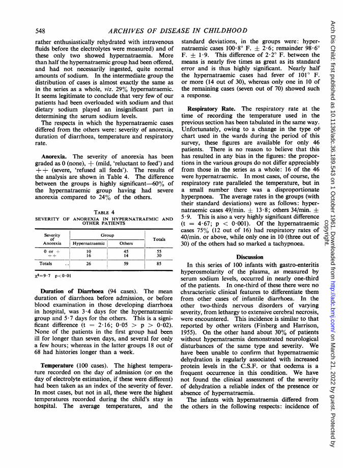
Anorexia. The severity of anorexia has beengraded as 0 (none), + (mild, 'reluctant to feed') and++ (severe, 'refused all feeds'). The results ofthe analysis are shown in Table 4. The differencebetween the groups is highly significant-60% ofthe hypernatraemic group having had severeanorexia compared to 24% of the others.
TABLE 4SEVERITY OF ANOREXIA IN HYPERNATRAEMIC AND
OTHER PATIENTS
Severity Groupof Totals
Anorexia Hypernatraemic Others
0 or + 10 45 55++ 16 14 30
Totals 26 59 85
x2=9 7 p<O-Ol
Duration of Diarrhoea (94 cases). The mean
duration of diarrhoea before admission, or beforeblood examination in those developing diarrhoeain hospital, was 3-4 days for the hypernatraemicgroup and 5-7 days for the others. This is a signi-ficant difference (t = 2 16; 0 05 > p > 0 02).None of the patients in the first group had beenill for longer than seven days, and several for onlya few hours; whereas in the latter groups 18 out of68 had histories longer than a week.
Temperature (100 cases). The highest tempera-ture recorded on the day of admission (or on theday of electrolyte estimation, if these were different)had been taken as an index of the severity of fever.In most cases, but not in all, these were the highesttemperatures recorded during the child's stay inhospital. The average temperatures, and the
standard deviations, in the groups were: hyper-natraemic cases 100.80 F. ± 2-6; remainder 98.60F. ± 1 9. This difference of 2- 2 F. between themeans is nearly five times as great as its standarderror and is thus highly significant. Nearly halfthe hypernatraemic cases had fever of 101 ' F.or more (14 out of 30), whereas only one in 10 ofthe remaining cases (seven out of 70) showed sucha response.
Respiratory Rate. The respiratory rate at thetime of recording the temperature used in theprevious section has been tabulated in the same way.Unfortunately, owing to a change in the type of)chart used in the wards during the period of thissurvey, these figures are available for only 46patients. There is no reason to believe that thishas resulted in any bias in the figures: the propor-tions in the various groups do not differ appreciablyfrom those in the series as a whole: 16 of the 46were hypernatraemic. In most cases, of course, therespiratory rate paralleled the temperature, but ina small number there was a disproportionatehyperpnoea. The average rates in the groups (withtheir standard deviations) were as follows: hyper-natraemic cases 49/min. + 13-8; others 34/min. ±5 - 9. This is aiso a very highly significant difference(t = 4 67; p < 0-001). Of the hypernatraemiccases 75% (12 out of 16) had respiratory rates of40/min. or above, while only one in 10 (three out of30) of the others had so marked a tachypnoea.
DiscussionIn this series of 100 infants with gastro-enteritis
hyperosmolarity of the plasma, as measured byserum sodium levels, occurred in nearly one-thirdof the patients. In one-third of these there were nocharacteristic clinical features to differentiate themfrom other cases of infantile diarrhoea. In theother two-thirds nervous disorders of varyingseverity, from lethargy to extensive cerebral necrosis,were encountered. This incidence is similar to thatreported by other writers (Finberg and Harrison,1955). On the other hand about 30% of patientswithout hypernatraemia demonstrated neurologicaldisturbances of the same type and severity. Wehave been unable to confirm that hypernatraemicdehydration is regularly associated with increasedprotein levels in the C.S.F. or that oedema is afrequent occurrence in this condition. We havenot found the clinical assessment of the severityof dehydration a reliable index of the presence orabsence of hypernatraemia.The infants with hypernatraemia differed from
the others in the following respects: incidence of
548
copyright. on M
arch 21, 2022 by guest. Protected by
http://adc.bmj.com
/A
rch Dis C
hild: first published as 10.1136/adc.36.189.543 on 1 October 1961. D
ownloaded from

HYPERNATRAEMIC DEHYDRATION IN GASTRO-ENTERITIS 549cerebral disturbances, short duration of diarrhoea,frequency of fever and tachypnoea and severity ofanorexia.
This account agrees substantially with that givenby Bowie, McKenzie and Hansen (1958) from whichwe quote: 'The clinical recognition of hyper-osmolarity in infantile gastro-enteritis is difficult . . .It is our impression that those showing the hyper-osmolar state had a short history of explosivediarrhoea and vomiting . .. Where gastro-enteritisis associated with hyperpyrexia, hyperventilation orseverely curtailed fluid intake the possibility ofhyperosmolarity developing is considerablyenhanced.'The pathogenesis of the condition does not seem
to be particularly mysterious. It is known (Weiland Wallace, 1956) that diarrhoeal stools are hypo-tonic so that profuse diarrhoea will, in the absenceof any compensating mechanism, lead to a greaterdeficit of water than of electrolyte, i.e. to a conditionof hypertonicity. The two possible compensatingmechanisms available are (1) an increase in theamount of water ingested to balance the losses and(2) differential excretion of electrolytes and water bythe kidneys resulting in the secretion of a con-centrated urine and maintenance of the tonicity ofthe body fluids. In infants neither of these mechan-isms is as effective as in adults: (1) Infants aredependent on others for their fluid intake and theneed for increased amounts of fluid may not beappreciated by the attendants. Even with anappreciation of these needs, severe anorexia maymake it impossible to get enough fluid into the child.This was an important factor in our patients.(2) Under the stress of dehydration, and perhapsaggravated by losses of electrolytes, especiallypotassium, in the stools, renal function becomesimpaired and the infantile kidneys become incapableof excreting a concentrated urine. The few studiesreported on hypematraemic infants (Weil andWallace, 1956) confirm the existence of this typeof dysfunction, manifested by the failure toexcrete as concentrated a urine as would have beenexpected. This temporary renal failure is madeworse by further losses of water from the skin andlungs through fever and hyperventilation. It mayalso contribute to further dehydration since thekidneys will require a greater volume of water forthe excretion of solutes than if they are functioningnormally.
In any event the development of hyperosmolarityis determined by disproportionate losses of waterrelative to sodium. This can occur withoutdiarrhoea, and, in fact, the highest level of serumsodium encountered in any case in the two years
and nine months of our survey was 235 mEq/l.(duplicate estimation) in an infant with congenitalpyloric stenosis who was constipated but dehydratedand wasted from persistent vomiting.With regard to treatment, recommendations have
varied from elaborate schedules (Skinner and Moll,1956) to statements that 'Patients with hyper-natraemia received the usual fluid therapy used inother cases of diarrhoea' (Darrow and Welsh, 1960).In general it is agreed that repair solutions shouldbe hypotonic and that the replacement of deficitsshould be spread over 48 hours. Our custom isto use 'fifth-normal saline in glucose water', i.e.a solution containing 0- 18% NaCl and 4-3%glucose, and half-strength plasma, as repair solutionsin all cases of diarrhoea requiring intravenous fluids.In the presence of hypernatraemia, we substitutevarying amounts of 5% glucose-water for the'fifth-normal saline', allow rather more than usualfor the repair of existing deficits and aim at a some-what less rapid correction of these deficits than inthe ordinary case. Serum sodium levels vary somuch, as do other features of these cases, that wethink it unwise to lay down definite rules.
SummaryA brief review of the recent literature on the
occurrence of hypernatraemic dehydration ininfantile gastro-enteritis is followed by short accountsof two patients with this condition.
Analysis of the case records of 100 consecutiveinfants with gastro-enteritis whose serum electro-lytes were determined reveals the following:
(1) An incidence of hypernatraemia similar to thatreported from other centres;
(2) An association between hypernatraemia and theoccurrence of neurological disturbances, similarto that reported by others, but neither as closenor as consistent as sometimes claimed;
(3) The importance of anorexia, fever and hyper-ventilation in the genesis of the condition.
Pathogenesis and treatment are briefly discussed.
REFERENCES
Bowie, M. D., McKenzie, D. and Hansen, J. D. L. (1958). Hyper-osmolarity in infantile gastro-enteritis. S. Afr. med. J., 32, 322.
Colle, E., Ayoub, E. and Raile, R. (1958). Hypertonic dehydration(hypernatremia): The role of feedings high in solutes. Pediatrics,22, 5.
Cooke, R. E. (1959). Parenteral fluid therapy. In Textbook ofPediatrics, ed. Nelson, W. E., 7th ed., pp. 183-98. Saunders,Philadelphia and London.and Ottenheimer, E. J. (1960). Clinical and experimental
interrelations of sodium and the central nervous system. InAdvances in Pediatrics, ed. Levine, S. Z., Vol. XI, pp. 81-145.The Year Book Publishers, Chicago.
Darrow, D. C. and Welsh, J. S. (1960). Recent experience in thetreatment of diarrhea in infants. J. Pediat., 56, 204.
copyright. on M
arch 21, 2022 by guest. Protected by
http://adc.bmj.com
/A
rch Dis C
hild: first published as 10.1136/adc.36.189.543 on 1 October 1961. D
ownloaded from

550 ARCHIVES OF DISEASE IN CHILDHOODFinberg, L. (1958). The possible role of the physician in causing
hypernatremia in infants dehydrated from diarrhea. Pediatrics,22, 2.
-and Harrison, H. E. (1955). Hypernatremia in infants. Anevaluation of the clinical and biochemical findings accompanyingthis state. Ibid., 16, 1.
Franz, M. N. and Segar, W. E. (1959). The association of variousfactors and hypernatremic diarrheal dehydration. Amer. J.Dis. Child., 97, 298.
Harrison, H. E. and Finberg, L. (1959). Hypernatremic dehydration.Pediat. Clin. N. Amer., Vol. 6, No. 1, p. 193.
Kerpel-Fronius, E. and Vonoczky, J. (1957). Significance of changesin the tonicity of body fluids in infantile diarrheal dehydration.Ann. Paediat. Fenn., 3, 403.
Rapoport, S. (1947). Hyperosmolarity and hyperelectrolytemia inpathologic conditions of childhood. Amer. J. Dis. Child.,74, 682.
Skinner, A. L. and Moll, F. C. (1956). Hypernatremia accompanyinginfant diarrhea. Ibid., 92, 562.
Tarail. R., Bass, L. W. and Runco, A. S. (1953). The frequency andnature of hypertonicity of the body fluids in infantile diarrhea.Ibid., 86, 658.
Weil. W. B. and Wallace, W. M. (1956). Hypertonic dehydrationin infancy. Pediatrics, 17, 171.
copyright. on M
arch 21, 2022 by guest. Protected by
http://adc.bmj.com
/A
rch Dis C
hild: first published as 10.1136/adc.36.189.543 on 1 October 1961. D
ownloaded from