Hydroxyl and molecular H2O diffusivity in a haploandesitic ...
13
Hydroxyl and molecular H 2 O diffusivity in a haploandesitic melt Huaiwei Ni a,b,⇑ , Zhengjiu Xu c , Youxue Zhang c a Bayerisches Geoinstitut, Universita ¨ t Bayreuth, 95440 Bayreuth, Germany b School of Earth and Space Sciences, University of Science and Technology of China, Hefei 230026, China c Department of Earth and Environmental Sciences, The University of Michigan, Ann Arbor, MI 48109, USA Received 8 December 2011; accepted in revised form 30 October 2012; Available online 15 November 2012 Abstract H 2 O diffusion in a haploandesitic melt (a high-silica and Fe-free andesitic melt, NBO/T = 0.173) has been investigated at 1 GPa in a piston-cylinder apparatus. We adopted a double diffusion couple technique, in which one couple was composed of a nominally anhydrous glass with 0.01 wt.% H 2 O and a hydrous glass with 5.7 wt.% H 2 O, and the other contained the same nominally anhydrous glass and a hydrous glass with 3.3 wt.% H 2 O. Both couples were annealed in a single experimental run and hence experienced exactly the same P–T history, which is crucial for constraining the dependence of H 2 O diffusivity on water content. H 2 O concentration profiles were measured by both Fourier transform infrared (FTIR) microspectroscopy and confo- cal Raman microspectroscopy. Nearly identical profiles were obtained from Raman and FTIR methods for profile length >1 mm (produced at 1619–1842 K). By contrast, for profile lengths <100 lm (produced at 668–768 K), FTIR profiles show marked convolution effects compared to Raman profiles. A comparison between the short FTIR and Raman profiles indicates that the real spatial resolution (FWHM) of FTIR analyses is about 28 lm for a 7 lm wide aperture on 200 lm thick glasses. While the short profiles are not reliable for quantitative modeling, the long diffusion profiles at superliquidus temperatures can be fit reasonably well by a diffusivity model previously developed for felsic melts, in which molecular H 2 O (H 2 O m ) is the only diffusive species and its diffusivity (D H 2 Om ) increases exponentially with the content of total water (H 2 O t ). However, there is noticeable misfit of the data at low H 2 O t concentrations, suggesting that OH diffusivity (D OH ) cannot be neglected in this andesitic melt at high temperatures and low water contents. We hence develop a new fitting procedure that simultaneously fits both diffusion profiles from a single experimental run and accounts for the roles of both OH and H 2 O m diffusion. With this procedure, D OH /D H 2 Om is constrained to be 0.1–0.2 at 1619–1842 K as H 2 O t concentration approaches zero. The obtained OH diffusivity is similar to fluorine diffusivity but is much higher than Eyring diffusivity. Ó 2012 Elsevier Ltd. All rights reserved. 1. INTRODUCTION As the most abundant and the most important volatile component in silicate melts, the diffusion of water has drawn substantial attention from volcanologists, geochem- ists and glass scientists (Shelby, 2008; Zhang and Ni, 2010). Since Zhang et al. (1991) laid out a theoretical basis for water diffusion, especially in demonstrating the dominating role of molecular H 2 O (H 2 O m ) rather than hydroxyl group (OH), water diffusivity has been experimentally determined for a variety of silicate melts (Zhang and Stolper, 1991; Nowak and Behrens, 1997; Zhang and Behrens, 2000; Freda et al., 2003; Liu et al., 2004; Behrens et al., 2004, 2007; Okumura and Nakashima, 2004, 2006; Ni and Zhang, 2008; Behrens and Zhang, 2009; Ni et al., 2009a,b; Wang et al., 2009; Persikov et al., 2010). It has been generally accepted that in felsic melts, water diffusivity increases strongly with increasing concentration of total water (H 2 O t ), but the relationship between diffusivity and H 2 O t concentration is less conclusive for silicate melts of interme- diate to mafic compositions. 0016-7037/$ - see front matter Ó 2012 Elsevier Ltd. All rights reserved. http://dx.doi.org/10.1016/j.gca.2012.10.052 ⇑ Corresponding author at: Bayerisches Geoinstitut, Universita ¨t Bayreuth, 95440 Bayreuth, Germany. Tel.: +49 921 55 3708; fax: +49 921 55 3769. E-mail address: [email protected] (H. Ni). www.elsevier.com/locate/gca Available online at www.sciencedirect.com Geochimica et Cosmochimica Acta 103 (2013) 36–48
Transcript of Hydroxyl and molecular H2O diffusivity in a haploandesitic ...

Available online at www.sciencedirect.com
www.elsevier.com/locate/gca
Geochimica et Cosmochimica Acta 103 (2013) 36–48
Hydroxyl and molecular H2O diffusivity in a haploandesitic melt
Huaiwei Ni a,b,⇑, Zhengjiu Xu c, Youxue Zhang c
a Bayerisches Geoinstitut, Universitat Bayreuth, 95440 Bayreuth, Germanyb School of Earth and Space Sciences, University of Science and Technology of China, Hefei 230026, China
c Department of Earth and Environmental Sciences, The University of Michigan, Ann Arbor, MI 48109, USA
Received 8 December 2011; accepted in revised form 30 October 2012; Available online 15 November 2012
Abstract
H2O diffusion in a haploandesitic melt (a high-silica and Fe-free andesitic melt, NBO/T = 0.173) has been investigated at1 GPa in a piston-cylinder apparatus. We adopted a double diffusion couple technique, in which one couple was composedof a nominally anhydrous glass with 0.01 wt.% H2O and a hydrous glass with 5.7 wt.% H2O, and the other contained the samenominally anhydrous glass and a hydrous glass with 3.3 wt.% H2O. Both couples were annealed in a single experimental run andhence experienced exactly the same P–T history, which is crucial for constraining the dependence of H2O diffusivity on watercontent. H2O concentration profiles were measured by both Fourier transform infrared (FTIR) microspectroscopy and confo-cal Raman microspectroscopy. Nearly identical profiles were obtained from Raman and FTIR methods for profile length>1 mm (produced at 1619–1842 K). By contrast, for profile lengths <100 lm (produced at 668–768 K), FTIR profiles showmarked convolution effects compared to Raman profiles. A comparison between the short FTIR and Raman profiles indicatesthat the real spatial resolution (FWHM) of FTIR analyses is about 28 lm for a 7 lm wide aperture on �200 lm thick glasses.
While the short profiles are not reliable for quantitative modeling, the long diffusion profiles at superliquidus temperaturescan be fit reasonably well by a diffusivity model previously developed for felsic melts, in which molecular H2O (H2Om) is theonly diffusive species and its diffusivity (DH2Om ) increases exponentially with the content of total water (H2Ot). However, thereis noticeable misfit of the data at low H2Ot concentrations, suggesting that OH diffusivity (DOH) cannot be neglected in thisandesitic melt at high temperatures and low water contents. We hence develop a new fitting procedure that simultaneously fitsboth diffusion profiles from a single experimental run and accounts for the roles of both OH and H2Om diffusion. With thisprocedure, DOH/DH2Om is constrained to be 0.1–0.2 at 1619–1842 K as H2Ot concentration approaches zero. The obtained OHdiffusivity is similar to fluorine diffusivity but is much higher than Eyring diffusivity.� 2012 Elsevier Ltd. All rights reserved.
1. INTRODUCTION
As the most abundant and the most important volatilecomponent in silicate melts, the diffusion of water hasdrawn substantial attention from volcanologists, geochem-ists and glass scientists (Shelby, 2008; Zhang and Ni, 2010).Since Zhang et al. (1991) laid out a theoretical basis forwater diffusion, especially in demonstrating the dominating
0016-7037/$ - see front matter � 2012 Elsevier Ltd. All rights reserved.
http://dx.doi.org/10.1016/j.gca.2012.10.052
⇑ Corresponding author at: Bayerisches Geoinstitut, UniversitatBayreuth, 95440 Bayreuth, Germany. Tel.: +49 921 55 3708; fax:+49 921 55 3769.
E-mail address: [email protected] (H. Ni).
role of molecular H2O (H2Om) rather than hydroxyl group(OH), water diffusivity has been experimentally determinedfor a variety of silicate melts (Zhang and Stolper, 1991;Nowak and Behrens, 1997; Zhang and Behrens, 2000;Freda et al., 2003; Liu et al., 2004; Behrens et al., 2004,2007; Okumura and Nakashima, 2004, 2006; Ni and Zhang,2008; Behrens and Zhang, 2009; Ni et al., 2009a,b; Wanget al., 2009; Persikov et al., 2010). It has been generallyaccepted that in felsic melts, water diffusivity increasesstrongly with increasing concentration of total water(H2Ot), but the relationship between diffusivity and H2Ot
concentration is less conclusive for silicate melts of interme-diate to mafic compositions.
H. Ni et al. / Geochimica et Cosmochimica Acta 103 (2013) 36–48 37
Water diffusivity in andesitic melts was reported to benearly independent of water content at superliquidustemperatures of 1558–1848 K (Behrens et al., 2004) andin the intermediate temperature range of 773–948 K(Okumura and Nakashima, 2006). Also at intermediatetemperatures, Okumura and Nakashima (2006) found thatwater diffusivity increases with increasing water content inbasaltic and dacitic melts, revealing an unusual behaviorof andesite melts. Contrary to previous reports (Behrenset al., 2004; Okumura and Nakashima, 2006), Ni et al.(2009a) and Persikov et al. (2010) demonstrated that, bothin the intermediate temperature range and at 1573 K, H2Ot
diffusivity is significantly enhanced by increasing H2Ot con-centration in haploandesitic melts. One may attribute thisdisagreement to the absence of iron in haploandesitic melts,but in both basaltic (Zhang and Stolper, 1991) and hap-lobasaltic (Persikov et al., 2010) melts, the increase of waterdiffusivity with increasing water content appears to be con-sistent. Whether or not water diffusivity depends on watercontent has a more profound importance than simply amathematical curiosity – if H2Ot diffusivity in andesiticmelts at superliquidus temperatures were indeed indepen-dent of H2Ot concentration as previously suggested(Behrens et al., 2004), this would imply that water diffusionin andesitic melts is not dominated by the transport ofH2Om as in the case of felsic melts (e.g., Zhang et al.,1991; Zhang and Behrens, 2000; Ni and Zhang, 2008; Niet al., 2009b).
All post-1990 water diffusion studies have used Fouriertransform infrared spectroscopy (FTIR), either by measur-ing a quenched diffusion profile (e.g., Zhang and Behrens,2000) or by monitoring in situ the change in bulk water con-tent of a dehydrating melt (e.g., Okumura and Nakashima,2004, 2006). FTIR measurement is known for its highreproducibility and sensitivity, and with careful calibrationit can also achieve high accuracy. However, due to the lim-ited spatial resolution of FTIR spectroscopy, convolution
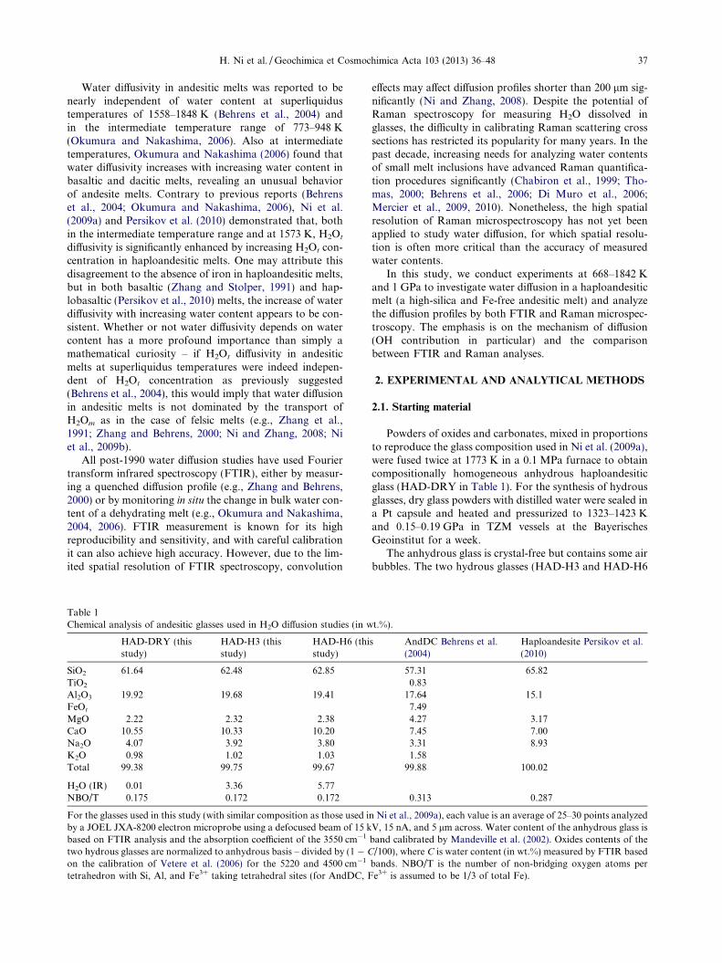
Table 1Chemical analysis of andesitic glasses used in H2O diffusion studies (in w
HAD-DRY (thisstudy)
HAD-H3 (thisstudy)
HAD-H6 (thistudy)
SiO2 61.64 62.48 62.85TiO2
Al2O3 19.92 19.68 19.41FeOt
MgO 2.22 2.32 2.38CaO 10.55 10.33 10.20Na2O 4.07 3.92 3.80K2O 0.98 1.02 1.03Total 99.38 99.75 99.67
H2O (IR) 0.01 3.36 5.77NBO/T 0.175 0.172 0.172
For the glasses used in this study (with similar composition as those used iby a JOEL JXA-8200 electron microprobe using a defocused beam of 15 kbased on FTIR analysis and the absorption coefficient of the 3550 cm�1
two hydrous glasses are normalized to anhydrous basis – divided by (1 � C
on the calibration of Vetere et al. (2006) for the 5220 and 4500 cm�1
tetrahedron with Si, Al, and Fe3+ taking tetrahedral sites (for AndDC, F
effects may affect diffusion profiles shorter than 200 lm sig-nificantly (Ni and Zhang, 2008). Despite the potential ofRaman spectroscopy for measuring H2O dissolved inglasses, the difficulty in calibrating Raman scattering crosssections has restricted its popularity for many years. In thepast decade, increasing needs for analyzing water contentsof small melt inclusions have advanced Raman quantifica-tion procedures significantly (Chabiron et al., 1999; Tho-mas, 2000; Behrens et al., 2006; Di Muro et al., 2006;Mercier et al., 2009, 2010). Nonetheless, the high spatialresolution of Raman microspectroscopy has not yet beenapplied to study water diffusion, for which spatial resolu-tion is often more critical than the accuracy of measuredwater contents.
In this study, we conduct experiments at 668–1842 Kand 1 GPa to investigate water diffusion in a haploandesiticmelt (a high-silica and Fe-free andesitic melt) and analyzethe diffusion profiles by both FTIR and Raman microspec-troscopy. The emphasis is on the mechanism of diffusion(OH contribution in particular) and the comparisonbetween FTIR and Raman analyses.
2. EXPERIMENTAL AND ANALYTICAL METHODS
2.1. Starting material
Powders of oxides and carbonates, mixed in proportionsto reproduce the glass composition used in Ni et al. (2009a),were fused twice at 1773 K in a 0.1 MPa furnace to obtaincompositionally homogeneous anhydrous haploandesiticglass (HAD-DRY in Table 1). For the synthesis of hydrousglasses, dry glass powders with distilled water were sealed ina Pt capsule and heated and pressurized to 1323–1423 Kand 0.15–0.19 GPa in TZM vessels at the BayerischesGeoinstitut for a week.
The anhydrous glass is crystal-free but contains some airbubbles. The two hydrous glasses (HAD-H3 and HAD-H6
t.%).
s AndDC Behrens et al.(2004)
Haploandesite Persikov et al.(2010)
57.31 65.820.83
17.64 15.17.494.27 3.177.45 7.003.31 8.931.58
99.88 100.02
0.313 0.287
n Ni et al., 2009a), each value is an average of 25–30 points analyzedV, 15 nA, and 5 lm across. Water content of the anhydrous glass isband calibrated by Mandeville et al. (2002). Oxides contents of the/100), where C is water content (in wt.%) measured by FTIR based
bands. NBO/T is the number of non-bridging oxygen atoms pere3+ is assumed to be 1/3 of total Fe).

38 H. Ni et al. / Geochimica et Cosmochimica Acta 103 (2013) 36–48
in Table 1) both contain small amounts of microcrystalsand bubbles, which are not expected to have a significanteffect on water diffusion (Zhang et al., 1991; Zhang andBehrens, 2000). FTIR analyses yield homogeneous totalwater (H2Ot) concentration in each glass (within 3% rela-tive): 0.01 wt.% in the nominally anhydrous glass usingthe calibration of the 3550 cm�1 band by Mandevilleet al. (2002), and 3.36 ± 0.05 wt.% and 5.77 ± 0.06 wt.%in the hydrous glasses using the calibration of the near-infrared (NIR) bands by Vetere et al. (2006). The anhy-drous composition of these glasses (Table 1), determinedby electron microprobe analyses, is similar to those usedin previous studies of viscosity (Richet et al., 1996) andH2O diffusivity (Ni et al., 2009a). But compared to theFe-bearing andesite (Behrens et al., 2004) and haploande-site (Persikov et al., 2010) with NBO/T (non-bridging oxy-gen atoms per tetrahedron) of �0.3, our glass compositionis more polymerized, with NBO/T of �0.173 and silica con-tent of �62 wt.% (Table 1).
2.2. Diffusion experiments
Glass cylinders of 2.5 mm diameter were prepared, with1.5 mm length for the anhydrous glass and 2.5 mm lengthfor the hydrous glasses. An anhydrous glass cylinder anda hydrous one, with their polished surfaces in contact,formed a diffusion couple. The diffusion couple was placedinside a Pt tube of 3.0 mm outer diameter and 0.2 mm wallthickness, and then the tube was sealed by welding.
Diffusion experiments were carried out in a 3/400 piston-cylinder apparatus at the Bayerisches Geoinstitut. A sketchof the sample assembly is illustrated in Fig. 1, involving atapered graphite heater and pressure media of talc, Pyrex
Fig. 1. Sample assembly for double diffusion couple experiments in3/400 piston-cylinder apparatus (drawn to scale). H2O concentrationof the hydrous half is different in the two couples.
glass and crushable alumina. Temperature was monitoredby a Pt–Pt90Rh10 thermocouple. Based on the temperaturedistribution for a straight graphite heater in a piston-cylin-der apparatus calibrated by Hui et al. (2008), Ni and Zhang(2008) applied temperature corrections of 29, 19–23, and 7–9 K for their H2O diffusion experiments under nominaltemperatures of 1873, 1573, and 673–873 K, respectively.For a tapered or stepped graphite heater, the central sec-tion, with a larger cross section area, has a smaller electricalresistance and dissipates less power as heat (Dunn, 1993).Therefore, the thermal gradient in our assembly using a ta-pered heater is expected to be lower than that in Ni andZhang (2008). In this study we have applied temperaturecorrections of 20, 15, and 5 K for nominal temperaturesof 1822, 1604, and 663–813 K, respectively (Table 2). The2r uncertainty in temperature is estimated to be 20 K forthe superliquidus runs (1604–1822 K) and 10 K for theintermediate temperature runs (663–813 K). In each run,two diffusion couples were loaded side by side; the only dif-ference between the two couples was the H2O concentrationin the hydrous half. This double diffusion couple design notonly promotes efficiency, but also guarantees that the sameP–T history is experienced by both diffusion couples, whichis useful for constraining the H2Ot dependence of water dif-fusivity. The sample assembly was first pressurized to1.0 GPa (after 9% friction correction). For the runs at663–813 K, temperature was increased to the target valueand maintained for 7–18 days. For the runs at 1604–1822 K, it was necessary to increase temperature rapidlyto minimize diffusion during heating. Therefore, heatingpower was manually increased so that a plateau tempera-ture was reached within one minute, at which the sampledwelled for several minutes before quenching. The completethermal history was recorded and an effective experimentalduration was inferred, using the method described in Zhangand Behrens (2000) and assuming an average activation en-ergy of 100 kJ/mol for water diffusion. Conditions of thediffusion experiments are summarized in Table 2.
Quenched samples were embedded in epoxy resin anddoubly polished to �500 lm thickness for Raman analyses,with the top surface approximately at the centerline of thesample. Subsequently the bottom surface of samples wasfurther polished till reaching �200 lm sample thicknessfor transmission FTIR analyses. After high temperature(1604–1822 K) runs, the small amounts of bubbles andmicrocrystals in starting glasses had disappeared, attributedto dissolution into the melt during heating and annealing,and the interface between the two halves of a diffusion coupleis difficult to recognize (Fig. 2a). By contrast, in intermediatetemperature experimental charges (Fig. 2b), the impurities ofstarting glasses continue to exist; the interface between twohalves remains straight, often with a �8 lm wide gap in be-tween, likely produced by contraction during quench.
2.3. Raman analyses
Confocal Raman analyses were performed with a HoribaJobin–Yvon LabRam HR800 spectrometer at the Bayreris-ches Geoinstitut, using a 514.5 nm argon ion laser of100 mW input power, a confocal hole of 300 lm diameter,

Fig. 2. Photomicrographs of two samples (positioned with increas-ing water content to the right) after diffusion experiments. (a)HAD-BGI-DC1a, quenched from 1619 K; (b) HAD-BGI-DC6b,quenched from 668 K.
Table 2H2O diffusion experiments in piston-cylinder apparatus at 1.0 GPa.
Run #a T0b (K) Tc (K) t0d (s) te (s) Initial H2Otf (wt.%) Final H2Ot
f (wt.%) Comment
HAD-BGI-DC1 a 1604 1619 ± 20 266 288 ± 10 0.01/5.75 0.02/5.75b 0.01/3.31 0.01/3.44
HAD-BGI-DC7 a 1822 1842 ± 20 149 175 ± 10 0.01/n.a. 0.02/5.77b 0.01/n.a. 0.02/3.33
HAD-BGI-DC2 a 813 818 ± 10 576,000 0.01/5.72 n.a. Crystallizationb 0.01/3.36 n.a. Crystallization
HAD-BGI-DC4 a 763 768 ± 10 828,000 0.01/5.82 n.a. Crystallizationb 0.01/3.40 0.02/3.36 Partial crystal
HAD-BGI-DC6 a 663 668 ± 10 1,555,200 0.01/5.74 0.02/5.72 Partial crystalb 0.01/3.38 0.02/3.38 Partial crystal
a Two diffusion couples in each experimental run.b Plateau temperature measured by Pt–Pt90Rh10 thermocouple.c Temperature corrected for thermal gradient (with estimated 2r error).d Dwelling duration at temperatures within 30 K (for HAD-BGI-DC1) or 50 K (for HAD-BGI-DC7) difference from the plateau
temperature.e Duration corrected for the diffusion during heating and cooling (with estimated 2r error).f Total H2O concentrations of two halves measured by FTIR before and after diffusion run.
H. Ni et al. / Geochimica et Cosmochimica Acta 103 (2013) 36–48 39
and an objective of 50� magnification (N.A. = 0.35). Foreach point of analysis, the laser beam was focused on thetop surface of the sample within an error of 2 lm. Becausethe analyzed glasses were transparent, a volume, instead ofan area, was selected by the confocal hole. The diameterof the selected volume was estimated to be 6 lm, about
the same as the hole diameter divided by the objective mag-nification. With a grating of 1800/mm, both the 200–1500 cm�1 region and the 2800–4000 cm�1 region were mea-sured for 2 � 18 s in a few windows.
A series of Raman spectra along a diffusion profile aredisplayed in Fig. 3a. The 2800–4000 cm�1 region containsthe 3550 cm�1 stretching band for H2O analyses. A cubicbaseline with base points at 2850, 2950, 3800 and3950 cm�1 was used to determine peak height. This is sim-ilar to the baseline adopted by Mercier et al. (2009),although they did not report the exact frequencies of theirbase points. For some spectra at low H2O concentration,the 2950 cm�1 base point had to be shifted to a higher fre-quency to produce a reasonable cubic baseline. The 200–1500 cm�1 region contains two prominent silicate networkbands at �500 and �1015 cm�1 and a weak band at�770 cm�1. Based on Mercier et al. (2009) and referencestherein, the low-frequency (LF) band at �500 cm�1 is re-lated to bridging oxygens and the high-frequency band at�1015 cm�1 is related to non-bridging oxygens. Weadopted a cubic baseline with base points at 250, 800,1200 and 1450 cm�1 to obtain the peak height of both theLF and HF bands. As haploandesite is an intermediatecomposition (rather than felsic or mafic), either the LF orHF band is strong enough for normalizing the H2O bandto minimize the influence of focusing error or fluctuationof the laser power. Fig. 4 shows a comparison between Ra-man intensity profiles based on H2O peak (I3550), H2O/LFpeak ratio (I3550/I500), and H2O/HF peak ratio (I3550/I1015),with I denoting the peak height at a certain wavenumberindicated by the subscript. Differences among the three pro-files are not large, yet the I3550/I1015 profile appears to besmoother than the other two profiles. Furthermore, Mercieret al. (2009) showed that for a fixed melt composition, peakheight ratio I3550/I1015 had a better linear correlation withH2O concentration than I3550/I500. Accordingly, we optedto use I3550/I1015 for the conversion of Raman data toH2O concentrations.

Fig. 3. Vibrational spectra along the diffusion profile in HAD-BGI-DC1a (T = 1619 K, t = 288 s, maximum H2Ot = 5.75 wt.%). (a)Smoothed Raman spectra. The indicated coordinates (x in lm) are consistent with the profiles in Figs. 4–7. Cubic baselines for the silicatenetwork bands at 500 and 1015 cm�1 and the H2Ot band at 3550 cm�1 are shown for selected spectra. (b) Smoothed FTIR spectra, verticallyshifted for clarity. Water content and coordinate for each spectrum are approximately the same as those for the Raman spectrum of the samecolor. Straight tangential baselines are applied to all three water bands. Peak heights (dashed lines) are adopted to compute water contents forboth Raman and FTIR. (For interpretation of the references to color in this figure legend, the reader is referred to the web version of thisarticle.)
40 H. Ni et al. / Geochimica et Cosmochimica Acta 103 (2013) 36–48
In converting the I3550/I1015 ratio to H2O concentration,we used the FTIR-determined H2Ot concentrations in theflat regions of the two halves of the diffusion couple (i.e.,the regions not affected by diffusion) as constraints. Follow-ing previous studies (e.g., Behrens et al., 2006; Di Muroet al., 2006), a linear correlation between I3550/I1015 andH2Ot (in wt.%) was assumed. Unlike FTIR that can be usedto independently quantify H2O species concentrations oncethe appropriate molar absorptivities are properly calibrated,Raman data must be calibrated for each specific profile.
2.4. FTIR analyses
Water contents of the starting glass cylinders were mea-sured by a Bruker IFS 120HR FTIR spectrometer at theBayerisches Geoinstitut, involving a NIR source, a CaF2
beamsplitter, and a liquid-nitrogen cooled MCT detector.
Diffusion profiles of doubly polished experimentalcharges were acquired using the Autoimage microscopeon a PerkinElmer Spectrum GX FTIR spectrometer atthe University of Michigan. Because a thicker samplewould reduce the spatial resolution due to beam divergencein the section, and a thinner sample would be prone tobreakage (especially when there were cracks) and weakeninfrared absorption, a thickness of �200 lm was chosenas a compromise for measuring diffusion profiles. For eachpoint, we made 64 scans through 7800–2000 cm�1 with arectangular aperture of 7 � 200 lm in the focus plane. Rep-resentative FTIR spectra along a diffusion profile togetherwith the baselines for three H2O bands are shown inFig. 3b.
At low H2Ot concentrations, the strong 3550 cm�1 band(Fig. 3b) is used to obtain H2Ot concentration with thecalibration of Mandeville et al. (2002). At high H2Ot

Fig. 4. Profiles using the 3550 cm�1 Raman band or Raman bandratios to characterize H2Ot for sample HAD-BGI-DC1a. Peakheights of the 3550 cm�1 H2O band (I3550) are shown in opensquares. I3550/I500 � 3500 and I3550/I1015 � 1850 are represented incrosses and solid circles, respectively, with I500 and I1015 being thepeak heights of the low-frequency (LF, 500 cm�1) and the high-frequency (HF, 1015 cm�1) silicate network bands. The I3550/I1015
profile, the smoothest and also the most consistent with the scaledFTIR data (solid curve), is used to compute H2Ot concentrationprofile, with the result referred to as the Raman data.
H. Ni et al. / Geochimica et Cosmochimica Acta 103 (2013) 36–48 41
concentrations, the 3550 cm�1 band becomes oversaturatedand the weaker NIR bands at 4500 cm�1 (representing OH)and 5220 cm�1 (representing H2Om) are well resolved.Hence, the heights of the two NIR bands are used to obtainOH and H2Om concentrations using the calibration of Ve-tere et al. (2006), which sum to the H2Ot concentration.In the transition from “low” to “high” H2Ot, which occursat 0.6 ± 0.2 wt.% H2Ot, both MIR and NIR calibrationmethods give similar H2Ot, verifying the self consistencyin the FTIR data along diffusion profiles.
3. RESULTS AND DISCUSSION
In total five experiments containing 10 diffusion couplesat 1.0 GPa were successfully performed (Table 2). However,both samples at 818 K (HAD-BGI-DC2a,b) and one sam-ple at 768 K (HAD-BGI-DC4a) showed heavy crystalliza-tion in the hydrous half, and could not be analyzed. Incomparison with previous dehydration experiments at0.1 GPa and 743–873 K (Ni et al., 2009a), the substantialcrystallization in this work can be attributed to the higherpressure (e.g., Hui et al., 2008; Ni and Zhang, 2008; Wanget al., 2009) and the higher water contents (3–6 wt.% versus2 wt.%). We measured four diffusion profiles from superliq-uidus runs (HAD-BGI-DC1a,b and HAD-BGI-DC7a,b)and three from intermediate temperature runs (HAD-BGI-DC4b and HAD-BGI-DC6a,b) using both Ramanand FTIR methods.
3.1. Comparison between FTIR and Raman
Sample preparation for Raman analyses is less time-con-suming than that for FTIR. Raman only requires polishingone side of a sample (although in this study we initially pol-ished both sides to 500 lm thickness), whereas FTIR profil-
ing requires doubly polishing the sample. Furthermore, it iscrucial for FTIR measurements to determine accurate sam-ple thickness along a diffusion profile, which is difficult inpractice if the sample contains many cracks or is not pol-ished evenly.
Consistent results are obtained from FTIR and Ramanfor the four diffusion profiles quenched from superliquidustemperatures. Two examples are shown in Fig. 5a and b.When a sample contains cracks, it appears that Ramanmeasurements are less affected than FTIR measurements(Fig. 5b). This difference is particularly important if thecracks are curved or the crack surfaces are not perpendicu-lar to the polished surfaces.
For the intermediate temperature runs with short diffu-sion profiles, however, differences between FTIR and Ra-man profiles are much more pronounced. Due to slowdiffusion at intermediate temperatures, and limitation offeasible experimental duration, the generated profiles areless than 100 lm long (Fig. 5c–e). We define the x-axis tobe along the traverse direction, the z-axis to be along thesample thickness direction, and the y-axis to be the thirdmutually perpendicular direction. Because the FTIR aper-ture is 7 lm (along x) by 200 lm (along y) and the measure-ment is in transmission mode with sample thickness ofabout 200 lm, the nominal spatial resolution for the IRmeasurement is 7 lm along the x-axis, 200 lm along they-axis, and 200 lm along the z-axis. Along the x-axis, whichis the most important for our measurements, the nominalspatial resolutions of the two analytical methods are com-parable: 6 lm for Raman and 7 lm for FTIR (aperturewidth in the focus plane). But compared to Raman profiles,FTIR profiles are smoother and longer, attributed to theconvolution effect due to the large incipient angle (17–36�)of the IR beam to the optical axis (the z-direction). Theconvolution effect causes the actual spatial resolution ofFTIR in this study to be much larger than 7 lm. In the y-and z-axes, the spatial resolution of Raman is also esti-mated to be 6 lm, but the spatial resolution of FTIR isabout 200 lm in our measurements so as to transmit en-ough photons and produce large enough absorbance.
Assuming that the convolution effect is negligible for theRaman profiles, we made an effort to estimate the actualspatial resolution of FTIR analyses by convoluting Ramanprofiles to match FTIR profiles. For each Raman profile, asmooth discretized profile at 1 lm interval was first created.The discretized profile was then numerically convolutedbased on the principle of Gaussian density function(Ganguly et al., 1988). For all three intermediate tempera-ture samples, using a Gaussian with a standard deviationof 12 lm to convolute the Raman profiles produces goodmatches with FTIR profiles (Fig. 5c–5e). If the spatial res-olution is defined to be the full width at half maximum(FWHM) of the Gaussian, the actual spatial resolution ofFTIR microspectroscopy is approximately 28 lm (2.35standard deviations). Ni and Zhang (2008) found that witha 20 lm wide aperture on a sample thickness of �200 lm,the actual spatial resolution of FTIR is about 30 lm.Therefore, reducing the aperture width from 20 to 7 lmdoes not appear to have improved the spatial resolutionof FTIR significantly.

Fig. 5. Comparison between FTIR (blue) and Raman (red) measurements. (a) Nearly identical profiles are obtained for a long profile withfew cracks in the sample; (b) The Raman profile is less susceptible to cracks than the FTIR profile; (c–e) For profiles with <100 lm length,FTIR profiles show a marked convolution effect compared to Raman profiles. Green curves represent profiles convoluted from Ramanprofiles to match FTIR profiles (see text for detail). (For interpretation of the references to color in this figure legend, the reader is referred tothe web version of this article.)
42 H. Ni et al. / Geochimica et Cosmochimica Acta 103 (2013) 36–48
3.2. The experiments at intermediate temperatures
As discussed above, because the H2Ot concentrationprofiles at intermediate temperatures of 668–768 K are<100 lm in length, the smooth profiles from FTIR analyses(Fig. 5c–e) do not reflect the true profile shape and length.Raman spectroscopy has better spatial resolution, but thequality of Raman spectra is significantly degraded by the
partial crystallization in the hydrous half, and by the gap(�8 lm wide) between the two halves. While the gap wasmost likely produced by contraction during quench, thepossibility of poor contact between the two halves cannotbe completely ruled out. Furthermore, the gap may havebeen widened during polishing, and there is no accurateway of subtracting the gap distance from the short Ramanprofiles (Fig. 5c–e).

H. Ni et al. / Geochimica et Cosmochimica Acta 103 (2013) 36–48 43
Along a water diffusion profile produced at someintermediate temperature, H2O speciation may reach equi-librium at high water contents, but there can be disequilib-rium at low H2Ot (Zhang et al., 1991). In fact, in the dryhalf of HAD-BGI-DC6a (18 days at 668 K), FTIR spectraof some points close to the gap contain the 5220 cm�1
H2Om band but the 4500 cm�1 OH band is negligible. Thissuggests that H2Om is the mobile species at 668 K, and thatthe conversion of H2Om to OH is too sluggish to reach equi-librium at low H2Ot.
In principle, all these problems could be alleviated byexperiments performed at higher temperatures. However,our attempt was unsuccessful: at 818 K nearly completecrystallization occurred in both diffusion couples in HAD-BGI-DC2. Considering the poor quality of both FTIRand Raman profiles and the complexities associated withthe intermediate temperature runs, we decided not to usethese profiles to obtain diffusivity. The intermediate temper-ature results are presented in this study only for underliningthe remarkable difference between Raman and FTIR, andfor estimating the actual spatial resolution for FTIRanalyses.
Fig. 6. (a) Fits of the FTIR profile for sample HAD-BGI-DC1a;and (b) inferred H2Ot diffusivity versus the mole fraction of H2Ot
based on different diffusivity models: error function (dashed greencurve); DH2Ot proportional to X (dashed cyan curve); speciation-based model assuming DH2Om = D0eaX and DOH = 0 (dashed purplecurve); modified speciation-based model assuming DH2Om = D0eaX
and DOH = constant (solid red curve). The modified speciation-based model produces the best fit. (For interpretation of thereferences to color in this figure legend, the reader is referred to theweb version of this article.)
3.3. A modified speciation-based diffusion model for
superliquidus experiments
We have focused on studying the four diffusion couplesquenched from 1619 and 1842 K. The obtained water con-centration profiles cannot be satisfactorily fit using an errorfunction (an example is shown in Fig. 6a). This suggeststhat H2Ot diffusivity in andesitic melt must be dependenton total H2O concentration, which is in contrast to Behrenset al. (2004) but is consistent with Persikov et al. (2010).Assuming H2Ot diffusivity proportional to H2Ot concentra-tion roughly fits the profiles, but there are considerable mis-fits at low H2Ot (Fig. 6a). The misfits cannot be attributedto the convolution effect, because these profiles are long andequivalent profiles have been obtained from Raman andFTIR analyses. A third model, first developed by Zhangand Behrens (2000) and later successfully applied in a seriesof studies (Liu et al., 2004; Ni and Zhang, 2008; Ni et al.,2009a,b), is applied, but the misfits persist (Fig. 6a). Thethird model is a mechanistic model that explicitly considersthe speciation reaction involving H2Om and OH, and as-sumes OH diffusivity to be negligible and H2Om diffusivityto increase exponentially with H2Ot concentration. Boththe mechanistic model and the proportionality model implynegligible H2Ot diffusivity as H2Ot concentration ap-proaches zero, which is the cause of the misfits at lowH2Ot. Therefore, with decreasing H2O concentration insuperliquidus andesitic melts, even though the H2Om/H2Ot ratio approaches zero and OH becomes the dominantspecies, H2Ot diffusivity does not become negligible, indi-cating that OH diffusivity cannot be neglected. Our experi-mental data hence offer the opportunity to constrain OHdiffusivity, which has never been resolved before.
In order to obtain OH diffusivity, we start from the gen-eral form of the H2O diffusion equation (Zhang et al.,1991),
@X@t¼ @
@xDH2Om
@X m
@xþ DOH
@X OH
2@x
� �; ð1Þ
where X = C/18.015/[C/18.015 + (100 � C)/33.84] (Niet al., 2009a) is the mole fraction of H2Ot in the haploande-sitic melt (C is H2Ot in wt.%), Xm and DH2Om are the molefraction and diffusivity of H2Om, XOH and DOH are themole fraction and diffusivity of OH, t is time and x is dis-tance. Because the temperature is high and hence equilib-rium for the speciation reaction H2Om + O = 2OH (whereO represents an anhydrous oxygen) is expected (e.g., Zhanget al., 1995), comparison with the diffusion equation for asimple component leads to
DH2Ot ¼ DH2Om
dX m
dXþ DOH
dX OH
2dX; ð2Þ
where DH2Ot is the apparent H2Ot diffusivity. The two differ-ential terms in Eq. (2) add up to unity, and the latter is asfollows (Wang et al., 2009):

44 H. Ni et al. / Geochimica et Cosmochimica Acta 103 (2013) 36–48
dX OH
2dX¼ 1� 2Xffiffiffiffiffiffiffiffiffiffiffiffiffiffiffiffiffiffiffiffiffiffiffiffiffiffiffiffiffiffiffiffiffiffiffiffiffiffiffiffiffiffiffiffiffiffiffiffiffiffiffi
4X ðX � 1Þð1� 4=KÞ þ 1p ; ð3Þ
in which K is the equilibrium constant for the H2O specia-tion reaction. For our haploandesitic melt (Ni et al., 2009a),
lnK ¼ 1:547� 2453=T ; ð4Þ
where T is temperature in K.Zhang et al. (1991) assumed DH2Om to be independent of
H2Ot concentration and DOH to be negligible in rhyoliticmelts. To reconcile new experimental data at higher H2Ot
concentrations (>3 wt.%), Zhang and Behrens (2000) re-vised the model of Zhang et al. (1991) and assumed instead
DH2Om ¼ D0eaX ; ð5Þ
with D0 and a being two constants at fixed T and P,whereas DOH was still assumed to be negligible.
Based on the fitting results discussed above and shownin Fig. 6, we find that DOH is not negligible in superliquidusandesitic melts. In principle, DOH may also depend on H2Ot
concentration (X), but the contribution of OH diffusion toDH2Ot is only expected to be important at low H2Ot. More-over, including the dependence of DOH on X would add oneadditional fitting parameter, which cannot be well resolvedbecause DH2Om also depends on X (i.e., it is possible to re-duce the dependence of DOH on X by increasing the depen-dence of DH2Om on X). Hence, we settle for the lessambitious goal of obtaining DOH only at low H2Ot concen-tration, and assume
DOH ¼ constant ðat fixed T and PÞ: ð6Þ
With this treatment, the dependence of DOH on H2Ot isconvoluted into that of DH2Om on H2Ot. For fitting a mea-sured diffusion profile, in addition to a small interfaceadjustment Dx0, three free parameters are allowed to vary:they are D0, a, and DOH/D0.
We have developed a multivariate least-squares fittingprocedure to simultaneously extract the parameters D0, a,DOH/D0 and Dx0. This modified speciation-based diffusivitymodel produces a much better fit than all previous models(Fig. 6a). The dependences of DH2Ot on H2Ot concentrationfor the new model as well as those for previous models are
Table 3Fitting parameters using the model DH2Om = D0eaX and DOH = constant.
Run # Analytical method T (K) a
HAD-BGI-DC1 a FTIR 1619 1b 2* 1
a Raman 1b 2* 1
HAD-BGI-DC7 a FTIR 1842 1b* 1
a Raman 1b* 1
Errors are given at 2r level. Equilibrium constant for reaction H2Om + O* Both profiles from a given experiment are regressed simultaneously.
shown in Fig. 6b for the charge HAD-BGI-DC1a. The ob-tained fitting parameters are summarized in Table 3. Thevalues of Dx0 are not reported because they are arbitrary(dependent on the initially chosen coordinates). In Table3, the errors of a and DOH/D0 are obtained purely from fit-ting, but the errors of lnD0 also incorporate the uncertain-ties of experimental durations.
In general, two diffusion couples in a single run have re-sulted in similar parameters, as do FTIR profile and Ra-man profile of the same sample. To fully exploit the factthat two diffusion couples experienced the same P–T–t his-tory, we have made a further effort to fit two profiles (eithertwo FTIR profiles or two Raman profiles) in a single runsimultaneously. This fitting procedure for both profileshas 5 free parameters: D0, a, DOH/D0, Dx01, and Dx02.The obtained best-fit parameters are also listed in Table 3(indicated in bold). Again, Dx01 and Dx02 are not reportedfor their insignificance. Calculated diffusion profiles usingthe best-fit parameters are plotted with measured profilesin Fig. 7, which match each other well in most cases.
Modeling two profiles simultaneously adds more con-straints to the diffusivity vs. H2Ot relationship. At1619 K, parameter a using the collective fitting procedureis 15.8 from FTIR profiles and 16.2 from Raman profiles.The difference between these two values is much smallerthan if each profile were treated separately. A similar phe-nomenon has been observed for the profiles at 1842 K,and a parameter a of �10.4 is extracted from both FTIRand Raman profiles. These values are much smaller thanthe a values of 75–85 at 743–873 K and 0.1 GPa in Niet al. (2009a). Therefore in haploandesitic melt, as temper-ature increases, the increase of H2Om diffusivity with H2Ot
concentration becomes less dramatic. A similar conclusionhas been reached by previous work on rhyolitic and daciticmelts (Zhang and Behrens, 2000; Ni and Zhang, 2008; Niet al., 2009b).
The DOH/D0 ratio has been constrained to be between0.1 and 0.2 with large relative errors (Table 3). This meansthat, at 1619–1842 K and extremely low H2Ot concentra-tion, OH diffusivity is lower than H2Om diffusivity probablyby less than an order of magnitude. This finding is not
lnD0 (D0 in m2/s) DOH/D0 R2
7.4 ± 2.1 �22.84 ± 0.16 0.24 ± 0.08 0.99978.3 ± 5.4 �22.93 ± 0.25 0.22 ± 0.10 0.99965.8 ± 2.1 �22.69 ± 0.15 0.18 ± 0.06 0.9995
8.7 ± 4.5 �22.64 ± 0.32 0.14 ± 0.13 0.99901.5 ± 6.6 �22.40 ± 0.32 0.09 ± 0.08 0.99866.2 ± 3.4 �22.41 ± 0.22 0.10 ± 0.07 0.9985
1.9 ± 5.5 �21.37 ± 0.42 0.16 ± 0.14 0.99919.6 ± 5.3 �21.02 ± 0.27 0.16 ± 0.08 0.99960.4 ± 4.6 �21.26 ± 0.35 0.18 ± 0.12 0.9987
1.0 ± 4.1 �21.19 ± 0.30 0.10 ± 0.08 0.99879.9 ± 7.3 �20.95 ± 0.39 0.10 ± 0.09 0.99920.4 ± 3.6 �21.16 ± 0.26 0.11 ± 0.07 0.9985
= 2OH is based on Ni et al. (2009a).
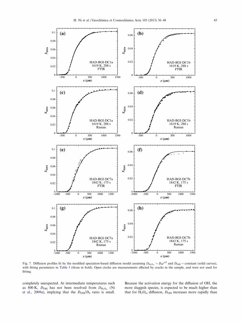
Fig. 7. Diffusion profiles fit by the modified speciation-based diffusion model assuming DH2Om = D0eaX and DOH = constant (solid curves),with fitting parameters in Table 3 (those in bold). Open circles are measurements affected by cracks in the sample, and were not used forfitting.
H. Ni et al. / Geochimica et Cosmochimica Acta 103 (2013) 36–48 45
completely unexpected. At intermediate temperatures suchas 800 K, DOH has not been resolved from DH2Om (Niet al., 2009a), implying that the DOH/D0 ratio is small.
Because the activation energy for the diffusion of OH, themore sluggish species, is expected to be much higher thanthat for H2Om diffusion, DOH increases more rapidly than

Fig. 8. Diffusivities of H2O species (circles and solid lines) innominally anhydrous haploandesitic melt at 1 GPa from theRaman data, with error bars at the 2r level. Fluorine diffusivitiesat 1 GPa (dashed lines) in anhydrous basaltic melt (Alletti et al.,2007) and Na-rich and K-rich phonolitic melts (Balcone-Boissardet al., 2009) are shown for comparison.
46 H. Ni et al. / Geochimica et Cosmochimica Acta 103 (2013) 36–48
DH2Om with increasing temperature. This explains the negli-gible DOH/D0 ratio at �800 K and resolvable DOH/D0 ratioat 1619–1842 K.
The relative contribution of OH diffusion to DH2Ot de-pends not only on DOH/D0 and a, but also on the differen-tial terms in Eq. (2), which further depend on theequilibrium constant K and H2Ot concentration. Ni et al.(2009a) found that K increases from rhyolitic to dacitic toandesitic melt at <900 K. If the compositional trend at<900 K can be extrapolated to higher temperatures, at a gi-ven H2Ot mole fraction (X), there is a higher percentage ofOH in andesitic melts than in felsic melts. At 1619–1842 K,if the K value from Eq. (4) is used to calculate XOH/(2X),OH will account for 100% of total H2O at X = 0, and morethan 70% at X = 0.104 (5.8 wt.% H2Ot). The differentialterm dXOH/(2dX), meaning the “weight” of DOH in DH2Ot ,is also higher in andesitic melt.
The successful resolution of DOH in this work, comparedto negligible DOH in earlier studies, can be attributed to thefollowing: (i) The “dry” half of the diffusion couple in thisstudy contains a very low concentration of H2Ot
(0.01 wt.%), for which the fraction of H2Om and its contri-bution to water diffusion are minimized. In some previousstudies (e.g., Zhang and Behrens, 2000), the “dry” halfoften contains >0.10 wt.% H2Ot, 10 times more than inthe present study. (ii) It is also possible that DOH/D0
in andesitic melt at superliquidus temperatures is greaterthan that in rhyolitic and dacitic melts.
3.4. Comparison between DOH and Eyring diffusivity
Zhang et al. (1991) assumed DOH to be similar to non-bridging oxygen diffusivity or Eyring diffusivity. The Eyringdiffusivity (DEyr) is related to melt viscosity (g) asDEyr = kT/gL, with k being the Boltzmann constant andL being the jump distance (�0.28 nm). Our new data allowus to examine whether the assumption of Zhang et al.(1991) is correct. At 1619 K and 60.1 wt.% H2Ot, the Eyr-ing diffusivity estimated from the viscosity model of Vetereet al. (2006) for andesitic melt is 1.6 � 10�13–2.0 � 10�13 m2/s. The DOH value at low H2Ot and 1619 Kcan be estimated from the values of DOH/D0 and D0 in thisstudy to be �2 � 10�11 m2/s, larger than DEyr by about twoorders of magnitude. The nonequivalence of DOH and DEyr
is not problematic, because for the two to be comparable, aprerequisite is that OH is always bonded to the networkformers (Si, Al and Fe3+). However, NMR studies havefound that OH can also be linked to alkalis or divalent cat-ions in both polymerized and depolymerized melts (Kohnet al., 1989; Xue and Kanzaki, 2004). These “free” OH sub-species are expected to have higher mobility than Si–OHand Al–OH, and their motion is not restricted by the pro-cess of viscous flow. The “free” OH subspecies are favoredby more depolymerized melts and a higher content of alka-line earth elements (Xue and Kanzaki, 2004). In our ratherpolymerized haploandesitic melt (NBO/T = 0.173),although the “free” OH subspecies may only account fora fraction of OH, they can still dominate OH diffusion,much the same way as a small fraction of H2Om dominatesH2Ot diffusion (Zhang et al., 1991).
3.5. Comparison between DOH and F diffusivity
The H2Om and OH diffusivities in nearly anhydroushaploandesitic melt obtained from the Raman data areshown in Fig. 8. H2Om has higher diffusivity than OH forbeing a neutral molecule and interacting less with the meltstructure. OH, similar to fluorine ion, has a valence of -1;their size is also comparable – for a coordination numberof 4, the ionic radii of OH and F are 0.135 and 0.131 nm,respectively (Shannon, 1976). Because the size and chargeof an ion play a major role in controlling its diffusivity(e.g., Zhang et al., 2010), we expect OH diffusivity and Fdiffusivity to be similar under the same conditions, espe-cially if OH� and F� are bonded to the same cations. F dif-fusivity has not been measured in andesitic melts, but asshown in Fig. 8, the DOH values at 1 GPa estimated inthe present study are close to the measured F diffusivitiesin basaltic and phonolitic melts at 1 GPa (Alletti et al.,2007; Balcone-Boissard et al., 2009). This good agreementsuggests that the OH diffusivity values constrained fromour fittings are reasonable.
3.6. Comparison with previous studies
H2Ot diffusivity at 1619–1842 K and 0–6 wt.% H2Ot canbe calculated using Eqs. (2)–(5) and the parameters re-ported in Table 3. Fig. 9 illustrates how DH2Ot varies withtemperature and water content based on the Raman data.
Because we only have data at two temperatures, the tem-perature dependence is not well constrained. Adopting theArrhenius relationship, the activation energy for DH2Ot atwater contents below 0.1 wt.% H2Ot is approximately146 kJ/mol, decreasing to 126 kJ/mol at 1 wt.% H2Ot, andto 75 kJ/mol at 5 wt.% H2Ot. For Fe-bearing andesiticmelts (AndDC in Table 1) at 1 GPa, Behrens et al. (2004)determined the activation energy at 1 wt.% H2Ot to be142 kJ/mol. Therefore, our roughly estimated activation

Fig. 9. Dependences of H2Ot diffusivity on (a) temperature; and (b)water content in the haploandesitic melt determined in this study(solid lines and curves), compared with H2Ot diffusivity in anFe-bearing andesitic melt (Behrens et al., 2004; open circles) and adifferent Fe-free andesitic melt (Persikov et al., 2010; dashed line).
H. Ni et al. / Geochimica et Cosmochimica Acta 103 (2013) 36–48 47
energies are reasonably close. Our DH2Ot data at 1 wt.%H2Ot are lower than those of Behrens et al. (2004) by a fac-tor of �2 (Fig. 9a). This difference may be attributed to thedifference in melt composition – the melt in Behrens et al.(2004) has lower SiO2 + Al2O3 content and higher NBO/T of 0.313 (Table 1). At temperatures >1300 K, H2O diffu-sivity tends to increase with increasing degree of melt depo-lymerization (Zhang and Ni, 2010).
Persikov et al. (2010) reported H2Ot diffusivity at 1573 Kand 0.1 GPa and up to 4 wt.% H2Ot in a different haplo-andesitic melt, with much higher alkalis (8.9 wt.% Na2O),similar SiO2 + Al2O3 (81 wt.%) and higher NBO/T (0.287)compared to the haploandesite in this study (Table 1).Fig. 9b shows that the diffusivities in Persikov et al.(2010) are higher than our values by a factor of 2–5. Weattribute this difference to the higher degree of depolymer-ization and the much higher alkali content of the meltinvestigated by Persikov et al. (2010). Wang et al. (2009)have shown that H2Ot diffusivity in peralkaline rhyoliticmelt is slightly larger than that in metaluminous rhyolite,supporting our interpretation. The pressure difference be-tween this study and that of Persikov et al. (2010) may alsocause some difference.
4. CONCLUSIONS
(1) Raman spectroscopy can be used to measure H2Oconcentration profiles in silicate melts once the con-centrations at the two ends are determined (e.g., byFTIR), and it can achieve a spatial resolution supe-rior to that of FTIR.
(2) The technique of double diffusion couples allowsmore accurate determination of the dependence ofH2O diffusivity on water contents in silicate melts.
(3) H2Ot diffusivity in haploandesitic melts increaseswith increasing H2Ot concentration.
(4) OH diffusivity in superliquidus haploandesitic meltsis 10–20% of H2Om diffusivity as water contentapproaches zero. OH diffusivity is similar to F diffu-sivity but is much higher than Eyring diffusivity.
ACKNOWLEDGMENTS
H.N. thanks H. Keppler for granting the access to hydrother-mal and Raman facilities, D. Krauße for probe analysis, H. Schu-lze, U. Dittmann, S. Ubelhack and H. Fischer for samplepreparation, and C. McCammon for suggestions on writing. Com-ments from three anonymous reviewers and E. Persikov have im-proved the manuscript. This work was supported by the visitorprogram of Bayerisches Geoinstitut, Germany, the RecruitmentProgram of Global Experts (Thousand Talents), China, and USNSF grant EAR-0838127.
REFERENCES
Alletti M., Baker D. R. and Freda C. (2007) Halogen diffusion in abasaltic melt. Geochim. Cosmochim. Acta 71, 3570–3580.
Balcone-Boissard H., Baker D. R., Villemant B. and Boudon G.(2009) F and Cl diffusion in phonolitic melts: influence of theNa/K ratio. Chem. Geol. 263, 89–98.
Behrens H. and Zhang Y. (2009) H2O diffusion in peralkaline toperaluminous rhyolitic melts. Contrib. Mineral. Petrol. 157,
765–780.
Behrens H., Zhang Y. and Xu Z. (2004) H2O diffusion in daciticand andesitic melts. Geochim. Cosmochim. Acta 68, 5139–5150.
Behrens H., Roux J., Neuville D. R. and Siemann M. (2006)Quantification of dissolved H2O in silicate glasses usingconfocal microRaman spectroscopy. Chem. Geol. 229, 96–112.
Behrens H., Zhang Y., Leschik M., Wiedenbeck M., Heide G. andFrischat G. H. (2007) Molecular H2O as carrier for oxygendiffusion in hydrous silicate melts. Earth Planet. Sci. Lett. 254,
69–76.
Chabiron A., Pfeifert C., Pironon J. and Cuney M. (1999)Determination of water content in melt inclusions by Ramanspectrometry. In Terra Nostra–Schriften der Alfred-Wegner-
Stiftung 99/6; ECROFI XV (European Current Research On
Fluid Inclusions), Abstr. Prog. (ed. H. Ristedt; guest eds. V.Luders, R. Thomas and A. Schmidt-Mumm). GeoForschungs-Zentrum, Potsdam. pp. 68–69.
Di Muro A., Villemant B., Montagnac G., Scaillet B. and ReynardB. (2006) Quantification of water content and speciation innatural silicic glasses (phonolite, dacite, rhyolite) by confocalmicroRaman spectroscopy. Geochim. Cosmochim. Acta 70,
2868–2884.
Dunn T. (1993) The piston-cylinder apparatus. In Experiments at
High Pressure and Applications to the Earth’s Mantle, vol. 21

48 H. Ni et al. / Geochimica et Cosmochimica Acta 103 (2013) 36–48
(ed. R. W. Luth). Mineral. Assoc. Canada Short Course
Handbook, pp. 39–94.
Freda C., Baker D. R., Romano C. and Scarlato P. (2003) Waterdiffusion in natural potassic melts. Geol. Soc. Spec. Publ. 213,
53–62.
Ganguly J., Bhattacharya R. N. and Chakraborty S. (1988)Convolution effect in the determination of compositionalprofiles and diffusion coefficients by microprobe step scans.Am. Mineral. 73, 901–909.
Hui H., Zhang Y., Xu Z. and Behrens H. (2008) Pressuredependence of the speciation of dissolved H2O in rhyoliticmelts. Geochim. Cosmochim. Acta 72, 3229–3240.
Kohn S. C., Dupree R. and Smith M. E. (1989) A multinuclearmagnetic resonance study of the structure of hydrous albiteglasses. Geochim. Cosmochim. Acta 53, 2925–2935.
Liu Y., Zhang Y. and Behrens H. (2004) H2O diffusion in daciticmelts. Chem. Geol. 209, 327–340.
Mandeville C. W., Webster J. D., Rutherford M. J., Taylor B. E.,Timbal A. and Faure K. (2002) Determination of molarabsorptivities for infrared absorption bands of H2O in andesiticglasses. Am. Mineral. 87, 813–821.
Mercier M., Di Muro A., Giordano D., Metrich N., Lesne P.,Pichavant M., Scaillet B., Clocchiatti R. and Montagnac C.(2009) Influence of glass polymerisation and oxidation onmicro-Raman water analysis in alumino-silicate glasses. Geo-
chim. Cosmochim. Acta 73, 197–217.
Mercier M., Di Muro A., Metrich N., Giordano D., Belhadj O. andMandeville C. W. (2010) Spectroscopic analysis (FTIR,Raman) of water in mafic and intermediate glasses and glassinclusions. Geochim. Cosmochim. Acta 74, 5641–5656.
Ni H. and Zhang Y. (2008) H2O diffusion models in rhyolitic meltwith new high pressure data. Chem. Geol. 250, 68–78.
Ni H., Liu Y., Wang L. and Zhang Y. (2009a) Water speciationand diffusion in haploandesitic melts at 743–873 K and100 MPa. Geochim. Cosmochim. Acta 73, 3630–3641.
Ni H., Behrens H. and Zhang Y. (2009b) Water diffusion in daciticmelt. Geochim. Cosmochim. Acta 73, 3642–3655.
Nowak M. and Behrens H. (1997) An experimental investigationon diffusion of water in haplogranitic melts. Contrib. Mineral.
Petrol. 126, 365–376.
Okumura S. and Nakashima S. (2004) Water diffusivity in rhyoliticglasses as determined by in situ IR spectrometry. Phys. Chem.
Mineral. 31, 183–189.
Okumura S. and Nakashima S. (2006) Water diffusion in basalticto dacitic glasses. Chem. Geol. 227, 70–82.
Persikov E. S., Newman S., Bukhtiyarov P. G., Nekrasov A. N.and Stolper E. M. (2010) Experimental study of water diffusionin haplobasaltic and haploandesitic melts. Chem. Geol. 276,
241–256.
Richet P., Lejeune A.-M., Holtz F. and Roux J. (1996) Water andthe viscosity of andesite melts. Chem. Geol. 128, 185–197.
Shannon R. D. (1976) Revised effective ionic radii and systematicstudies of interatomic distances in halides and chalcogenides.Acta Crystallogr. A32, 751–767.
Shelby J. E. (2008) A limited review of water diffusivity andsolubility in glasses and melts. J. Am. Ceram. Soc. 91, 703–708.
Thomas R. (2000) Determination of water contents of granite meltinclusions by confocal laser Raman microprobe spectroscopy.Am. Mineral. 85, 868–872.
Vetere F., Behrens H., Holtz F. and Neuville D. R. (2006) Viscosityof andesitic melts-new experimental data and a revised calcu-lation model. Chem. Geol. 228, 233–245.
Wang H., Xu Z., Behrens H. and Zhang Y. (2009) Water diffusionin Mount Changbai peralkaline rhyolitic melt. Contrib. Min-
eral. Petrol. 158, 471–484.
Xue X. and Kanzaki M. (2004) Dissolution mechanisms of water indepolymerized silicate melts: constraints from 1H and 29SiNMR spectroscopy and ab initio calculations. Geochim. Cos-
mochim. Acta 68, 5027–5057.
Zhang Y. and Behrens H. (2000) H2O diffusion in rhyolitic meltsand glasses. Chem. Geol. 169, 243–262.
Zhang Y. and Ni H. (2010) Diffusion of H, C, and O componentsin silicate melts. Rev. Mineral. Geochem. 72, 171–225.
Zhang Y. and Stolper E. M. (1991) Water diffusion in a basalticmelt. Nature 351, 306–309.
Zhang Y., Stolper E. M. and Wasserburg G. J. (1991) Diffusion ofwater in rhyolitic glasses. Geochim. Cosmochim. Acta 55, 441–
456.
Zhang Y., Stolper E. M. and Ihinger P. D. (1995) Kinetics of thereaction H2O + O = 2OH in rhyolitic and albitic glasses:preliminary results. Am. Mineral. 80, 593–612.
Zhang Y., Ni H. and Chen Y. (2010) Diffusion data in silicatemelts. Rev. Mineral. Geochem. 72, 311–408.
Associate editor: Peter Ulmer


![Central Metabolism Cofactor Biosynthesis · ppp9 pi h h2o ppi h h2o h2o dad-5 h[p] atp adp h pi h2o succoa lipoate atp glx 2p4c2me xu5p-D h2o cbl1 ppi h[e] h2o h dad-5 gthrd asp-L](https://static.fdocuments.in/doc/165x107/5f47678d7025ea6bb340bf3d/central-metabolism-cofactor-biosynthesis-ppp9-pi-h-h2o-ppi-h-h2o-h2o-dad-5-hp.jpg)