hydrophobic and fibrillar microporous.pdf
4
Hydrophobic and fibrillar microporous polyetherurethane urea prosthesis: an ESCA study on the internal external surfaces of explanted and grafts Marie Therrien, Robert Guidoin, AlainAdnotand Roy&on Paynter* Biomaterials Unit, St-Franqois d’Assise Hospital and Departments of Surgery and Chemical Engineering, Lava1 University, Quebec City, Quebec G 1 K 7P4, Canada (Received 13 March 1989; revised 18 May 1989; accepted 2 1 June 1989) The ESCA study gives a good qualitative and quantitative elemental analysis of internal and external surfaces of foreign materials. Microporous hydrophobic Mitrathane@ (a polyetherurethane urea) grafts were implanted as blood conduits in dogs for up to 6 month. Surface analysis of explanted grafts demonstrated the presence of different contaminants: sodium, chlorine, silicon, in patent grafts, i.e. those implanted for 1 month and less. The sulphur probably comes from the presence of proteins on the surface of the polymer and the high level of nitrogen is also protein-related. At 6 month implantation, the grafts were occluded and a decrease of proteins on the surface was observed. The values of N/C and O/C ratios are also reported. For the virgin material, these ratios correspond to the quantity of hard and soft segments; but, for the explanted grafts, these parameters are also influenced by the presence of proteins due to the VersaclearP washing which did not wash away all the proteins on the surface of the polymer. The SEM photographs showed a certain degradation of polyurethane after 6 month of implantation. However, by ESCA study, it is difficult to compare the surface of virgin and explanted grafts because it is masked by the presence of proteins. Keywords: Vascular prostheses, ESCA, biodegradation Textile polyester grafts, either woven or knitted and expanded polytetrafluoroethylene (ePTFE) prostheses have a good success record for the replacement of medium and large diameter arteries’ but no synthetic vascular graft has yet been able to challenge the performance of the autologous saphenous vein for the repair of vessels <6 mm in diameter’. Possessing outstanding in vitro properties, relatively blood-compatible and flexible segmented poly- urethanes have been involved in the production of small diameter blood conduits3-5. Matrix Medica Inc. (Wheat Ridge, CO, USA) has developed hydrophilic and hydrophobic polyetherurethane urea prostheses (PEUU). The porosity of the first one is produced by the control of phase separation during manufacturing6, ‘. The second one was fabricated by electrostatic spinning of the PEUU on a rotating mandril. The pores in the wall of the hydrophilic model were not connected; on the opposite, those of the hydrophobic one were (Figures 1 and 2). An X-ray photo-electron spectro- Correspondence to Dr R. Guidoln. *Present address: INRS-Energle, C.P. 1020, Varennes, Quebec City, Quebec JOL 2PO. Canada. scopy study of hydrophilic Mitrathane@ has already been published on the hydrophilic model*. To obtain a better understanding of the in vivo behaviour of polyurethane prostheses, we implanted the Mitrathane microporous hydrophobic polyurethane graft in dogs as infrarenal aorta substitutes for periods of up to 6 month. This paper focuses on the results of ESCA analysis on the internal and external surfaces of explanted grafts. MATERIALS AND METHODS Synthetic material Hydrophobic microporous polyurethane vascular grafts 5 mm in diameter were provided by Matrix Medica Inc. These were fabricated by electrostatic spinning of poly- urethane elastomer on a rotating mandril, a method adapted from that previously described by Annis et a/.3. The prostheses were implanted in dogs as infrarenal aorta substitutes for the following prescheduled periods: 24 h, 1 wk. 1 month and 6 month. For each period of time, implantations were performed in duplicate. Results which have been reported elsewhere9 can be summarized as 0 1989 Butterworth Et Co (Publishers) Ltd. 0142-9612/89/080517-04$03.00 Biomatenals 1989, Vol 10 October 517
Transcript of hydrophobic and fibrillar microporous.pdf

Hydrophobic and fibrillar microporous polyetherurethane urea prosthesis: an ESCA study on the internal external surfaces of explanted
and grafts
Marie Therrien, Robert Guidoin, Alain Adnot and Roy&on Paynter* Biomaterials Unit, St-Franqois d’Assise Hospital and Departments of Surgery and Chemical Engineering, Lava1 University,
Quebec City, Quebec G 1 K 7P4, Canada
(Received 13 March 1989; revised 18 May 1989; accepted 2 1 June 1989)
The ESCA study gives a good qualitative and quantitative elemental analysis of internal and external surfaces of foreign materials. Microporous hydrophobic Mitrathane@ (a polyetherurethane urea) grafts were implanted as blood conduits in dogs for up to 6 month. Surface analysis of explanted grafts demonstrated the presence of different contaminants: sodium, chlorine, silicon, in patent grafts, i.e. those implanted for 1 month and less. The sulphur probably comes from the presence of proteins on the surface of the polymer and the high level of nitrogen is also protein-related. At 6 month implantation, the grafts were occluded and a decrease of proteins on the surface was observed. The values of N/C and O/C ratios are also reported. For the virgin material, these ratios correspond to the quantity of hard and soft segments; but, for the explanted grafts, these parameters are also influenced by the presence of proteins due to the VersaclearP washing which did not wash away all the proteins on the surface of the polymer. The SEM photographs showed a certain degradation of polyurethane after 6 month of implantation. However, by ESCA study, it is difficult to compare the surface of virgin and explanted grafts because it is masked by the presence of proteins.
Keywords: Vascular prostheses, ESCA, biodegradation
Textile polyester grafts, either woven or knitted and expanded
polytetrafluoroethylene (ePTFE) prostheses have a good
success record for the replacement of medium and large
diameter arteries’ but no synthetic vascular graft has yet
been able to challenge the performance of the autologous
saphenous vein for the repair of vessels <6 mm in
diameter’. Possessing outstanding in vitro properties,
relatively blood-compatible and flexible segmented poly-
urethanes have been involved in the production of small
diameter blood conduits3-5.
Matrix Medica Inc. (Wheat Ridge, CO, USA) has
developed hydrophilic and hydrophobic polyetherurethane
urea prostheses (PEUU). The porosity of the first one is
produced by the control of phase separation during
manufacturing6, ‘. The second one was fabricated by
electrostatic spinning of the PEUU on a rotating mandril. The
pores in the wall of the hydrophilic model were not
connected; on the opposite, those of the hydrophobic one
were (Figures 1 and 2). An X-ray photo-electron spectro-
Correspondence to Dr R. Guidoln.
*Present address: INRS-Energle, C.P. 1020, Varennes, Quebec City, Quebec
JOL 2PO. Canada.
scopy study of hydrophilic Mitrathane@ has already been
published on the hydrophilic model*.
To obtain a better understanding of the in vivo
behaviour of polyurethane prostheses, we implanted the
Mitrathane microporous hydrophobic polyurethane graft in
dogs as infrarenal aorta substitutes for periods of up to 6
month. This paper focuses on the results of ESCA analysis on
the internal and external surfaces of explanted grafts.
MATERIALS AND METHODS
Synthetic material
Hydrophobic microporous polyurethane vascular grafts
5 mm in diameter were provided by Matrix Medica Inc.
These were fabricated by electrostatic spinning of poly-
urethane elastomer on a rotating mandril, a method adapted
from that previously described by Annis et a/.3. The
prostheses were implanted in dogs as infrarenal aorta
substitutes for the following prescheduled periods: 24 h,
1 wk. 1 month and 6 month. For each period of time,
implantations were performed in duplicate. Results which
have been reported elsewhere9 can be summarized as
0 1989 Butterworth Et Co (Publishers) Ltd. 0142-9612/89/080517-04$03.00
Biomatenals 1989, Vol 10 October 517
ESCA analysis on explanted grafts: M. Therrien et al.
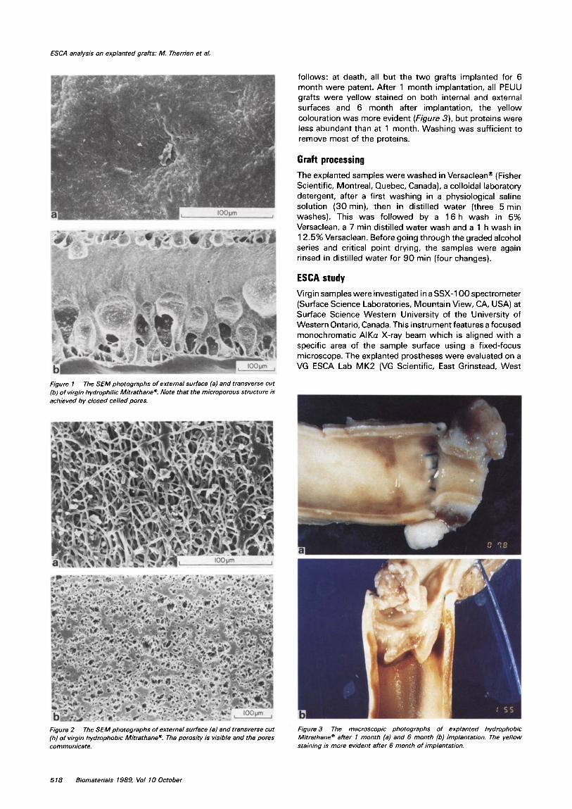
follows: at death, all but the two grafts implanted for 6 month were patent. After 1 month implantation, all PEUU grafts were yellow stained on both internal and external surfaces and 6 month after implantation, the yellow colouration was more evident (Figure 3), but proteins were less abundant than at 1 month. Washing was sufficient to remove most of the proteins.
Graft processing
The explanted samples were washed in Versaclean@ (Fisher Scientific, Montreal, Quebec, Canada), a colloidal laboratory detergent, after a first washing in a physiological saline solution (30 min), then in distilled water (three 5 min washes). This was followed by a 16 h wash in 5% Versaclean, a 7 min distilled water wash and a 1 h wash in 12.5% Versaclean. Before going through the graded alcohol series and critical point drying, the samples were again rinsed in distilled water for 90 min (four changes).
ESCA study
Virgin samples were investigated in a SSX-100 spectrometer (Surface Science Laboratories, Mountain View, CA, USA) at Surface Science Western University of the University of Western Ontario Canada. This instrument features a focused monochromatic AlKa X-ray beam which is aligned with a specific area of the sample surface using a fixed-focus microscope. The explanted prostheses were evaluated on a VG ESCA Lab MK2 (VG Scientific, East Grinstead, West
Figure 1 The SEM photographs of external surface (a) and transverse cut (b) of virgin hydrophilic Mitrathane? Note that the microporous structure is achieved by closed ceiled pores.
Figure 2 The SEM photographs of external surface (a) and transverse cut (b) of virgin hydrophobic Mitrathane? The porosity is visible and the pores communicate.
Figure 3 The macroscopic photographs of explanted hydrophobic Mitrathane@ after I month (a) and 6 month (b) implantation. The yellow staining is more evident after 6 month of implantation.
518 Biomaterials 1989, Vol 10 October
ESCA analysis on explanted grafts: M. Therrien et al.
Sussex, UK) which features a MgKa beam at Lava1 University.
RESULTS
In Table 1, the values for the apparent surface composition of the prostheses are reported. Carbon, nitrogen and oxygen are the major components of the polyurethane and sodium, chlorine, silicon and sulphur are the minor elements deposited on the surfaces. These components are due to contamination during the preparation of the grafts and/or to implantation.
The N/C and O/C ratios were calculated. The values for the virgin and explanted polyurethane are presented in Table 2.
In Tab/e 3, the estimated compositions of reference proteins are reported. The percentage of carbon, nitrogen, oxygen and sulphur are presented for a comparison with the values of polyurethane.
DISCUSSION
Blood polyurethane interactions are affected by the surface
Table 1 Apparent surface composition of prostheses
Time of Surface C N 0 Na Cl SI S implantation
0 Internal 80 2 17 - - 1 - External 79 2 19 - P’ P’ -
24 h
1 wk
1 month
6 month
Internal 71 8 20 0.1 - - 0.5 External 70 10 19 - - - 0.4
Internal External 73 7 19 - - 0.2 0.2
Internal 69 10 20 0.2 0.1 - 0.2 External 75 6 19 0.2 - 0.1 0.2
Internal 76 4 19 - - - - External 74 6 20 - - 0.2 -
*Present
Table 2 Results of N/C and O/C ratios of prostheses
Time of Surface C N 0 N/C o/c Implantation
0
24 h
1 wk
1 month
6 month
Internal External
Internal External
Internal External
Internal External
Internal External
80 2 17 0.03 0.21 79 2 19 0.03 0.24
71 8 20 0.1 1 0.28 70 10 19 0.14 0.27
73 7 19 0.10 0.26
69 10 20 0.14 0.29 75 6 19 0.08 0.25
76 4 19 0.05 0.25 74 6 20 0.08 0.27
Table 3 Estimated composition of reference materials (proteinsJ
Protein C N 0 S
AlbumIn Haemoglobin Fibrinogen y-globulin
ND: no data.
65 14 18 2 65 14 18 3 67 12 20 1 63 14 23 ND
properties of the polymer. ESCA studies allow a surface analysis of virgin and explanted samples; their depth of analysis is between 1 and 10 nm. The basis of the surface analysis of polyurethanes is that the surface properties of a polyurethane represent the sum of the effects of all the chemical groups on the surface.
An ESCA study gives a good qualitative and quantitative elemental analysis of the internal and external surfaces of polyurethane grafts. In Tab/e 1, the percentage of elements is reported. The first columns (C, N and 0) represent concentrations of major elements and other columns (Na .S) show concentrations of minor components calculated from survey scans. Sodium and chlorine are common contaminants on many biomaterials. Silicon may originate from polysiloxane, a mould-release agent used during the manufacturing of the graft. The presence of sulphur probably comes from the proteins on the surface of the prosthesis. If we compare the percentages of sulphur on virgin and explanted hydrophobic Mitrathane, we see some differences that indicate the possible presence of proteins. Furthermore, the concentration of nitrogen is higher for explanted polyurethane than for virgin polyurethane and sulphur seems more abundant on explanted grafts. Also, it can be seen that the silicon found as a contaminant on the virgin material is masked by adsorbed proteins on the surfaceof explanted polyurethane. Finally, uponexamination of Tab/e 3, we can conclude that the values for plasma proteins are near the values of explanted Mitrathane.
For a period of 6 month implantation, a diminution of proteins on the surface was observed. Parallel histological studies9 showed fibrin and infiltration of blood elements into the Mitrathane graft at 24 h, and at 1 wk. At 1 month, a thin internal capsule was present on the graft flow surface of the prosthesis. However, at 6 month, a complete occlusion occurred. Most thrombi were anchored to the anastomoses and were found to be non-adherent to the graft surface. This‘ is an explanation for the diminution of proteins.
Polyurethane is composed of soft and hard segments. Soft segments come from polyether groups and hard segments from urethane-urea groups. Biocompatibility of biomaterials is due in part to alternation of soft and hard segments. If the polymer has a lot of soft segments, its surface is exposed to adsorption of proteins and confor- mational variations of fibrinogen and to a degradation of the polymer. On the other hand, if a polymer has more hard segments, the biocompatibility is much better because the secondary amide groups, present in hard segments, form reversible hydrogen bonds with peptide bonds in fibrinogen”.
In Tab/e 2, the values for N/C and O/C ratios are reported. For the virgin sample, the N/C ratio corresponds to quantity of hard segments and the O/C ratio to the quantity of soft segments. The soft segment concentration is high for the virgin Mitrathane graft and the flow surface (internal) of the prosthesis has more hard segments. Now, thecomparison of these values (O/C and N/C) for the virgin and explanted grafts is difficult because, for the explanted polyurethane grafts, these results correspond rather to the presence of proteins. For this reason, it is not yet possible to comment on the increase or decrease of soft and hard segments on the surface of explanted polyurethane grafts because the protein layer masks the surface of the polymer.
CONCLUSION
The use of ESCA appeared to be the most appropriate method for analysis of the internal and external surfaces of
Biomaterials 7989, Vol 10 October 519

ESCA analysis on explanted grafts: M. Therrien er al.
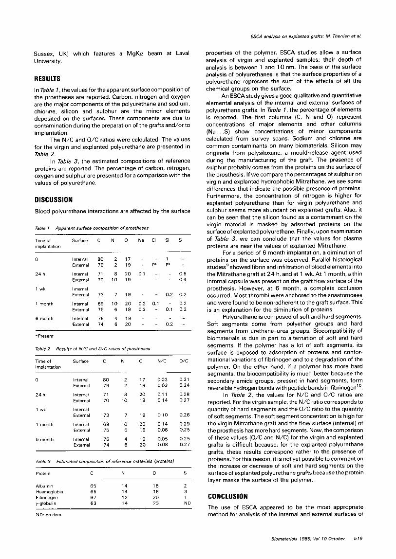
Figure 4 The SEM photograph of external surface of hydrophobic Mitrarhane’ after 6 monrh of implantation. Nora the presence of biological debris on the flow surface and some broken fibriis in the PU srrucrure.
vascular grafts because the penetration is only l-l 0 nm. The results for the virgin and explanted hydrophobic Mitrathane confirmed that these prostheses were submitted to a surface degradation and/or an adsorption of proteins during the implantation (Figure 4). The Versaclean washing seems to be the best detergent for removal of most of the proteins without changing the surface of polymer’. The scanning electron microscopy (SEM) photographs showed a certain degradation of polyurethane after 6 month implan- tation. However, by ESCA study, it is difficult to compare the surface of virgin and explanted grafts because it is masked by the presence of proteins. To complete these findings, other possible experimentation and analysis would be necessary such as Fourier Transform Infrared-Attenuated Total Reflectance (FTIR-ATR) (a surface analysis method with a depth of 1000 nm) to see the infrared bands of proteins and polymer. Differential Scanning Calorimetry (DSC) could also be used to determine the glass transition temperatures and melting temperatures of virgin and explanted polyurethane which would characterize the
polymer and allow a better understanding of the phenomenon of degradation.
ACKNOWLEDGEMENTS
This work was supported by the Medical Research Council of Canada (Grant MA 9429). The authors are indebted to K. Horth and Y. Marois for their collaboration. The grafts were kindly provided by Matrix Medica Inc.
REFERENCES
1
2
3
4
5
6
7
8
9
10
DeBakey, M., The development of vascular surgen/, Am. J. Surg. 1979.137.697-738 Reichle. F.A., Criteria for evaluation of new arterial prosthesis by comparing vein with Dacron femoro-popliteal bypass, Surg. Gyn. Obsrer. 1978. 146. 7 14-720 Annis, D., Bomat, A., Edwards, R.O., Higham, A.. Loveday, B. and Wilson, J.. An elastomeric vascular prosthesis, Trans. Amer. Sot. Artif lnr. Org. 1978, 24, 209-2 14 Leidner, J., Wong, E.W.C., MacGregor, D.C. and Wilson, G.J.. A novel process for the manufacturing of porous grafts: process description and product eva1uation.J. Biomed. Marer. Res. 1983,17,229-247 Hess, F.. Jerusalem, D.. Braun, B. and Grande, P.. Evaluation of the patency rate of fibrous microvascular polyurethane prostheses after implantation in the rat aorta, Microsurgery 1983, 4, 178-l 81 Gilding, D.K., Reed, A.M., Askill, I.N. and Eriana, S., Mitrathane? a new polyether-urethane-urea for critical medical applications, Trans. Amer. Sot. Artif lnr. Org. 1984, 30. 57 l-576 Ives. CL., Zamora. J.L.. Eskin, S.G., Weilbaecher, D.G., Gao, Z.R., Noon, G.P. and DeBakey, M.E., In viva investigation of a new elastomeric vascular graft (Mitrathane”), Trans. Amer. Sot. Anif lnr. Org. 1984,30, 587-590 Paynter. R.W., Martz, H. and Guidoin, R., An X-ray photoelectron spectroscopy study of the external surface of explanted mrcroporous polyurethane vascular prostheses, Biomaterials 1987, 8, 94-99 Marois. Y., Guidoin. R.. Bayer, D.. Assayed, F., Doillon, C., F’aynter, R. and Marois, M., In viva evaluation of hydrophobic and fibrillar microporous polyetherurethane urea graft, Biomaterials 1989, 10, 521-531 Stupp. S.I.. Kauffman. J.W. and Carr, S.H., Interactions between segmented polyurethane surfaces and the plasma protein fibrinogen, J. Biomed. Mar. Res. 1977, 11, 237-250
520 Biomaterials 1989, Vol 10 October