Hydrophilic Pyrazine Dyes as Exogenous Fluorescent...
11
Published: June 13, 2011 r2011 American Chemical Society 5048 dx.doi.org/10.1021/jm200257k | J. Med. Chem. 2011, 54, 5048–5058 ARTICLE pubs.acs.org/jmc Hydrophilic Pyrazine Dyes as Exogenous Fluorescent Tracer Agents for Real-Time Point-of-Care Measurement of Glomerular Filtration Rate Raghavan Rajagopalan,* William L. Neumann, † Amruta R. Poreddy, Richard M. Fitch, John N. Freskos, Bethel Asmelash, Kimberly R. Gaston, Karen P. Galen, Jeng-Jong Shieh, and Richard B. Dorshow Covidien Pharmaceuticals, 675 McDonnell Boulevard, Hazelwood, Missouri 63042, United States ’ INTRODUCTION The assessment of renal function is a critical part of patient care, and the accurate and real-time measurement and monitor- ing of renal function is essential for minimizing the risk of kidney failure due to chronic or acute clinical, physiological, and patho- logical conditions. Glomerular filtration rate (GFR) is now widely accepted as the best indicator of renal function, and current clinical guidelines advocate its use in the staging of kidney disease. 1 Endogenous plasma creatinine assay, 2 and theoretical equations 3 based on the plasma creatinine concentration, continue to be the most common clinical method of assessing GFR despite the known limitations. 4 6 However, the GFR value obtained by these methods is frequently misleading because it is affected by age, state of hydration, renal perfusion, muscle mass, dietary intake, and many other anthropometric and clinical variables. Moreover, creatinine is partially cleared by tubular secretion along with glomerular filtration and, as Diskin 5 recently re- marked, “Creatinine clearance is not and has never been synon- ymous with GFR, and all of the regression analysis will not make it so because the serum creatinine depends upon many factors other than filtration.” The optimum measure of GFR is by the use of exogenous tracer agents. 7 However, the infrequently used conventional GFR agents such as inulin, 8 iothalamate, 9,10 and 99 m Tc-DTPA 11 suffer from various undesirable properties (such as radioactivity, need for ionizing radiation, laborious ex-vivo handling of blood and urine samples, and lack of consistent and reliable supply) that render them unsuitable for real-time point-of-care renal function monitoring. Thus, to overcome these various limitations, there has been considerable effort directed at developing fluorescent GFR tracer agents. 12 15 On the basis of the considerable empirical knowledge that hydrophilic and anionic substances are preferentially cleared through the renal system, 16,17 we had originally proposed a two-pronged approach for the rational design of fluorescent exogenous GFR tracer agents. 13 The first method involves enhancing the fluorescence of known GFR tracer agents that are intrinsically poor fluorescent emitters such as lanthanide metal complexes, and the second involves transforming highly fluorescent dyes (which are intrinsically lipophilic) into hydro- philic, anionic species to force them to clear via the kidneys. In the first approach, we have prepared and evaluated euro- pium DTPA complexes endowed with various molecular “an- tenna” to induce ligand-to-metal fluorescence resonance energy transfer (FRET). 13 Some success was obtained in that one of these metal complexes exhibited a 2700-fold increase in euro- pium fluorescence (compared to Eu DTPA) and underwent clearance exclusively through the kidneys. In this paper, we present, for the first time, the successful results of developing highly fluorescent exogenous GFR tracer agents based on the second approach. 18 The key requirements for the rational design of exogenous fluorescent tracer agents are: (a) excitation and emission occur- ring in the visible region (λ g ∼425 nm), (b) having highly hydrophilic neutral or anionic character, (c) very low or no plasma protein binding, (d) no in vivo metabolism, and (e) Received: March 4, 2011 ABSTRACT: Various hydrophilic pyrazine-bis(carboxamides) derived from 3,5-diamino-pyrazine-2,5-dicarboxylic acid bearing neutral and anionic groups were prepared and evaluated for use as fluorescent glomerular filtration rate (GFR) tracer agents. Among these, the dianionic D-serine pyrazine derivatives 2d and 2j, and the neutral dihydroxypropyl 2h, exhibited favorable physico- chemical and clearance properties. In vitro studies show that 2d, 2h, and 2j have low plasma protein binding, a necessary condition for renal excretion. In vivo animal model results show that these three compounds exhibit a plasma clearance equivalent to iothalamate (a commonly considered gold standard GFR agent). In addition, these compounds have a higher urine recovery compared to iothalamate. Finally, the plasma clearance of 2d, 2h, and 2j remained unchanged upon blockage of the tubular secretion pathway with probenecid, a necessary condition for establishment of clearance via glomerular filtration only. Hence, 2d, 2h, and 2j are promising candidates for translation to the clinic as exogenous fluorescent tracer agents in real-time point-of-care monitoring of GFR.
Transcript of Hydrophilic Pyrazine Dyes as Exogenous Fluorescent...
Published: June 13, 2011
r 2011 American Chemical Society 5048 dx.doi.org/10.1021/jm200257k | J. Med. Chem. 2011, 54, 5048–5058
ARTICLE
pubs.acs.org/jmc
Hydrophilic Pyrazine Dyes as Exogenous Fluorescent Tracer Agentsfor Real-Time Point-of-Care Measurement of GlomerularFiltration RateRaghavan Rajagopalan,* William L. Neumann,† Amruta R. Poreddy, Richard M. Fitch, John N. Freskos,Bethel Asmelash, Kimberly R. Gaston, Karen P. Galen, Jeng-Jong Shieh, and Richard B. Dorshow
Covidien Pharmaceuticals, 675 McDonnell Boulevard, Hazelwood, Missouri 63042, United States
’ INTRODUCTION
The assessment of renal function is a critical part of patientcare, and the accurate and real-time measurement and monitor-ing of renal function is essential for minimizing the risk of kidneyfailure due to chronic or acute clinical, physiological, and patho-logical conditions. Glomerular filtration rate (GFR) is now widelyaccepted as the best indicator of renal function, and current clinicalguidelines advocate its use in the staging of kidney disease.1
Endogenous plasma creatinine assay,2 and theoretical equations3
based on the plasma creatinine concentration, continue to be themost common clinical method of assessing GFR despite theknown limitations.4�6 However, the GFR value obtained bythese methods is frequently misleading because it is affected byage, state of hydration, renal perfusion, muscle mass, dietaryintake, and many other anthropometric and clinical variables.Moreover, creatinine is partially cleared by tubular secretionalong with glomerular filtration and, as Diskin5 recently re-marked, “Creatinine clearance is not and has never been synon-ymous with GFR, and all of the regression analysis will not makeit so because the serum creatinine depends upon many factorsother than filtration.”
The optimum measure of GFR is by the use of exogenoustracer agents.7 However, the infrequently used conventionalGFR agents such as inulin,8 iothalamate,9,10 and 99 mTc-DTPA11
suffer from various undesirable properties (such as radioactivity,need for ionizing radiation, laborious ex-vivo handling of bloodand urine samples, and lack of consistent and reliable supply) thatrender them unsuitable for real-time point-of-care renal functionmonitoring. Thus, to overcome these various limitations, there
has been considerable effort directed at developing fluorescentGFR tracer agents.12�15
On the basis of the considerable empirical knowledge thathydrophilic and anionic substances are preferentially clearedthrough the renal system,16,17 we had originally proposed atwo-pronged approach for the rational design of fluorescentexogenous GFR tracer agents.13 The first method involvesenhancing the fluorescence of known GFR tracer agents thatare intrinsically poor fluorescent emitters such as lanthanidemetal complexes, and the second involves transforming highlyfluorescent dyes (which are intrinsically lipophilic) into hydro-philic, anionic species to force them to clear via the kidneys. Inthe first approach, we have prepared and evaluated euro-pium�DTPA complexes endowed with various molecular “an-tenna” to induce ligand-to-metal fluorescence resonance energytransfer (FRET).13 Some success was obtained in that one ofthese metal complexes exhibited a 2700-fold increase in euro-pium fluorescence (compared to Eu�DTPA) and underwentclearance exclusively through the kidneys. In this paper, wepresent, for the first time, the successful results of developinghighly fluorescent exogenous GFR tracer agents based on thesecond approach.18
The key requirements for the rational design of exogenousfluorescent tracer agents are: (a) excitation and emission occur-ring in the visible region (λ g ∼425 nm), (b) having highlyhydrophilic neutral or anionic character, (c) very low or noplasma protein binding, (d) no in vivo metabolism, and (e)
Received: March 4, 2011
ABSTRACT: Various hydrophilic pyrazine-bis(carboxamides) derived from3,5-diamino-pyrazine-2,5-dicarboxylic acid bearing neutral and anionic groupswere prepared and evaluated for use as fluorescent glomerular filtration rate(GFR) tracer agents. Among these, the dianionic D-serine pyrazine derivatives2d and 2j, and the neutral dihydroxypropyl 2h, exhibited favorable physico-chemical and clearance properties. In vitro studies show that 2d, 2h, and 2jhave low plasma protein binding, a necessary condition for renal excretion. In vivo animal model results show that these threecompounds exhibit a plasma clearance equivalent to iothalamate (a commonly considered gold standard GFR agent). In addition,these compounds have a higher urine recovery compared to iothalamate. Finally, the plasma clearance of 2d, 2h, and 2j remainedunchanged upon blockage of the tubular secretion pathway with probenecid, a necessary condition for establishment of clearance viaglomerular filtration only. Hence, 2d, 2h, and 2j are promising candidates for translation to the clinic as exogenous fluorescent traceragents in real-time point-of-care monitoring of GFR.

5049 dx.doi.org/10.1021/jm200257k |J. Med. Chem. 2011, 54, 5048–5058
Journal of Medicinal Chemistry ARTICLE
clearance exclusively via glomerular filtration (often demon-strated by equality of plasma clearance with and without a tubularsecretion inhibitor such as probenecid19). The selection of thelead clinical candidate(s) may be based on secondary considera-tions such as the ease of synthesis, lack of toxicity, and stability.The secondary screening criteria should further take into accountthe tissue optics properties and the degree of extracellulardistribution of the fluorescent tracers. Volume of distribution isan important parameter in the assessment of hydration state ofthe patient, whereas the absorption/emission properties provideessential information for the design of the probe.
Pyrazines are a class of photostable small molecules havinghighly desirable photophysical properties useful for in biome-dical applications. Pyrazine derivatives containing electronwithdrawing groups at the 2,5 positions and electron donatinggroups at the 3,6 positions such as 3,6-diamino-2,5-pyrazine-dicarboxylic acid (1) and the corresponding amides stronglyabsorb and emit in the blue to orange regions with a largeStokes shift on the order of ∼100 nm and with fluorescencequantum yields of about 0.4.20,21 Conversion of the carboxylgroup in 1 to the secondary amide derivatives produces abathochromic shift of about 40 nm, and alkylation of the aminogroup results in further red-shift of about 40 nm. Hence, thepyrazine scaffold presents an attractive opportunity to “tune”the electronic and renal clearance properties at once byintroducing hydrophilic substituents.
The pyrazine-dicarboxylic acid (1) and its known simplecarboxamide analogues are lipophilic and are insoluble in waterand, therefore, unsuitable as possible GFR tracer agents in thisform. However, the diacid 1 is an excellent scaffold for furthermodification, and several hydrophilic amides bearing variousneutral and anionic substituents (Figure 1) were designed,
synthesized, and evaluated for use as GFR tracer agent candidates.These pyrazine derivatives may be divided into two generalcategories. Compounds 2a�e bear primary amino groups andabsorb radiation in the blue region of the electromagneticspectrum (Table 1). Compounds 2f�j are N-alkylated pyrazinesand absorb radiation in the green region of the spectrum.
’RESULTS AND DISCUSSION
Syntheses. The neutral polyhydroxy derivatives 2a and 2bwere prepared by condensing the diacid 1 with protectedaminoethanol 3a or aminopropanediol 3b respectively by thestandard carbodiimide coupling method, followed by deprotec-tion with hydrochloric acid (Scheme 1). The dianionic serinederivatives 2c and 2dwere prepared by coupling the diacid 1withL- or D-serine benzyl ester hydrochloride (3c or 3d), followed byhydrogenolysis in the presence of 10%Pd�C as catalyst atatmospheric pressure. The tetraanionic aspartate derivative 2ewas prepared by coupling the diacid 1 with dibenzyl aspartate 3e,followed by transfer hydrogenation with ammonium formate and10% Pd�C.Alkyl substitution on the amino groups of the pyrazine scaffold
1 was known to significantly increase the wavelength of bothabsorption and emission maxima, but the existing base-inducedalkylation methods resulted in rather poor yields of the desired
Table 1. Absorption, Emission, and Plasma Protein Binding
compd λAbs (nm) λEm (nm) PPB (%)a
2a 436 558 12
2b 432 558 2
2c 435 559 0
2d 435 557 0
2e 436 558 0
2f 490 599 74
2g 486 600 15
2h 484 594 6
2i 486 597 4
2j 488 597 0
iothalamate NAb NA 10c
aMeasured to (1%. bNot applicable. cReference 23.
Figure 1. Pyrazine-based GFR tracer agents.

5050 dx.doi.org/10.1021/jm200257k |J. Med. Chem. 2011, 54, 5048–5058
Journal of Medicinal Chemistry ARTICLE
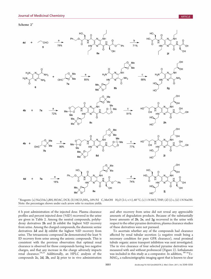
products. Consequently, a convenient and robust reductiveamination method was developed for the synthesis of N-alkylatedpyrazines,22 which was adopted for the preparation of the analogues2f�j (Scheme 2). The neutral N-alkyalted pyrazine derivative 2gwasprepared by the reductive amination of D-glyceraldehyde acetonide(7a) with 3,6-diamino-2,5-dicarboxamide (6), followed by de-protection of the isopropylidene group with aqueous HCl. Thebis(propyl) derivative 2f, was prepared by reductive amination ofpropionaldehyde with bis-benzyl ester 4d, followed by subse-quent transfer hydrogenation of the intermediate 8a. The neutral2h and 2i were made available by the initial reductive alkylationof the bis-acetonide 4b with 7a or L-glyceraldehyde acetonide(7b), respectively, followed by acidic hydrolysis of the acetonidegroups. Finally, a similar reductive alkylation of 4d with 7aafforded 8e, which was then treated with aqueous HCl to removethe acetonide groups followed by base hydrolysis of benzyl estersleading to the dianionic compound 2j.In Vitro Studies.Absorption (λabs) and emission (λem)maxima
and plasma protein binding (PPB) were determined for all
compounds prior to in vivo evaluation (Table 1). Compounds2a�e exhibited absorption maxima in the range of 430�440 nmand emission maxima in the range of 550�560 nm. Compounds2f�j exhibited absorption maxima in the range of 480�490 nmand emission maxima in the range of 590�600 nm. Thefluorescence quantum yield of 2c in DMSO was measured tobe 0.42.With the exception of 2f and 2g, all compounds exhibitedlow plasma protein binding (PPB), comparable to or lower (i.e.,better) than iothalamate. As would be expected, the introductionof hydrophobic propyl group in 2f resulted in high PPB.Surprisingly, the two regioisomeric compounds 2b and 2gexhibited measurably different PPB. It is possible that theprimary carboxamide, which contains an additional site forhydrogen bonding at the amide site in 2g is contributing to thehigher interaction with the protein.In Vivo Studies. All in vivo studies were performed in the
Sprague�Dawley rat model. Plasma concentrations of the traceragents were determined by HPLC at 0, 2, 6, and 24 h postadministration of the injected dose. Urine was collected through
Scheme 1a
aReagents: (a) Et3N, EDC 3HCl, HOBt 3H2O, DMF; (b) 4 N HCl in dioxane; (c) EDC 3HCl, HOBt 3H2O, DMF; (d) 1 N HCl, THF; (e) DIPEA,EDC 3HCl, HOBt 3H2O, DMF; (f) H2, 10% Pd�C, EtOH�H2O (3:1, v/v); (g) HCO2NH4, 10% Pd�C, MeOH�H2O (2:1, v/v), 60 �C. Note: thepercentages shown under each arrow refer to reaction yields.
5051 dx.doi.org/10.1021/jm200257k |J. Med. Chem. 2011, 54, 5048–5058
Journal of Medicinal Chemistry ARTICLE
6 h post administration of the injected dose. Plasma clearanceprofiles and percent injected dose (%ID) recovered in the urineare given in Table 2. Among the neutral compounds, polyhy-droxy derivatives 2h and 2i exhibit the highest %ID recoveryfrom urine. Among the charged compounds, the dianionic serinederivatives 2d and 2j exhibit the highest %ID recovery fromurine. The tetraanionic compound 2e demonstrated the least %ID recovery from urine among the anionic compounds. This isconsistent with the previous observation that optimal renalclearance is observed for those compounds having two negativecharges, and that any increase in the charge adversely impactsrenal clearance.24,25 Additionally, an HPLC analysis of thecompounds 2c, 2d, 2h, and 2j prior to in vivo administration
and after recovery from urine did not reveal any appreciableamounts of degradation products. Because of the substantiallylower amounts of 2b, 2e, and 2g recovered in the urine withrespect to the other pyrazine derivatives, plasma clearance studiesof these derivatives were not pursued.To ascertain whether any of the compounds had clearance
affected by renal tubular secretion (a negative result being anecessary condition for pure GFR clearance), renal proximaltubule organic anion transport inhibition was next investigated.The in vivo clearance of four selected pyrazine derivatives wasmeasured with and without probenecid (Figure 2). Iothalamatewas included in this study as a comparator. In addition, 99 mTc-MAG3, a radioscintigraphic imaging agent that is known to clear
Scheme 2a
aReagents: (a)Na(OAc)3BH,HOAC,DCE; (b)HCO2NH4, 10% Pd�C,MeOH�H2O (2:1, v/v), 60 �C; (c) 1NHCl, THF; (d) (i) c, (ii) 1NNaOH.Note: the percentages shown under each arrow refer to reaction yields.

5052 dx.doi.org/10.1021/jm200257k |J. Med. Chem. 2011, 54, 5048–5058
Journal of Medicinal Chemistry ARTICLE
via the tubular secretion pathway,19,25 was employed as a positivecontrol.Probenecid significantly decreased the clearance rate of 99
mTc-MAG3 (p = 0.001)26 as expected from a non-GFR agent.Probenecid did not significantly affect the clearance rate ofiothalamate (p > 0.05), also as expected from a GFR agent.Compounds 2h and 2j did not exhibit differences in clearancerates (p > 0.05). However, a statistically significant difference(p < 0.05) was detected in the clearance rate of the L-serineenantiomer 2c with respect to the presence or absence ofprobenecid, whereas the D-serine enantiomer 2d did not displaya significant difference (p > 0.05). Therefore, renal tubularsecretion is not a significant elimination pathway for compounds2d, 2h, and 2j in the rat.Noninvasive real-time fluorescence monitoring of the clear-
ance of compound 2h is shown in Figure 3.27 Each panel containsimages of two mice. The mouse on the left was administered300 μL of a 2 mM solution (in PBS) of compound 2h. Themouse on the right received 200 μL of PBS only. The twoadministrations were given simultaneously. Compound 2h isseen to distribute quickly throughout the body and then clearsfrom the body and concentrates in the abdomen. Surgery after
the 60 min time point verified that the highly fluorescent spot inthe abdomen was the bladder.To follow the time dependence of the fluorescence quantita-
tively, an arbitrary region of interest (ROI) was drawn encom-passing the area around the bladder and an approximately equalarea was drawn near the shoulder on the series of optical imagesof the mouse injected with compound 2h from 0 to 70 min. As afunction of time, the fluorescence signal is seen to monotonicallyincrease over the bladder region as expected (Figure 4). Over theshoulder region, this signal is seen to increase post administrationof tracer agent and then wash out as a function of time as it isremoved from the body by the renal system. This fluorescencedecease is consistent with the decrease in plasma concentrationof 2h as indicated in Table 2.
’CONCLUSION
On the basis of the fluorescence properties, plasma proteinbinding data, the injected dose recovered in urine, the plasmaclearance data, and the renal tubular secretion studies, thepyrazine serine derivatives 2d, 2h, and 2j are viable candidatesfor translation to the clinic as exogenous fluorescent tracer agentsfor the measurement of GFR. In the rat animal model, thesecompounds display superior properties compared to iothala-mate, which is currently an accepted standard for the measure-ment of GFR. Formal preclinical development studies are inprogress and will be reported elsewhere.
’EXPERIMENTAL SECTION
Chemistry. Unless otherwise noted, all solvents and reagents wereused as supplied. Organic extracts were dried over either anhyd Na2SO4
or anhyd MgSO4 and filtered using a fluted filter paper (P8) or a frittedglass funnel. Solvents were removed on a rotary evaporator underreduced pressure. Analytical TLC was performed on Analtech silicagel GF plates (250 μm), and the components were visualized by UVlight. Flash chromatography was carried out either using EMD silica gel60 (40�63 μm) or C18 silica gel (YMCODS-A 120 Å I 25/40) in glasscolumns. Automated flash chromatography was carried out on a TeldyneIsco CombiFlash Rf system using prepacked silica gel columns. RP-LC/MS (ESI, positive ion mode) analyses were carried out on either a BDSHypersil C18 3 μm (50 mm � 4.6 mm) or a ThermoElectron HypersilGold C18 3 μm (4.6 mm� 50 mm) column. Compounds were injected
Table 2. %ID Recovery in Urine, and Plasma Clearance Half-Livesa
compd
%ID recovered
in urine at 6 h
plasma half-lifeb
(min)
2b 61( 3 (3)c NDd
2c 82( 7 (3) 36( 6 (8)
2d 90( 1 (3) 29( 1 (6)
2e 61( 16 (3) ND
2g 25( 4 (3) ND
2h 88( 2 (3) 19( 1 (6)
2i 87( 4 (3) ND
2j 85( 2 (3) 20( 1 (5)
iothalamate 80( 2 (6) 32( 2 (4)aThe values are given as mean ( SEM (standard error of mean).bTerminal phase half-life from two compartment modeling. cNumbersin parentheses indicate number of test animals. dNot determined.
Figure 2. Clearance of 99 mTc-MAG3 with and without probenecid, and clearance of several pyrazine compounds and iothalamate comparator with andwithout probenecid.

5053 dx.doi.org/10.1021/jm200257k |J. Med. Chem. 2011, 54, 5048–5058
Journal of Medicinal Chemistry ARTICLE
using a gradient condition (5 to 50�95%B/6�10 min) with a flow rateof 1 mL/min (mobile phase A, 0.05% TFA in H2O; mobile phase B,0.05% TFA in CH3CN). Preparative RP-HPLC was carried out using aWaters XBridge Prep C18 5 μm OBD 30 mm � 150 mm or 19 � 250mm column [λmax, PDA (200�800 nm); flow, 50 mL/min; gradient,5�50% B/10�17 min; mobile phase A, 0.1% TFA in H2O; mobilephase B, 0.1% TFA in CH3CN]. RP-HPLC analyses were carried outusing a Phenomenex Luna 5 μm C18(2) 100 Å 250 mm � 4.6 mmcolumn [detection, UV; flow, 1 mL/min; gradient, 5/10% B to 50/90%B/20 min; mobile phase A, 0.1% TFA in H2O; mobile phase B, 0.1%TFA in CH3CN)], and the chromatographic purities of all the com-pounds were >95%. UV/vis and fluorescence spectra were measured ona Shimadzu UV-3101 PC and Jobin Yvon Fluorolog-3 spectrometers,respectively. NMR spectra were recorded on either a Varian VNMRS-500 spectrometer. 1H Chemical shifts are expressed in parts per million(δ) relative to TMS (δ = 0) as an internal standard. 13C Chemical shiftsare referenced to either TMS (δ = 0) or the residual solvent peaks in the
spectra. Coupling constants (J) are reported in Hz. HRMS (ESI) datawas obtained on a ThermoFisher LTQ-Orbitrap mass spectrometerequipped with an IonMax electrospray ionization source in FTMSmodewith resolution g30K. Elemental analyses were carried out by AtlanticMicrolab, Inc., Norcross, GA.3,6-Diamino-N2,N5-bis(2-tert-butoxyethyl)pyrazine-2,5-
dicarboxamide (4a). A 100 mL round-bottom flask equipped witha magnetic stir bar was charged with diacid 1 (0.420 g, 2.10 mmol),2-tert-butoxyethylamine 3HCl (3a, 0.717 g, 4.66 mmol), EDC 3HCl(0.894 g, 4.66 mmol), HOBt 3H2O (0.630 g, 4.66 mmol), and Et3N(0.630 g, 6.36 mmol) in anhyd DMF (40 mL). The reaction mixturewas stirred overnight at rt and concentrated in vacuo to ∼10 mLsolution that was partitioned between EtOAc (125 mL) and satdNaHCO3 (100 mL). The organic solution was washed with 10%aqueous citric acid (100 mL) and brine (50 mL). The solution wasconcentrated and purified via flash chromatography over silica gelusing EtOAc�hexanes (2:1, v/v) as eluent to give 4a (0.290 g, 35%).1HNMR (DMSO-d6) δ 8.30 (t, J = 5.9 Hz, 2 H), 6.57 (s, 4H), 3.42 (dt,J = 5.3 Hz, 4 H), 3.36 (dt, J = 5.9 Hz, 4 H), 1.14 (s, 18 H). 13C NMR(DMSO-d6) δ 164.8, 146.1, 126.1, 72.5, 59.7, 39.5, 27.2. HRMS (ESI)m/z calcd for C18H33N6O4 (M +H)+ 397.2558, found 397.2558; calcdfor C18H32N6O4Na (M + Na)+ 419.2377, found 419.2376.3,6-Diamino-N2,N5-bis(2-hydroxyethyl)pyrazine-2,5-di-
carboxamide (2a). A 100 mL round-bottom flask equipped with amagnetic stir bar was charged with the diether 4a (0.233 g, 0.588mmol) and 4 N HCl�dioxane (8 mL) and stirred at rt overnight. Thereaction was concentrated in vacuo, and the crude product (∼0.170 g)was purified via RP-HPLC (5�50%B). The product containingfractions were concentrated in vacuo and vacuum-dried to afford 2a(0.082 g, 49%) as an orange solid. 1H NMR (DMSO-d6) δ 8.30 (t J =5.9 Hz, 2H), 7.60 (s, 4 H), 4.83 (t, J = 5.9 Hz, 2 H) 3.45 (dt, J = 5.4 Hz,4 H), 3.35 (dt, J = 5.9 Hz, 4 H). 13C NMR (DMSO-d6) δ 165.4, 146.1,126.7, 60.0, 41.8. HRMS (ESI) m/z calcd for C10H17N6O4 (M + H)+
285.1306, found 285.1307; calcd for C10H16N6O4Na (M + Na)+
307.1125, found 307.1126.3,6-Diamino-N2,N5-bis[(2,2-dimethyl-1,3-dioxolan-4-yl)-
methyl]pyrazine-2,5-dicarboxamide (4b). A mixture of diacid 1(0.350 g, 1.77 mmol), 2,2-dimethyl-1,3-dioxolane-4-methanamine (3b,0.933 mL, 7.20 mmol), HOBt 3H2O (0.812 g, 5.30 mmol), and EDC 3HCl (1.02 g, 5.32mmol) were stirred together in DMF (20mL) for 16 h
Figure 3. In vivo time-dependent fluorescence detection (with pixelintensity range bar graph). The mouse on the left was treated withcompound 2h, and the mouse on the right was treated with PBS; (a)preadministration, (b) 10 min post administration, (c) 20 min postadministration, (d) 30 min post administration, (e) 60 min postadministration. The ratio between most intense and least intensefluorescence in the 60 min post administration image was approximatelya factor of 100.
Figure 4. Fluorescence measured over the bladder (dashed line) andshoulder region (solid line) as a function of time (data was normalized topeak value of each ROI).
5054 dx.doi.org/10.1021/jm200257k |J. Med. Chem. 2011, 54, 5048–5058
Journal of Medicinal Chemistry ARTICLE
at rt. The mixture was concentrated to dryness, and the residue waspartitioned between EtOAc and water. The layers were separated, andthe EtOAc solution was washed with satd NaHCO3 and brine. TheEtOAc solution was concentrated to afford the diastereomeric bis-amide4b (0.665 g, 88%) as a yellow solid. 1H NMR (CDCl3) δ 8.38 (t, J = 5.8Hz, 2 H), 6.55 (s, 4 H), 4.21 (quintet, J = 5.8 Hz, 2 H), 3.98 (dd, J = 8.4Hz, 6.3 Hz, 2 H), 3.65 (dd, J = 8.4 Hz, J = 5.8 Hz, 2 H), 3.39 (apparentquartet�diastereotopic mixture, J = 5.9 Hz, 4 H), 1.35 (s, 6 H), 1.26 (s,6 H). 13C NMR (CDCl3) δ 165.7, 146.8, 126.8, 109.2, 74.8, 67.2, 42.2,41.1, 27.6, 26.1. RP-LC/MS (ESI) m/z 425.4 (M + H)+ (tR = 4.00 min,5�95%B/6 min). HRMS (ESI) m/z calcd for C18H28N6O6Na (M +Na)+ 447.1963, found 447.1960. Anal. Calcd for C18H28N6O6: C, 50.93;H, 6.65; N, 19.80. Found: C, 51.35; H, 7.12; N, 19.59.3,6-Diamino-N2,N5-bis(2,3-dihydroxypropyl)pyrazine-2,5-
dicarboxamide (2b). The above bis-amide 4b was dissolved in THF(100 mL) and treated with 1.0 N HCl (2 mL). After hydrolysis wascomplete, the mixture was treated with K2CO3 (1 g) and stirred for 1 hand filtered through a plug of C18 silica gel using methanol. The filtratewas concentrated to dryness and the residue was triturated with MeOH(50 mL). The solids were filtered and discarded, and the residue wastreated with ether (50 mL). The precipitate was collected by filtrationand dried under high vacuum. This material was purified by radial flashchromatography using CHCl3�MeOH (19:1 to 1:1, v/v) to afford 2b(0.221 g, 36%) as an orange solid. 1H NMR (DMSO-d6) δ 8.00 (br m,6 H), 5.39 (br s, 2 H), 4.88 (br s, 2 H), 3.63�3.71 (complex m, 2 H),3.40 (dd, J = 11.1, 5.1 Hz, 2H), 3.28 (dd, J = 11.1, 6.6 Hz, 2 H), 2.92 (dd,J = 12.6, 3.3 Hz, 2 H), 2.65 (dd, J = 12.6, 8.4 Hz, 2 H). RP-LC/MS (ESI)m/z 345 (M + H)+ (tR = 4.13 min, 5�95% gradient acetonitrile in 0.1%TFA over 10 min on a 30 mm column). HRMS (ESI) m/z calcd forC12H20N6O6Na (M + Na)+ 367.1337, found 367.1336.3,6-Diamino-N2,N5-bis[(S)-1-(benzyloxy)-3-hydroxy-1-ox-
opropan-2-yl]pyrazine-2,5-dicarboxamide (4c). A 250 mLround-bottom flask equipped with a Claisen adapter and an additionfunnel was charged with L-serine benzyl ester hydrochloride (3c, 4.87 g,21.0 mmol) and anhydrous DMF (100 mL) was cannulated into it. Theresulting colorless solution was cooled in an ice-bath and stirred for 15min under N2 atmosphere. Then DIPEA (3.83 mL, 22.0 mmol) wasadded dropwise via addition funnel over a 30 min period, and after 30min, the cooling bath was removed and diacid 1 (1.98 g, 10.0 mmol) wasadded in one portion. The brick-red suspension was allowed to stir for 30min before the addition of HOBt 3H2O (3.37 g, 22.0 mmol) in oneportion. After another 15min, the reaction flask was once again cooled inan ice-bath, and EDC 3HCl (4.22 g, 22.0 mmol) was added in portionsover a 15 min period. The resulting somewhat lighter and brownsuspension was slowly allowed to warm to rt and stirred overnight(ca. 17 h) under N2. The reaction mixture was diluted with EtOAc(500 mL) and transferred to a 1 L separatory funnel. The solution waswashed with milli-Q H2O (150 mL), and the aq phase was furtherextracted with EtOAc (100 mL). The combined organic extracts weresuccessively washed with 150 mL portions of water, 0.50 M KHSO4,water, satd NaHCO3 (�2), water, and brine. Removal of the solventfollowed by drying under high vacuum gave 4.75 g of the crude productas an orange fluffy solid, which upon flash chromatography over silica gelusing CHCl3MeOH (19:1, v/v) as eluant (sample adsorbed on silica gelwas loaded on the column), afforded the bis-amide 4c (4.34 g, 79%) asan orange solid: Rf 0.40 [CHCl3�MeOH (9:1, v/v)]. 1H NMR(DMSO-d6)) δ 8.56 (d, J = 8.3 Hz, 2 H, exchangeable with D2O),7.40�7.31 (m, 10 H), 6.76 (s, 4 H, exchangeable with D2O), 5.37 (t, J =5.6Hz 2H), 5.20 (distorted AB pair, 4 H), 4.67�4.64 (dt, J = 8.3, 3.8 Hz,2 H), 3.97�3.93 (m, 2 H), 3.81�3.77 (m, 2 H). 13C NMR (DMSO-d6)δ 170.1, 164.9, 146.4, 135.8, 128.4, 128.0, 127.6, 125.9, 66.2, 61.1, 54.4.RP-LC/MS (ESI) m/z 553.3 (M + H)+ (tR = 4.24 min, 15�95%B/6 min). Anal. Calcd for C26H28N6O8: C, 56.52; H, 5.11; N, 15.21.Found: C, 56.53; H, 5.05; N, 15.16.
3,6-Diamino-N2,N5-bis[(S)-1,3-dihydroxy-1-oxopropan-2-yl]pyrazine-2,5-dicarboxamide (2c). The bis-amide 4c (3.87 g,7.00 mmol) was weighed into a 500 mL round-bottom flask equippedwith a Claisen adapter and anhyd EtOH (225 mL), and milli-Q H2O(50 mL) were added. The resulting orange suspension (partiallysoluble) was briefly sonicated and 10% Pd�C (0.39 g) was added as aslurry in H2O (25 mL). The reaction mixture was thoroughly purgedfirst with argon and then withH2 and stirred under H2 atmosphere (slowbubbling) at rt for 5.5 h. The reaction mixture was once again purgedwith Ar, and the catalyst was removed by filtration over a Celite bed. Thebed was thoroughly washed with EtOH�H2O (400 mL; 1:1, v/v), andthe combined filtrates were concentrated in vacuo and further driedunder high vacuum. The resulting orange residue (2.45 g) was trituratedwith CH3CN (100 mL; sonicated briefly to get the material off the wallsof the flask) to give 2c (2.29 g, 88%) as a red powder. 1HNMR (DMSO-d6) δ 8.46 (d, J = 8.3 Hz, 2 H, exchangeable with D2O), 6.78 (br s, 4 H,exchangeable withD2O), 4.48�4.45 (dt, J= 8.3, 3.9Hz, 2H; collapses toa triplet at δ = 4.47 ppm with J = 3.7 Hz upon D2O exchange), 3.88 (dd,J = 10.9, 3.8 Hz, 2 H), 3.74 (dd, J = 10.9, 3.6 Hz, 2 H). 13C NMR(DMSO-d6) δ 171.5, 164.6, 146.3, 125.9, 61.1, 54.2. RP-LC/MS (ESI)m/z 373.3 (M + H)+ (tR = 2.62 min, 5�95%B/6 min). Anal. Calcdfor C12H16N6O8: C, 38.71; H, 4.33; N, 22.57. Found: C, 38.67; H, 4.50;N, 22.27.3,6-Diamino-N2,N5-bis[(R)-1-(benzyloxy)-3-hydroxy-1-ox-
opropan-2-yl]pyrazine-2,5-dicarboxamide (4d). The reactionof diacid 1 (9.91 g, 50.0 mmol) with D-serine benzyl ester hydrochloride(3d, 24.33 g, 105.0 mmol) in the presence of DIPEA (19.16 mL,110.0 mmol), HOBt 3H2O (17.61 g, 115.0 mmol), and EDC 3HCl(22.05 g, 115.0 mmol) in DMF (300 mL) was carried out overnight(ca. 14 h) as described for the preparation of 4c. The dark solution wasconcentrated to a syrupy residue under high vacuum (bath temp 60 �C)that was partitioned between EtOAc and milli-Q H2O (400 mL each).The layers were separated and the aqueous layer was further extractedwith EtOAc (3 � 200 mL), and the combined EtOAc extracts weresuccessively washed with 0.50MKHSO4, saturated NaHCO3, H2O, andbrine (250 mL each). Removal of the solvent left an orange slush thatwas dried under high vacuum overnight to give 23.7 g of an orange solid.The crude product was subjected to flash chromatography over silica gelusing CHCl3 to CHCl3�MeOH (97:3, v/v) as eluant in a gradientfashion (sample adsorbed on silica gel was loaded on the column; carriedout in 4 runs) to give the bis-amide 4d (19.6 g, 71%) as an orange solid:Rf 0.45 [CHCl3�MeOH (9:1, v/v)]. 1H NMR (DMSO-d6) δ 8.56 (d,J = 8.0 Hz, 2 H, exchangeable with D2O), 7.40�7.33 (m, 10 H), 6.76 (s,4 H, exchangeable with D2O), 5.37 (t, J = 5.5 Hz, 2 H), 5.20 (distortedAB pair, 4 H), 4.66�4.63 (dt, J = 8.0, 4.0 Hz, 2 H), 3.97�3.93 (m, 2 H),3.81�3.77 (m, 2 H). 13C NMR (DMSO-d6) δ 170.1, 164.9, 146.4,135.8, 128.4, 128.0, 127.6, 125.9, 66.2, 61.1, 54.4. RP-LC/MS (ESI)m/z553.3 (M + H)+ (tR = 4.44 min, 5�95%B/6 min). Anal. Calcd forC26H28N6O8: C, 56.52; H, 5.11; N, 15.21. Found: C, 56.39; H, 5.11;N, 14.99.3,6-Diamino-N2,N5-bis[(R)-1,3-dihydroxy-1-oxopropan-2-
yl]pyrazine-2,5-dicarboxamide (2d). The bis-amide 4d (7.74 g,14.0 mmol) was hydrogenated in the presence of 10% Pd�C (0.774 g)in EtOH�H2O (560 mL; 3:1, v/v) for 5 h as described for thepreparation of 2c. After a similar work up and isolation procedure, 2d(4.89 g, 94%) was obtained as an orange powder. 1H NMR (DMSO-d6)δ 8.46 (d, J = 8.3 Hz, 2 H, exchangeable with D2O), 6.78 (br s, 4 H,exchangeable withD2O), 4.48�4.45 (dt, J= 8.1, 3.9Hz, 2H; collapses toa triplet at δ = 4.47 ppm with J = 3.7 Hz upon D2O exchange), 3.88 (dd,J = 11.1, 3.9 Hz, 2 H), 3.74 (dd, J = 11.1, 3.7 Hz, 2 H). 13C NMR(DMSO-d6) δ 171.6, 164.7, 146.4, 125.9, 61.2, 54.3. RP-LC/MS (ESI)m/z 373.2 (M + H)+ (tR = 2.86 min, 5�95% B/6 min). Anal. Calcdfor C12H16N6O8: C, 38.71; H, 4.33; N, 22.57. Found: C, 38.44; H,4.51: N, 22.33.

5055 dx.doi.org/10.1021/jm200257k |J. Med. Chem. 2011, 54, 5048–5058
Journal of Medicinal Chemistry ARTICLE
3,6-Diamino-N2,N5-bis[(R)-1,2-(dibenzyloxycarbonyl)ethyl]-pyrazine-2,5-dicarbox-amide (4e). A mixture of sodium salt ofdiacid 1 (0.600 g, 2.48 mmol), D-Asp(OBn)-OBn 3 p-TsOH salt (3e,2.43 g, 5.00 mmol), HOBt 3H2O (0.919 g, 6.00 mmol) and EDC 3HCl(1.14 g, 5.95 mmol) in DMF (50 mL) was treated with Et3N (4 mL).The resulting mixture was stirred overnight at rt The reaction mixturewas concentrated, and the residue was partitioned between water andEtOAc. The EtOAc layer was separated and washed successively withsatd sodium bicarbonate, water, and brine. The EtOAc solution wasconcentrated, and the residue was purified by flash chromatography[SiO2, CHCl3�MeOH (50/1 to 10/1, v/v] to afford the bis-amide 4e(1.15 g, 58% yield) as a yellow foam. 1H NMR (CDCl3) δ 8.61 (d, J =8.4 Hz, 2 H), 7.29�7.39 (m, 20 H), 5.85 (br s, 4 H), 5.22 (ABq, J = 10.0Hz,ΔνAB = 17.3Hz, 4H), 5.10 (ABq, J = 12.2Hz,ΔνAB = 34.3Hz, 4H),5.06�5.09 (m, 2 H), 3.11 (ABq of d, J = 17.0, 5.1 Hz, ΔνAB = 77.9 Hz,4 H). 13C NMR (CDCl3) δ 170.7, 170.7, 165.4, 147.0, 135.7, 135.6,129.0, 128.9, 128.8, 128.75, 128.7, 126.9, 68.0, 67.3, 49.1, 37.0. RP-LC/MS (ESI) m/z 789 (M + H)+ (tR = 5.97 min, 50�95% gradientacetonitrile in 0.1% TFA over 10 min on a 250 mm column).3,6-Diamino-N2,N5-bis[(R)-1,2-(dicarboxy)ethyl]pyrazine-
2,5-dicarboxamide (2e). To a well stirred solution of tetra-benzylester 4e (0.510 g, 0.650mmol) in THF (20mL) andwater (10mL)wereadded 10% Pd/C (0.50 g) and ammonium formate (1.00 g, 15.9 mmol).The resulting mixture was heated to 60 �C for 2 h and allowed to coolto rt. The mixture was filtered through Celite and concentrated. Theresulting material was purified by reverse phase medium pressurechromatography (C18, 10�70% manual gradient acetonitrile in 0.1%TFA) to afford 2e (0.138 g, 54%) as an orange solid. 1H NMR (DMSO-d6) δ 8.62 (d, J = 8.4 Hz, 2 H), 6.67 (br s, 4 H), 4.73 (dt, J = 8.4, 5.4 Hz,2 H), 2.74�2.88 (complex m, 4 H). 13C NMR (DMSO-d6) δ 172.6,165.2, 147.0, 126.6, 60.8, 49.1. RP-LC/MS (ESI) m/z 429 (M + H)+
(tR = 4.01min, 5�95% gradient acetonitrile in 0.1%TFA over 10min ona 250 mm column). HRMS (ESI) m/z calcd for C14H14N6O10 (M �2 H)2� 213.0391, found 213.0388.N2,N5-Bis[(R)-1-(benzyloxy)-3-hydroxy-1-oxopropan-2-
yl]-3,6-bis(propylamino)pyrazine-2,5-dicarboxamide (8a).To a solution of the bis-benzyl ester 4d (0.890 g, 1.61 mmol) andpropionaldehyde (5, 0.460 mL, 6.44 mmol) in anhyd DCE (65 mL) wasadded HOAc (0.368 mL, 6.44 mmol) with stirring at 0 �C under argonatmosphere. Then Na(OAc)3BH (1.36 g, 6.44 mmol) was introduced insmall portions and the reddish suspension was slowly allowed to warm tort and stirred overnight (ca. 24 h) under argon. The reaction wasquenched by a slow addition of satd NaHCO3 at 0 �C and extracted withCHCl3 (150 mL). The CHCl3 extracts were successively washed withH2O and brine (50mL each). Removal of the solvent gave 1.04 g of a redsolid, which upon flash chromatography over silica gel [CHCl3�MeOH(39:1, v/v)] afforded 8a (0.800 g, 78%) as a dark-red solid. 1H NMR(CDCl3) δ 8.68 (d, J = 7.4 Hz, 2 H), 7.60 (t, J = 5.6 Hz, 2 H), 7.37�7.33(m, 10 H), 5.26 (s, 4 H), 4.78�4.75 (dt, J = 7.3, 3.7 Hz, 2 H), 4.08 (dd,J = 6.1, 3.9 Hz, 4 H), 3.39�3.28 (m, 4 H), 2.64 (t, J = 6.2 Hz, 2 H),1.65�1.61 (m, 4 H), 0.97 (t, J = 7.4 Hz, 6 H). 13C NMR (CDCl3) δ169.9, 166.5, 146.0, 135.1, 128.7, 128.6, 128.2, 125.8, 67.6, 63.6, 55.1,42.9, 22.7, 11.7. RP-LC/MS (ESI) m/z 637.4 (M + H)+ (tR = 5.13 min,5�95% B/6 min). Anal. Calcd for C32H40N6O8: C, 60.37; H, 6.33; N,13.20. Found: C, 60.49; H, 6.42; N, 13.13.N2,N5-Bis[(R)-1,3-dihydroxy-1-oxopropan-2-yl]-3,6-bis-
(propylamino)pyrazine-2,5-dicarboxamide (2f). A solution ofthe bis-benzyl ester 8a (0.700 g, 1.10 mmol) in MeOH (150 mL) waspurged with argon and a slurry of 10% Pd/C (0.350 g) in water (8 mL)was added. Then ammonium formate (0.480 g, 7.61 mmol) was added,and the resulting mixture was heated to 60 �C and stirred for 1 h. Thereaction mixture was allowed to cool to room temperature, filteredthrough Celite, and concentrated on a rotary evaporator. The crudeproduct (0.559 g) was subjected to purification by preparative RP-HPLC
(Sunfire Prep C18 OBD 5 μm 50 mm � 10 mm, 5�95%B/13 min) togive 2f (0.356 g, 71%) as dark-red powder. 1HNMR (DMSO-d6) δ 8.54(d, J = 7.8 Hz, 2 H), 4.94�4.87 (m, 4H), 4.77 (dd, J = 10.8, 5.8 Hz, 2H),4.45�4.42 (dt, J = 8.1, 3.8 Hz, 2 H), 3.89 (dd, J = 11.0, 3.7 Hz, 2 H), 3.79(dd, J = 11.0, 3.4 Hz, 2 H), 3.40�3.31 (m, 4 H), 1.61�1.56 (m, 4 H),0.94 (t, J = 7.4 Hz, 6 H). 13C NMR (DMSO-d6) δ 172.0, 165.5, 145.9,126.1, 61.4, 54.7, 42.7, 22.8, 11.9. RP-LC/MS (ESI) m/z 457.4 (M +H)+ (tR = 3.77 min, 5�95% B/6 min). HRMS (ESI) m/z calcd forC18H29N6O8 (M + H)+ 457.2041, found 457.2044.3,6-Bis{[(S)-2,2-dimethyl-1,3-dioxolan-4-yl]methylamino}-
pyrazine-2,5-dicarboxamide (8b). The reaction of 3,6-diamino-pyrazine-2,5-dicarboxamide (6, 0.196 g, 1.00 mmol) with (R)-(+)-2,2-dimethyl-1,3-dioxolane-4-carbaldehyde (7a, 0.390 g, 3.00 mmol) in thepresence of HOAc (0.180 g, 3.00 mmol) and Na(OAc)3BH (0.633 g,3.00 mmol) in anhyd DCE (10 mL) was carried out overnight asdescribed for the preparation of 8a. A similar work up afforded 8b (0.36g, 85%) as a red solid, which was used without further purification. 1HNMR (CDCl3) δ 7.95 (br t, 2 H), 7.62 (br, 2 H), 5.60 (br, 2H), 4.37 (m,21H), 4.12 (m, 2 H), 3.81 (m, 2 H), 3.58 (m, 4H), 1.55 (s, 6 H), 1.38 (s,6 H). 13C NMR (CDCl3) δ 168.1, 146.1, 126.1, 109.4, 74.9, 67.3, 43.4,27.1, 25.5. RP-LC/MS (ESI) m/z 425.4 (M + H)+.3,6-Bis[(S)-2,3-dihydroxypropylamino]pyrazine-2,5-di-
carboxamide (2g). The bis-acetonide 8b (0.36 g, 0.85 mmol) wasdissolved in THF (5 mL) and treated with 1 N HCl (1 mL). Thereaction mixture was stirred overnight, neutralized with 1 N NaOH(1 mL), and evaporated to dryness. The crude product was subjected toflash chromatography over C18 silica gel using water as eluent to afford2g (0.200 g, 68%). 1H NMR (DMSO-d6) δ 7.91 (br t, 2 H), 7.60 (br,2 H), 4.75 (br, 2 H), 4.60 (m, 2 H), 3.60 (m, 4 H), 3.20 (m, 4 H). 13CNMR (DMSO-d6) δ 167.9, 145.5, 125.8, 70.4, 63.8, 43.6. HRMS (ESI)m/z calcd for C12H21N6O6 (M + H)+ 345.1517, found 345.1457.N2,N5-Bis[(2,2-dimethyl-1,3-dioxolan-4-yl)methyl]-3,6-
bis{[(S)-2,2-dimethyl-1,3-dioxolan-4-yl]methylamino}pyra-zine-2,5-dicarboxamide (8c). The reaction of bis-acetonide 4b(1.72 g, 4.05 mmol) with aldehyde 7a (2.64 g, 20.3 mmol) in thepresence of HOAc (1.13 mL, 19.6 mmol) and Na(OAc)3BH (4.18 g,19.7 mmol) in anhyd DCE (50 mL) was carried out overnight (ca. 22 h)as described for the preparation of 8a. The reaction was found to beincomplete (RP-LC/MS) at this stage and subsequently treated withadditional aldehyde 7a (0.70 g, 5.38 mmol), HOAc (0.300 mL, 5.20mmol), and Na(OAc)3BH (1.10 g, 5.19 mmol) and continued over-night. After a similar work up described in the prepaaration of 8a, thecrude product (4.30 g) obtained was subjected to automated flashchromatography [column: RediSep Rf 330 g silica gel; λmax, 280 nm;flow, 80 mL/min; gradient, 0�2% B/40 min (mobile phase A, CHCl3;mobile phase B, MeOH)] to afford 8c (1.90 g, 72%) as an orangepowder. 1HNMR (CDCl3) δ 8.10 (t, J = 6.0Hz, 2H), 8.06 (t, J = 6.0Hz,2 H), 4.36�4.31 (m, 4 H), 4.09�4.06 (m, 4 H), 3.79�3.76 (m, 2 H),3.74�3.67 (m, 2 H), 3.66�3.52 (m, 8 H), 1.49 (s, 3 H), 1.48 (s, 3 H),1.45 (s, 6 H), 1.37 (s, 6 H), 1.36 (s, 6 H). RP-LC/MS (ESI) m/z 653.3(M + H)+ (tR = 4.83 min, 5�95% B/6 min). Anal. Calcd forC30H48N6O10: C, 55.20; H, 7.41; N, 12.88. Found: C, 55.14; H, 7.37;N, 12.60.N2,N5-Bis(2,3-dihydroxypropyl)-3,6-bis[(S)-2,3-dihydrox-
ypropylamino]pyrazine-2,5-dicarboxamide (2h). To an or-ange solution of tetra-acetonide 8c (3.68 g, 5.64mmol) in THF (50mL)was added 1.0 N HCl solution (45 mL) and stirred for 2 h at rt in anatmosphere of argon.Most of the THFwas removed; the aqmixture wasneutralized with 1.0 N NaOH, and evaporated to dryness. The residuethus obtained was further dried under high vacuum to give the crudeproduct (5.86 g), which upon purification by gravity chromatographyover C18 silica gel (gradient elution with water to 35%MeOH in water),afforded 2h (2.52 g, 91%) as a red powder. 1HNMR (DMSO-d6) δ 8.50(t, J = 5.9 Hz, 2 H, exchangeable with D2O), 7.99 (t, J = 5.7 Hz, 2 H,

5056 dx.doi.org/10.1021/jm200257k |J. Med. Chem. 2011, 54, 5048–5058
Journal of Medicinal Chemistry ARTICLE
exchangeable with D2O), 4.91 (dd, J = 4.9, 1.3 Hz, 2 H, exchangeablewith D2O), 4.81 (d, J = 4.9 Hz, 2H, exchangeable with D2O), 4.69�4.63(m, 4 H, exchangeable with D2O), 3.66�3.56 (m, 6 H), 3.47�3.31 (m,10 H), 3.28�3.19 (m, 4 H). 13C NMR (DMSO-d6) δ 165.9, 145.9,126.4, 70.8, 70.41, 70.36, 64.6, 64.31, 64.29, 44.15, 44.13, 42.9. RP-HPLC (254 nm) 97.3% (tR = 11.76 min, 5�50% B/20 min). RP-LC/MS (ESI)m/z 493.4 (M+H)+ (tR = 3.00min, 5�95%B/6min). HRMS(ESI) m/z calcd for C18H32N6O10Na (M + Na)+ 515.2072, found515.2059. Anal. Calcd for C18H32N6O10 3H2O: C, 42.35; H, 6.71; N,16.46. Found: C, 42.31; H, 6.58; N, 16.43.N2,N5-Bis[(2,2-dimethyl-1,3-dioxolan-4-yl)methyl]-3,6-
bis{[(R)-2,2-dimethyl-1,3-dioxolan-4-yl]methylamino}pyr-azine-2,5-dicarboxamide (8d). The reaction of bis-acetonide 4b(1.00 g, 2.36 mmol) with (S)-2,2-dimethyl-1,3-dioxolane-4-carbalde-hyde (7b, 0.920 g, 7.07 mmol) in the presence of HOAc (0.400 mL,6.94mmol) andNa(OAc)3BH (1.46 g, 6.89mmol) in anhydDCE (40mL)was carried out overnight (ca. 22 h) as described for the preparation of 8c[including further treatment with additional reagents aldehyde 7b(0.920 g, 7.07 mmol), HOAc (0.400 mL, 6.94 mmol), and Na-(OAc)3BH (1.46 g, 6.89 mmol) overnight]. After a similar work up,the crude product (1.45 g) was subjected to an automated flash chroma-tography [column: RediSep Rf 120 g silica gel; λmax, 280 nm; flow,40 mL/min; gradient, 0�2% B/40 min, 2%B/90 min (mobile phase A,CHCl3; mobile phase B, MeOH)] to afford 8d (1.38 g, 90%) as anorange�red powder. 1H NMR (CDCl3) δ 8.10 (t, J = 6.0 Hz, 2 H), 8.06(t, J = 6.0Hz, 2H), 4.36�4.31 (m, 4H), 4.09�4.06 (m, 4H), 3.79�3.76(m, 2 H), 3.74�3.70 (m, 2 H), 3.66�3.52 (m, 8 H), 1.49 (s,3 H), 1.48 (s, 3 H), 1.45 (s, 6 H), 1.37 (s, 6 H), 1.36 (s, 6 H). RP-LC/MS (ESI) m/z 653.5 (M + H)+ (tR = 4.83 min, 5�95%B/6 min).Anal. Calcd for C30H48N6O10: C, 55.20; H, 7.41; N, 12.88. Found: C,54.98; H, 7.50; N, 12.73.N2,N5-Bis(2,3-dihydroxypropyl)-3,6-bis[(R)-2,3-dihydrox-
ypropylamino]pyrazine-2,5-dicarboxamide (2i). The depro-tection of tetra-acetonide 8d (1.00 g, 1.53 mmol) with 1.0 N HCl(15 mL) in THF (25 mL) was carried out for 5 h as described for thepreparation of 2h. After a similar work up, the crude product (2.01 g)was subjected to purification by gravity chromatography over C18 silicagel (gradient elution with water to 25%MeOH in water) to give 2i (0.58g, 77%) as an orange powder. 1HNMR (DMSO-d6) δ 8.50 (t, J = 5.8Hz,2 H, exchangeable with D2O), 7.99 (t, J = 5.4 Hz, 2H, exchangeable withD2O), 4.91 (d, J = 3.4, Hz, 2 H), 4.80 (d, J = 4.7 Hz, 2 H, exchangeablewith D2O), 4.68�4.63 (m, 4 H, exchangeable with D2O), 3.63�3.55(m, 6 H), 3.47�3.31 (m, 10 H), 3.27�3.19 (m, 4 H). 13C NMR(DMSO-d6) δ 165.9, 145.94, 145.93 126.4, 70.8, 70.41, 70.36, 64.7,64.31, 64.29, 44.14, 44.12, 42.9. RP-HPLC (254 nm) 98.2% (tR = 11.78min, 5�50%B/20 min). RP-LC/MS (ESI) m/z 493.3 (M + H)+ (tR =2.97min, 5�95%B/6min). HRMS (ESI)m/z calcd for C18H32N6O10Na(M + Na)+ 515.2072, found 515.2069. Anal. Calcd for C18H32N6O10 3H2O: C, 42.35; H, 6.71; N, 16.46. Found: C, 42.75; H, 6.94; N, 16.53.N2,N5-Bis[(R)-1-(benzyloxy)-3-hydroxy-1-oxopropan-2-yl]-
3,6-bis{[(S)-2,2-dimethyl-1,3-dioxolan-4-yl]methylamino}-pyrazine-2,5-dicarboxamide (8e).The reaction of bis-benzyl ester4d (0.50 g, 0.905 mmol) with aldehyde 7a (0.71 g, 5.43 mmol) in thepresence of HOAc (0.31 mL, 5.43 mmol) and Na(OAc)3BH (1.15 g,5.43 mmol) in anhyd DCE (20 mL) was carried out overnight asdescribed for the preparation of 8c [including further treatment withadditional reagents aldehyde 7a (0.71 g, 5.43 mmol), HOAc (0.71 g,5.43 mmol), and Na(OAc)3BH (1.15 g, 5.43 mmol) overnight]. After asimilar work up, the crude product (0.80 g) was subjected to anautomated flash chromatography [column: RediSep Rf 40 g silica gel;λmax, 280 nm; flow, 15 mL/min; gradient, 0�3% B/30 min, 3%B/50min (mobile phase A, CHCl3; mobile phase B, MeOH)] to afford 8e(0.13 g, 18%) as an orange�red powder. 1H NMR (CDCl3) δ 8.62(d, J = 7.8 Hz, 2 H), 7.91 (t, J = 6.1 Hz, 2 H), 7.37�732 (m, 10 H), 5.25
(s, 4 H), 4.83�4.80 (dt, J = 7.6, 3.8 Hz, 2 H), 4.33�4.29 (m, 2 H), 4.11(dd, J = 11.5, 4.1 Hz, 2 H), 4.04�3.99 (m, 4 H), 3.89 (dd, J = 8.6, 4.7 Hz,2 H), 3.77�3.72 (dt, J = 14.0, 6.7 Hz, 2 H), 3.44�3.39 (dt, J = 13.7, 5.6Hz, 2 H), 3.17 (br, 2 H), 1.48 (s, 6 H), 1.35 (s, 6 H). 13CNMR (CDCl3)δ 169.9, 165.8, 146.1, 135.2, 128.7, 128.5, 128.2, 125.9, 109.6, 75.4, 67.5,67.1, 63.4, 54.9, 42.9, 27.2, 25.4. RP-LC/MS (ESI)m/z 781.4 (M + H)+
(tR = 4.80 min, 5�95%B/6 min). HRMS (ESI) m/z calcd forC38H48N6O12Na (M + Na)+ 803.3222, found 803.3223; calcd forC38H49N6O12 (M + H)+ 781.3403, found 781.3412.N2,N5-Bis[(R)-1,3-dihydroxy-1-oxopropan-2-yl]-3,6-bis-
[(S)-2,3-dihydroxypropylamino]pyrazine-2,5-dicarboxa-mide (2j). To a solution of bis-acetonide 8e (0.075 g, 0.096 mmol) inTHF (3 mL) was added 1.0 NHCl solution (0.5 mL) and stirred for 4 hat rt under argon atmosphere. Most of the THFwas removed, and the aqreaction mixture was neutralized with 1.0 N NaOH solution andevaporated to dryness. RP-LC/MS analysis of at this stage indicatedpartial hydrolysis of benzyl ester groups even though the aliquot prior toneutralization with NaOH showed the presence of single peak withmolecular ion at m/z 701.3 (M+H)+ corresponding to the desiredproduct. To complete the ester hydrolysis, the above residue wasdissolved in THF�H2O (2 mL; 1:1, v/v) and treated with 1.0 N NaOH(1 mL) for 1 h at rt. The reaction mixture was neutralized with 1.0 NHCl, evaporated to dryness, and the crude product (0.200 g) wassubjected to purification by gravity chromatography over C18 silica gel(gradient elution with water to 20%MeOH in water) to afford 2j (0.037g, 74%) as a red powder. 1HNMR (DMSO-d6)δ 8.89 (d, J= 5.7Hz, 2H,exchangeable with D2O), 7.90 (t, J = 5.7 Hz, 2 H, exchangeable withD2O), 5.47 (br s, 2 H, exchangeable with D2O), 4.89 (d, J = 5.1 Hz, 2 H,exchangeable with D2O), 4.75 (t, J = 5.7 Hz, 2 H, exchangeable withD2O), 3.84�3.80 (dt, J = 7.4, 5.5 Hz, 2 H), 3.77�3.72 (m, 4 H),3.52�3.23 (m, 10 H). 13C NMR (DMSO-d6) δ 172.67, 165.2, 145.9,126.5, 70.3, 64.5, 62.7, 55.4, 44.6. RP-HPLC (280 nm) 98.2% (tR = 12.31min, 5�50%B/20 min). RP-LC/MS (ESI) m/z 521.3 (M + H)+ (tR =3.03 min, 5�95%B/6 min). HRMS (ESI) m/z calcd for C18H27N6O12
(M � H)� 519.1692, found 519.1680; calcd for C18H26N6O12 (M �2H)2� 259.0810, found 259.0802.Measurement of Photophysical Properties and Protein
Binding. In general, each test compound was dissolved in PBS buffer toform a 2 mM stock solution. The UV absorbance properties weredetermined on a 100 μM solution in PBS using a UV-3101PCUV�vis�NIR scanning spectrophotometer system from Shimadzu.The fluorescence properties (λex, λem, and intensity maximum at λem)were determined on a 10 μM solution in PBS using a Fluorolog-3spectrofluorometer system from Jobin Yvon Horiba. The percentplasma protein binding was determined on a 20 μM compound solutionin rat plasma incubated at 37 �C for 1 h. The separation of free frombound was made using an Amicon Centrifree YM-30 device(Regenerated Cellulose 30000 MWCO) and a Z400K refrigerateduniversal centrifuge from Hermle. The concentration of protein-freewas determined via HPLC analysis using a set of external calibrationstandards and fluorescence detection.Injected Dose Recovery in Urine Studies. Recovery of the
injected dose in urine studies were conducted in either conscious oranesthetized Sprague�Dawley rats. The test compound (1mL, 2mM inPBS) was administered by tail vein injection into conscious, restrainedrats, with subsequent collection of urine at the time points of 2, 4, and 6 hpost injection. Themetabolic cages were washed with water to maximizethe recovery of urine discharged at each time point. Alternatively, ratswere anesthetized with 100 mg/kg Inactin intraperitoneally, a tracheatube was inserted to maintain adequate respiration, and 1 mL of testcompound was injected into the lateral tail vein. Rats were placed on37 �C heating pad during the entire experiment. At 6 h post injection,the abdomen was opened, and the urine was removed from the bladderusing a 21 gauge needle and a 3 cc syringe. Quantitation of each compound

5057 dx.doi.org/10.1021/jm200257k |J. Med. Chem. 2011, 54, 5048–5058
Journal of Medicinal Chemistry ARTICLE
in urine was performed via HPLC analysis using a set of external calibra-tion standards and fluorescence detection. The percent recovery of com-pound in urine at each time point was calculated based on the balanceof mass.Invasive Pharmacokinetic Studies. Male Sprague�Dawley rats
(330�380 g) were anesthetized by Inactin (IP). Rats were surgicallyinstrumented with a trachea tube (PE-190) to facilitate breathing andfemoral artery and vein catheters (PE-50 filledwith 20 units/mLheparinizedsaline) for blood sampling and drug administration, respectively. Afteradministration of 1 mL of a 2 mM solution of agent, approximately 200μL of blood was sampled and placed into a heparinized tube (MicrotainerBrandTubew/LithiumHeparin, BD365971) at 0, 1, 6, 12, 18, 30, 45, 60, 90,120min.The concentrationof compound in each centrifugedplasma samplewas determined via HPLC analysis using a set of external calibrationstandards and fluorescence detection. The resulting pharmacokinetic para-meters of the compound were analyzed using WinNonLin pharmacokineticmodeling software (Pharsight, Mountainview, CA).Probenecid Inhibition Studies. Two groups of 4�6 rats male
Sprague�Dawley rats were treated in the same manner as describedabove in the invasive pharmacokinetic studies. The treatment groupreceived 70 mg/kg probenecid 10 min prior to injection of the testcompound; this administration was flushed with 0.2 mL NaCl. Thecontrol group was treated with the vehicle (PBS).Fluorescence Detection Studies. A Maestro in vivo imaging
system fromCRi was employed. Twomice were anesthetized and had theirabdomens shaved and remaining hair removed with depilatory. A home-made catheter (30 gauge needle and PE 10 tubing) was put into the lateraltail vein for administration of the test materials. The green filter set of theMaestro instrument was used. A DyCE collection was set up with a 600 sduration at 1 frame every 3 s with a 2.5 ms exposure. A second time serieswas set up to acquire 1 frame every 60 s for a 3600 s duration. The collectionwas started approximately 10 s before the compounds were injected.
’AUTHOR INFORMATION
Corresponding Author*Phone: 314-654-3800. Fax: 314-654-8900. E-mail: [email protected].
Present Addresses†Department of Pharmaceutical Sciences, Southern Illinois Uni-versity, 220 University Park Drive, Edwardsville, Illinois, 62026,United States.
’ACKNOWLEDGMENT
We thankDr. Carlos Rabito of theHarvardUniversityMedicalSchool and Massachusetts General Hospital, Professor ThomasDowling of the University ofMaryland, andDrs. SevagDemirjianand Joseph Nally of the Cleveland Clinic Foundation for theirinsightful discussions and advice. We thank our Covidien Phar-maceutical colleagues Christopher R. DeFusco, Jolette Wojdyla,TimMarzan, MicahWilcox, Tasha Shoenstein, Rana Kumar, andBich Vu for their experimental and analytical support. We thankour Covidien Pharmaceutical colleague Brian Donley for his helpwith the statistical analysis of the probenecid inhibition study.Wethank Dr. Bryan Q. Spring of Wellman Center of Photomedicinefor his help in imaging processing.
’ABBREVIATIONS USED
GFR, glomerular filtration rate; %ID, percent injected dose; PPB,plasma protein binding; PBS, phosphate buffered saline; DTPA,diethylenetriaminepentaacete
’REFERENCES
(1) Ekanoyan, G.; Levin, N. W. In Clinical Practice Guidelines forChronic Kidney Disease: Evaluation, Classification, and Stratification (K/DOQI); National Kidney Foundation:Washington, D.C., 2002; pp 1�22.
(2) Rule, A. D.; Larson, T. S.; Bergstralh, E. J.; Slezak, J. M.; Jacobsen,S. J.; Cosio, F. G. Using serum creatinine to estimate glomerularfiltration rate: accuracy in good health and in chronic kidney disease.Ann. Intern. Med. 2004, 141, 929–937.
(3) Stevens, L. A.; Levey, A. S. Clinical implications of estimatingequations for glomerular filtration rate.Ann. Intern.Med. 2004, 141, 959–961.
(4) Bellomo, R.; Ronco, C.; Kellum, J. A.; Mehta, R. L.; Palevsky, P.Acute Dialysis Quality Initiative workgroup. Acute renal failure—definition, outcome measures, animal models, fluid therapy, and in-formation technology needs: the Second International ConsensusConference of the Acute Dialysis Quality Initiative (ADQI) Group.Crit. Care 2004, 8, R204–R212.
(5) Diskin, C. J. Creatinine and GFR: an imperfect marriage ofconvenience. Nephrol., Dial., Transplant. 2006, 21, 3338–3339.
(6) Carrie, B. J.; Goldbetz, H. V.; Michaels, A. S.; Myers, B. D.Creatinine: an inadequate filtration marker in glomerular disease. Am.J. Med. 1980, 69, 177–182.
(7) Stevens, L. A.; Levey, A. S. Measured GFR as a confirmatory testfor estimated GFR. J. Am. Soc. Nephrol. 2009, 20, 2305–2313.
(8) Sturgeon, C.; Sam, A. D.; Law, W. R. Rapid determination ofglomerular filtration rate by single-bolus inulin: a comparison ofestimation analyses. J. Appl. Physiol. 1998, 84, 2154–2162.
(9) Wilson, D. M.; Bergert, J. M.; Larson, T. H.; Leidtke, R. R. GFRdetermined by nonradiolabeled iothalamate using capillary electrophor-esis. Am. J. Kidney Dis. 1997, 39, 646–652.
(10) Guesry, P.; Kaufman, L.; Orloff, S.; Nelson, J. A.; Swann, S.;Holliday, M. Measurement of glomerular filtration rate by fluorescentexcitation of non-radioactive meglumine iothalamate. Clin. Nephrol.1975, 3, 134–138.
(11) Lewis, N.; Kerr, R.; Van Buren, C. Comparative evaluation ofurographic contrast media, inulin, and 99mTc-DTPA clearance methodsfor determination of glomerular filtration rate in clinical transplantation.Transplantation 1989, 48, 790–796.
(12) Rabito, C .A.; Chen, Y.; Schomacker, K. T.; Modell, M. D.Optical, real-time monitoring of the glomerular filtration rate. Appl. Opt.2005, 44, 5956–5965.
(13) Chinen, L.; Galen, K. P.; Kuan, K. T.; Dyszlewski, M. E.; Ozaki,H.; Sawai, H.; Pandurangi, R. S.; Jacobs, F. G.; Dorshow, R. B.;Rajagopalan, R. Fluorescence-enhanced europium complexes for theassessment of renal function. J. Med. Chem. 2008, 51, 957–962.
(14) Yu, W.; Sandoval, R. M.; Molitoris, B. A. Rapid determinationof renal filtration using an optical ratiometric imaging approach. Am.J. Physiol. Renal Physiol. 2007, 292, F1873–F1880.
(15) Schock-Kusch, D.; Sadick, M.; Henninger, N.; Kraenzlin, B.;Claus, G.; Kloetzer, H.-M.; Weiss, C.; Pill, J.; Gretz, N. Transcutaneousmeasurement of glomerular filtration rate using FITC-sinistrin in rats.Nephrol., Dial., Transplant. 2009, 24, 2997–3001.
(16) Roch-Ramel, F.; Besseghir, K.; Murer, H. Renal excretion andtubular transport of organic anions and cations. In Handbook ofPhysiology, Section 8, Neurological Physiology, Vol. II; Windhager,E. E., Ed.; Oxford University Press: New York, 1992; pp 2189�2262.
(17) Roch-Ramel, F.; De Broe, M. E. Renal handling of drugs andxenobiotics. In Clinical Nephrotoxins, 2nd ed.; De Broe, M. E., Porter,G. A., Bennet, W. M., Verpooten, G. A., Eds.; Kluwer AcademicPublishers: Dordrect, Netherlands, 2003; pp 21�46.
(18) Preliminary results of the work were presented in part at theSPIE, Biophotonics West annual conference, San Jose, CA, 2008.Dorshow, R. B.; Asmelash, B.; Chinen, L. K.; Debreczeny, M. P.; Fitch,R.M.; Freskos, J. N.; Galen, K. P.; Gaston, K. R.; Marzan, T. A.; Poreddy,A. R.; Rajagopalan, R.; Shieh, J.-J; Neumann, W. L. New molecularprobes for the continuous monitoring of renal function. In MolecularProbes for Biomedical Applications II, Vol. 6867; Achilefu, S., Barnhop,D. J., Raghavachari, R., Eds.; SPIE Press: Bellingham, WA, 2008.

5058 dx.doi.org/10.1021/jm200257k |J. Med. Chem. 2011, 54, 5048–5058
Journal of Medicinal Chemistry ARTICLE
(19) Fritzberg, A. R.; Kasina, S.; Eshima, D.; Johnson, D. L. Synthesisand biological evaluation of technetium-99m-MAG3 as a hippuranreplacement. J. Nucl. Med. 1986, 27, 111–116.(20) Shirai, K.; Yanagisawa, A.; Takahashi, H.; Fukunishi, K.;Matsuoka,
M. Synthesis and fluorescent properties of 2,5-diamino-3,6-dicyanopyrazinedyes. Dyes Pigm. 1998, 39, 49–68.(21) Kim, J. H.; Shin, S. R.; Matsuoka, M.; Fukunishi, K. Self-
assembling of aminopyrazine fluorescent dyes and their solid statespectra. Dyes Pigm. 1998, 39, 341–357.(22) Poreddy, A. R.; Asmelash, B.; Neumann,W. L.; Dorshow, R. B. A
highly efficient method for the N-alkylation of aminopyrazines: Synthesisof hydrophilic red fluorescent dyes. Synthesis 2010, 2383–2392.(23) Prueksaritanont, T.; Lui, C. Y.; Lee, M. G.; Chiou, W. L. Renal
and non-renal clearances of iothalamate. Biopharm. Drug Dispos. 1986,7, 347–355.(24) Bormans, G.; Cleynhens, B.; Adriaens, P.; de Roo, M.; Verbrug-
gen, A. Synthesis and labeling characteristics of 99mTc-mercaptoacetyltri-peptides. J. Labelled Compound. Radiopharm. 1993, 33, 1065–1078.(25) Bubeck, B.; Brandau, W.; Weber, E.; Kaelbe, T.; Parekh, N.;
Georgi, P. Pharmacokinetics of technetium-99m-MAG3 in humansJ. Nucl. Med. 1990, 31, 1285–1293.(26) Two-sample t-test. Weiss, N. A. Introductory Statistics, 8th ed.;
Addison-Wesley: Reading, MA, 2007.(27) Dorshow, R. B.; Bugaj, J. E.; Burleigh, B. D.; Duncan, J. R.;
Johnson, M. A.; Jones, W. B. Noninvasive fluorescence detection ofhepatic and renal function. J. Biomed. Opt. 1998, 3, 340–345.


![Rotational Ligand Dynamics in Mn[N(CN) 2 ] 2 .pyrazine](https://static.fdocuments.in/doc/165x107/56815d96550346895dcbb50b/rotational-ligand-dynamics-in-mnncn-2-2-pyrazine.jpg)