Gold nanoparticle-based colorimetric and electrochemical ...

HAL Id: hal-01150369https://hal-univ-rennes1.archives-ouvertes.fr/hal-01150369
Submitted on 18 Nov 2015
HAL is a multi-disciplinary open accessarchive for the deposit and dissemination of sci-entific research documents, whether they are pub-lished or not. The documents may come fromteaching and research institutions in France orabroad, or from public or private research centers.
L’archive ouverte pluridisciplinaire HAL, estdestinée au dépôt et à la diffusion de documentsscientifiques de niveau recherche, publiés ou non,émanant des établissements d’enseignement et derecherche français ou étrangers, des laboratoirespublics ou privés.
Hydrogen bioelectrooxidation on goldnanoparticle-based electrodes modified by Aquifex
aeolicus hydrogenase: Application to hydrogen/oxygenenzymatic biofuel cells
Karen Monsalve, Magali Roger, Cristina Gutierrez-Sanchez, Marianne Ilbert,Serge Nitsche, Deborah Byrne-Kodjabachian, Valérie Marchi, Elisabeth. Lojou
To cite this version:Karen Monsalve, Magali Roger, Cristina Gutierrez-Sanchez, Marianne Ilbert, Serge Nitsche, et al..Hydrogen bioelectrooxidation on gold nanoparticle-based electrodes modified by Aquifex aeolicus hy-drogenase: Application to hydrogen/oxygen enzymatic biofuel cells. Bioelectrochemistry, Elsevier,2015, 106 (Part A), pp.47-55. 10.1016/j.bioelechem.2015.04.010. hal-01150369

AC
CEPTED
MAN
USC
RIP
T
ACCEPTED MANUSCRIPT
Hydrogen bioelectrooxidation on gold nanoparticle-based electrodes
modified by Aquifex aeolicus hydrogenase: application to hydrogen/oxygen
enzymatic biofuel cells
Karen Monsalve(a)
, Magali Roger(a)
, Cristina Gutierrez-Sanchez(a)
, Marianne Ilbert(a)
,
Serge Nitsche(b)
, Deborah Byrne-Kodjabachian(c)
, Valérie Marchi(d)
, Elisabeth Lojou(a)*
(a) Bioénergétique et Ingénierie des Protéines, UMR 7281, CNRS-AMU, 31 Chemin
Aiguier, 13009 Marseille, France.
(b) CINaM, Campus de Luminy, Case 913, 13288 Marseille Cedex 9, France
(c) IMM CNRS-AMU, 31 chemin Joseph Aiguier, 13009 Marseille, France
(d) Université Rennes 1, Institut des Sciences Chimiques de Rennes, CNRS UMR 6226
Campus de Beaulieu, 35042 Rennes, France
* Corresponding author: Tel : +33 491164524; fax : +33 491164097; e-mail address :

AC
CEPTED
MAN
USC
RIP
T
ACCEPTED MANUSCRIPT
Abstract
For the first time, gold nanoparticle-based electrodes have been used as platforms for efficient
immobilization of the [NiFe] hydrogenase from the hyperthermophilic bacterium Aquifex
aeolicus. AuNPs were characterized by electronic microscopy, dynamic light scattering and
UV-Vis spectroscopy. Two sizes around 20.0±5.3 nm and 37.2±4.3 nm nm were synthesized.
After thiol-based functionalization, the AuNPs were proved to allow direct H2 oxidation over
a large range of temperatures. A high current density up to 1.85±0.15 mA.cm-2
was reached at
the smallest AuNPs, which is 170 times higher than the one recorded at the bare gold
electrode. The catalytic current was especially studied as a function of the AuNP size and
amount, and procedure for deposition. A synergetic effect between the AuNP porous deposit
and the increase surface area was shown. Compared to previously used nanomaterials such as
carbon nanofibers, the covalent grafting of the enzyme on the thiol-modified gold
nanoparticles was shown to enhance the stability of the hydrogenase. This bioanode was
finally coupled to a biocathode where BOD from Myrothecium verrucaria was immobilized
on AuNP-based film. The performance of the so-mounted H2/O2 biofuel cell was evaluated,
and a power density of 0.25 mW.cm-2
was recorded.
Keywords: Gold nanoparticles; Hydrogenase; Bilirubin oxidase; Direct electron transfer,
Enzymatic H2/O2 biofuel cell.

AC
CEPTED
MAN
USC
RIP
T
ACCEPTED MANUSCRIPT
1. Introduction
Enzymatic biofuel cells (EBFCs) have emerged as sustainable biodevices alternative to
low temperature proton membrane exchange fuel cells for small portable electrical
alimentation [1-3]. A new generation of EBFCs has been developed very recently based on
[NiFe] hydrogenases and multicopper oxidases such as bilirubin oxidases as efficient
biocatalysts for H2 oxidation and O2 reduction respectively [4]. Thanks to the use of inhibitor-
tolerant and thermostable enzymes immobilized on 3D-carbon networks, power densities in
the range of the mW/cm2 were reached at neutral pH over a large range of temperatures [5-8].
Some limitations were however highlighted which need to be overcome before EBFCs can be
used in commercial devices. Especially, mass transfer processes require design and modeling
of the porous electrodes, and stability of the biohybrids is to be circumvented. One more key
requirement to improve enzyme connection, then to yield higher current densities, is the
accurate knowledge of the electrically connected enzymes at the electrochemical interface.
This will also provide the fundamental missing data which are necessary to understand than
remediate the instability of the electrocatalytic signal.
In the search for efficient electron transfer between enzymes and electrified interfaces,
nanoparticles have attracted increasing interest. Due to quantum size effect, nanoparticles
display physical properties that are different from bulk metal [9]. It is particularly important
when they are used in electrochemistry because they exhibit size-dependent surface
adsorption properties and charge donation/acceptance capabilities which determine the
electrocatalytic pathways and kinetics [10]. Long distance electron transfer can be affected in
case of very small size nanoparticles which approach the effective electron tunneling
distances (< 5 nm). In bioelectrochemistry, AuNP variable size and electronic properties are
expected to provide versatile building blocks as well as large surface area-to-volume ratios
suitable for high enzyme loading. The activity, stability and electron transfer properties may
be altered at nanostructured interfaces compared to flat surfaces, especially when the
curvature of the nanoparticle is comparable to the size of the enzyme [11-13]. Because gold
nanoparticles (AuNPs) with controlled sizes can be quite easily prepared and functionalized
by versatile thiol chemistry, AuNP films on electrochemical interfaces have been targeted. It
was demonstrated that AuNPs can act as conductive wires between the enzymes and the
electrode. Long range electron transfer and efficient catalysis were highlighted for various
proteins and enzymes immobilized on AuNP films, such as heme proteins including
membrane cytochrome oxidases [14-17], azurin, a blue copper protein [18], glucose oxidase
[19, 20], and sulfite oxidase [21]. Porous 3D-networks of AuNPs obtained by drop casting of
concentrated gold colloids were shown to enhance electrocatalysis by bilirubin oxidase
(BOD) [22, 23], cellobiose dehydrogenase [24], and laccase [25-28]. Sugar/O2 BFCs were
accordingly constructed with AuNP-based bioanode and biocathode [29-31].
Aquifex aeolicus [NiFe] membrane bound hydrogenase (Aa MbH1) is one of the identified
hydrogenases which present O2-, CO- and temperature tolerances [32-34]. Direct electrical
connection of this enzyme was already shown on graphite, carbon nanotubes, carbon
nanoparticles and carbon nanofibers (CNFs) [35-37]. Thiol modified gold electrodes were
also studied as platforms for hydrogenases [38-39]. Electron transfer proceeds from the
[NiFe] active site buried inside the large subunit to the surface of the enzyme via a conductive
line of three FeS clusters. Combining electrochemistry, Atomic Force Microscopy,
Polarization Modulation Infrared Reflection Adsorption Spectroscopy (PMIRRAS) and
molecular dynamics at self-assembled-monolayers on gold, it was demonstrated that the
transmembrane helix close to the surface FeS electron relay and surrounded by detergent
controlled the immobilization of the enzyme [38, 40]. Decrease of the catalytic current with

AC
CEPTED
MAN
USC
RIP
T
ACCEPTED MANUSCRIPT
time was however often observed. But because the amount of enzyme effectively participating
to the current is unknown, the reasons for such a decrease are difficult to establish. Release
but also change in orientation or in structural conformation of the enzyme upon time, applied
potential or environmental conditions may account for the signal evolution. One elegant way
would be to couple electrochemistry to other methods such as Quartz Crystal Microbalance
(QCM), Surface Plasmon Resonance (SPR) and surface spectroscopies (SEIRA, SERRS or
PMIRRAS for example), which most often rely on gold substrates. In this context, it would be
of high interest to increase the signal/noise ratio by enzyme immobilization on NPs. We
report here the first step toward this objective. The direct electrocatalytic oxidation of
hydrogen by Aa MbH1 immobilized on AuNP deposited on gold electrodes is demonstrated
for the first time. The influence of AuNP film structure on both the amount of electrically
connected enzymes and the electron transfer rate is studied. The bioanode is coupled to a
biocathode based on BOD from Myrothecium verrucaria (Mv BOD) also immobilized on
AuNP-based film, and the performance of the so-mounted H2/O2 EBFC is evaluated.
Promising results are obtained which compare well to the previous H2/O2 EBFC based on
carbon nanomaterials.
2. Experimental
2.1. Chemicals and materials
All solutions were prepared with Milli-Q water (18.2 MΩ.cm). Biphenyl-4,4’-dithiol
(BPDT), 3-mercaptopropionic acid (3-MPA), 6-mercaptohexanoic acid (6-MHA), 4-
aminothiophenol (4-ATP) for gold electrode or AuNP functionalization were prepared to a
final concentration of 5 mM in 90/10 v/v ethanol/water solutions. 2,2'-azino-bis(3-
ethylbenzothiazoline-6-sulphonic acid) (ABTS) was used as a substrate for bilirubin oxidase
activity. 50 mM 4-(2-hydroxyethyl)piperazine-1-ethanesulfonic acid (HEPES) buffer pH 7.2,
and 10 mM phosphate buffer pH 6 were used for hydrogenase solution deposited on pyrolytic
graphite (PG) and gold electrodes respectively. Bilirubin oxidase solution was prepared in 10
mM phosphate buffer pH 7. Covalent grafting of the enzymes was realized with 14 mM 1-(3-
dimethylamino-propyl)-3-ethylcarbodiimide (EDC) and 21 mM N-hydroxysuccinimide
(NHS) in the presence of 10 mM morpholino-ethanesulphonic acid (MES) buffer pH 6. Gold
(III) chloride solution 30 wt. % and sodium citrate were used for AuNP synthesis. CNF were
synthesized as in [39] and prepared in solution (50:50) of Milli-Q water and
dimethylformamide to a final concentration of 4 mg.mL-1
and sonicated for 30 min. n-
Dodecyl α-D-maltoside (DDM) with a critical micelle concentration (CMC) of 0.18 mM at
25°C was diluted in water. It was quantified using thin layer chromatography as described in
Ciaccafava et al. [35]. All chemicals were purchased from Sigma-Aldrich. Aa MbH1 was
purified as described in Luo et al. [32]. Mv BOD was a gift from Amano Enzyme Inc.
(Nagoya, Japan). Purity of the enzymes was checked on 12% SDS-PAGE gel.
2.2. Instrumentation and measurement procedures
Electrochemical experiments were performed using a potentiostat from Autolab with
Nova software. The Ag/AgCl (NaCl sat.) reference electrode was separated from the
electrolyte using a side junction maintained at room temperature. A polycrystalline gold
electrode from Materials Mates was the working electrode (projected surface area A=0.0078
cm2). Unless specified, all current densities reported in this paper were calculated using the
real gold electroactive surface area obtained by integration of the gold oxide reduction peak at
+0.9 V, taking into account a charge of 390 µC.cm-2
for the reduction of a gold oxide
monolayer [41] (the electroactive surface area of the gold electrodes are between 2 and 5

AC
CEPTED
MAN
USC
RIP
T
ACCEPTED MANUSCRIPT
times higher than the projected geometric area according to the electrode). Measurements of
the electroactive gold surface of the bare gold electrode (AuE) and of the gold nanoparticle
modified gold electrode (AuNP/AuE) were done by cyclic voltammetry in 0.05 M H2SO4,
under N2 and room temperature. The gold electroactive surface increase due to nanoparticle
casting was defined as the ratio between the surface developed by the gold nanoparticles and
the bare gold surface. It is denoted AuNPs/AuE. For the biofuel cell measurement, the
electrodes were placed at 6 cm from the Nafion®
membrane (Nafion® 117 from DUPONT-
USA) separating the compartments. The biofuel cell performances were examined with a
constant supply of substrate of 100 % H2 and 100 % O2 for anode and cathode respectively.
Gas bubbling at an optimized flow rate of 5 cm3/s was maintained into the electrolyte solution
to limit substrate depletion. Each half-cell was independently thermo-regulated. The cell
current and voltage were measured by polarization curves, after stabilization of the system.
Scan rate was 3 mV/s. All the experiments are at least three times replicated.
Transmission Electron Microscopy (TEM) was performed with the high transmission
resolution electron microscope JEM 3010 (JEOL HRTEM). 1 µL of AuNP suspensions were
deposited on 300 mesh copper grid carbon film and let dry. Scanning Electron Microscopy
(SEM) was performed with the high scanning resolution microscope JSM 6320F (JEOL
FEGSEM). Three successive castings of 1µL of AuNP dispersion were deposited on a flat
gold support to mimick the gold deposit on the gold electrode and let dry. Average AuNP
diameter and standard deviations were calculated from each sample using ImageJ software.
Dynamic Light Scattering (DLS) experiments were performed using a Zetazizer Nano
Series (Malvern Instruments, London, UK). The AuNPs were analyzed in a disposable micro-
cuvette ZEN0040 after 2 min equilibration within the instrument at 25°C. All measurement
conditions were optimized automatically by the instrument software. The results are reported
as the average of 3 measurements consisting of 11 runs each with a run duration of 10
seconds. The size determination in polydisperse samples was determined by the distribution
analysis based on Multiple Narrow Modes non-negative least squares analysis in high
resolution with 300 classes to give a more detailed spectrum.
UV-visible experiments were recorded using a Cary-Win UV-visible
spectrophotometer.
2.3. Nanoparticle synthesis
AuNP synthesis was performed by citrate reduction of HAuCl4 in water as previously
described [42]. Briefly, 12.5 mL of 38.8 mM sodium citrate were added to 125 mL of boiling
1mM HAuCl4 solution under vigorous stirring leading to nanoparticle formation. After 15 min
of reaction, the reactants were let to cool down at room temperature. The deep red color of
AuNPs in water reflects the Surface Plasmon Band (SPB), a broad absorption band in the
visible region around 520 nm, whose intensity decreases and position increases with the size
of the NP [43, 44]. AuNP size was followed by UV spectrophotometry and additionally
confirmed by DLS and TEM. To increase the number of AuNPs per volume, the AuNP
solution was centrifuged (15 min, 10 000g) in 1.5 mL Eppendorf tubes; then 98% of the
remaining supernatant volume was thrown away. The precipitant was suspended by
ultrasonication and stored at 4°C. To prepare larger nanoparticles, less amount of the sodium
citrate reducing agent (19 mM) was used while keeping the same auric chloride concentration.
2.4. Electrode preparation
Gold electrode surface (AuE) was cleaned by immersion in Regia water (3:1
concentrated HCl:HNO3), rinsed with Milli-Q water and then polished successively with 1,
0.3 and 0.04 µm alumina slurry (ESCIL, Lyon, France) on a cloth polishing pad (PRESI).

AC
CEPTED
MAN
USC
RIP
T
ACCEPTED MANUSCRIPT
During the polishing step intervals the electrodes were rinsed with Milli-Q water, then
electrodes were dipped in a Milli-Q ultrasonic water bath for few seconds. Finally,
electrochemical cleaning was done by cycling in 0.05 M H2SO4 solution between -0.35 V and
+1.5 V at 0.1 V/s until reproducible voltammograms were obtained. To obtain nanostructured
AuNP/AuE electrodes, 1µL of concentrated AuNPs was cast on the surface of a previously
cleaned AuE and evaporated. This procedure was repeated consecutively. To determine the
increase in the electroactive gold surface cyclic voltammetry measurements were performed
in 0.05 M H2SO4 solution between -0.35V and +1.5V at a scan rate of 0.1 V/s under N2
atmosphere until stable voltammograms were obtained (around 30 cycles). The AuE or
AuNP/AuE electrodes were then immersed in 5 mM thiolated compound solutions for 12
hours for chemical functionalization. The thiol-modified electrodes were thoroughly rinsed
with ethanol then with Milli-Q water to remove physically absorbed thiols.
Aa MbH1 and Mv BOD were either physically adsorbed to the thiol layer or covalently
bound with 5.5µL of 14 mM EDC and 4.5 µL of 21 mM NHS. The mixture was left for 90
min at 4°C, then the enzyme-modified electrodes were rinsed with Milli-Q water to remove
non covalently attached enzymes.
CNF/PG electrodes were prepared by three successive deposits of 5µL CNF solution.
Between each layer the deposit was dried at 60°C for 5 min. Current densities for the CNF/PG
electrodes were calculated using the geometric area of the PG electrode (0.0706 cm2).
Electrochemical experiments were carried out in 10 mM phosphate buffer, pH 6 and 50
mM HEPES buffer, pH 7.2 for Au-based electrodes and PG-based electrodes respectively.
3. Results and Discussion
3.1. Characterization of AuNP deposit on gold electrodes
The morphology and size distribution of the AuNPs were evaluated by DLS, HRTEM
and UV-visible spectroscopy (Figure 1). DLS experiments showed that the AuNP preparation
is stable over a 6 month storage period. According to the size distribution by mass, the AuNP
preparation synthesized with 38.8 mM sodium citrate contains two populations around 27 nm
and 140 nm (Figure 1A). 94 % of the population is 26.6±2 nm however. The surface plasmon
band in UV-Visible spectra is obtained at 524 nm (Figure 1B), as expected for AuNPs with a
size range around 25 nm [43]. TEM images of AuNP solutions reveal a non-strictly uniform
size most probably due to some aggregation during solvent evaporation. The AuNPs are
however well resolved with average diameter size of 20.0±5.3 nm (Figure 1C). As
hydrodynamic diameters from DLS measurements represent the size of particles continuously
moving in the solution and as it takes into account the citrate-coating corona around the
particles, larger size dimensions are generally obtained than observed by microscopy where
the samples are fixed and dried. The three methods are then in good agreement. For the
nanoparticles synthesized with 19.4 mM sodium citrate, the plasmon band shifted towards
532 nm (Figure 1B). In accordance with this plasmon band shift, larger size nanoparticles are
observed by TEM yielding AuNPs with average size of 37.2±4.3 nm (Figure 1D).
To modify the bare gold electrode (AuE), 1 µL of concentrated AuNPs was deposited,
followed by evaporation. Increasing cycles of “casting-evaporation” were repeated, and the
consecutive increases in the electroactive surface area were evaluated using cyclic
voltammetry (CV). Typical CVs obtained by consecutive AuNP colloid drops on the AuE are
shown in Figure 2A. The amount of charge under the gold oxide reducing peak at + 0.9 V vs.
Ag/AgCl increases with increasing castings. From integration of this reduction peak, the real
electroactive surface area is evaluated taking into account a theoretical charge of 390±10
µC.cm-2
for the reduction of a gold oxide monolayer [41]. When reported to the real

AC
CEPTED
MAN
USC
RIP
T
ACCEPTED MANUSCRIPT
electroactive surface of the unmodified AuE evaluated similarly than the modified electrode, a
linear relationship is obtained at least for up to four drop casting layers (Figure 2B). This
suggests that all the AuNPs participate to the electroactive surface. A value for AuNPs/AuE
of more than 50 is reached after four drop castings. Similarly, Murata et al. showed that even
with 15 drop castings of AuNPs almost all the AuNPs were interconnected [45].
The AuNPs/AuE developed surface was further analyzed using SEM (Figure 2C). A
three dimensional nanostructured network develops in which well-defined spherically-shaped
AuNPs separated by nanoholes are observed. The average size of the AuNPs is 22 ± 3.2 nm
which agrees with the size previously determined. Both the preservation of size and
morphology of the AuNPs and the porous nature of the deposit are expected to be of great
interest for enzyme electrochemistry. Enzyme attachment will take benefit of the AuNP
property, while the porosity will help mass transport of substrates.
3.2. Electroenzymatic oxidation of H2 on AuNPs
CVs of direct H2 oxidation by Aa MbH1 directly adsorbed on one casting of AuNPs on
a gold electrode are shown in Figure 3. Because this hydrogenase is O2-tolerant, a high
current for H2 oxidation can be obtained under H2 atmosphere with the electrochemical cell
directly on the bench. H2 was first maintained in over pressure above the electrolyte (Figure 3,
curve a). A plateau shape was recorded which is very much like the shape previously
observed by modification of a graphite electrode by CNFs [37]. We demonstrated that this
particular shape was related to mass transport limitation inside the mesoporous CNF film, and
could be circumvented by bubbling H2 inside the electrolyte. AuNP deposit generates the
same limitation since recording the CV with continuous H2 flow inside the electrolyte results
in an increase in the catalytic current and the appearance of a classical CV shape for H2
oxidation by adsorbed [NiFe] hydrogenase (Figure 3, curve b) [33]. This classic bell shape is
characterized at pH 6 and 60°C by an onset potential of - 0.5 V vs. Ag/AgCl for H2 oxidation
in relation with the redox potentials of the FeS cluster, an increase in current as the catalysis
proceeds, followed by a decrease of the current at potentials higher than - 0.2 V vs. Ag/AgCl
related to the formation of an inactive state of the enzyme. This is a reversible process as H2 is
again oxidized on the reverse scan. No reduction of protons can be observed as expected for
O2-tolerant hydrogenases [33]. The addition of a redox mediator in solution (methylene blue
is suitable for mediated oxidation using this typical hydrogenase [38]) resulted in a very small
additional catalytic current. Most hydrogenase molecules are thus electrically connected to the
AuNPs. As expected, no oxidative currents can be detected under N2 or in the absence of
hydrogenase (Figure 3, curve c and SI 1).
Previous studies dedicated to Aa MbH1 immobilization on thiol-based self-assembled-
monolayer emphasized that efficient catalytic H2 oxidation can be obtained either with amino-
or carboxylic-end functions [38]. This behavior was rationalized by taking into account the
low value of the dipole moment of the protein which furthermore presents a large variation in
direction [40]. Similarly, immobilization of the hydrogenase on 4-ATP or 3-MPA modified
AuNPs allows direct and efficient H2 oxidation (data not shown). The affinity of the
hydrogenase for both positively and negatively charged interfaces is thus preserved at the
nanoparticles. In this work, 4-ATP was preferred over 3-MPA to functionalize the
nanoparticles because of a higher stability over time under the reducing experimental
conditions that are required for the catalytic H2 oxidation by the hyperthermophilic
hydrogenase. This is in agreement with the previous study dedicated to hydrogenases from
Desulfovibrio on gold electrodes [46]. Compared to the signal obtained at the bare AuE,
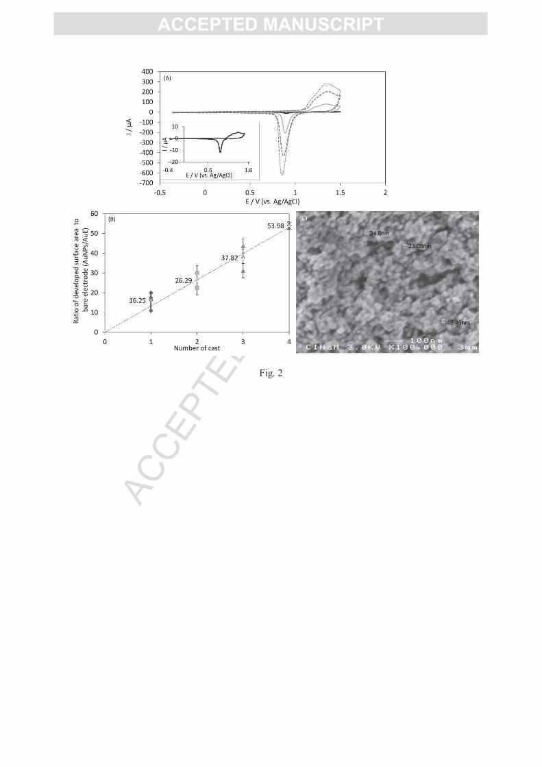
AuNP nanostructure induces a great enhancement of the catalytic current. The higher the
AuNP developed surface, the higher the catalytic current (Figure 4A). Current density for H2

AC
CEPTED
MAN
USC
RIP
T
ACCEPTED MANUSCRIPT
oxidation reaches 1.85 ± 0.15 mA.cm-2
for AuNPs/AuE around 50, which is up to 170 times
higher than at the bare AuE. Although the electroactive surface is greatly increased, and even
for the highest AuNPs/AuE values, no non catalytic signals could be observed under N2 atm.
Horse heart cytochrome c (cyt c) was also adsorbed on AuNP deposits modified by 6-
MHA (Table 1 in SI). The AuNP modified gold electrode was incubated with 10 µL of 50 µM
cytochrome c for 1 h at 4°C. A well defined redox wave developed at 0 V vs. Ag/AgCl
characteristic of the FeIII
/FeII transition of the hemic center. The increase in the peak currents,
either anodic or cathodic, was almost proportional to the increase in the surface area, hence
denoting that it was mostly related to more proteins immobilized on a larger electroactive
surface area. The same linear relation between the amount of cyt c and the number of deposits
was mentioned by Murata et al. [22]. In contrast, the authors reported that O2 reduction by
bilirubin oxidase (BOD) rapidly reached a saturation value, suggesting that the difference in
size between cyt c and BOD could control the immobilization process in the depths of the
AuNP assembly. To have a better understanding of the behavior of hydrogenase, the catalytic
current density was reported to the electroactive surface developed by the AuNPs (Figure 4B)
calculated for each electrode by CV and peak integration as described above. Three domains
can be clearly defined as a function of AuNPs/AuE. For the lowest AuNPs/AuE, between 1
and 10, the increase in the catalytic current is simply related to the increase in the surface
area, as denoted by the constancy of the current densities reported to the electroactive area
developed by the AuNPs. In this first domain, the current density reported to the surface
developed by the AuNPs is in the order of 10 µA.cm-2
. This value is very close to the current
density obtained at the bare AuE. This most probably reflects the first step of AuNP
deposition on the electrode as a rather flat deposit. A second domain can be observed for
higher AuNPs/AuE, i.e. between 10 and up to 25. An enhancement of the current density
much above the enhancement of the surface is observed as highlighted in Figure 4B. In this
domain, the current density reported to the AuNP developed surface area gradually increases
up to 0.08 mA.cm-2
. A synergic effect between the increase in the electroactive surface and
the morphology of the AuNP film may account for that phenomenon. This step involves most
probably the formation of the microporous structure as shown in Figure 2C, which is
favorable to a high amount of connected hydrogenase displaying a high electron transfer rate.
The third domain concerns AuNPs/AuE between 25 and 50, where the current density
reported to the surface developed by the AuNPs decreases then tends to stabilize as the value
AuNPs/AuE increases. In this step, as the thickness increases, it can be hypothesized that the
structure of the deposit becomes less porous, thus less adapted to a high efficiency of the
enzyme.
When the hydrogenase is adsorbed on higher size AuNPs obtained by using a lower
citrate concentration, the catalytic current is lower as already noticed for other enzymes such
as cytochrome bo3 oxidase [17] or laccase [25] (Figure SI 2). A lower increase in the surface
area was also measured. AuNPs/AuE was experimentally always inferior to 6. The catalytic
current reported to the surface area developed by the AuNPs is close to 10 µA.cm-2
, falling
into the current density range obtained for the same AuNPs/AuE domain with the smaller size
AuNPs. As shown in Figure SI 2 whatever the nanoparticle size, no shift in the catalytic
potential can be observed. It thus appears that the size of the AuNP has little influence on the
electron transfer rate. In a recent work, Shleev’s group investigated the influence of NP size
on the electron transfer rate for O2 reduction by BOD [47]. The main conclusion was that the
use of NPs with size higher than the enzyme dimension induced no influence on the electron
rate. The same conclusion can be drawn from our experiments. One must suspect however
that the formation of a microporous structure with suitable cavities for hydrogenase
entrapment may help in the enhancement of the electrocatalysis once a certain thickness of

AC
CEPTED
MAN
USC
RIP
T
ACCEPTED MANUSCRIPT
AuNPs is reached. Other methods than electrochemistry are now necessary to confirm this
assumption.
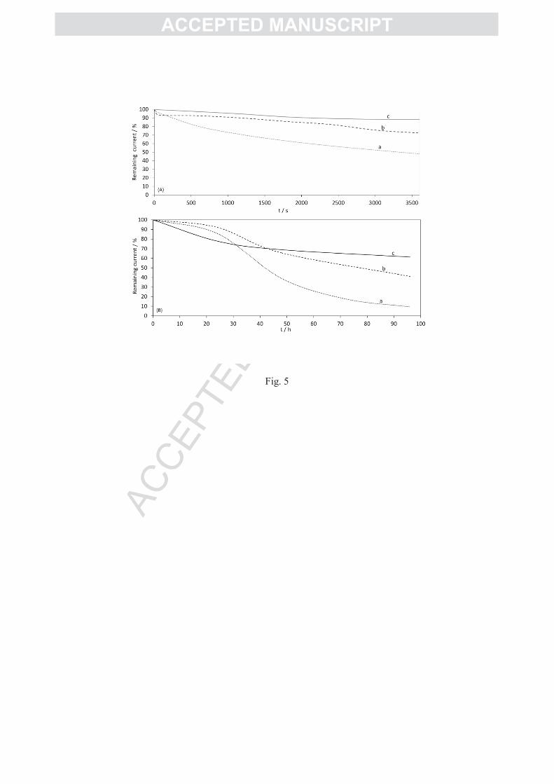
The stability of the AuNP/Aa MbH1 biohybrid was first followed over one hour by
chronoamperometry at a potential of -0.3 V vs. Ag/AgCl (Figure 5). During this short period,
all the weakly attached materials, AuNPs and enzyme-AuNPs are expected to contribute to
the current loss. It is observed that 50% of the initial catalytic current is lost after continuous
working of the enzyme absorbed on the AuNP modified electrode at 60°C and under H2
atmosphere (Figure 5A, curve a). If a covalent attachment is done between the hydrogenase
and the amino group of the 4-ATP layer via EDC/NHS coupling there is an improvement of
the stability of the bioelectrode. The current decrease is only 25% (Figure 5A, curve b).
Because Aa MbH1 is a membrane bound hydrogenase, it is extracted from the cell membrane
using the neutral detergent DDM. We previously demonstrated that the amount of detergent
was crucial for enzyme stability, while a high amount of detergent might affect the
electrochemical signal [39]. In this work, an optimized amount of DDM in the enzyme
solution was found to be close to 3 times the CMC (i.e. 0.54 mM). In these conditions, the
current loss is only 11 % after one hour of continuous catalysis (Figure 5A, curve c). Stability
improvement by covalent attachment of the enzyme then DDM addition, suggests that
catalytic current decrease originates from multiple factors, including enzyme leaching and
enzyme activity loss.
Temperature is a key factor for the catalytic reaction but also for the stability of the
whole system. Chronoamperometry experiments were thus recorded at -0.3V vs. Ag/AgCl
during consecutive increments of temperatures (Figure SI 3). It can be observed that the
enzyme/AuNP biohybrid can work in the full range of temperatures from 30°C to 70°C. A
good stability is obtained at 30°C. The current obtained at 70°C is at least four times higher
than at the lowest temperature, but it is also less stable mostly because of the instability of the
thiol-AuNP architecture at high temperatures. As the temperature decreases back to 60°C, the
stability of the system is recovered describing a current more than twice the current recorded
at 30°C.
We also carried out hydrogenase immobilization on AuNPs attached to the AuE via
dithiol bridges (BPDT) (Figure SI 4). Because in this case only an AuNP monolayer was
expected to be formed, AuNPs/AuE value was much less yielding values around 1.4. The
catalytic process was also very much like the process obtained by immobilization of Aa
MbH1 on an adsorbed layer of AuNPs directly onto the gold electrode. This confirms that the
use of the BPDT as a linker does not preclude electron transfer because of the high
conductance of the linker [18]. AuNPs are expected to be strongly attached to the gold surface
through the BPDT linker in comparison with AuNPs simply adsorbed. However, the stability
of the two bioelectrodes followed by chronoamperometry over 1 h was very similar,
suggesting that the attachment of the AuNPs on the gold electrode is not the limiting factor.
The stability of the AuNP/hydrogenase biohybrid was then followed over several days
by checking the maximum CV current for H2 oxidation each day (Figure 5B). The modified
electrodes and enzyme solution were daily preserved at 4°C in 10mM phosphate buffer pH 6,
previously saturated with N2 to remove O2 traces all along the experiments. After 24h the
AuNP electrodes keep 20% of the initial current, and after 30 h the current tends to stabilize.
During the following days only a slight change is recorded, and the current remains more than
60% of the initial current after 4 days. The AuNP-based bioelectrode appears to be more
stable than the previous biohybrid developed in our lab which was based on hydrogenase
immobilization in carbon nanofiber network [37] (Figure 5B). This carbon material was
proved to be very efficient for direct enzymatic H2 oxidation leading to current densities
higher than 4.5 mA.cm-2
(based on the geometric area). However, the CNF/Aa MbH1
bioelectrode was shown to be poorly stable with time, losing 50% of current after 48 h, and

AC
CEPTED
MAN
USC
RIP
T
ACCEPTED MANUSCRIPT
90% after 4 days. The absence of covalent attachment between the CNFs and the enzyme can
mainly account for this instability. But it is worth noting that the evolution of the catalytic
current on CNFs is identical to the evolution of the current recorded with a PG electrode daily
freshly modified by the enzyme, and very different from the one recorded at the AuNP
modified electrode. This most probably reflects that the AuNP nanostructure is more suitable
for enzyme protection than the CNF film.
3.3. H2/O2 biofuel cell
AuNP based electrodes were used to build a biofuel cell operating with Aa MbH1 at the
anode and Mv BOD at the cathode (Figure 6). The cell configuration was previously described
[6, 8]. The temperature of each half cell, separated by a Nafion membrane, can be
independently regulated. The performances of the biocathode and the bioanode were first
evaluated in the fuel cell configuration (Figure 6A). To balance the cathodic and anodic sides,
AuNPs/AuE values of around 15 and 40 were used for H2 oxidation and O2 reduction
respectively. A high current density at a temperature of 60°C in the anodic compartment was
recorded for H2 oxidation by hydrogenase, in agreement with the one obtained in the
conventional three electrode configuration. Immobilization of Mv BOD on AuNP based
electrodes was previously studied [22, 30]. Direct electron transfer for O2 reduction was
reported in the case of the commercially available Mv BOD on unmodified AuNPs or AuNPs
modified by carboxylate-terminated SAMs. Accordingly, we verify in this work that
modification of the AuNPs by 3-MPA allows efficient immobilization of Mv BOD. Current
densities in the order of 0.4 mA.cm-2
with an onset around + 0.5 V are obtained for O2
reduction at a temperature of 25°C and in condition of O2 bubbling inside the electrolyte of
the cathodic compartment (Figure 6A). We noted however that increasing the temperature
progressively to 40°C in the cathodic compartment resulted in a progressive decrease of the
catalytic activity, as expected for a non thermostable enzyme such as Mv BOD.
No covalent attachment was done for the biocathode. Actually, the addition of
EDC/NHS resulted in a strong decrease of the catalytic current. Some structural
rearrangements were previously suggested in order to explain the decrease in the electron
transfer rate after covalent BOD immobilization on gold electrodes [48]. We performed both
SDS-PAGE gels and ABTS activity in agarose gels in the presence of Mv BOD with
increasing EDC/NHS concentrations (i.e. 6/10, 14/21 and 20/30 mM) (data not shown). These
gels proved that at least under these experimental conditions no denaturation of the protein
occurs. The loss of activity in the present work might imply either structural rearrangement or
release of BOD after EDC/NHS treatment on AuNPs modified by MPA. Pita et al. also
immobilized Mv BOD on AuNPs treated through a mixture of MPA and diazonium salts. No
deactivation of the enzyme upon covalent attachment was reported, but it was noted that the
MPA modification alone resulted in the disappearance of the catalytic signal in serum media
[23]. This was attributed to the lower stability of the MPA modification compared to the one
prepared by diazonium salt reduction. Accurate assessment is however needed for which
coupled spectroscopy/electrochemistry methods would be of great interest.
The open circuit voltage of the biofuel cell was 1.08±0.05 V. Temperature was
maintained at 60 °C and 25 °C in the anodic and cathodic compartment respectively. The
polarization curve for the biofuel cell is shown in Figure 6B. When decreasing the cell
voltage, a sharp increase in the current can be observed at around 0.4 V. The voltage of each
electrode was concomitantly monitored during the cell polarization. Because the cathode is
the limiting electrode in the system, the BOD bioelectrode varied much more quickly than the
hydrogenase bioelectrode. As shown in Figure 6B, the biocathode reaches negative values in
the same domain where a sudden increase of the current in the polarization curve is observed.

AC
CEPTED
MAN
USC
RIP
T
ACCEPTED MANUSCRIPT
At these potentials O2 can be directly reduced at some bare parts of the AuNP electrode,
producing hydrogen peroxide. Consequently, whereas the bioanode current was almost
unchanged after the polarization experiment, the biocathode lost around 50% in current
density (Figure 6A). Attempt to increase the stability of the biocathode by co-immobilization
of catalase with Mv BOD as proposed in [49] did not result in an improved stability.
Nevertheless, at a cell voltage of 0.8 V a power density of 0.25 mW.cm-2
is reached (Figure
6C). This power density compares well with the previous power densities reported with the
same enzymes immobilized at carbon nanotube networks [8]. In the absence of enzymes, a
power density less than 2 µW.cm-2
is obtained (Figure SI 5). Immobilization of enzymes on
AuNPs has been already demonstrated to enhance the power densities of sugars/O2 EBFC
[22]. But this is the first time that a H2/O2 biofuel cell based on hydrogenase immobilization
on AuNPs is reported. Our results prove that H2/O2 biofuel cells can be efficient in many
electrode configurations such as carbon nanomaterials but also metal nanoparticles.
4. Conclusion
In this work, the O2- CO- and temperature-tolerant [NiFe] hydrogenase from Aquifex
aeolicus hyperthermophilic bacterium was immobilized on gold nanoparticle deposits. For the
first time, it is shown that direct H2 enzymatic oxidation is very efficient on such
nanostructured interfaces with no need of any redox mediator and over a large range of
temperatures. It is underlined that the microporosity of the AuNP film allows both an
enhancement of the electroenzymatic activity beyond the surface enhancement and
stabilization with time of the enzyme. Combined with Mv BOD at the cathode, a biofuel cell
was designed able to deliver a power density of 0.25 mW.cm-2
. Future work will focus on
immobilization of thermostable enzymes on AuNPs for O2 reduction at high temperatures.
This work provides the first investigation of enzymatic H2 oxidation on nanoparticles which is
deisrable to develop coupled methods involving electrochemistry and spectroscopy. This is in
progress in the laboratory with the aim to determine the key factors controlling the stability of
a bioelectrode as a function of various experimental conditions.
Acknowledgments
The authors thank P. Infossi, Drs M. Guiral, and M.T. Giudici-Orticoni (BIP,
Marseille, France) for fruitful discussions, Région Provence-Alpes-Côte d’Azur, Région
Aquitaine and ANR for financial support.

AC
CEPTED
MAN
USC
RIP
T
ACCEPTED MANUSCRIPT
References
[1] D. Leech, P. Kavanagh, W. Schuhmann, Enzymatic fuel cells: recent progress,
Electrochim. Acta 84 (2012) 223-234.
[2] S. Cosnier, A. Le Goff, M. Holzinger, Toward glucose biofuel cells implanted in human
body for powering artificial organs: review, Electrochem. Commun. 38 (2014) 19-23.
[3] D. Pakratov, P. Falkman, Z. Blum, S. Shleev, A hybrid electric power device for
simultaneous generation and storage of electric energy, Energ. Environ. Sci. 7 (2014) 989-993
[4] A. de Poulpiquet, D. Ranava, K. Monsalve, MT. Giudici-Orticoni, E. Lojou, Biohydrogen
for a new generation of H2/O2 biofuel cells: a sustainable energy perspective,
ChemElectroChem. 1 (2014) 1724-1750.
[5] L. Xu, F. Armstrong, Optimizing the power of enzyme-based membrane-less hydrogen
fuel cells for hydrogen-rich H2-air mixtures, Energ. Environ. Sci. 6 (2013) 2166-2171.
[6] A. de Poulpiquet, A. Ciaccafava, R. Gadiou, S. Gounel, M.T. Giudici-Orticoni, N. Mano,
E. Lojou, Design of a H2/O2 biofuel cell based on thermostable enzymes, Electrochem. Com.
42 (2014) 72-74.
[7] S. Krishnan, F. Armstrong, Order-of-magnitude enhancement of an enzymatic hydrogen-
air fuel cell based on pyrenyl carbon nanostructures, Chem. Sci. 3 (2012) 1015-1023.
[8] A. Ciaccafava, A. de Poulpiquet, V. Techer, M.T. Giudici-Orticoni, S. Tingry, C.
Innocent, E. Lojou, An innovative powerful and mediatorless H2/O2 biofuel cell based on an
outstanding bioanode, Electrochem. Commun. 23 (2012) 25-28.
[9] M.C. Daniel, D. Astruc, Gold nanoparticles: assembly, supramolecular chemistry,
quantum-size-related properties, and applications toward biology, catalysis, and
nanotechnology, Chem. Rev. 104 (2004) 293-346.
[10] S. Chen, Y. Liu, Electrochemistry at nanometer-sized electrodes, Phys. Chem. Chem.
Phys. 16 (2014) 635-652.
[11] U. Jensen, E. Ferapontova, D. Sutherland, Quantifying protein adsorption and function at
nanostructured materials: enzymatic activity of glucose oxidase at GLAD structured
electrodes, Langmuir 28 (2012) 11106-11114.
[12] R. Villalonga, P. Diez, P. Yanez-Sedeno, J. Pingarron, Wiring horseradish peroxidase on
gold nanoparticles-based nanostructured polymeric network for the construction of
mediatorless hydrogen peroxide biosensor, Electrochim. Acta 56 (2011) 4672-4677.
[13] R. Villalonga, P. Diez, M. Eguilaz, P. Martinez, J. Pingarron, Supramolecular
immobilization of xanthine oxidase on electropolymerized matrix of functionalized hybrid
gold nanoparticles/single-walled carbon nanotubes for the preparation of electrochemical
sensors, Appl. Mat. Interfaces 4 (2012) 4312-4319.

AC
CEPTED
MAN
USC
RIP
T
ACCEPTED MANUSCRIPT
[14] T. Meyer, F. Melin, H. Xie, I. von des Hocht, S. Choi, M. Noor, H. Michel, R. Gennis, T.
Soulimane, P. Hellwig, Evidence for distinct electron transfer processes in terminal oxidases
from different origin by means of protein film voltammetry, J. Am. Chem. Soc. 136 (2014)
10854-10857.
[15] P. Jensen, Q. Chi, F. Grumsen, J. Abad, A. Horsewell, D. Schiffrin, J. Ulstrup, Gold
nanoparticles assisted assembly of a heme protein for enhancement of long-range interfacial
electron transfer, J. Phys. Chem. C 111 (2007) 6124-6132.
[16] T. Meyer, J. Gross, C. Blanck, M. Schmutz, B. Ludwig, P. Hellwig, F. Melin,
Electrochemistry of cytochrome c1, cytochrome c552 and CuA from the respiratory chain of
Thermus thermophilus immobilized on gold nanoparticles, J. Phys. Chem. B 115 (2011)
7165-7170.
[17] F. Melin, T. Meyer, S. Lankiang, S. Choi, R. Gennis, C. Blanck, M. Schmutz, P.
Hellwig, Direct electrochemistry of cytochrome bo3 oxidase at a series of gold nanoparticles-
modified electrodes, Electrochem. Commun. 26 (2013) 105-108.
[18] P. Jensen, Q. Chi, J. Zhang, J. Ulstrup, Long-range interfacial electrochemical electron
transfer of Pseudomonas aeruginosa azurin-gold nanoparticles hybrid systems, J. Phys.
Chem. C 113 (2009) 13993-14000.
[19] J. Holland, C. Lau, S. Brozik, P. Atanassov, S. Banta, Engineering of glucose oxidase for
direct electron transfer via site-specific gold nanoparticles conjugation, J. Am Chem. Soc. 133
(2011) 19262-19265.
[20] U. Jensen, E. Ferapontova, D. Sutherland, Quantifying protein adsorption and function at
nanostructured materials: enzymatic activity of glucose oxidase at GLAD structured
electrodes, Langmuir 28 (2012) 11106-11114.
[21] S. Frasca, O. Rojas, J. Salewski, B. Neumann, K. Stiba, I. Weidinger, B. Tiersch, S.
Leimkühler, J. Koetz, U. Wollenberger, Human sulfite oxidase electrochemistry on gold
nanoparticles modified electrode, Bioelectrochem. 87 (2012) 33-41.
[22] K. Murata, K. Kajiya, N. Nakamura, H. Ohno, Direct electrochemistry of bilirubin
oxidase on three-dimensional gold nanoparticles electrodes and its application in a biofuel
cell, Energ. Environ. Sci. 2 (2009) 1280-1285.
[23] M. Pita, C. Gutierrez-Sanchez, M. Toscano, S. Shleev, A. de Lacey, Oxygen biosensor
based on bilirubin oxidase immobilized on a nanostructured gold electrode, Bioelectrochem.
94 (2013) 69-74.
[24] H. Matsumura, R. Ortiz, R. Ludwig, K. Igarashi, M. Samejima, L. Gorton, Direct
electrochemistry of Phanerochaete chrysosporium cellobiose dehydrogenase covalently
attached on gold nanoparticles modified solid gold electrodes, Langmuir 28 (2012) 10925-
10933.

AC
CEPTED
MAN
USC
RIP
T
ACCEPTED MANUSCRIPT
[25] C. Guttierez-Sanchez, M. Pita, C. Vaz-Dominguez, S. Shleev, A. De Lacey, Gold
nanoparticles as electronic bridges for laccase-based biocathodes, J. Am. Chem. Soc. 134
(2012) 17212-17220.
[26] C. Lanzellotto, G. Favero, M. Antonelli, C. Tortolini, S. Cannistraro, E. Coppari, F.
Mazzei, Nanostructured enzymatic biosensor based on fullerene and gold nanoparticles:
preparation, characterization and analytical applications, Biosensors Bioelec. 55 (2014) 430-
437.
[27] D. Brondani, N. de Souza, B. Souza, A. Neves, I. Vieira, PEI-coated gold nanoparticles
decorated with laccase: a new platform for direct electrochemistry of enzymes and biosensing
applications, Biosensors Bioelec. 42 (2013) 242-247.
[28] V. Krikstolaityte, A. Barrantes, A. Ramanavicius, T. Arnebrant, S. Shleev, T. Ruzgas,
Bioelectrocatalytic reduction of oxygen at gold nanoparticles modified with laccase,
Bioelectrochem. 95 (2014) 1-6.
[29] K. Murata, M. Suzuki, K. Kajiya, N. Nakamura, H. Ohno, High performance bioanode
based on direct electron transfer of fructose dehydrogenase at gold nanoparticles-modified
electrodes, Electrochem. Commun. 11 (2009) 668-671.
[30] X. Wang, M. Falk, R. Ortiz, H. Matsumura, J. Bobacka, R. Ludwig, M. Bergelin, L.
Gorton, S. Shleev, Mediatorless sugar/oxygen enzymatic fuel cells based on gold
nanoparticles-modified electrodes, Biosensors Bioelec. 31 (2012) 219-225.
[31] P. Lamberg, S. Shleev, R. Ludwig, T. Arnebrant, T. Ruzgas, Performance of enzymatic
fuel cell in cell culture, Biosensors Bioelec. 55 (2014) 168-173.
[32] X. Luo, P. Tron-Infossi, M. Brugna, M.T. Giudici-Orticoni, E. Lojou, Physicochemical
key parameters for direct catalytic oxidation of hydrogen by hyperthermophilic [NiFe]
hydrogenase immobilized at gold and carbon nanotubes-modified electrodes, J. Biol. Inorg.
Chem. 14 (2009) 1275-1288.
[33] M. Pandelia, V. Fourmond, P. Tron-Infossi, E. Lojou, P. Bertrand, C. Leger, MT.
Giudici-Orticoni, W. Lubitz, Membrane-bound hydrogenase I from the hyperthermophilic
bacterium Aquifex aeolicus: enzyme activation, redox intermediates and oxygen tolerance, J.
Am. Chem. Soc. 132 (2010) 6991-7004.
[34] E. Lojou, Hydrogenases as catalysts for fuel cells: strategies for efficient immobilization
at electrode interfaces, Electrochim. Acta 56 (2011) 10385.
[35] A. De Poulpiquet, A. Ciaccafava, K. Szot, B. Pillain, P. Infossi, M. Guiral, M. Opallo,
MT. Giudici-Orticoni, E. Lojou, Exploring properties of a hyperthermophilic membrane-
bound hydrogenase at carbon nanotube modified electrodes for a powerful H2/O2 biofuel cell,
Electroanalysis 25(2013) 685-695.
[36] K. Szot, A. De Poulpiquet, A. Ciaccafava, H. Marques, M. Jonsson-Niedziolka, J.
Niedziolka-Jonsson, F. Marken, E. Lojou, M. Opallo, Carbon nanoparticulate films as

AC
CEPTED
MAN
USC
RIP
T
ACCEPTED MANUSCRIPT
effective scaffolds for mediatorless bioelectrocatalytic hydrogen oxidation, Electrochim. Acta
111 (2013) 434-440.
[37] A de Poulpiquet, H. Marques-Knopf, V. Wernert, R. Gadiou, MT. Giudici-Orticoni, E.
Lojou, Carbon Nanofiber Mesoporous Films: Efficient Platforms for Bio-Hydrogen Oxidation
in Biofuel Cells, Phys. Chem. Chem. Phys. 16 (2014) 1366-1378.
[38] A. Ciaccafava, P. Infossi, M. Ilbert, M. Guiral, S. Lecomte, MT. Giudici-Orticoni, E.
Lojou, Electrochemistry, AFM and PM-IRRAS spectroscopy of immobilized hydrogenase:
role of a trans-membrane helix on enzyme orientation for efficient H2 oxidation, Angew.
Chem. Int. Ed. 51(2012)953-956.
[39] A. Ciaccafava, A. De Poulpiquet, P. Infossi, S. Robert, R. Gadiou, M.T. Giudici-
Orticoni, S. Lecomte, E. Lojou, A friendly detergent for H2 oxidation by Aquifex aeolicus
membrane-bound hydrogenase immobilized on graphite and SAM-modified gold electrodes,
Electrochim. Acta 82 (2012) 115-125.
[40] F. Oteri, A. Ciaccafava, A. de Poulpiquet, E. Lojou, M. Baaden, S. Sacquin-Mora,
Fluctuations in the dipole moment of membrane-bound hydrogenase from Aquifex aeolicus
account for its adaptability to charged electrodes, Phys. Chem. Chem. Phys. 16 (2014) 11318-
11322.
[41] S. Trasatti, O. Petrii, Real surface area measurements in electrochemistry, Pure Appl.
Chem. 63 (1991) 711-734.
[42] G. Frens, Controlled Nucleation for the Regulation of the Particle Size in Monodisperse
Gold Suspensions, Nature Phys. Sci. 241 (1973) 20-22.
[43] W. Haiss, N. Thanh, J. Aveyard, D. Fernig, Determination of size and concentration of
gold nanoparticles from UV-Vis spectra, Anal. Chem. 79 (2007) 4215-4221.
[44] S. Link, M. A. El-Sayed, Size and temperature dependence of the Plasmon absorption of
colloidal gold nanoparticles, J. Phys. Chem. B 103 (1999) 4212-4217.
[45] K. Murata, K. Kajiya, Y. Suga, T. Watanabe, N. Nakamura, H. Ohno, A simple
fabrication method for three-dimensional gold nanoparticles electrodes and their application
to the study of the direct electrochemistry of cytochrome c, Electroanalysis 22 (2010) 185-
190.
[46] O. Rüdiger, C. Gutiérrez-Sánchez, D. Olea, I. Pereira, M. Vélez, V.M. Fernández, A.L.
De Lacey, Enzymatic Anodes for Hydrogen Fuel Cells based on Covalent Attachment of Ni-
Fe Hydrogenases and Direct Electron Transfer to SAM-Modified Gold Electrodes,
Electroanalysis, 22 (2010) 776-783.
[47] D. Pankratov, R. Sundberg, D. B. Suyatin, J. Sotres, A. Barrantes, T. Ruzgas, I.
Maximov, L. Montelius, S. Shleev, The influence of nanoparticles on enzymatic
bioelectrocatalysis, RSC Adv. 4 (2014) 38164-38168.

AC
CEPTED
MAN
USC
RIP
T
ACCEPTED MANUSCRIPT
[48] K. Singh, T. McArdle, P. Sullivan, C. Blanford, Sources of activity loss in the fuel cell
enzyme bilirubin oxidase, Energ. Environ. Sci. 6 (2013) 2460-2464.
[49] S. Shleev, G. Shumakovich, O. Morozova, A. Yaropolov, Stable 'Floating' Air Diffusion
Biocathode Based on Direct Electron Transfer Reactions Between Carbon Particles and High
Redox Potential Laccase, Fuel Cells, 10 (2010) 726-733.

AC
CEPTED
MAN
USC
RIP
T
ACCEPTED MANUSCRIPT
Legends
Figure 1: Size distribution and morphology of the gold nanoparticles. (A) AuNP hydrodynamic size
distribution by DLS; (B) UV-visible spectra of AuNPs obtained with 38.8 mM (bold line), and 19.4
mM (dot-dashed line) sodium citrate; TEM images of AuNPs synthesized with 38.8 mM (C) and 19.4
mM (D) sodium citrate.
Figure 2: (A) CVs of gold electrodes modified with consecutive AuNP drop castings: 1 (grey line), 3
(dashed line), 4 (dotted line) deposits; Inset: CV of the bare polycrystalline gold electrode; (B)
AuNPs/AuE values as a function of AuNP casting number. 0.05 M H2SO4; scan rate 0.1V/s under N2
atmosphere and room temperature. (C) SEM image of the top view of the AuNP modified gold
surface.
Figure 3: CVs for H2 oxidation by Aa MbH1 immobilized on a gold electrode modified with one
AuNP deposit: (a) H2 in over pressure above the electrolyte; (b) H2 in continuous flow in the
electrolyte solution; (c) H2 is replaced by N2. 10 mM phosphate buffer, pH 6, 60°C, 5 mV/s.
Figure 4: (A) H2 catalytic currents for increasing AuNPs/AuE values (a) 28, (b) 38 and (c) 50. Inset:
H2 oxidation by Aa MbH1 absorbed on a bare gold electrode (B) Catalytic current densities for H2
oxidation reported to the electroactive surface area of the AuNP deposit as a function of AuNPs/AuE.
10 mM phosphate buffer, pH 6, 60°C under H2 flow, 5mV/s.
Figure 5: (A) H2 oxidation current loss with the Aa MbH1 immobilized on 4-ATP modified AuNPs:
(a) without EDC/NHS (dotted line), (b) with EDC/NHS (dashed line); (c) with EDC/NHS and 3CMC
DDM addition (solid line). E = -0.3 V vs. Ag/AgCl, 10 mM phosphate buffer, pH 6, 60°C under H2
flow; (B) long term H2 oxidation current loss with the Aa MbH1 immobilized on (a) CNF modified PG
electrode (dotted line), (b) freshly daily adsorbed on bare PG electrode (dashed line), (c) covalently
bounded to 4-ATP modified AuNP gold electrode (solid line); 60°C under H2 flow, 5 mV/s.
Figure 6: (A) Direct H2 oxidation and O2 reduction at AuNP nanostructured electrodes in the fuel cell
configuration, before (solid line) and after (dashed line) cell measurements; (B) Polarization curve of
AuNP-based H2/O2 biofuel cell (black line) and cathode potential evolution during the polarization
experiment (grey line); (C) Operational performance of the AuNP-based H2/O2 biofuel cell: power
density (grey line) and cell voltage (black line) as a function of the current density. 10mM phosphate
buffer pH 7, under continuous H2 (bioanode) or O2 (biocathode) flow, 3 mV/s.

AC
CEPTED
MAN
USC
RIP
T
ACCEPTED MANUSCRIPT
Supplemental Informations
Surface
Increment
Ipa / A Ipc / A Γ /
mol.cm-2
Em / V rE Γ
Increment
1 3.5 10-9
5.7 10-9
7.6 10-12
0.004 0.027 1
20 7.9 10-8
7.8 10-8
1.3 10-10
0.009 0.002 17.7
36 2.3 10-7
2.4 10-7
3.9 10-10
0.001 0.002 51.5
Table 1: Electrochemistry of Horse heart cytochrome c at 6-MHA-AuNP modified gold electrode. Ipa
and Ipc: anodic and cathodic peak currents respectively, Γ: protein surface coverage, Em : average
redox potential, DE : potential difference between the anodic and cathodic peak potentials. 10 mM
phosphate buffer pH 6, 0.1V/s.
Figure SI 1: CVs for H2 oxidation on AuNP deposit in the absence of Aa MbH1; 10 mM phosphate
buffer pH 6, 60°C under continuous H2 flow, 5 mV/s.
Figure SI 2: Comparative CVs for H2 oxidation by Aa MbH1 covalently immobilized on (a) 25 nm
AuNPs and AuNPs/AuE of 14 (solid line) and (b) 35 nm AuNPs and AuNPs/AuE of 3 (dashed line).
10 mM phosphate buffer pH 6, 60°C under continuous H2 flow, 5 mV/s.
Figure SI 3: Chronoamperometry measurement at -0.3V vs. Ag/AgCl for H2 oxidation current at
consecutive temperature increasing conditions. 10 mM phosphate buffer pH 6, 60°C under continuous
H2 flow.
Figure SI 4: CVs for H2 oxidation by Aa MbH1 covalently immobilized on a gold electrode modified
with AuNPs on a BPDT layer (dashed line), or AuNPs directly adsorbed on the gold electrode (solid
line). 10 mM phosphate buffer pH 6, 60°C under continuous H2 flow, 5 mV/s.
Figure SI 5: Polarization and power curves on AuNPs in the absence of enzymes. 10mM phosphate
buffer pH 7, under continuous H2 (bioanode) or O2 (biocathode) flow, 3 mV/s.

AC
CEPTED
MAN
USC
RIP
T
ACCEPTED MANUSCRIPT
Fig. 1
AC
CEPTED
MAN
USC
RIP
T
ACCEPTED MANUSCRIPT
Fig. 2

AC
CEPTED
MAN
USC
RIP
T
ACCEPTED MANUSCRIPT
Fig. 3
AC
CEPTED
MAN
USC
RIP
T
ACCEPTED MANUSCRIPT
Fig. 4
AC
CEPTED
MAN
USC
RIP
T
ACCEPTED MANUSCRIPT
Fig. 5

AC
CEPTED
MAN
USC
RIP
T
ACCEPTED MANUSCRIPT
Fig. 6

AC
CEPTED
MAN
USC
RIP
T
ACCEPTED MANUSCRIPT
Highlights
- 20.0±5.3 and 37.2±4.3 nm gold nanoparticles were deposited on a gold electrode
- O2- and CO-tolerant [NiFe] hydrogenase was immobilized on the AuNP deposits
- Direct H2 oxidation was obtained with current densities up to 1.85±0.15 mA.cm-2
- A biofuel cell was designed delivering 0.25 mW.cm-2