Hunagxian Ju Molecular imprinting: a dynamic technique for...
19
REVIEW Vivek Babu Kandimalla Æ Hunagxian Ju Molecular imprinting: a dynamic technique for diverse applications in analytical chemistry Received: 29 February 2004 / Revised: 22 June 2004 / Accepted: 29 July 2004 / Published online: 8 October 2004 ȑ Springer-Verlag 2004 Abstract Continuous advances in analyzing complex matrices, improving reliability and simplicity, and per- forming multiple simultaneous assays with extreme sensitivity are increasing. Several techniques have been developed for the quantitative assays of analytes at low concentrations (e.g., high-pressure liquid chromatogra- phy, gas chromatography, immunoassay and the poly- merase chain reaction technique). To achieve highly specific and sensitive analysis, high affinity, stable, and specific recognition agents are needed. Although bio- logical recognition agents are very specific and sensitive they are labile and/or have a low density of binding sites. During the past decade molecular imprinting has emerged as an attractive and highly accepted tool for the development of artificial recognition agents. Molecular imprinting is achieved by the interaction, either nonco- valent or covalent, between complementary groups in a template molecule and functional monomer units through polymerization or polycondensation. These molecularly imprinted polymers have been widely em- ployed for diverse applications (e.g., in chromatographic separation, drug screening, chemosensors, catalysis, immunoassays etc.) owing to their specificity towards the target molecules and high stability against physico- chemical perturbations. In this review the advantages, applications, and recent developments in molecular imprinting technology are highlighted. Keywords Molecular imprinting Æ Analytical chemistry Æ Catalysis Æ Solid-phase extraction Æ Sensors Introduction In view of the challenges in environmental, medicine, food process industries, molecular biology, security, and defense areas there is a great need for analytical methods with high sensitivity and good veracity. Biological rec- ognition agents such as antibodies, enzymes, and other receptor molecules have been widely employed in ana- lytical and diagnostic practices [1, 2]. Although these agents are highly specific and sensitive, they are labile, expensive, and have a low density of binding sites. Hence there is a significant demand for robust and stable receptor molecules that can mimic biorecognition ele- ments such as antibodies and enzymes. The developing technique of molecular imprinting provides a promising and advantageous alternative to overcome the problems associated with biomolecules. Molecular imprinting is a dynamic methodology used to design new polymeric receptors against to wide range of target molecules. It was first reported in 1949 by adsorbing different dyes in silica [3, 4]. Originally it was introduced as a means to create binding sites in synthetic polymers. The technique has now matured and has become established in several disciplines owing to its ability to form stable, robust materials with molecular selectivity for a wide variety of compounds. It has also become an attractive method for the preparation of sensor components and catalysts [5–7]. Generally molecularly imprinted polymers (MIPs) are synthesized from functionalized monomers through processes such as radical polymerization or polycon- densation. Compared to biomolecules, molecular im- prints (MIs) are highly stable and convenient for the development of new analytical methods, which can even perform under relatively harsh conditions. Since the 1990s, great progress in analytical methods, sensors, and catalytic and membrane materials has been made, in which MIPs provide opportunities for advancements. MIs have been successfully developed for the binding of drugs [8], herbicides [9, 10], amino acids and their derivatives [11], peptides [12], proteins [13–19], nucleo- V. B. Kandimalla Æ H. Ju (&) Department of Chemistry, Key Laboratory of Analytical Chemistry for Life Science (Chinese Ministry of Education), Nanjing University, Nanjing 210093, China E-mail: [email protected] Tel.: +86-25-83593593 Fax: +86-25-83593593 Anal Bioanal Chem (2004) 380: 587–605 DOI 10.1007/s00216-004-2793-9
Transcript of Hunagxian Ju Molecular imprinting: a dynamic technique for...

REVIEW
Vivek Babu Kandimalla Æ Hunagxian Ju
Molecular imprinting: a dynamic technique for diverse applicationsin analytical chemistry
Received: 29 February 2004 / Revised: 22 June 2004 / Accepted: 29 July 2004 / Published online: 8 October 2004
� Springer-Verlag 2004
Abstract Continuous advances in analyzing complexmatrices, improving reliability and simplicity, and per-forming multiple simultaneous assays with extremesensitivity are increasing. Several techniques have beendeveloped for the quantitative assays of analytes at lowconcentrations (e.g., high-pressure liquid chromatogra-phy, gas chromatography, immunoassay and the poly-merase chain reaction technique). To achieve highlyspecific and sensitive analysis, high affinity, stable, andspecific recognition agents are needed. Although bio-logical recognition agents are very specific and sensitivethey are labile and/or have a low density of binding sites.During the past decade molecular imprinting hasemerged as an attractive and highly accepted tool for thedevelopment of artificial recognition agents. Molecularimprinting is achieved by the interaction, either nonco-valent or covalent, between complementary groups in atemplate molecule and functional monomer unitsthrough polymerization or polycondensation. Thesemolecularly imprinted polymers have been widely em-ployed for diverse applications (e.g., in chromatographicseparation, drug screening, chemosensors, catalysis,immunoassays etc.) owing to their specificity towardsthe target molecules and high stability against physico-chemical perturbations. In this review the advantages,applications, and recent developments in molecularimprinting technology are highlighted.
Keywords Molecular imprinting Æ Analyticalchemistry Æ Catalysis Æ Solid-phase extraction ÆSensors
Introduction
In view of the challenges in environmental, medicine,food process industries, molecular biology, security, anddefense areas there is a great need for analytical methodswith high sensitivity and good veracity. Biological rec-ognition agents such as antibodies, enzymes, and otherreceptor molecules have been widely employed in ana-lytical and diagnostic practices [1, 2]. Although theseagents are highly specific and sensitive, they are labile,expensive, and have a low density of binding sites. Hencethere is a significant demand for robust and stablereceptor molecules that can mimic biorecognition ele-ments such as antibodies and enzymes. The developingtechnique of molecular imprinting provides a promisingand advantageous alternative to overcome the problemsassociated with biomolecules. Molecular imprinting is adynamic methodology used to design new polymericreceptors against to wide range of target molecules. Itwas first reported in 1949 by adsorbing different dyes insilica [3, 4]. Originally it was introduced as a means tocreate binding sites in synthetic polymers. The techniquehas now matured and has become established in severaldisciplines owing to its ability to form stable, robustmaterials with molecular selectivity for a wide variety ofcompounds. It has also become an attractive method forthe preparation of sensor components and catalysts[5–7]. Generally molecularly imprinted polymers (MIPs)are synthesized from functionalized monomers throughprocesses such as radical polymerization or polycon-densation. Compared to biomolecules, molecular im-prints (MIs) are highly stable and convenient for thedevelopment of new analytical methods, which can evenperform under relatively harsh conditions. Since the1990s, great progress in analytical methods, sensors, andcatalytic and membrane materials has been made, inwhich MIPs provide opportunities for advancements.MIs have been successfully developed for the binding ofdrugs [8], herbicides [9, 10], amino acids and theirderivatives [11], peptides [12], proteins [13–19], nucleo-
V. B. Kandimalla Æ H. Ju (&)Department of Chemistry,Key Laboratory of Analytical Chemistry for Life Science(Chinese Ministry of Education),Nanjing University, Nanjing 210093, ChinaE-mail: [email protected].: +86-25-83593593Fax: +86-25-83593593
Anal Bioanal Chem (2004) 380: 587–605DOI 10.1007/s00216-004-2793-9

tides [20], and nucleotide bases [21]. MIPs have beenused as tailor-made separation materials, antibody andreceptor mimics in assay systems, biomimetic recogni-tion elements in biological sensors, and artificial enzymesystems for catalytic applications. In recent years severalreviews on MIPs have appeared with specific appliedareas (e.g., catalysis, chromatography, and biomimeticsensors) [19, 22–38]. Here we present an overview ofmolecular imprinting. It is extremely difficult to includeall aspects of molecular imprinting and cover every re-port; however, we have tried to incorporate all impor-tant aspects. This article highlights the advantages,applications, and recent developments in MIP technol-ogy.
Advantages of molecular imprints over biomolecules
Biological recognition agents such as antibodies [39–43],enzymes [44–46], plant or animal tissues [47], and cells[48, 49] have been widely used as the affinity sensingagents in analytical biochemistry, biosensors, diagnostickits, and other applications. These molecules are highlyspecific to the target molecules, but are highly fragile andunstable. The production of homogeneous biomoleculesis difficult, and sometimes requires animals (i.e., inantibody production). Maintenance of animals andproduction of antibodies are laborious and complicated,and technical skills are necessary [50]. Purification ofantibodies and enzymes is tedious and expensive, andthey often require special handling methods. Regenera-tion of biomolecules is also difficult and limited. Some-times they lose their activity within few reuse cycles,which leads to inconsistency of accuracy and increasescost per analysis. A wider acceptance of biosensortechnology is particularly impaired by the limited sta-bility of the biological component immobilized onto atransducer surface. MIs are synthetic receptor sites andhave high physical and chemical resistance towardsvarious external degrading factors. Hence MIs arehighly appealing alternatives. The functional groups ofMIs can easily be modified (present inside the cavity) toincrease the affinity or catalytic activity of receptor sites.They can be easily regenerated without loss of affinity,and hence are highly useful for continuous use and flowinjection analysis. MIs are stable towards a wide rangeof solvents, metal ions, and acid treatments [51, 52]. MIswith homogeneous quality and affinity can be synthe-sized in short span of time without any immunization orinvolvement of animals. MIPs are highly thermostable,can be used over a range of temperatures [53, 54], andare stored at ambient temperature and in dry statewithout loss of performance, whereas biomolecules areusually used under controlled conditions and theirstorage is also difficult. Owing to their advantageousproperties over natural biological recognition agents,MIPs therefore offer great potential for various appli-cations in diagnostics, separation, purification, andquantification processes etc.
However, MIPs possess many disadvantages: forexample, it is hard to completely remove the templatefrom MIPs; the imprinted polymer is insoluble; and thepolymer contains many imprinted cavities of which onlysome are really good and match the template molecule[55]. Although many developed polymers performeffectively in separation and sensing, there has been noreal rationality in the design and synthesis of MIPs.There is no ideal or effective procedure for the design ofMIPs against microbial cells and macromolecules (bio-polymers) [26]. To overcome these limitations newfunctional monomers and polymerization methods arebeing investigated. These improvements would be highlybeneficial for nearly all potential applications. In thisdirection, new MIP formats such as hierarchicalimprinting and grafting techniques have been developed[56]. More recently, combinatorial and computationalapproaches have also appeared for the development ofhighly specific MIPs. Some of the developments inmolecular imprinting techniques are discussed in detailin the section.
Imprinting
In the past few years, molecular imprinting technologyhas developed into a viable approach for mimickingnatural recognition entities, such as antibodies [57] andbiological receptors such as enzymes [58, 59]. MIPs havebeen utilized as a molecular recognition membrane orlayer on chemical-sensing systems in combination withtransducers such as quartz crystal microbalances(QCMs) [60], surface plasmon resonance devices [61],field-effect devices [62], conductometry [63], or impedo-metric determination [64]. Imprinting of molecules oc-curs by the polymerization of functional and cross-linking monomers in the presence of a template/targetligand. In this process, the template is initially allowed toestablish bond formation with polymerizable function-ality, and the resulting complexes are subsequently co-polymerized with cross-linkers into a rigid polymer(Fig. 1). Finally the polymer is ground into micrometer-sized particles, and the template molecules are removedfrom the imprint by using selective solvent systems. Theresultant imprints possess a steric (size and shape) andchemical (spatial arrangements or complementaryfunctionality) memory for the template. These enableMIPs to rebind the same target molecule from a mixture[14] provided that the binding sites of the molecularreceptors and the guest molecules complement eachother in size, shape, and chemical functionality. Delaneyet al. [64] reported a MIP for desmetryn and charac-terized this MIP by impedance spectroscopy. The elec-trodes coated with the MIP displayed fairly specificbinding of desmetryn, which was detected by the de-crease in the electrode capacitance. Only small capaci-tative effects were observed on addition of terbumetonor atrazine. The same kind of specific recognition abili-ties were also observed in the case of parathion and
588

penicillin MIPs [64–68]. Imprinting can be achieved byeither noncovalent or covalent interactions. The mostwidely used strategy, pioneered by Mosbach [69, 70], isbased on noncovalent interactions between specificfunctional groups on the polymerizable monomers andthe template in order to position the monomers in aspecific spatial orientation prior to polymerization. Afterpolymerization and removal of the template, the func-tional groups of the polymeric matrix can then bind thetarget through the same noncovalent interactions. High-affinity binding sites can be generated using the nonco-valent imprinting strategy; however, the limitation isthat the template and target must form a sufficientnumber of noncovalent intermolecular interactions.Wulff [71, 72] introduced a covalent molecular imprint-ing method, which utilized reversible covalent bondingbetween a polymerizable monomer and a templatemolecule. After polymerization, these bonds werecleaved to liberate the template and subsequently re-formed in order to selectively bond the target. Covalentimprinting strategy is very stable and selective. However,the number of functional groups to react with templatein the imprint is limited. At high concentrations veryrigid imprint formation occurs. For practical repetitiveuse the cleavage and rebinding may be limited andproblematic due to the limited interactions.
To overcome problems associated with covalent andnoncovalent approaches, Whitcombe et al. [73] reportedan alternative approach, which had advantageouscharacteristics of both covalent and noncovalent
imprinting strategies. In this approach, they employed(4-vinyl)phenylcarbonate ester as a functional monomer,which functioned as the covalently bond templatemonomer but was easily and efficiently cleaved hydro-lytically with the loss of CO2. Different functionalmonomers, such as methacrylic acid (MAA) [74, 75]and Cu[1–4-vinylbenzyl]-1,4,7-triazacyclononane]SO4
(STACNCu) [76], have been employed in order to gethighly specific affinity and stable MIPs. A combinationof allylamine and tris(4-vinylphenyl)boroxine (TVPhB)was used in the development of a biomimetic receptorsystem for the determination of sialic acid based onmolecular imprinting. Allylamine can interact with sialicacid through an ionic or hydrogen bond and the for-mation of a fluorescent compound [77]. Piletsky et al.[36] used MAA and diethyl aminoethylmethacrylate(DEAEM) as a functional monomers for the design ofnoncovalently interacted MIPs against atrazine. TheDEAEM MIP showed good conductometric measure-ments and low detection limits [78]. Parathion molecu-larly imprinted sol–gel thin films were developed usingaminopropyltriethoxysilane (APTES) and phenyltri-methoxysilane (PTMOS) [79]. Some of the mono-mers were specifically designed to allow fluorescenceself-signaling in the presence of analytes, for example, amixture of trans-4-[p-(N,N-dimethylamino)styryl]-N-vi-nylbenzylpyridinium chloride (DMASVBP, a signalingmonomer) and 2-hydroxyethyl methacrylate (HEMA)[80]. Acryloyl–cyclodextrins were synthesized as func-tional vinyl monomers, and various antibiotics andoligopeptides were molecularly imprinted in water [81].
Although several monomers have been used, the mostwidely used functional monomer is MAA, because it canform hydrogen bonds with a number of chemicalstructures, and the binding and removal of guest mole-cules can be performed under mild conditions. Thefunctional monomer usually includes two functionalgroups. At one end, it interacts with the templatethrough noncovalent interactions (e.g., hydrogen bond-ing, van der Waals forces, or hydrophobic interactions)or reversible covalent interactions. At the other end ofthe monomer (i.e., the end that is not interacting withthe template) there is a group that is able to bindcovalently with the cross-linker. The cross-linker poly-merizes the monomers around the template with cova-lent binding and holds them in place after the template isremoved. If the entire binding sites are covered withpolymer, then it may be difficult or impossible to removethe template from the imprint. This problem could beminimized by selecting the appropriate cross-linker andconcentration [82, 83]. An excess of cross-linking agentis generally used to increase the rigidity of the imprintedmatrix, but this sometimes has an adverse effect on theinteraction between the template and the matrix,resulting in low recognition capacity [84]. The widelyemployed cross-linkers are ethyleneglycol dimethacry-late (EDMA) and trimethylolpropane trimethacrylate.
In addition to high-affinity binding sites, the numberof binding sites is also very important to achieve the best
Fig. 1 Schematic representation of the preparation of MIs
589

MIs. The successful in vitro preparation of molecularlyimprinted monoliths is primarily dependent upon thechoice and molar ratios of monomer, cross-linker,porogen, and template molecule, and the polymerizationtemperature. The optimum levels of these ingredients arevery important for obtaining finely cross-linked, highaffinity, and stable MIPs. Moreover, the selectivity ofthe imprints depends both on the orientation of thefunctional groups inside the cavities and the shape ofthe cavities [85–87]. The dominant factor, however, isthe former [86]. The relationships between the imprintsand template molecules have been reported by studying43 compounds in their respective MIs [88]. The goodMIPs exhibit high levels of binding affinity and selec-tivity that are typical of antibodies. Katz and Davis [89]reported organic-functionalized, amorphous, micropo-rous silicas for molecular imprinting. In their study, thetriethoxysilane was incorporated into the silica frame-work during sol–gel synthesis, and subsequent removalof the aromatic core created a cavity with spatiallyorganized aminopropyl groups covalently anchored tothe pore walls. Zimmerman et al. [90] reported amolecular imprinting inside dendrimers, which aremacromolecules of highly regular structure consisting ofa polyfunctional central core covalently linked to layersof repeating units. The template molecules and core usedwere tetrakis-meso(3,5-dihydroxyphenyl)-porphyrin inwhich dendrons were covalently attached through esterlinks. The outer shell of the dendrimer was polymerizedintramolecularly, and the template was then removed.The binding site contained eight precisely positionedcarboxyl groups. This imprinted dendrimer showedselectivity to rebind structural isomers of the templatesuch as tetrakis-meso(2,6-dihydroxyphenyl)-porphyrin.The main advantage with dendrimer imprints is that thetemplate can be easily removed completely, which is notalways possible with traditional, highly cross-linkedMIPs.
To obtain the optimized MIPs for a given targetanalyte, combinatorial approaches toMIP synthesis havebeen developed [25, 91, 92], in which the ingredients of theimprinting recipe, in particular the kind and molar ratioof the functional monomers, are varied using automatedprocedures. Cederfur et al. [92] developed a MI libraryagainst the b-lactam antibiotic penicillin G. This librarywas constructed by combining various functionalizedmonomers and cross-linkers and varying the stoichiom-etry and the concentration of the components in the pre-polymerization mixtures. Among these polymers, onesynthesized from MAA and trimethylolpropane tri-methacrylate as the functionalized monomer and cross-linker, respectively, showed the highest selectivity forpenicillin G. The best MIP against penicillin G wasscreened through radioligand competitive binding assay.The cross-reactivities of penicillin G MIP with otherb-lactam antibiotics were also studied: cross-reactivitywas shown to be 15, 16, and 19% with penicillin-V,ampicillin, and amoxicillin, respectively. Nafcillin,cephapirin, chloramphenicol, tetracycline, dapsone, and
erythromycin showed much lower cross-reactivity (0.01–0.1%). The selection of MIPs from the MI library ishighly convenient and effective for the isolation of high-affinity MIPs against target molecules. RecentlySubrahmanyam et al. [93] developed a method thatincluded computational screening (Bite-and-Switch) of avirtual library of functional monomers against a targetmolecule followed by selection of those able to form thestrongest complex with the template. The computation-ally designed polymer showed superior selectivity incomparison to the polymer prepared by using a tradi-tional approach. These polymers exhibited an affinitycomparable with antibodies. This approach can be usedfor the design of assays and sensors exhibiting greaterselectively for amino-containing substances.
Advances in molecular imprinting technology
There are only a few examples of non-cross-linked MIPsin the literature. Recently, Kobayashi et al. [94, 95] re-ported a phase-inversion molecular imprinting method,in which linear polymer was used as a matrix, and thetemplate molecule information could be encoded by thephase-inversion process. Phase-inversion molecularimprinting of theophylline was performed by usingacrylonitrile (AN) copolymers with acrylic acid (AA)(P(AN-co-AA)) and MAA (P(AN-co-MAA). These co-polymers were synthesized in the presence of differentconcentrations of the theophylline template. The poly-mer solutions were cast and phase-inversed in water toprepare theophylline-imprinted copolymers. To confirmthe polymer specificity towards the template, differentstructurally related compounds such as 2-hydroxyethy-L-theophylline, theobromine, lumazine, purine, anduracil were checked. MAA imprinted polymer showedhigh selectivity for the theophylline. Recently Lim et al.[96] reported another non-cross-linked MIP for estrone.In this study, an aromatic polyimide was used as theimprinted matrix. Polyimide imprint exhibited goodthermal stability and mechanical strength. The urethanebond formed between the template and polyimide ma-trix was highly stable at room temperatures and could becleaved at elevated temperatures. This type of thermallyreversible interaction between template and MIs ishighly useful for the development of highly sensitivesensors and effective separation processes.
Shi et al. [97] reported a novel imprinting method forprotein molecules. This method employed radio-fre-quency glow-discharge plasma deposition to formpolymeric thin films around proteins coated withdisaccharide molecules. The disaccharides were cova-lently attached to the polymer film, creating polysac-charide-like cavities that exhibited highly selectiverecognition for a variety of template proteins, includingalbumin, immunoglobulins G, lysozyme, ribonuclease,and streptavidin (Fig. 2). In this process, initially desiredprotein molecules were adsorbed on a mica surface anda sugar layer of 10–50 A was spin-casted over the
590

adsorbed protein molecules. Finally, plasma depositionof C3F6 was used to form a 10- to 30-nm-thick fluoro-polymer thin film. The resulting plasma was fixed to aglass cover slip with epoxy resin and oven-cured. Themica was pealed off and the obtained sample was soakedin NaOH/NaClO solution to remove the template mol-ecules (protein). The amount of protein adsorbed washigher on the protein imprints than on a mica-imprintedcontrol surface (without template protein). Throughcompetitive adsorption, AFM, and time-of-flight sec-ondary ion mass spectrometry, the binding efficiencyand selectivity were confirmed. By using this method itcould be possible for the development of highly selectiveMIPs for the proteins and high molecular weight com-pounds [97].
In general molecular imprinting methods, the tem-plate removal results in MIs containing nanometer-sizedbinding sites in addition to larger sized pores. In order toaccess the host-binding site the guest molecules mustpenetrate pores whose sizes are difficult to controlindependently from the generation of the imprinted site.Another possible limitation is aggregation of templatesin the pre-polymerization mixtures [98, 99]. To over-come these limitations, an advantageous alternative isthe immobilization of template on a surface of porousdisposable solid material that acts as a mold to create adesired and homogeneous porosity. In addition to this,an oriented immobilization of the imprinted moleculealso results in a better orientation homogeneity of thebinding sites. In this field, Yilmaz et al. [100] developedthe first MIP against theophylline. 8-Carboxypropyl-theophylline was immobilized on a silica surface func-tionalized with aminopropyl groups through solid-phasepeptide synthesis. Acetic anhydride was added at the endof coupling reaction to block remaining free amino-
propyl groups. Imprints were designed by using immo-bilized theophylline with trifluoromethylacrylic acid as anegatively charged monomer, divinylbenzene as a non-polar cross-linker, and 2,2¢-azobis(2,4-dimetylvaleroni-tirile) as an initiator (Fig. 3). Finally silica wascompletely removed by treatment with aqueous hydro-fluoric acid. The porosity of the polymer was determinedby nitrogen adsorption and desorption analysis. A nar-row pore size distribution was obtained with a meanpore diameter of 254 and 257 A and pore surface area of35 and 31 m2 g�1 for the imprinted and nonimprintedpolymers, respectively. No larger pores or mesoporeswere detected. This theophylline-imprinted polymershowed high selectivity over the related compounds suchas theobromine and caffeine. The Sellergren group [101,102] reported hierarchically imprinted polymers againstnucleotides and peptides. In recent years, hierarchicallyimprinted polymers have gained considerable impor-tance, as they allow the preparation of uniformly shapedand sized MIs with narrow pore size distribution andsurface-confined binding sites. Template precursors suchas 9-(2-bromoethyl) adenine and 6-chloro 2,4-diamino-pyrididine were immobilized by reaction with aminogroups of porous silica particles (11.5-nm average porediameter). The resulting nucleotide immobilized poresilica particles were completely filled with MAA,EDMA, and initiator by repeated vacuum–nitrogenpurge cycles. The polymerization was then carried outby heating the particles at 60�C (for 20 h), and the silicatemplate was subsequently dissolved by treatment of thecomposite particles in (NH4)HF2 solution for 96 h. Theresulting polymer imprinted particles were similar to themirror image of the original silica template (Fig. 4). Thisimprinted polymer showed mesoporosity, narrow poresize distribution, and low bulk swelling for the template
Fig. 2 Mechanism for the design of MIs for proteins (adapted fromref. [97])
Fig. 3 Schematic representation of the molecular imprintingapproach employing immobilized templates and a sacrificial solidsupport (adapted from ref. [100])
591

and analogues in acetonitrile/acetic acid (99:1 v/v) mo-bile phase [101]. Most of the molecular imprinting re-ports are confined to the small molecules such as drugs,amino acids, and nucleotides. Recently Titirici andSellergren [102] reported hierarchically imprinted poly-mers for the peptide recognition. In this approach thepeptide epitopes were synthesized by standard solid-phase synthesis on the surface of porous silica support.These immobilized peptides were used as templates forthe generation of hierarchically imprinted polymermatrices. Dai et al. [103] developed a hierarchically im-printed sorbent for the separation of metal ions.
Applications of molecular imprinting technology
Purification and separation
In the pharmaceutical industry there is a great deal ofinterest in and necessity for the separation of enantio-merically pure compounds and other molecules. Mostof the work on MIPs has focused on the separation ofcompounds [104], particularly from mixtures of race-mates [105], using these polymers as stationary phases inHPLC and in capillary electrochromatography [106]owing to their inherent stability, low cost, and ease ofpreparation [107]. In addition, MIPs have demonstratedshowed high stereoselectivity and enantioselectivity for anumber of compounds such as amino acid derivatives[108], peptides [12, 109], amino alcohol [110], and anti-inflammatory agents [111].
The extraction and concentration of specific resi-dues from complex mixtures like milk, serum, andurine are very tedious and laborious processes, andaccuracy is also limited. For the isolation of residuesfrom biological and food samples, specific and stableartificial receptor molecules are very convenient andadvantageous. Muldoon and Stanker [112] developedan atrazine MIP for the extraction of atrazine frombeef liver extracts. The column had a retention bindingcapacity of 19 lM of atrazine per gram of MIP fromchloroform. For comparative analysis, purified andunpurified beef liver extracts were analyzed by bothreversed-phase HPLC and ELISA. The use of molec-ular imprinted solid-phase extraction (MISPE) im-
proved the accuracy and precision of the HPLCmethod and lowered the limit of detection (5 ngmL�1). The average atrazine recovery was determinedby HPLC in beef liver homogenates spiked to levels of5–500 ng mL�1 and found to be 88.7% followingMISPE and 60.9% for the unpurified extracts, whereasthe recovery determined by ELISA was 92.8% fol-lowing MISPE and 79.6% for the unpurified extracts.Unpurified sample extracts interfered with both theHPLC and ELISA methods [112].
Solid-phase extraction (SPE) is quite convenientcompared with liquid/liquid extraction (LLE) owing tolow solvent consumption and the possibility of auto-mation. Sellergren [113] reported a pentamidine (PAM)-selective imprinted dispersion polymer (MAA andEDMA) for selective enrichment and analysis of PAMpresent in real samples at low concentrations. Samplesorption, determination, and desorption studies werecarried out using HPLC, in which the column was filledwith PAM-imprinted polymers. At the physiologicalconcentration of 30 nM, an enrichment factor of 54 wasobtained by using a PAM-selective polymer, whereas theenrichment factor on a benzamidine-imprinted referencepolymer was only 14. MIP-based SPE appears to be anattractive alternative to the LLE techniques presentlyemployed, and the separation of closely structurally re-lated enantiomers with great efficiency could also bepossible.
Dela et al. [114] developed two MIs for 2,5-dimeth-ylpyrazine (DMP) and 2,3,5-trimethylpyrazine (3MP),which are major flavor compounds associated withvarious seed and grain food-processing operations,including roasting and drying. These imprints are usefulfor the selective isolation and enrichment of flavorcompounds. SPE of flavonoids was reported by Moli-nelli et al. [115]. Flavonoids and related compounds,present in fruits or finished products such as wine, areresponsible for the color, fragrance, and, to some extent,the taste and quality of the wine. In this study, MIPsselective for quercetin were prepared and applied as thematerial for SPE in offline separations. The MIPsshowed excellent selectivity toward quercetin and abinding capacity of 0.4 mg quercetin per gram polymerwith a recovery of 98.2%. The produced MIPs werecheap and thermally and mechanically stable.
Fig. 4 Schematicrepresentation of hierarchicallyimprinted polymer againstadenine (adapted from ref.[101])
592

Ochratoxin is a well-known natural contaminant oncereals, rice, peanuts, coffee beans, cottonseed, anddecaying vegetation, which eventually enters the foodchain. As it is a toxic compound even at very low con-centrations, there are great need to separate and quan-tify it from food materials. Baggiani et al. [116] reporteda molecular imprinted polymer that recognized myco-toxins such as ochratoxin-A. MIPs were prepared usingthe mimic N-(4-chloro-1-hydroxy-2-naphthoylamido)-L-phenylalanine as a template. The binding properties ofthe template towards the ochratoxin-A and several re-lated molecules were measured by eluting with acetoni-trile and chloroform in an HPLC column packed withthe imprinted polymer. Molecular imprint polymerbound both the template and ochratoxin-A throughhydrogen bond interactions and a steric fit effect inchloroform and acetonitrile. This molecular imprintpolymer may be used for the solid-phase extraction ofochratoxin-A from real samples. In another study [117],ochratoxin-A MIP particles were synthesized from N-phenylacrylamide and slurry-packed into a microcolumnfor selective SPE of ochratoxin-A from wheat extracts.Pulsed elution (PE) using methanol/triethylamine(99:1 v/v) allowed good quantitative desorption ofochratoxin-A. The MISPE–PE method, with fluores-cence detection at kex=385 and kem=445 nm, afforded adetection limit of 5.0 ng mL�1. The recovery of och-ratoxin-A from wheat extracts was 103±3%. Eachmolecularly imprinted SPE–PE analysis took less than5 min to complete.
Molecularly imprinted polymers have been used forthe SPE and quantification of environmental pollutantssuch as pesticides, herbicides, and phenolic compounds.Sulfonylureas are a class of herbicides which are highlytoxic compounds for mammals. Recently, Zhu et al.[118] reported an MIP for the solid-phase extraction ofsulfonyl ureas. MIP cartridges were developed by usingmetsulfuron-methyl (MSM; as template), 2-(trifluorom-ethyl)-acrylic acid (TFMAA; as monomer), DVB, and2,2¢-azobisisobutyronitrile (AIBN). About 96% recov-eries were reported from river water and rainwaterspiked with sulfonlyureas at 50 ng L�1. The MIPsexhibited good stability and selectivity even after 200enrichment and desorption studies. In another study, 4-nonylphenols (4-NP) were extracted from environmentalwater samples by using noncovalently imprinted MIP asa selective SPE sorbent, coupled online to a liquidchromatographic system. 4-NP is highly toxic, even atlow concentrations, especially to aquatic organisms. Inthis study 4-NP was selectively extracted from riverwater samples, spiked with the 11 environmental pro-tection agency phenolic compounds at microgram perliter levels [119].
Membrane-based chemical separations of biomedi-cal and pharmaceutical constituents are an emergingresearch area and industrial technology. By using ul-trathin films of MIPs, the target compounds can beseparated specifically and quickly. Hong et al. [120]designed an ultrathin film composite across the
microporous support for the separation of the bron-chodilator theophylline. This thin film exhibited goodselectivity and separation for the print molecule (the-ophylline) relative to a chemically very similar com-peting molecule (caffeine). MIs have also been used forthe removal of secondary metabolites or by-productsby specific adsorption from fermented broths. Thepigments, N-glutamyl-rubropuctamine and N-glutamyl-monascorubramine, were successfully separated from afermented broth of Monascus sp. by extracting intoethyl acetate [121]. The imprints showed very fastbinding and desorption capacity and can be reused.Succinyl L-tyrosine is a by-product from the fermen-tation-based synthesis of clavulanic acid, a b-lactamaseinhibitor [122]. A noncovalent MIP against succinylL-tyrosine was designed and used successfully to sep-arate succinyl L-tyrosine from the fermented broth.
Naproxen [(+)-2-(6-methoxynaphthyl)propanoic acid]is a nonsteroidal anti-inflammatory drug. Since thispropanoic acid contains one chiral center, it has twoenantiomers which must be considered separately interms of pharmacokinetics and toxicology. Kempe andMosbach [123] separated racemic naproxen by using amolecular imprinting strategy in which 4-vinylpyridinewas used as a functional monomer. Lei and Tan [124]reported another chiral stationary phase for the enan-tioselective separation of naproxen using molecularlyimprinted columns in which acrylamide and EDMAwere co-polymerized in the presence of (S)-naproxen.Racemic naproxen was efficiently resolved on the MIPwith affinity chromatography equilibrium constants of34.45 and 17.69 in the mobile phase of THF/heptaneand acetonitrile, respectively (for (S)-naproxen). Suc-cessful separation of nicotine from tobacco smoke hasbeen reported [125], in which the MI was prepared usingMAA and EDMA and chloroform as a porogen. Thisimprinted polymer showed good binding capacity of90 mg g�1 for nicotine and is much more capable ofremoving nicotine from tobacco smoke than commercialfilter tips.
Schweitz et al. [126] reported the separation of the(S)-ropivacaine enantiomers, bupivacaine and mepiva-caine, using MIP CEC columns. The influences of sev-eral parameters on the ability of the resultant imprintedcapillary column to resolve rac-ropivacaine were inves-tigated. Highly selective MIP was obtained using MAAand EDMA as monomer and cross-linker respectively.The parameters affecting the characteristics of the MIP,including the type and amount of functional and cross-linking monomers, the molar ratio of the imprint mol-ecule to the monomers, and the type of porogen, werethoroughly studied. Optimization of the volume ratio oftoluene to isooctane in the porogen was reported to beimportant for obtaining columns with optimal flow-through properties. In the absence of isooctane thepolymer became very dense, and hydrodynamic pump-ing was not possible. Higher amounts of isooctane led tothe polymer having a soft gel-like appearance ratherthan being a stable monolith [126].
593

Cheong et al. [127] developed a noncovalent polymerimprint for the separation of testosterone. In this study,the influences of various polymerization conditions onthe binding strength and selectivity of the template wereexamined. The efficacy of covalent and noncovalentimprinting methods was also compared. After prepara-tion of methacrylate imprints, particles were wet-sievedin EtOH and the 25- to 45-lm fraction was packed into100·4.6-mm (i.d.) HPLC column, and analyses wasperformed with a UV-Vis detector (set at 225 nm forthese compounds) with MeCN as eluent. This MIshowed the highest selectivity towards the testosteroneamong closely related molecules like b-estrodiol.
Silica particles have been widely used as a stationaryphase in thin-layer chromatography (TLC). Krilz et al.[128] reported a MIP-based TLC method for theseparation of D and L-phenylalanine anilide as modelcompounds. MIPs were prepared in acetonitrile byphoto-initiation at 4�C using a system of EDMA as across-linker and MAA as a functional monomer. Non-imprinted polymers were prepared in the absence oftarget molecules. The polymers were ground for 20 minin a mechanical mortar, and the particles were slurred inacetonitrile and then left to allow sedimentation. Theparticles were again washed with 20% acetic acid inmethanol and finally in acetone. After drying, fine whitepolymer particles were used as a stationary phase inTLC. Polymer particles and plaster of Paris were slurredin a solvent containing distilled water and ethanol. Aftersonication, the slurry was poured on a carborundum-sanded microslide glass (76·26 mm2) and allowed to dryat room temperature overnight. The TLC plates coatedwith imprinted particles showed clear separation ofD- and L-phenylalanine anilide [128]. No separationoccurred TLC plates coated with nonimprinted parti-cles. This study clearly indicated that MIP could beeffectively used for the separation and manufacture ofoptically pure drugs.
Haginaka and Kagawa [129] prepared uniformlysized MIPs for tert-butoxycarbonyl-L-Trp (Boc-L-Trp)using EDMA as the cross-linker, and MAA and/or4-vinylpyridine (4-VPY) as the functional monomers orwithout use of a functional monomer. The Boc-L-Trp-imprinted EDMA polymers were able to recognizeBoc-L-Trp by virtue of its molecular shape and separateBoc-Trp enantiomers in the hydro-organic mobilephase. Besides the molecular shape recognition, thehydrogen-bonding interaction of Boc-Trp with a func-tional group in the polymers and hydrophobic interac-tions with the polymer backbones were also involved inthe recognition of Boc-Trp enantiomers on the im-printed MAA-co-EDMA and 4-VPY-co-EDMA poly-mers when a mixture of phosphate buffer andacetonitrile was used as the mobile phase. The hydrogen-bonding interaction was dominant when acetonitrileonly was used as the mobile phase. The Boc-L-Trp-im-printed 4-VPY-co-EDMA polymers showed the highestretention and enantioseparation factors for Boc-Trpunder optimized HPLC conditions. The baseline sepa-
ration of Boc-Trp enantiomers was obtained within10 min on the imprinted 4-VPY-co-EDMA polymers.Rachkov et al. [130] prepared a MIP (MAA, EGDMA,2,2¢-azobis(2,4-dimethylvaleronitrile) (ABDV)) by anepitope approach for peptide separation in aqueous-richmobile phases. Equimolar mixtures of the template Tyr-Pro-Leu-Gly amide (YPLG) and Gly-Leu-Tyr (GLY)were efficiently resolved in peptide concentrations in therange of up to 75 lM. It is noteworthy that oxytocin (apeptide bearing the same three-amino-acid C-terminusPLG sequence as the template) can be also recognized bythe MIP and efficiently separated from its mixture withtocinoic acid, which has a structure containing the samecyclic part as that of oxytocin, but without the keyC-terminus moiety.
Yilmaz et al. [131] reported a chromatographicenantioseparation of racemic isoproterenol, a b-adren-ergic agonist, and then compared the results to thoseobtained with conventional irregularly shaped MIPparticles. Both materials (i.e., silica-MIP composite andspherical MIP) showed lower back-pressures, highermass transfers, and higher plate numbers than the tra-ditional particles. However, a significant peak bandbroadening and tailing was still observed. Sellergren andShea have studied how various factors affecting thestability of the monomer template assembly (e.g., choiceof solvent, polymerization temperature, and type ofcross-linking) influence the morphology and separationperformance of the polymers imprinted with L-phenyl-alanine anilide. These authors also studied the equilib-rium isotherms of the two enantiomers of phenylalanineanilide (PA) by conventional frontal analysis at threedifferent pHs on a thermally treated imprinted station-ary phase selective for the L enantiomer [132]. The sat-uration capacity is smaller for D-PA than for L-PA, thetemplate. The analytical separation of D-PA rather thanfor L-PA is best at pH 3.0 (separation factor 2.82) with ashort retention time. A good compromise between theresolution and the saturation capacity is obtained atpH 5.8. Poor analytical and preparative results are ob-tained at pH 7.0 (separation factor 1.32). At this pH, theisotherm remains nearly linear in the whole concentra-tion range accessible to measurement. The authorsfound that the number of nonselective sites increasedwith increasing mobile phase pH slightly faster than thenumber of selective sites as a result of the different pKranges for the two types of sites. Moreover, the bindingenergy and the homogeneity of the selective sites de-creased with increasing pH. The same group [133]developed a method for online cleanup and high-throughput analysis of triazines in real samples. TheMIP column selectively retains the triazine analytes andthe residual matrix is separated completely. The cleanedand enriched extract is subsequently eluted to an HPLCcolumn and analyzed by LC-MS. The setup includesa complete online analysis cycle including multidimen-sional SPE and separation. The detection time is lessthan 15 min. Terbuthylazine, atrazine, propazine,simazine, ametryn, prometryn, irgarol, and also the
594

metabolites deethylatrazine and deisopropylatrazinehave been determined without any matrix interferences.Nonspecific interactions with the polymer are also verymuch less (<1%). More than 300 enrichment and des-orption cycles MIP showed good performance. Sell-ergren et al. developed MIPs for the selective solid-phaseextraction and separation of nicotine [134], chlorotri-azine [135], terbutylazine [136], and phenytoin [137]from real samples.
Most of the reports on MIPs are confined to thoseinvolving preparation of cross-linked macroporousmonolith which are then ground and sieved to obtainsmall particles (lm). This preparation is time-consumingand yields of only moderate amounts of useful imprintedparticles. One of the disadvantages with these particles istheir irregular shape, which is inconvenient for efficientpacking in the chromatographic separation (e.g., HPLCand LC) columns. Sometimes the cavities may be rup-tured in the grinding process. A good alternative to thisapproach is the preparation of MIP microspheres. Yeet al. [138] presented MIP microspheres with goodselectivity and affinity against theophylline, caffeine, and17-estradiol. The microspheres were prepared usingthese three molecules as templates with dilute solutionsof MAA and trimethylolpropane trimethacrylate. Theirselectivity and binding specificity were confirmed usingradioligand binding analysis. Very high binding speci-ficity was retained when the molar ratio of functionalmonomer cross-linker was controlled between 1:1 and2.3:1. Molecularly imprinted microspheres are highlydesirable and could be employed in many applicationssuch as competitive ligand binding assays, solid-phasemicroextraction (SPME), sensor development usingdeposited microspheres, and capillary electrochroma-tography. The ion-imprinting method is a useful tech-nique for the preparation of adsorbents for theseparation of metal ions. Recently Say et al. reportedpreconcentration of copper (II) ions from aqueoussolutions using ion-selective imprinted polymer mi-crobeads [139]. Microbeads were prepared usingpoly(ethylene glycol dimethacrylate–methacryloylami-dohistidine/Cu(II)) and metal complexing though adispersion polymerization technique. A maximum of48 mg g�1 Cu(II) was adsorbed onto the imprintedbeads. The pH significantly affected the adsorptioncapacity of the imprinted microbeads. Under acidicconditions, the beads showed very low affinity, whereasthe maximum affinity was reported at pH 7.0. The im-printed microbeads showed excellent selectivity for thetarget ion, Cu(II), even in the presence of Zn2+, Ni2+,and Co2+ ions. These beads can easily be regenerated byusing 0.1 M EDTA solution.
MIPs have been prepared in the bead formthrough suspension polymerization techniques [140,141], core-shell emulsion polymerization [142], or dis-persion/precipitation polymerization techniques [137,143]. Sometimes the spherical particles produced possessnarrow size distribution with improved recognition orkinetic properties. These methods make it difficult to
obtain the desired particle morphologies, and tedioustrial and effort is often needed to arrive at an acceptablecompromise. To overcome these problems MIPs havebeen prepared as grafted coatings on silica supports[144], organic polymer supports [145], and on the wallsof fused silica capillaries [146]. In these methods, prior topolymerization, the polymerizable double bonds mustfirst be formed, which can add to the growing polymerchains in solution, thus linking them to the surface.However, it is difficult to control the thickness of thepolymer layer, and capillary forces upon evaporation ofthe solvent may cause incomplete wetting of the surface.Moreover, the maximum density of grafted polymerchains is limited here due to kinetic and steric factors. Toobtain the grafting thin films of MIPs Sulitzky et al.[147] immobilized surface-bound free radical initiatorson silica particles though covalent and noncovalentmethods prior to the polymerization. This method pro-vided the fine-tuning of layer thickness with improvedkinetic properties or enhanced capacity in chromato-graphic or sensor applications. By using this method,thin films for L-phenylalanine anilide, with, ca. 0.8-nmaverage film thickness and 10-nm average pore diameteron silica were prepared. These particles resulted in highcolumn efficiencies with plate numbers (N) for the im-printed enantiomer of ca. 700 m�1 and for the antipodeof ca. 24,000 m�1. In other studies [148, 149], MIP filmsgrafted on porous and/or nonporous silica particles wereused as stationary phases in capillary electrochroma-tography for the enantioseparation of L-phenylalaninieanilide. The capillaries packed with silica particles con-taining grafted films of imprinted polymer were stableand could be reproducibly prepared with acceptableinter-capillary and inter-run reproducibility.
Separation of bioactive compounds using MIPs
Molecularly imprinted polymers have been used for thedirect extraction of pharmacophoric compounds fromherbs [150]. Molecular imprints were prepared usingquercetin, a highly active compound in the falvonoidfamily, and used for the extraction of structural analo-gous from the hydrolyzate of ginko leaves (Chinesetraditional herb). By using these MIP cartridges, quer-cetin and kaempferol were successfully separated. Withthe same quercetin MIP, Zhu and Xu [151] and Zhuet al. [152] selectively separated inhibitors of epidermalgrowth factor receptor (EGFR) from Caragana jubataextracts. Quercetin is a natural protein kinase inhibitoreven at micromolar concentration. The EGFR belongsto the class of trans-membrane growth factor receptorprotein tyrosin kinases and can be inhibited by querce-tin. Generally EGFR is overexpressed in a large per-centage of clinical cancers and is associated with poorprognosis. In this study MIPs were used as stationaryphases to extract two main anti-EGFR inhibitors,piceatannol and butein, from the plant extract withmethanol as a mobile phase. These studies indicated that
595

MIP could be successfully used for drug discovery andscreening, and also could avoid the tediousness andinefficiencies of traditional isolation.
Screening of bioactive molecules using MIPs
MIPs have also been employed for the screening ofdrugs and inhibitors and for other biomedical applica-tions instead of highly sensitive and costly biomacro-molecules. Mosbach et al. [153] developed an ‘‘anti-idiotypic’’ approach using imprinted polymer for thetemplate-guided synthesis of bioactive molecules. Theseauthors prepared an imprinted polymer using a Kallik-rein inhibitor as a model target and used this MIP tofacilitate the synthesis of new inhibitors (mimics). Kal-likrein inhibitors possess the common feature of a pos-itively charged amino or guanidine group connected to ahydrophobic moiety; the two residues resemble the sidechains of the peptide sequence Phe-Arg, which binds tothe S2–S1 pocket of the enzyme’s active site. A repre-sentative nonpeptidic inhibitor is 2-(4-amidinophenyla-mino)-4-chloro-6-phenylethylamino-triazine, which hasbeen chosen as a template for MIP preparation. For theimprint development (2-trifluoromethyl)acrylic acid(TFMAA) and divinylbenzene were employed as afunctional monomer and cross-linker, respectively. TheTFMAA formed strong ionic interactions with theamidine group, while DVB provided the possibility ofp–p stacking with the aromatic moieties in the template.The resultant imprints resembled the active center oftissue kallikrein enzyme and specifically bind 2-(4-ami-dinophenylamino)-4-chloro-6-phenylethylamino-triazine.This imprint was further challenged with other artifi-cially synthesized inhibitors for the drug screening. Inanother study, a steroid library composed of 22 closelyrelated compounds was screened for estrogen specificity[154]. Further isolation, identification, and drugscreening can be done in the future by designing moreinhibitors of active MIP sites.
Sensors
Biological recognition molecules such as enzymes, anti-bodies, and receptors have been widely employed asaffinity recognition molecules for the preparation ofbiosensors. Artificial receptors such as MIPs are cur-rently under investigation in numerous applicationsowing to their stability and advantageous features [155].Some reports on the design and preparation of atrazineMIPs are available [156–160]. Shoji et al. [161] reportedan atrazine sensor based on a MIP-modified gold elec-trode. The atrazine sensor was successfully fabricated bydirectly polymerizing the atrazine-imprinted polymerusing MAA and EDMA onto the surface of a goldelectrode. By introducing LiCl into the MIP, atrazinewas reduced at �800 mV vs. Ag/AgCl at pH 3.0. Thiselectrode exhibited a good sensitivity for atrazine com-
pared to other triazines. Piletsky et al. [162] reported aconductometric sensor for the analysis of atrazine usingMIP as a recognition agent. The MIP was prepared byusing MAA and EDMA in the presence of atrazine as atemplate. This sensor was able to analyze atrazine in therange of 0.01–0.5 mg L�1, and the membrane was highlystable for up to 4 months without loss of sensitivity. Apiezoelectric sensor has been reported by Chianella et al.[163] for the analysis of microsystin LR, a highly toxiccompound produced by freshwater cyanobacteria,present in aqueous samples. Microsystin MIs were usedfor solid-phase extraction from the water samples andrecognition receptors in piezoelectric sensor. The use ofMIP-SPE provided up to 1,000-fold preconcentration,which was sufficient for achieving the required detectionlimit (1 nM) for microcystin LR in drinking water. Theminimum detectable amount of toxin for this sensor was0.35 nm.
Online clean-up and determination of chloramphe-nicol from opthalmic and spiked milk samples were re-ported by Mena et al. [164]. Chloramphenicol is anantibiotic with a wide range of bioactivity. Chloram-phenicol was eluted from the MIP microcolumn byusing methanol; a good recovery of 96.4% from aninitial concentration of 9.7 g L�1 was obtained. Detec-tion of chloramphenicol was carried out by square-wavevoltammetry at electrochemically activated carbon fibermicroelectrodes. Good linearity for chloramphenicolwas obtained between the concentrations of 3.0·10�8and 1.0·10�5 mol L�1. Rodrıguez et al. [165] reporteda competitive flow-through assay for the analysis ofchloramphenicol using a MIP as recognition phase.Chloramphenicol-imprinted polymers were designedusing a mixture of diethylaminoethylmethacrylate andvinylpyridine as functional monomers, EDMA as cross-linking agent, and tetrahydrofuran as the porogen. Thesample throughput of this system was 5–6 samples perhour with a detection of limit down to 100 lg mL�1.
Kroger et al. [166] reported a molecular imprintingscreen-printed electrode-based biomimitic sensor for thedetection of 2,4-dichlorophenoxyacetic acid (2,4-D) usingdifferential pulse voltammetry. The screen-printed elec-trodes were coated with a homogeneous thin layer ofpolymer and covered with an agarose layer to protect themolecular imprint layer from the washing steps. Theanalysis of competitive binding strategy was performedwith an irrelevant electrochemically active probe. Thescreen-printed biomimitic sensor showed a good cali-bration curve for the analysis of 2,4-D over the lMrange.Thin films of a molecularly imprinted sol–gel polymer forparathion have been developed by Marx et al. [65]. Thefilms were cast onto glass substrates or glassy carbonelectrodes and were used to detect parathion in aqueoussolutions. Gas-phase binding measurements were per-formed on coated QCM resonators, and the liquid-phaseexperiments were carried out using GC-FPD and cyclicvoltammetry. To develop parathion MIs, functional sil-anes containing phenyl and primary amine groups havebeen employed owing to their ability to interact non-
596
covalently with parathion within the polymer film andprovide the chemical anchors for rebinding. The im-printed films showed good selectivity towards the para-thion from the mixture of organophosphorus pesticides(OPs). However, in the gas phase relatively high non-specific binding was reported compared to the liquidphase as a result of the difference in the nature ofhydrogen bonds in the gas and liquid phases. Watermolecules assist the binding of the solvated parathionmolecule in the liquid phase, which stabilizes the complexformed between parathion and the matrix [65]. This typeof MIP could be very useful for the construction ofchemosensors for the quantification of OPs.
The use of molecular imprints as the recognitionagent in an ELISA plate assay format has been reportedfor epinephrine analysis [167]. MIs against epinephrinewere coated on microplate wells by polymerization of 3-aminophenylboronic acid as a functional monomer inthe presence of ammonium persulfate. After polymeri-zation, brown transparent films were formed on thewells and washed thoroughly with 10 mM HCl solutionand then deionized water. The resultant polymer-bind-ing properties were tested in an enzyme-linked assayusing competition between free ligand and HRP-Nconjugate. The obtained MI exhibited high specificitytowards the epinephrine. The pH and concentration ofbuffer showed considerable effects on the polymeraffinity and on the strength of the electrostatic andreversible covalent interactions [167]. It is anticipatedthat MIP-coated microplates could be particularly use-ful for the development of diagnostic assays and drugscreening. Corticosteroids are involved in metabolism ofanti-inflammatory action, electrolyte and water balance,and various functions of the nervous system. Radioim-munoassay and enzyme-linked immunosorbent assaysare widely employed for the selective recognition of aparticular corticosteroid. There are a multitude of ste-roids with very similar structures. Hence, for the specificquantification of steroids, highly selective, sensitive, andstable molecular recognition agents are necessary.
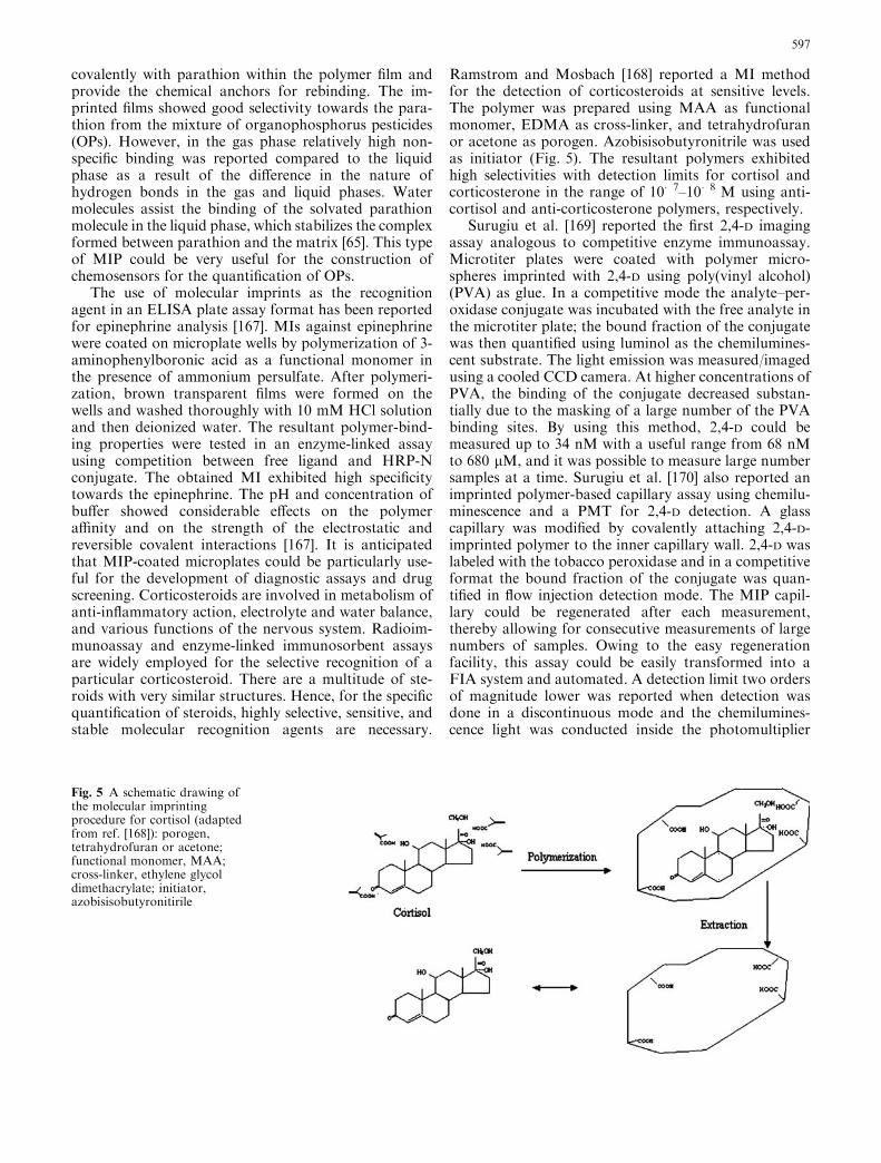
Ramstrom and Mosbach [168] reported a MI methodfor the detection of corticosteroids at sensitive levels.The polymer was prepared using MAA as functionalmonomer, EDMA as cross-linker, and tetrahydrofuranor acetone as porogen. Azobisisobutyronitrile was usedas initiator (Fig. 5). The resultant polymers exhibitedhigh selectivities with detection limits for cortisol andcorticosterone in the range of 10�7–10�8 M using anti-cortisol and anti-corticosterone polymers, respectively.
Surugiu et al. [169] reported the first 2,4-D imagingassay analogous to competitive enzyme immunoassay.Microtiter plates were coated with polymer micro-spheres imprinted with 2,4-D using poly(vinyl alcohol)(PVA) as glue. In a competitive mode the analyte–per-oxidase conjugate was incubated with the free analyte inthe microtiter plate; the bound fraction of the conjugatewas then quantified using luminol as the chemilumines-cent substrate. The light emission was measured/imagedusing a cooled CCD camera. At higher concentrations ofPVA, the binding of the conjugate decreased substan-tially due to the masking of a large number of the PVAbinding sites. By using this method, 2,4-D could bemeasured up to 34 nM with a useful range from 68 nMto 680 lM, and it was possible to measure large numbersamples at a time. Surugiu et al. [170] also reported animprinted polymer-based capillary assay using chemilu-minescence and a PMT for 2,4-D detection. A glasscapillary was modified by covalently attaching 2,4-D-imprinted polymer to the inner capillary wall. 2,4-D waslabeled with the tobacco peroxidase and in a competitiveformat the bound fraction of the conjugate was quan-tified in flow injection detection mode. The MIP capil-lary could be regenerated after each measurement,thereby allowing for consecutive measurements of largenumbers of samples. Owing to the easy regenerationfacility, this assay could be easily transformed into aFIA system and automated. A detection limit two ordersof magnitude lower was reported when detection wasdone in a discontinuous mode and the chemilumines-cence light was conducted inside the photomultiplier
Fig. 5 A schematic drawing ofthe molecular imprintingprocedure for cortisol (adaptedfrom ref. [168]): porogen,tetrahydrofuran or acetone;functional monomer, MAA;cross-linker, ethylene glycoldimethacrylate; initiator,azobisisobutyronitirile
597

tube by an optical fiber bundle. A dynamic range ofdetection from 5 pg mL�1–100 ng mL�1 (22.5 pM–450 nM) was obtained in this manner.
A highly sensitive piezoelectric sensor incorporating aMIP as the recognition element has been reported forthe detection of 2-methylisoborneol (MIB) [171]. MIBand geosmin are off-flavor compounds produced by avariety of microorganisms, which cause odor problemsin drinking water and in fish [172, 173]. Highly sensitivedetection limits down to 10 ng mL�1 were obtained dueto the intercalation of a nylon layer between the QCMand the MI. The sensitivity of these devices has beenimproved by 20-fold while maintaining selectivity incontrast to a previous report [174]. For the design ofMIP-based sensors during the past few years, mass-sensitive acoustic transducers, such as the surface-acoustic wave oscillator [175, 176], the QCM [174, 177],or the Love-wave oscillator [178], have been employed.Ultrathin films of titanium oxide gel imprinted with 4-(4-propyloxyphenylazo)benzoic acid (C3AzoCO2H) wereprepared by repeated immersion of a gold-coated quartzcrystal microbalance (QCM) electrode or a quartz platein solutions of C3AzoCO2H and titanium butoxide(Ti(O-nBu)4) in toluene/ethanol [179]. The templatingazobenzene carbodylic acid reacted with titaniumbutoxide though covalent bonding in the film. Thetemplate molecules were completely removed from thethin film using diluted aqueous ammonia. Binding anddesorption studies were done by measuring frequencychanges in the presence of template molecules. Rebind-ing of template molecules is fast (<1 min).
Dickert et al. reported some MIPs for the detection ofmicrobial cells in aqueous samples using QCMs [180–182] and this author also recently published a review onsensor strategies for microorganism detection [183].Biological recognition materials like antibodies showedpromising sensitivity towards the detection of cells. Butthe main limitation was the limited stability of theresulting sensor layers. Dickert and Hayden [184] re-solved this problem by employing artificial recognitionelements such as MIPs. Based on this strategy, yeastimprints were made on QCM electrode surfaces usingthe titania oxide sol–gel stamping method. The yeast-imprinted sensor coatings showed a regular honeycomb-like surface with 1-lm depth. The yeast cells can bemeasured up to 21 g L�1 in growth media [182]. Thiskind of detection system is very useful for the fermen-tation and biotechnological industries for cell countingand contamination detection. Viruses are smaller thanbacterial and yeast cells and cannot be observed orenumerated with light microscopes. Recently Dickertet al. [185] reported a bioimprinted QCM for thedetection tobacco mosaic virus (TMV) in aqueous media(pnat sap). By using this sensor, TMV was detectedquickly in the range of 100 ng mL�1–1 mg mL�1, andimprints can be easily regenerated and reused. In thefuture, MIPs for the specific detection of cell surfacemarkers of microbial cells may be developed along theselines.
Leung et al. [186] reported a MI for 3-chloro-1,2-propanediol (3-MCPD). The presence of chloropropa-nol in foods is carcinogenic. According to the EuropeanCommunity (EC) the limit on 3-MCPD in acid-hydro-lyzed vegetable proteins and soya sauces is 0.02 mg kg�1
(dried weight). GC is widely employed for the analysis ofchloropropanols; however, this procedure requiresderivatization schemes, which is a time-consuming pro-cess and requires the use of further solvents. Sometimesincomplete derivatization leads to false results. Hence,for industrial use, chemosensors are highly desirable. 3-MCPD imprints have been successfully developed using4-vinylphenylboronic acid as the functional monomer[186]. By using this imprint as a recognition element,potentiometric response of the glass electrode is rea-sonably linear over the range 0–3.2 mM for 3-MCPD.However, in the case of real samples some problems likeblocking of sites by the interferences present in the realsample (soya sauce) were encountered. A few reviewshave been published on applications of molecularimprinting technology in food analysis [58, 187].
Fluorescence-based sensors are highly sensitive andconvenient for analysis. The use of external fluorescence(conjugates) is presently a practiced method. In this kindof approach where direct analyses of target moleculesare possible, changes of fluorescence intensity appear inthe presence of target molecules without any use ofexternal reagents or conjugates. Brune et al. [188] re-ported that a mutant of Escherichia Coli phosphatebinding protein labeled with a fluorescent dye adjacentto its binding site exhibited a large increase in fluores-cence upon binding of inorganic phosphate. The samekind of labeling studies were conducted for the deter-mination of maltose [189] and glucose [190]. However,the design of fluorescent MIs has been hampered by thelack of suitable fluorescent tags, which should respondto the binding event with significant fluorescence inten-sity changes. Gao et al. [191] synthesized a fluorescentmonomer, which allowed for the preparation of fluo-rescent sensors for cis-diols using molecular imprintingmethod. Using this monomer an imprint polymer for D-fructose was prepared. The resultant polymer showedsignificant enhancement of fluorescence intensity uponbinding with D-fructose. The enhancement was depen-dent on template concentration. It was also reportedthat the fluorescent monomer developed could be usedfor the preparation of fluorescent sensors of other sugarsand biologically important catecholamines, which alsohave a cis-diol moiety. In another study, a fluorescentadenosine 3¢,5¢-cyclic monophosphate (cAMP)-im-printed polymer was prepared by polymerization oftrimethylolpropane trimethacrylate, 2-hydroxyethylmethacrylate (HEMA), and the fluorescent functionalmonomer in the presence of cAMP. Methanol was usedas the solvent porogen because of its high solubility ofcAMP and all monomers [192]. The recognition prop-erties of the cAMP-imprinted polymer were studied inaqueous media by fluorescence spectroscopy. This fluo-rescent molecular imprinted polymer displayed a
598

quenching of fluorescence in the presence of aqueouscAMP, whereas almost no effect was observed in thepresence of the structurally similar molecule, cGMP.The association constant for the binding of cAMP to theimprinted polymer was determined to be in the order of105 M�1. These selective fluorescent polymer imprintswill be very useful for the development of fluorescentchemosensors for the aqueous detection of cAMP.Homocysteine is a nonessential thiol-containing aminoacid formed as an intermediate during the metabolism ofmethionine. Blood plasma of a normal adult contains 5–15 lM of total homocysteine. Homocysteine is toxic toendothelial cells of blood vessels, and an elevated plasmalevel of homocysteine can induce vascular dysfunctionsthat may lead to arteriosclerosis, stroke, and cerebro-vascular, peripheral vascular, and coronary heart dis-eases. The clinical determination of homocysteine reliesmostly on chromatography, GC-MS and HPLC, capil-lary electrophoresis, and immunoassay. These methodsare time-consuming and expensive. Chow et al. [193]reported a fluorescence-based method for the determi-nation of homocysteine present in blood plasma. In theirwork, MIP was developed using N-(1-pyrenyl)maleim-idyl-dl-homocysteine (PM-H) or N-(1-pyrenyl)maleim-idyl-dl-cysteine (PM-C) as template and MAA,trimethylolpropane trimetharcylate (TRIM), and 1-az-obiscyclohexanecarbonitrile (ABCN) as recognitionmatrices.
A molecularly imprinted sol–gel material for thedetection and quantification of DDT was reported byGrahm et al. [194]. Both covalent and noncovalentstrategies were employed; however, good selectivity andbinding were obtained though covalent imprints as aresult of the lack of strong intermolecular interactionsbetween the sol–gel polymer and DDT. A covalentimprinting strategy was employed by generating a sac-rificial spacer through the reaction of two 3-iso-cyanatopropyltriethoxysilanes with one of two differenttemplate molecules: 4,4-ethylenedianiline (EDA) or 4,4-ethylidenebisphenol (EBP). After formation of the sol–gel, the bonds linking the spacer template to the matrixwere cleaved in a manner that generated a pocket of theappropriate size bordered by amine groups. The bondswere aided in the binding of DDT through weakhydrogen-bonding interactions. These imprints wereable to bind selectively with DDT. For construction ofthe chemosensor, an environmentally sensitive fluores-cent probe, 7-nitrobenz-2-oxa-1,3-diazole (NBD), waslocated adjacent to the DDT binding site. Fluorescencefluctuations occurred at the binding site in response tothe presence or absence of DDT. By using this fluores-cent MI, 50 pg mL�1 of DDT could be detected with aresponse time of <60 s. Repeated measurements couldalso be made with the same sensing films after regener-ation with acetone. However, this fluorescent MIshowed significant problems such as minimal increase influorescence intensity and limited dynamic range of thesensor as a result of the improper positioning of theNBD fluorophore in close proximity to the binding sites.
A conductometric chemosensor based on MIPs hasalso been reported. In this study benzyltriphenylphos-phonium was taken as a model analyte (template).Molecular imprinted polymer against the templatemolecule was prepared by photoinitiated polymerizationat 366 nm with template, MAA, EDMA, and AIBNinitiator in acetonitrile. The binding ability of the MIPwas checked by measuring the absorbance at 268 nm[195]. The imprinted polymer exhibited improved con-ductivity compared to nonimprinted polymer on theelectrode surface. Hedborg et al. [62] reported an inte-grated sensor based on a MIP, a capacitance sensorconsisting of a field-effect capacitor covered with a thinphenylalanine anilide imprinted polymer membrane [24].Panasyuk et al. [196] reported a capacitative detectionmethod in conjunction with imprinted electropolymer-ized polyphenol layers on gold electrodes. Lin andYamada [197] reported a chemiluminescence flow-through sensor for 1,10-phenanthroline based on thecombination of molecular imprinting and chemilumi-nescence. A ternary complex 4-vinylpyridine–Cu(II)–1,10-phenanthraline was synthesized and used as func-tional monomer for the 1,10-phenanthroline imprintdevelopment in combination with styrene and divinyl-benzene and packed into the column. The polymercontaining ternary complex is an efficient catalyst for thedecomposition of H2O2. When analyte and H2O2 passedinto the column with the buffer stream, these werecomplexed by the pyridine–Cu(II) binding sites andencountered H2O2 molecules. During the H2O2 decom-position, superoxide radical ion is formed which reactswith 1,10-phenanthraline and liberates a chemilumines-cence signal. The 1,10-phenanthraline is destroyed dur-ing the chemiluminescent reaction, liberating the bindingsite for another analyte molecule.
Kugimiya and Takeuchi [61] reported a MIP for thedetection of sialic acid and ganglioside. Various se-quences of oligosaccharides, which contain sialic acid atthe non-reducing end in glycoproteins and glycolipids,play important roles as cellular adhesion receptors andin molecular interaction. These oligosaccharides havebeen used as markers of biological information. In thisstudy, an SPR-based sensing system for detection ofganglioside (GM1), a glycolipid containing sialic acid atthe non-reducing end, was developed. On the surface ofan SPR chip, sialic acid MIP was coated through UVirradiation initiation and used for the detection of gan-glioside (GM1). The performance of MIP-SPR chips inthe presence of other structurally related compoundswas also investigated. The resonance angle changes as afunction of concentration and was linear from 0.1 to1.0 mg mL�1 of GM1. The estimation of creatininelevels is very important in the clinical diagnosis of renal,thyroid, and muscular malfunctions. Higher levels mayindicate diabetic nephropathy, eclampsia, musculardystophy, pre-eclampsia, pyelonephritis, reduced renalblood flow, urinary tract obstruction etc. The currentlyavailable spectrophotometic and enzymatic detectiontechniques suffer from interference with compounds
599

present in the biological fluids. Recently [198] a capaci-tative creatinine sensor was reported based on aphotografted MIP using a gold electrode. Creatininebinding was detected by a decrease in the electrodecapacitance. The sensor response is reversible and highlyselective. No response to the addition of sodium chlo-ride, creatine, urea, or glucose was detected. The detec-tion limit for creatinine is 10 lM, which is optimal formedical applications.
Catalysis
In many cases, enzymes exhibit low catalytic activitiesdue to the presence of organic solvents, inhibitors, and/or complex mixtures and perturbations in the tempera-ture and solution pH. These problems may be avoidedby employing synthetic biomimitic catalytic counter-parts [199] instead of biomolecules such as enzymes andcatalytic antibodies [200]. The catalytic counterparts canbe synthesized by tuning the enzyme active site throughmolecular imprinting with substrates or their transitionstate analogues (TSAs) [201–203]. For the preparationof catalytically active MIs, a cavity has to first be madewith a defined shape corresponding to the shape of thesubstrate or, even better, to the shape of the transitionstate of the reaction. At the same time, functional groupsare incorporated that act as binding sites, coenzymeanalogs, or catalytic sites within the cavity and in a de-fined stereochemical manner [26]. These artificial poly-meric catalysts are more durable and more resistant toharsh environments than biomolecules [204, 205], thusthey may be highly advantageous for industrial contin-
uous transformation and/or conversion reactions.Beach and Shea [206] reported a molecular imprintedcatalyst (MIC) that could catalyze the dehydrofluori-nation of 4-fluoro-4-(p-nitrophenyl)butan-2-one [206].2-Benzylmalonic acid was used as a template to orientthe N-(2-aminoethyl)methacrylamide monomers prior topolymerization. Polymerization was carried out in DMFsolvent in the presence of EDMA and 2,2¢-azobisi-sobutyronitrile. These polymers are insoluble, open-cellmacroporous solids with internal surface areas. Finallythe template molecules were removed from the cavitieswith dilute NaOH solution. The recovery of templateswas 78–85% [150]. In order to direct proper positioningof two amino groups in the catalytic cavity, differentdicarboxylic acid template molecules were employed.The catalytic activity of the imprint was greatly influ-enced by the position of carboxylic groups on the tem-plate. The most active imprinted polymer was achievedwith benzylmalonic acid as a template (Fig. 6). Thisapproach highlighted the possibility of changing theposition of functional groups in the active sites as astrategy for developing high-activity MIP catalysts.Sellergren and Shea [207] and Sellergren et al. [208] re-ported a highly cross-linked polymer catalyst for thehydrolysis of N-tert-butoxycarbonyl phenyl alanine-p-nitrophenyl ester. The cross-linked polymer had appro-priately placed hydroxyl, imidazole, and carboxylfunctional groups to mimic the amino acid residues ofchymotrypsin. One catalyst was constructed in which achiral phosphonate analog of D-phenylalanine was usedas the template. The two other catalysts were controlpolymers, one in which the carboxylic acid groups wererandomized using an achiral template and without the
Fig. 6 Catalyst design for thedehydrofluorination preparedby imprinting (adapted fromref. [206]): a reactionmechanism, b catalystpreparation through MIP
600

tetrahedral phosphonate, and the other in which thephenolimidazole functionality was removed. The maxi-mum rate enhancement for the hydrolysis of Boc-D-PheONP observed with the polymers was tenfold overreaction in solution without polymers. The controlpolymers showed less activity, approximately 5.7-fold orless over the reaction in solution and complete loss ofenantioselectivity. The polymer catalyst showed a 1.85-fold rate enhancement of the D-isomer over the L-isomer;the control polymers showed no preference for oneisomer over the other.
Leonhardt and Mosbach [209] reported an imprintedmatrix with esterolytic activity, in which cobalt(II) ionswere used to coordinate catalytically active vinylimi-dazole groups and the template during the imprintingprocess. The introduction of the substrates (p-nitrophe-nyl esters of methionine or leucine) to the sites resultedin an accelerated and substrate-specific hydrolysis ofactivated amino acid analogues [209]. Wulff et al. [210]used N,N¢-diethyl(4-vinylphenyl)amidine (DEVPA) as afunctional monomer to bind to phosphonate TSA-im-print molecules. The authors reasoned that a comple-mentary shape to the transition state analogue itselfmight not be sufficient for catalysis, and that appropri-ately positioned amidine functionality similar to that ofarginine had to be introduced for efficient catalysis tooccur. Furthermore, the efficiency could be improved ifthe noncovalent bonds had sufficiently high associationconstants to give 1:1 complexes of template and bindingsite, an interaction termed the ‘‘stoichiometric nonco-valent interaction.’’ The double salt bridge of theDEVPA–phosphonate TSA complex provided an ‘ox-yanionic hole’ for TS stabilization similar to that foundin serine proteases. Basic hydrolysis of an aryl ester wasshown to be accelerated >100-fold in the presence of theMIP catalyst. The catalyst exhibited Michaelis–Mentonkinetics (Km=0.60 mM; kcat=0.8·10�4 min�1). Thesame group also studied in the possibility of usingDEVPA to catalyze the hydrolysis of carbonates andcarbamates. Imprinted polymers were prepared in bulkand in suspension using cross-linker EDMA, functionalmonomers MMA and DEVPA, and diphenyl phosphateas template molecule in the presence of acetonitrile(MeCN), cyclohexanol-n-dodecanol, or toluene asporogen. Under pseudo-first-order conditions, thehydrolysis of diphenyl carbonate and diphenyl carba-mate showed rate enhancements of 588 and 1,435 times,respectively, compared to the rates of the uncatalyzedreactions. Relative to that of the control polymer, theMIP catalyst showed rate enhancements of 10 and 24times, respectively [211]. Thus, MIP catalysts showedlarger rate accelerations than catalytic antibodies.
There is significant importance in developing mate-rials that effectively adsorb and/or degrade toxic andpesticide compounds [212–214]. Phosphotriesterase iscapable to hydrolyze a variety of phosphodiester bondsin the detoxification of pesticides and chemical warfareagents [215, 216]. In addition to enzymatic methods,nonenzymatic phosphoester bond-cleaving methods
have also been reported [217–219]. Meng et al. [212]constructed an artificial phosphotriesterase by the co-polymerization of 4(5)-vinylimidazole–Zn2+–MAAcluster with a divinylbenzene polymer. Compared withthe spontaneous hydrolysis, the resulting polymer cata-lyst showed 105-fold rate acceleration towards thehydrolysis of diethyl p-nitrophenyl phosphate (parao-xon). The catalytic activity of the MIC prepared in thisstudy was fivefold higher than the polymer catalyst co-polymerized with Co2+–imidazole cluster [220, 221].The MIC showed a turnover rate of 7.4·10�2 s�1 to-wards the hydrolysis of paraoxon [212]. By employingthe MIC as catalyst, an amperomertic sensor was con-structed, which showed a detection limit of 0.1 mM forparaoxon with an excellent stability even at highertemperatures. Morihara’s group reported extensivestudies on molecular imprinting with TSA on acidicallyhydrolyzed silica gel surfaces doped with Al3+. TheseAl3+ ions provided Lewis acid centers for catalysis, andimprinting created binding sites complementary to thetemplate on silica gel surface [222–226]. The selectivityand catalytic activity relied on a combination of imprintmolecule shape and Lewis base activity of the aluminatecomponents of the particles. Markowitz et al. [227] re-ported surface imprinted silica particles for the catalyticactivity. A surfactant derivative of a a-chymotrypsintransition state analogue (TSA) was molecularly im-printed on surfaces using organosilanes N-(3-aminoe-thylaminomethyl)-phenyltrimethoxysilane (PEDA),carboxyethylsilanetriol (CTES), and N-(3-triethoxysi-lylpropyl)-4,5-dihydroimidazole (IPTES) added alongwith tetraethoxysilane (TEOS) to imprint the chemicalfunctionality. Surfaces imprinted with N-a-decyl-D-phenylalanine-2-aminopyridine enantioselectively cata-lyzed the hydrolysis of benzoyl-L-arginine-p-nitroanilide(L-BAPNA). No hydrolysis of D-BAPNA was observed.The catalytic activity of surface-imprinted particlesformed with TEOS and PEDA was significantly greaterthan that of surface-imprinted particles formed withTEOS and IPTES. Recently Wulff [26] published anexcellent review on MICs. Although MICs were highlystable under different harsh conditions, their catalyticactivities were low compared to biocatalysts (e.g., en-zymes). The exact mimic of enzyme catalytic sites wasquite difficult due to its complexity of structure andinteractions. For the improvement of catalytic activity ofMICs, intense research, new polymers systems, highlyselective substrates and/or TSAs, and new strategies arenecessary. An overview of various applications of MIPsare listed in Table 1.
Conclusions
The increased demand for medical, environmental, andindustrial analyses during the last decade has created aneed for quick and specific devices/receptors for theseparations and analyses of various compounds inaqueous and non-aqueous phases. Molecular recogni-
601
tion agents with high affinity, selectivity, and stabilityare therefore needed to meet this demand. Biologicalrecognition agents such as antibodies and enzymes playa pivotal role in analytical and diagnostic applications;however, their instability and irreversible deactivationlimit their application and long-term use in biosensorsand separation processes. The high selectivity, affinity,stability, and integrity of MIPs provide good alterna-tives to the biological recognition agents. Most molec-ular imprinted polymers are applied mainly forchromatographic uses because they are rigid gel matri-ces. However, the utilization of MIP potentials in sep-aration and purification technology is still at a juvenilestage. Furthermore, efforts are needed to develop newMIP methods towards high molecular weight targets
such as biopolymers and microorganisms. There is agreat demand for new functional monomers and cross-linkers to aid the design of high affinity imprints for awide variety of compounds. Easy-to-use instruments andtechniques suitable for onsite use by unskilled personsare required. Hence, for the construction of real-timeand highly sensitive chemosensors, self-signaling poly-mer imprints and new mechanisms are also highly nec-essary. One more obstacle in the development ofbiosensors is the lack of suitable interface between theMIP and the sensor element; hence further work has tobe intensified in this direction for the effective utilizationof MIPs.
Acknowledgements We are gratefully acknowledge the financialsupport of the Distinguished Young Scholar Fund to HX Ju(20325518), the National Natural Science Foundation of China(20275017), the Specialized Research Fund for the Excellent YoungTeachers from the Chinese Ministry of Education, the ScienceFoundation of Jiangsu (BS2001063), and the Key Research Projectof Jiangsu Institute of Cancer Research.
References
1. Killard, AJ, Deasy B, Kennedy RO, Smyth MR (1995) TrendsAnal Chem 14:257–262
2. Freitag R (1999) J Chromatogr B 722:2793. Dickey FH (1949) Proc Natl Acad Sci USA 35:227–2294. Dickey FH (1955) J Phys Chem 59:695–7075. Jenkins AL, Uy OM, Murray GM (1999) Anal Chem 71:373–
3786. Lahav M, Kharitonov AB, Katz O, Kunitake T, Willner I
(2001) Anal Chem 73:720–7237. Wulff G, Gross T, Schonfeld R (1997) Angew Chem Int Ed
Engl 36:1962–19648. Vlatakis G, Andersson LI, Muller R, Mosbach K (1993)
Nature 361:645–6479. Andersson LA (1996) Anal Chem 68:111–11710. Kugimiya A, Matsui J, Takeuchi T, Yano K, Muguruma H,
Elgersma A, Karube I (1995) Anal Lett 28:2317–232311. Andersson LA, O’Shannessy DJ, Mosbach K (1990) J Cro-
matogr 513:167–16912. Ramstrom O, Nicholls IA, Mosbach K (1994) Tetrahedron
Asymmetry 5:649–65613. Rachkov A, Minoura NB (2001) Biochim Biophy Acta
1544:255–26014. Wulff G (1995) Angew Chem Int Ed Engl 34:1812–183215. Ansell RJ, Ramstrom O, Mosbach K (1996) Clin Chem
42:1506–151216. Shea KJ (1994) Trends Polym Sci 2:166–17317. Sellergren B (1997) Trends Anal Chem 16:310–32018. Kempe M, Mosbach K (1995) J Chromatogr A 691:317–32319. Schweitz L, Anderson LI, Nilsson S (1998) J Chromatogr A
817:5–1320. Shea KJ, Spivak DA, Sellergren B (1993) J Am Chem Soc
115:3368–336921. Mathew J, Buchardt O (1995) Bioconjugate Chem 6:524–52822. Vidyasankar S, Arnold FH (1995) Curr Opin Biotechnol
6:218–22423. Takeuchi T, Haginaka J (1999) J Chromatogr B 728:1–2024. Cormack PAG, Mosbach K (1999) Reactive Funct Polym
41:11525. Haupt K, Mosbach K (2000) Chem Rev 100:2495–250426. Wulff G (2002) Chem Rev 102:1–2727. Peppas NA, Huang Y (2002) Pharm Res 19:579–58928. Ramstrom O, Ansel RJ (1998) Chirality 10:195–20929. Vallano P T, Remcho VT (2000) J Chromatogr A 887:125–135
Table 1 List of some applications of molecular imprinting tech-nology
Template/target Application References
Nicotine Extraction [228, 229]Caffeine Assay and sensor [138, 230]Glucose Sensor [231, 232]Fructose andgalactose
Separation [87]
Aspartame Detection andcatalyst
[233]
Fructose Separation [86]Monosodiumglutamate
Separation [234]
Amino acids Separation [235]Vitamin K1 Sensor [236]OP pesticide Detection and sensor [220, 221, 223]Triazine Sensor [237]Atrazine Sensor and extraction [112, 162]DDT Detection and sensor [194]Penicillin G Separation [238]Ampicillin Analysis [239]Ephedrine Separation [240]Anesthetics Separation [241]Cephalexin Extraction [242]Chloramphenicol Analysis [165]Tylosin Detection [243]Cholesterol Sensor [244]Cholesterol Catalyst [245]Steroid Catalyst [246]Morphine Sensor [247]Menthol Analysis [248]Peptides and proteins Recognition [13]Bovine serum albumin Catalyst [249, 250]Bentazone Extraction [251]Cu2+ Detection [252]Zn2+ Detection [253, 254]RNase A Separation [18]Nucleotide bases Separation [255]4-Nitrophenyl acetate Catalyst [256]Benzamidine, pentamidine Separation [257]L-Phenylanilide Separation [258, 259]Propranolol Separation [146, 260]Amino acid phenylesters Catalyst [139]Sulfonamides Separation [261]Biotin Assay [262]Fructosyl valine Catalyst [263]Hydroxycoumarin Extraction [264]Polycyclic aromatichydrocarbons
Sensor [265, 266]
Fenvalerate Sensor [267]
602

30. Schweitz L, Spegel P, Nilsson S (2001) Electrophoresis22:4053–4063
31. Wulff G, Knorr K (2001) Biosepara 10:257–27632. Lanza F, Sellergren B (2001) Chromatographia 53:599–61133. Martin-Esteban A (2001) Fresenius J Anal Chem 370:795–80234. Piletsky SA, Turner APF (2002) Electroanal 14:317–32335. Ye L, Haupt K (2004) Anal Bioanal Chem 378:1887–189736. Piletsky SA, Panasyuk, TL, Piletskaya EV, Nicholls IA,
Ulbricht M (2003) J Membr Sci 157:263–27237. Tada M, Iwasawa Y (2003) J Mol Catal A 199: 115–12038. Nicholls IA, Rosengren JP (2001) Bioseparation 10:301–30539. Stefan RI, Girmai BR, Frederick VSJ, Hassan YAE (2003)
Talanta 59:883–88740. Mallat E, Barcelo D, Barzen C, Gauglitz G, Abuknesha R
(2001) Trends Anal Chem 20:124–13241. Du D, Yan F, Liu S, Ju HX (2003) J Immunol Methods
283:67–7542. Ju HX, Yan GF, Chen F, Chen HY (1999) Electroanalysis
11:124–12843. Dai Z, Yan F, Chen J, Ju HX (2003) Anal Chem 75:5429–
543444. Liu SQ, Yu JH, Ju HX (2003) Electroanal Chem 540:61–7745. Yu JH, Liu SQ, Ju HX (2003) Biosens Bioelectron 19:401–40946. Thakur MS, Karanth NG (2003) In: Malhotra BD, Turner
APF (eds) Advances in biosensors, vol 5. Elsevier, Amster-dam, pp 131–160
47. Rekha K, Gouda MD, Thakur MS, Karanth NG (2000)Biosens Bioelectron 15:499–502
48. Yu JH, Ju HX (2003) Anal Chim Acta 486:209–21649. Liu SQ, Ju HX (2003) Analyst 124:1420–142450. Brooker G, Harper JF, Terasaki WL, Moylan RD (1979) In:
Brooker G, Greengard T, Robison GA (eds) Advances incyclic nucleotide research. Raven, New York, pp 1–11
51. Andersson LI, Ekberg B, Mosbach K (1993) Bioseparationand catalysis in molecularly imprinted polymers. In: Ngo TT(ed) Molecular interactions in bioseparations. Plenum, NewYork pp, 383–395
52. Kriz D, Ramstrom O, Svensson A, Mosbach K (1995) AnalChem 67:2142–2144
53. Fischer L, Muller R, Ekberg B, Mosbach K (1991) J AmChem Soc 113:9358–9360
54. Mulchandani A, Kaneva I, Chen W (1998) Anal Chem70:5042–5046
55. Haupt K (2002) Nat Biotechnol 20:881–88556. Titirici MM, Sellergren B (2004) Anal Bioanal Chem 378:
1913–192157. Haupt K, Mosbach K (1998) Trends Biotechnol 16:468–47558. Whitcombe MJ, Alexander C, Vulfson EN (1997) Trends
Food Sci Technol 8:140–14559. Kempe M, Mosbach K (1995) J Chromatogr A 694:3–1360. Kugimiya A, Yoneyama H, Takeuchi T (2000) Electroanalysis
12:1322–132661. Kugimiya A, Takeuchi T (2001) Biosens Bioelectron 16:1059–
106262. Hedborg E, Winquist F, Lundstrom I, Andersson LI, Mos-
bach K (1993) Sens Actuators A 36–38:796–79963. Sergeyeva TA, Piletsky SA, Brovko AA, Slinchenko EA,
Sergeeva LM, Elskaya AV (1999) Anal Chim Acta 392:105–111
64. Delaney TP, Mirsky VM, Ulbricht M, Wolfbeis OS (2001)Anal Chim Acta 435:157–162
65. Marx S, Zaltsman A, Turyan I, Mandler D (2004) Anal Chem76:120–126
66. Cederfur J, Yuxin P, Meng Z, Kempe M (2003) J Comb Chem5:67–72
67. Levi R, McNiven S, Piletsky SA, Cheng S-H, Yano K, Kar-ube I (1997) Anal Chem 69:2017–2021
68. Mayes AG, Mosbach K (1997) Trends Anal Chem 16:321–33269. Ekberg B, Mosbach K (1989) Trends Biotechnol 7:92–9670. Mosbach K (1994) Trends Biochem Sci 19:9–1471. Wulff G, Sarhan A (1995) Angew Chem Int Ed Engl 34:1812–
1832
72. Wulff G (1993) Trends Biotechnol 11:85–8773. Whitcombe MJ, Rodriguez ME, Villar P, Vulfson EN (1995) J
Am Chem Soc 117:7105–711174. Kriz D, Mosbach K (1995) Anal Chim Acta 300:71–7575. Lai EPC, Fafara AVA, Van der Noot M, Polsky B (1998) Can
J Chem 76:265–26976. Chen G, Guan Z, Chen CT, Fu L, Sundaresan V, Arnold FH
(1997) Nat Biotechnol 15:354–35777. Piletsky SA, Piletskaya EV, Yano K, Kugimiya A, Elgersma
AV, Levi R, Kahlow U, Takeuchi T, Karube I, Panasyuk TI,Elskaya AV (1996) Anal Lett 29:157–170
78. Piletsky SA, Piletskaya EV, Elgersma AV, Yano K, Karube I,Parhometz YP, Elskaya AV (1995) Biosens Bioelectron10:959–964
79. Marx S, Zaltsman A, Turyan I, Mandler D (2004) Anal Chem76:120–126
80. Turkewitsch P, Wandelt B, Darling GD, Powell WS (1998)Anal Chem 70:2025–2030
81. Ansanumar H, Akiyama T, Kajiya K, Hishiya T, KomiyamaM (2001) Anal Chim Acta 435:25–33
82. Wulff G, Vietmeier J, Poll HG (1987) Makromol Chem188:731–740
83. Wulff G, Kemmerer R, Vietmeier J, Poll HG (1982) J Chim6:681–683
84. Marty JD. Tizra M, Mauzac M, Rico-Lattes I, Lattes A(1999) Macromolecule 32:8674–8677
85. O’Shannessy DJ, Andersson LI, Mosbach K (1989) J MolRecognit 2:1–9
86. Wulff G, Haarer J (1991) Makromol Chem 192:1329–133187. Wulff G, Schauhoff S (1991) J Org Chem 56:395–40088. Sun BW, Yang ML, Li YZ, Chang WB (2003) Acta Chim Sin
61:878–88489. Katz A, Davis ME (2000) Nature 403:286–28990. Zimmerman SC, Wendland MS, Rakow NA, Zharov L, Su-
slick S (2002) Nature 418:399–40391. Takeuchi T, Fukuma D, Matsui J (1999) Anal Chem 71:285–
29092. Cederfur J, Pei Y, Zihui M, Kempe M (2003) J Comb Chem
5:67–7293. Subrahmanyam S, Piletsky SA, Piletska EV, Chen B, Karim
K, Turner APF (2001) Biosens Bioelectron 16:631–63794. Kobayashi T, Fukaya T, Abe M, Fujii N (2002) Langmuir
18:2866–287295. Kobayashi T, Reddy PS, Fujii N (2002) Eur Polym J 38:779–
78196. Lim CH, Ki CD, Kim, TH, Chang Ji Y (2004) Macromolecule
37:6–897. Shi H, Wei-Bor T, Michael D, Ferrari GS, Ratner BD (1999)
Nature 398:593–59798. Andersson HS, Karisson JG, Pieltsky SA, Koch-Schmidt AC,
Mosbach K, Nicholls IA (1999) J Chormatogr A 848:39–4999. Katz A, Davis ME (1999) Macromolecules 32:4113–4121100. Yilmaz E, Haupt K, Mosbach K (2000) Angew Chem Int Ed
Eng 39:2115–2118101. Titirici MM, Hall AJ, Sellergren B (2002) Chem Mater 14:21–
23102. Titirici MM, Sellergren B (2004) Anal Bioanal Chem
378:1913–1921103. Dai S, Burleigh MC, Ju YH, Gao HJ, Lin JS, Branes C, Xue
ZL (2000) J Am Chem Soc 122:992–993104. Remcho VT, Tan ZJ (1999) Anal Chem 71:A248–A255105. Kempe M, Mosbach K (1995) J Chromatogr A 694:3–13106. Schweitz L, Andersson LI, Nilsson S (1998) J Chromatogr A
817:5–13107. Kriz D, Mosbach K (1994) Anal Chim Acta 300:71–75108. Sellergren BA (1994) In: Subramanian G (ed) Practical ap-
proach to chiral separations by liquid chromatography.VCH,Weinhein pp 69–75
109. Kempe M (1996) Anal Chem 68:1948–1953110. Fishcher L, Muller R, Ekberg B, Mosbach K (1991) J Am
Chem Soc 113:9358–9360111. Kempe M, Mosbach K (1994) J Chromatogr A 664:276–279
603

112. Muldoon MT, Stanker LH (1997) Anal Chem 69:803–808113. Sellergren B (1994) Anal Chem 66:1578–1582114. Dela CE, Muguruma H, Jose W, Pedersen H (1999) Anal Lett
32:841–854115. Molinelli A, Weiss R, Mizaikoff B (2002) J Agric Food Chem
50:1804–1808116. Baggiani C, Giraudi G, Vanni A (2002) Bioseparation 10:389–
394117. Zhou SN, Lai EPC, Miller JD (2004) Anal Bioanal Chem
378:1903–1906118. Zhu Q-Z, Degelmann P, Niessner R, Knopp D (2002) Environ
Sci Technol 36:5411–5420119. Masque N, Rosa M, Marce RM, Francesc BF, Cormack
PAG, Sherrington DC (2000) Anal Chem 72:4122–4126120. Hong JM, Anderson PE, Qian J, Martin CR (1998) Chem
Mater 10:1029–1033121. Ju JY, Shin CS, Whitcombe MJ, Vulfson EN (1999) Bio-
technol Bioeng 64:232–239122. Yu Y, Ye L, De Biasi V, Mosbach K (2002) Biotechnol Bio-
eng 79:23–28123. Kempe M, Mosbach K (1994) J Chromatogr A 664:276–279124. Lei JD, Tan TW (2002) Biochem Eng J 11:175–179125. Liu Y, Liu XL, Wang JD (2003) Anal Lett 36:1631–1645126. Schweitz L, Andersson LI, Nilsson S (1997) J Chromatogr A
792:401–409127. Cheong SH, McNiven S, Rachkov A, Levi R, Yano K,
Karube I (1997) Macromolecules 30:1317–1322128. Kriz D, Kriz CB, Andersson LI, Mosbach K (1994) Anal
Chem 66:2636–2639129. Haginaka J, Kagawa C (2004) Anal Bioanal Chem 378:1907–
1912130. Rachkov A, Minoura N, Shimizu T (2001) Anal Sci 17:i609–
i612131. Yilmaz E, Ramstrom O, Moller P, Sanchez D, Mosbach K
(2002) J Mater Chem 12:1577–1581132. Chen Y, Kele M, Quinones I, Sellergren B, Guiochon G
(2001) J Chromatogr A 927:1–17133. Koeber R, Fleischer C, Lanza F, Boos KF, Sellergren B,
Barcelo D (2001) Anal Chem 73:2437–2444134. Mullet WM, Lai EPC, Sellergren B (1999) Anal Commun
35:217–220135. Ferrer I, Lanza F, Tolokan A, Horvath V, Sellergren B,
Horvai G, Barcelo D (2000) Anal Chem 72:3934–3941136. Horvath V, Pap T, Tolokan A, Horvai G, Sellergren B (2002)
J Chromatogr A 973:1–12137. Bereczki A, Tolokan G, Horvai V, Horvath F, Lanza HAJ,
Sellergren B (2001) J Chromatogr A 930:31–38138. Ye L, Weiss R, Mosbach K (2000) Macromolecules 33:8239–
8245139. Emgenbroich M, Wulff G (2003) Chem Euro J 9:4106–4117140. Mayes AG, Mosbach K (1996) Anal Chem 68:3769–3774141. Strikovsky AG, Kasper D, Gruen M, Green HJ, Wulff G
(2000) J Am Chem Soc 122:6295–6296142. Perez N, Whitcombe MJ, Vulfson EN (2000) J Appl Polym
Sci 77:1851–1859143. Sellergren B (1994) Chromatogr A 673:133–138144. Arnold FH, Plunkett S, Dhal PK, Vidyasankar S (1995)
Polym Prepr 36:97–98145. Piletsky SA, Matuschewski H, Schedler U, Wilpert A, Piletska
EV, Thiele TA, Ulbricht M (2000) Macromolecules 33:3092–3098
146. Schweitz L, Andersson LI, Nilsson S (1997) Anal Chem69:1179–1183
147. Sulitzky C, Ruckert B, Andrew J, Hall A J, Lanza F, KandKU, Sellergren B (2002) Macromolecules 35:79–91
148. Quaglia M, Lorenzi ED, Sulitzky C, Massolini G, Sellergren B(2001) Analyst 126:1495–1498
149. Quaglia M, Lorenzi ED, Sulitzky C, Caccialanza G, Seller-gren B (2003) Electrophoresis 24:952–957
150. Xie J, Zhu L, Luo H, Zhou L, Li C, Xu X (2001) J Chro-matogr A 934:1–11
151. Zhu L, Xu X (2003) J Chromatogr A 991:151
152. Zhu L, Chen L, Xu X (2003) Anal Chem 75:6381–6387153. Mosbach K, Yu Y, Andersch J, Ye L (2001) J Am Chem Soc
123:12420–12421154. Ye L, Yu Y, Mosbach K (2001) Analyst 126:760–765155. Piletsky SA, Piletska EV, Bossi A, Karim K, Lowe P, Turner
APF (2001) Biosens Bioelectron 16:701–707156. Matsui J, Kubo H, Takeuchi T (1998) Anal Sci 14:699–702157. Siemann M, Andersson LI, Mosbasch K (1996) J Agric Food
Chem 44:141–145158. Muldoon MT, Stanker LH (1995) J Agric Food Chem
43:1424–1427159. Matsui J, Miyoshi Y, Doblhoff-Dier O, Takeuchi T (1995)
Anal Chem 67:4404–4408160. Matsui J, Doblhoff-Dier O, Takeuchi T (1995) Chem Lett
20:489–491161. Shoji R, Takeuchi T, Kubo I (2003) Anal Chem 75:4882–4886162. Piletsky SA, Piletskata EV, Elgersma AV, Yano K, Karube I,
Parhometz YP, ElSkaya AV (1995) Biosens Bioelectron10:959–964
163. Chianella I, Piletsky S, Tothill IE, Chen B, Turner APF (2003)Biosens Bioelectron 18:119–127
164. Mena ML, Agui L, Martinez-Ruiz P, Yanez-Sedeno P, Re-viejo AJ, Pingarron JM (2003) Anal Bioanal Chem 376:18–25
165. Rodrıguez JLS, Dıaz-Garcıa ME (2001) Biosens Bioelectron16:955–961
166. Kroger S, Turner APF, Mosbach K, Haupt K (1999) AnalChem 71:3698–3702
167. Piletsky SA, Piletska EV, Chen B, Karim K, Weston D,Barrett G, Lowe P, Turner APF (2000) Anal Chem 72:4381–4385
168. Ramstrom O, Ye L, Mosbach K (1996) Chem Biol 3:471–477169. Surugiu I, Danielsson B, Ye L, Mosbach K, Haupt K (2001)
Anal Chem 73:487–491170. Surugiu I, Svitel J, Ye L, Haupt K, Danielsson B (2001) Anal
Chem 73:4388–4392171. Ji HS, McNiven S, Kyong-Hoon L, SaitoT, Ikebukuro K,
Karube I (2000) Biosens Bioelectron 15:403–409172. Borjesson TS, Stollman UM, Schnurer JL (1993) J Agric
Food Chem 42:104–106173. Pollak FC, Berger RG (1996) Appl Environ Microbiol
62:1295–1299174. Ji HS, McNiven S, Ikebukuro K, Karube I (1999) Anal Chim
Acta 390:93–100175. Dickert FL, Tortschanoff M, Bulst WE, Fischerauer G (1999)
Anal Chem 71:4559–4563176. Dickert FL, Forth P, Lieberzeit P, Tortschanoff M (1998)
Fresenius J Anal Chem 360:759177. Liang C, Peng H, Bao X, Nie L, Yao S (1999) Analyst
124:1781–1785178. Jakoby B, Ismail GM, Byfield MP, Vellekoop MJ (1999) Sens
Actuators A 76:93–97179. Lee SW, Ichinose I, Kunitake T (1998) Langmuir 14:2857–
2863180. Hayden O, Dickert FL (2001) Adv Mater 13:1480–1483181. Hayden O, Bindeus R, Dickert FL (2003) Meas Sci Technol
14:1876–1881182. Dickert FL, Hayden O, Lieberzeit P, Haderspoeck C, Bindeus
R, Palfinger C, Wirl B (2003) Synth Met 138: 65–69183. Dickert FL, Lieberzeit P, Hayden O (2003) Anal Bioanal
Chem 377:540–549184. Dickert FL, Hayden O (2002) Anal Chem 74:1302–1306185. Dickert FL, Hayden O, Bindeus R, Mann KJ, Blaas D,
Waigmann E (2004) Anal Bioanal Chem 378:1929–1934186. Leung, MKP, Chiu BKW, Lam MHW (2003) Anal Chim
Acta 491:15–25187. Ramstrom O, Skudar K, John Haines J, Patel P, Bruggemann
O (2001) J Agric Food Chem 49:2105–2114188. Brune M, Hunter JL, Hiwekk SA, Martin SR, Hazkett TL,
Corrie JET, Webb MR (1998) Biochemistry 37:10370–10380189. Gilardi G, Zhou Q, Hibbert L, Cass AE (1994) Anal Chem
66:3840–3847190. Marvin JS (1997) Proc Natl Acad Sci USA 94:4366–4371
604

191. Gao S, WangW, Wang B (2001) Bioorg Chem 29:308–320192. Turkewitsch P, Wandelt B, Darling GD, Powell WS (1998)
Anal Chem 70:2025–2030193. Chow CF, Michael HW, Lam MHW, Leung MKP (2002)
Anal Chim Acta 466:17194. Graham AL, Carlson CA, Edmiston PL (2002) Anal Chem
74:458195. Kriz D, Kempe M, Mosbach K (1996) Sens Actuators B
33:178–181196. Panasyuk TL, Mirsky VM, Piletsky SA, Wolfbeis OS (1999)
Anal Chem 71:4609–4613197. Lin JM, Yamada M (2001) Analyst 126:810–815198. Delaney TP, Mirsky VM, Wolfbeis OS (2002) Electroanalysis
14:221–225199. Lee D, Choi YK, Kim MJ (2000) Org Lett 2:2553–2555200. Shokat KM, Leumann CJ, Sugasawara R, Schultz PG (1994)
Nature 338:269–270201. Dabulis K, Klibanov AM (1993) Biotechnol Bioeng 41:566–
571202. Mishra P, Griebenow K, Klibanov AM (1996) Biotechnol
Bioeng 52:609–614203. Rich JO, Dordick JS (1997) J Am Chem Soc 119:3245–3252204. Williamson G, Vallejo J (1997) Int J Biol Macromol 21:163–
167205. Craig DB, Arriaga EA, Wong JCY, Lu H, Dovichi NJ (1996)
J Am Chem Soc 118:5245–5253206. Beach JV, Shea KJ (1994) J Am Chem Soc 116:379–380207. Sellergren B, Shea KJ (1994) Tetrahedron Asymmetry 5:1403–
1406208. Sellergren B, Karmalkar RN, Shea KJ (2000) J Org Chem
65:4009–4027209. Leonhardt A, Mosbach K (1987) React Polym 6:28–32210. Wulf G, Gross T, Schonfeld R (1997) Angew Chem Int Ed
Engl 36:1962–1964211. Strikovsky AG, Kasper D, Grun M, Greens G, Hradil J,
Wulff G (2000) J Am Chem Soc 122:6295–6296212. Meng Z, Yamazaki T, Sode K (2003) Biotechnol Lett
25:1075–1080213. Sogorb MA, Vilanova E (2002) Toxicol Lett 128:215–228214. Gaberlein S, Spener F, Zaborosch C (2000) Appl Microbiol
Biotechnol 54:652–655215. Dumas DP, Durst HD, Landis WG, Raushel FM, Wild JR
(1990) Arch Biochem Biophys 277:155–159216. Rastogi VK, DeFrank JJ, Cheng T, Wild JR (1997) Biochem
Biophys Res Commun 241:294–296217. Koper O, Lucas E, Klabunde KJ (1999) J Appl Toxicol
19:S59–S62218. Lucas EM, Klabunde KJ (1999) Nanostruct Mater 12:179–
181219. Jenkins AL, Yin R, Jensen JL (2001) Analyst 126:798–802220. Yamazaki T, Yilmaz E, Mosbach K, Sode K (2001) Anal
Chim Acta 435:209–214221. Yamazaki T, Meng Z, Mosbach K, Sode K (2001) Electro-
chemistry 69:96–101222. Morihara K, Doi Y, Takiguchi M, Shimada T (1993) Bull
Chem Soc Jpn 66:2977–2982223. Matsuishi T, Shimada T, Morihara K (1994) Bull Chem Soc
Jpn 67:748–756224. Morihara K, Lijima T, Usui H, Shimada T (1993) Bull Chem
Soc Jpn 66:3047–3052225. Shimada T, Kurazono R, Morihara K (1993) Bull Chem Soc
Jpn 66:836–840226. Matsuishi T, Shimada T, Morihara K (1992) Chem Lett
10:1921–1924227. Markowitz MA, Paul R, Kust PR, Klaehna J, Deng G, Ga-
bera BP (2001) Anal Chim Acta 435:177–185228. Zander A, Findlay P, Renner T, Sellergren B (1998) Anal
Chem 70:3304–3314229. Liu Y, Liu XL, Wang JD (2003) Chin J Anal Chem 31:1202–
1206230. Liang C, Peng H, Bao X, Nie L, Yao S (1999) Analyst
124:1781–1785
231. Chen GH, Guan ZB, Chen CT, Fu LT, Sundaresan V, ArnoldFH (1997) Nat Biotechnol 15:354–357
232. Malitesta C, Losito I, Zambonin PG (1999) Anal Chem71:1366–1370
233. Ye L, Ramstrom O, Ansell R, Mansson MO, Mosbach K(1999) Biotechnol Bioeng 64:650–655
234. Vidyasankar S, Ru M, Arnold FH (1997) J Chromatogr A775:51–63
235. Lin JM, Nakagama T, Uchiyama K, Hobo T (1996) Chro-matographia 43:585–591
236. Andersson L, Mandenius CF, Mosbach K (1988) TetrahedronLett 29:5437–5440
237. Piletsky SA, Piletskaya EV, Elskaya AV, Levi R, Yano K,Karube I (1997) Anal Lett 30:445–450
238. Skudar K, Bruggemann O, Wittelsberger A, Ramstrom O(1999) Anal Commun 36:327–331
239. Lubke C, Lubke M, Whitcombe MJ, Vulfson EN (2000)Macromolecules 33:5098–5105
240. Ramstrom O, Yu C, Mosbach K (1996) J Mol Recognit9:691–693
241. Schweitz L, Andersson LI, Nilsson S (1997) J Chromatogr A792:401–409
242. Lai EPC, Wu SG (2003) Anal Chim Acta 481:165–174243. Siemann M, Andersson LI, Mosbach K (1997) J Antibiot
50:88–92244. Piletsky SA, Piletskaya EV, Sergeyeva TA, Panasyuk TL,
Elskaya AV (1999) Sens Actuators B 60:216–220245. Kim JM, Ahn K-D, Wulff G (2000) Macromol Chem Phys
202:1105–1108246. Alexander C, Smith CR, Whitcombe MJ, Vulfson EN (1999) J
Am Chem Soc 121:6640–6651247. Kriz D, Ramstrom O, Svensson A, Mosbach K (1995) Anal
Chem 67:2142–2144248. Pinel C, Loisil P, Gallezot P (1997) Adv Mater 9:582–589249. Ohya Y, Miyaoka J, Ouchi T (1996) Macromol Rapid
Commun 17:871–874250. Slade CJ. (2000) J Mol Catal B 9:97–100251. Baggiani C, Trotta F, Giraudi G, Giovannoli C, Vanni A
(1999) Anal Commun 36:263–266252. Koide Y, Senba H, Shosenji H, Maeda M, Takagi M (1996)
Bull Chem Soc Jpn 69:125–130253. Yoshida M, Uezu K, Goto M, Nakashio F (1996) J Chem Eng
Jpn 29:174–176254. Uezu K, Nakamura H, Kanno J, Sugo T, Goto M, Nakashio
F (1997) Macromolecules 30:3888–3891255. Spivak DA, Shea KJ (1998) Macromolecules 31:2160–2165256. Kawanami Y, Yunoki T, Nakamura A, Fujii K, Umano K,
Yamauchi H, Masuda K (1999) J Mol Catal A 145:107–109
257. Nilsson K, Lindell J, Sellergin NB (1994) J Chromatogr A680:57–61
258. Lin J-M, Nakagama T, Uchiyama T, Hobo J (1997) LiqChromatogr Rel Technol 20:1489–1506
259. Lin J-M, Nakagama T, Uchiyama Y, Hobo T (1997) BiomedChromatogr 11:298–302
260. Walshe M, Garcia E, Howarth J, Smyth MR, Kelly M T(1997) Anal Commun 34:119–122
261. Zheng N, Fu Q, Li YZ, Chang WB, Wang ZM, Li TJ (2001)Microchem J 69:55–59
262. Takeuchi T, Dobashi A, Kimura K (2000) Anal Chem72:2418–2422
263. Yamazaki T, Ohta S, Yanai Y, Sode K (2003) Anal Lett36:75–89
264. Walshe M, Howarth J, Kelly MT, Opkennedy R, Smyth MR(1997) J Pharm Biomed Anal 16:319–325
265. Dickert FL, Achatz P, Halikias K (2001) Fresenius J AnalChem 371:11–15
266. Stanley S, Percival CJ, Auer M (2003) Anal Chem 75:1573–1577
267. Gong JL, Gong FC, Kuang Y, Zeng GM, Shen GL, Yu RQ(2004) Anal Bioanal Chem 379:302–307
605


![Analytica Chimica Acta - sklac.nju.edu.cnsklac.nju.edu.cn/hxju/lunwenlunzhu/paper2014/470 Anal Chim Acta Zhu... · instrumentation [11–13]. For the past fewyears, avarietyof amplification](https://static.fdocuments.in/doc/165x107/5c65ec8509d3f230488b5a64/analytica-chimica-acta-sklacnjuedu-anal-chim-acta-zhu-instrumentation.jpg)