

Human tear film and meibum. I. Very long chain wax esters ... · Epoxidation of MGS. ......
41
1 Human tear film and meibum. I. Very long chain wax esters and (O-acyl)-omega- hydroxy fatty acids of meibum. Igor A. Butovich 1, 2, 3 , Jadwiga Wojtowicz 1 , Mike Molai 1 . 1 Department of Ophthalmology and 2 Graduate School of Biomedical Sciences, UTSouthwestern Medical Center, 5323 Harry Hines Blvd, Dallas, TX, 75390, USA 3 Contact information: Address: Department of Ophthalmology, UT Southwestern Medical Center, 5323 Harry Hines Blvd, Dallas, TX, 75390, USA Phone: 214-648-3523 Fax: 214-648-9061 Email: [email protected] Running title: Very long chain lipids of human meibum List of abbreviations: OAHFA- O-acetylated ω-hydroxy fatty acids APCI - atmospheric pressure chemical ionization CE - cholesteryl ester(s) Cer - ceramide(s) DAG - diacylglycerol(s) FA - fatty acid(s) FAl - fatty alcohol(s) GC-MS - gas chromatography HPLC - high performance liquid chromatography MAG - monoacylglycerol(s) MGS - meibomian gland secretions MS n - ion trap mass spectrometry NP HPLC – normal phase HPLC RP HPLC – reversed phase HPLC RT - retention time(s) SIM - selected ion monitoring TF - tear film TFLL - tear film lipid layer TIC - total ion chromatogram VLC - very long chain WE - wax ester(s) by guest, on June 14, 2018 www.jlr.org Downloaded from
Transcript of Human tear film and meibum. I. Very long chain wax esters ... · Epoxidation of MGS. ......

1
Human tear film and meibum. I. Very long chain wax esters and (O-acyl)-omega-hydroxy fatty acids of meibum.
Igor A. Butovich1, 2, 3, Jadwiga Wojtowicz1, Mike Molai1.
1Department of Ophthalmology and 2Graduate School of Biomedical Sciences, UTSouthwestern Medical Center, 5323 Harry Hines Blvd, Dallas, TX, 75390, USA
3Contact information: Address: Department of Ophthalmology, UT Southwestern Medical Center, 5323 Harry Hines Blvd, Dallas, TX, 75390, USA Phone: 214-648-3523 Fax: 214-648-9061 Email: [email protected] Running title: Very long chain lipids of human meibum List of abbreviations: OAHFA- O-acetylated ω-hydroxy fatty acids APCI - atmospheric pressure chemical ionization CE - cholesteryl ester(s) Cer - ceramide(s) DAG - diacylglycerol(s) FA - fatty acid(s) FAl - fatty alcohol(s) GC-MS - gas chromatography HPLC - high performance liquid chromatography MAG - monoacylglycerol(s) MGS - meibomian gland secretions MSn - ion trap mass spectrometry NP HPLC – normal phase HPLC RP HPLC – reversed phase HPLC RT - retention time(s) SIM - selected ion monitoring TF - tear film TFLL - tear film lipid layer TIC - total ion chromatogram VLC - very long chain WE - wax ester(s)
by guest, on June 14, 2018w
ww
.jlr.orgD
ownloaded from

2
Abstract.
Human meibum was targetly analyzed for the presence of intact WE and related
compounds by using reversed phase high-performance liquid chromatography in combination
with ion trap mass spectrometry. The major detected WE were based on C18:n (n=1-4)
unsaturated fatty acids (FA) ranking in the following order of abundance: C18:1>C18:2>C18:3>C18:4.
The major fatty alcohols (FAl) found in WE were of saturated nature and varied from C18:0 to
C28:0. The three most abundant species were C18:1-FA esters of C24:0, C25:0, and C26:0-FAl.
Typically, a major compound based on C18:1-FA and a saturated FAl was accompanied by a few
related compound based on a C18:2, C18:3, and C18:4-FA. Contrary to the previous reports, no
epoxy-WE or epoxy-FA were detected in fresh and 1-year old meibum samples.
More than twenty (O-acyl)-ω-hydroxy-FA (OAHFA) were observed. The main OAHFA
were based on very long chain ω-hydroxy-FA (C30:1, C32:1, and C34:1, correspondingly) acetylated
through their ω-hydroxyls by a C18:1-FA. Due to their amphiphilic anionogenic nature, OAHFA
may be responsible for stabilization of the tear film lipid layer by creating an interface between
the vast pool of strictly nonpolar lipids of meibum (WE, CE, etc) and the aqueous subphase
beneath it – a role previously attributed to phospholipids.
Key words:
Meibomian gland secretions
Tear film lipid layer
Tears
High pressure liquid chromatography
Ion trap mass spectrometry
Very long chain fatty acids and alcohols
Epoxides
Lipids
by guest, on June 14, 2018w
ww
.jlr.orgD
ownloaded from

3
Introduction.
Meibum is a lipid-rich secretion that is produced by Meibomian glands located at the ocular
lid margins of both upper and lower eyelids of various mammals and humans. Meibum is an
intrinsic part of the human tear film (TF), the main role of which is to protect the ocular
surface from dehydration. Among other proposed functions of the tear film are antimicrobial,
lubricatory, and nutritional ones. For general information on meibum and TF, the reader is
advised to read earlier comprehensive reviews published by various laboratories during the
last few decades (1-8). In TF, meibum is believed to form its outermost layer which retards
evaporation of water from the bulk of TF and from the ocular surface beneath it (7). Yet
another function of meibum is to form a hydrophobic barrier along the margins of the eyelids
(a.k.a. Marx’s line) to contain TF at, and prevent it from leaking out of, the designated ocular
surface area (2). These protective functions imply a very hydrophobic nature of meibum.
Indeed, the major meibum components were identified as various wax esters (WE) and
cholesteryl esters (CE) with long chain and very long chain FA (9-11). The information on
acylglycerol content of meibum is more limited as no definite structural information on the
putative meibomian acylglycerols is currently available (12, 13). Interestingly, lipidomic
analyses of human meibum demonstrated that its composition is distinctively different from
that of sebum. For example, free cholesterol, squalene, ceramides (Cer) and typical
phospholipids were shown to be minor components of meibum (9-11, 14, 15), while they
dominated in the skin (16, 17). Meibum has been analyzed using various experimental
techniques, discussion of which goes beyond the scope of this manuscript. Recently, we
published preliminary results on the lipid composition of human meibum and tears (9-11,
15). The major method employed in these studies was normal phase high performance liquid
chromatography in combination with atmospheric pressure chemical ionization mass
spectrometry (NP HPLC-MS). The main advantage of the NP HPLC is its ability to separate
different classes of lipids. For example, mono-, di-, and triacyl glycerols (MAG, DAG, and
TAG, correspondingly) elute as three distinctively different groups of HPLC peaks, and so
do other groups of lipids (free FA and their amides and esters; simple Cer and acyl-Cer, etc.).
However, under the conditions of NP HPLC, certain classes of lipids, mostly of a very
hydrophobic nature, have very short retention times (RT) and tend to coelute. The coelution
by guest, on June 14, 2018w
ww
.jlr.orgD
ownloaded from

4
of various compounds of CE, WE, and TAG families observed in our earlier experiments
with meibomian lipids made unambiguous characterization of these particular lipid classes
somewhat difficult and necessitated elaborate fragmentation studies designed to overcome
this problem(15). In this manuscript, we will concentrate on lipidomic analysis of various
species of WE and related compounds by utilizing reversed-phase (RP) HPLC-MS, which
was to provide better separation of very hydrophobic analytes detected in normal human
meibum. This would enable us to compare them side-by-side with authentic lipid standards,
where available.
Materials and Methods.
Materials, Reagents and Equipment.
Authentic WE, FAl and FA derivatives were purchased from Sigma-Aldrich (St. Louis, MO)
and Nu-Chek Prep, Inc. (Elysian, MN). HPLC or spectroscopy grade solvents used for
making lipid stock solutions and HPLC eluents were manufactured by Burdick & Jackson
(Muskegon, MI) or Sigma-Aldrich. For reversed-phase HPLC experiments, a C18 Hypersil
Gold column (2.1x150mm, 5µm; manufactured by ThermoFisher Scientific, Waltham, MA)
was used. Chromatographic separation experiments were conducted on an Alliance 2695
HPLC Separations Module (Waters Corp., Milford, MA). The chromatograph was interfaced
to an ion trap mass spectrometer (an LCQ Deca XP Max from ThermoFisher) equipped with
an atmospheric pressure chemical ionization (APCI) ion source and operated under an
XCalibur software (from the same manufacturer).
Sample collection.
The samples of normal human meibum were collected from both lower and upper eyelids of
seven volunteers (three males and four females) using a platinum spatula, dried, and stored
exactly as described earlier (9-11, 15). The samples were used for structural analyses of
meibomian lipids only – no conclusions about the effects of gender, age, type of diet etc.
were to be made. Just before analyses, each sample was dissolved in an appropriate amount
of a n-hexane:propan-2-ol (1:1, v/v) solvent mixture HP to make a ~1mg/mL sample stock
by guest, on June 14, 2018w
ww
.jlr.orgD
ownloaded from

5
solution. The study was approved by the UTSouthwestern Medical Center Institutional
Review Board and was conducted in accordance with the principles of the Declaration of
Helsinki and Good Clinical Practice (GLP).
Epoxidation of MGS.
Unsaturated compounds of MGS (~0.1 mg, dry weight) were epoxidized by incubating the
sample for 15-min in 1 mL of 3% solution of peroxyacetic acid in solvent HP at 70oC under
nitrogen. Then, the solvent was evaporated to dryness at 35oC under a stream of nitrogen.
Synthesis of (O-oleoyl)-16-hydroxypalmitic acid.
10 µmoles (~3 mg) of oleoyl chloride were dissolved in 2 mL of CHCl3 in a glass reaction
vial and 10 µmoles (~2.7 mg) of 16-hydroxypalmitic acid were added to the mixture. Then,
the vial was sealed, the mixture was warmed up to 70ºC in a dry block heater and kept at that
temperature upon occasional vortexing for 30 min. The reaction product – (O-oleoyl)-16-
hydroxypalmitic acid – was analyzed by RP HPLC-MS. The compound was detected as a
prominent ion m/z 535.6 (theoretical mass-to-charge ratio 535.5). Its retention time was about
4.4 min. In an MS2 experiment 535.6@50V, it produced two prominent product ions m/z 271
and 281 – anions of 16-hydroxypalmitic and linoleic acid, correspondingly.
Sample analyses using reversed phase HPLC-MS.
The samples were analyzed by RP HPLC-MS using a slightly modified version of a protocol
described in our recent paper on CE in meibum (11). Dry meibum samples were dissolved in
the solvent HP to yield ~1 mg/mL stock sample solutions. A 2 mL HPLC-MS-certified vial
with a silicon-free PTFE cap was used. A C18 Hypersil Gold column was equilibrated with
an acetonitrile : propan-2-ol : 5 mM aqueous ammonium formate = 45:50:5 (vol/vol/vol)
solvent mixture at 35°C. The flow rate was maintained at 0.2 mL/min during the entire
experiment. A sample aliquot (from 0.5 to 7 µL; larger injection volumes severely degrade
the shapes of the HPLC peaks) was injected using an autoinjector, and the elution started by
linearly changing the eluent’s composition from the original one to 5:90:5 over the course of
35 min. Then, the elution continued isocratically for another 10 min, after which the solvent
by guest, on June 14, 2018w
ww
.jlr.orgD
ownloaded from

6
composition was changed back to the original 45:50:5 over the next 1 min, and the column
was re-equilibrated for another 14 min with the initial solvent mixture.
The entire effluent was directed to the ion source of the mass spectrometer. As WE easily
form adducts with protons, ammonium, sodium, and potassium ions (9, 10), the detection
was conducted in the positive ion mode. The following MS parameters were typically used:
vaporization temperature 350°C, sheath gas (nitrogen) 40 arbitrary units; source voltage +5
kV; capillary temperature 350°C; capillary voltage between +2 and +10 V; tube lens offset -
30V. The activation time was set at 2×100 ms, while the scanning range was between m/z
205 and 2000. The RT and MS signals of the WE species detected in meibum and tear
samples were compared with those of authentic WE standards where available. When
necessary, fragmentation of the analytes in MSn experiments was performed to verify their
structural assignments. NP HPLC analyses of samples were performed exactly as described
in our earlier publications (9, 10).
Results.
All seven samples of human meibum demonstrated very similar patterns with respect to
the compounds discussed below. Our current results concern only structural characterization
of the compounds – quantitation of the compounds would require the use of authentic lipid
standards to generate corresponding calibration curves. For the vast majority of the meibum
compounds, those standards are currently unavailable from any source. The experiments are
in progress to address this issue.
Very long chain wax esters.
Due to the extremely complex composition of lipid samples extracted from human tissues,
their total ion chromatograms (TIC) recorded in the m/z range of 205 to 2000 a.m.u. are often
diffused and lack well-defined HPLC peaks. However, under the conditions of NP HPLC
analysis, most of the detected compounds coelute forming one easily detectable, but
unresolved, TIC HPLC peak with RT of about 3.6 min (Fig. 1A, trace TIC). Note the
complexity of the lipid profiles visible in a relatively narrow MS range where typical WE are
visible (Fig. 1B). Extraction of a signal of a particular ion from TIC, or running a selected ion
monitoring (SIM) experiment, often dramatically improves the HPLC pattern, allowing one
by guest, on June 14, 2018w
ww
.jlr.orgD
ownloaded from

7
to clearly see the selected analytes in complex mixtures (11). Previously, we reported on the
presence of a range of oleic acid-based WE with m/z values between 535 and 673 or so in
meibum (9, 10). However, extraction of the signals of these ions from TIC and plotting them
as individual chromatograms (Fig. 1A, traces m/z 563.5, 619.5, and 673.5) demonstrated that
they all coeluted. We speculated that separation of WE might become possible under the
conditions of RP HPLC. Thus, we utilized a previously described protocol which worked
well for CE (11). Though TIC RP HPLC of meibum produced a rather complex and diffused
pattern (Fig. 2A), extraction of the individual ions of expected meibum WE resulted in a
much clearer picture with sharp HPLC peaks of individual compounds (Figs. 2B-D).
The RT of some of the meibomian lipids were compared with those of corresponding
authentic WE standards (Fig. 3). This, along with the observed m/z values of the parent
compounds for the WE standards (Figs. 3A, 3C, and 3E) and meibum sample (Figs. 3B, 3D,
and 3F) provided a solid basis for the correct identification of the analytes in human samples.
At least three compounds detected in meibum matched standard WE, namely stearyl oleate
(SO), docasonyl oleate (DO), and behenyl oleate (BO). However, as of now the longest
oleic-acid WE available commercially is BO (C40H78O2, isotopic molecular mass 590). To
expand the range of WE standards, we synthesized in-house two new oleic acid-based WE
with C26:0 (hexacosanol) and C28:0 (octacosanol) FAl components. These compounds were
produced in a reaction of oleoyl chloride with corresponding FAl (the details of the
procedure are to be published elsewhere). The newly synthesized compounds had the same
RT as the corresponding compounds from meibum (Figs. 4A-D), and produced the same
mass spectra (Figs. 4E-H), which closely matched theoretical mass spectra of hexacosanyl
oleate and octacosanyl oleate (not shown). Finally, fragmentation of the corresponding
parent (M+H)+ ions in MS/MS experiments revealed reassuringly similar patterns of
characteristic product ions for natural compounds and matching WE standards (not shown;
see (10) for more details). Thus, one can conclude that our preliminary data on the major
unsaturated WE found in human meibum was confirmed in our new RP HPLC-MS
experiments.
However, oleic acid-based WE (C18:1-WE) were not the only type of WE found in human
meibum. Most of the major detected WE species were accompanied by a group of relatively
minor, but still visible compounds which apparently differed from the major compound in
by guest, on June 14, 2018w
ww
.jlr.orgD
ownloaded from

8
terms of their unsaturation. As an example, a partial MS spectrum of three major WE species
with m/z values of 619.6, 633.6 and 647.5 is presented in Fig. 5A. These three compounds
were tentatively identified as C18:1-based esters of C24:0, C25:0, and C26:0 alcohols (10) with the
total number of carbon atoms being 42, 43, and 44, respectively. From Fig. 5A, one can see
that each of those C18:1-based WE has an escort of less saturated homologues M-2, M-4, and
M-6 (where M is the molecular mass of the main C18:1-based WE), presumably of C18:2, C18:3,
and C18:4 nature (Figs. 5B-5E). In all likelihood, the latter three are polyunsaturated linoleic,
linolenic, and stearidonic acids, routinely found in mammals. These are considered essential
dietary FA vital for normal human metabolism.
The homologous ions were fragmented in MS2 experiments in order to verify their
structures, . As an example, let's consider a family of ions m/z 641.5 (I), 643.5 (II), 645.5
(III), and 647.5 (IV). Fragmentation of these ions produced a repeatable pattern of product
ions: m/z 277, 259, and 241 (I), 279, 261, and 243 (II), 281, 263, and 245 (III), and 283, 265,
and 247 (IV) (Fig. 6). The product ions of meibum ion m/z 647.5 were identical to those of
authentic hexacosanyl oleate: ion m/z 283 was that of a proton adduct of oleic acid, while
ions m/z 265 and 247 were its mono- and di-dehydrated products. Similarly, ion m/z 281 and
its product ions belonged to linoleic acid, ion m/z 279 – to linolenic acid, while 277 – to
stearidonic acid. Interestingly, with the increase in the degree of unsaturation, the compounds
became progressively less stable revealing more fragmentation of their FA chains (compare
the spectra of diene III and tetraene I, Figs. 6A and 6B). Thus, one can conclude that the
meibum compounds that produced ions m/z 641, 643, 645, and 647 belong to the family of
hexacosanyl esters of those C18:n FA differing in the degree of the number of their double
bonds (n=1–4). Similar results were obtained for other families of ions presented in Figs. 1B
and 5A. Interestingly, single ion monitoring experiments revealed another correlation, this
time between the number of double bonds in the molecules and the number of HPLC peaks
detected for each ion (Fig. 7). Thus, monoene IV produced one prominent HPLC peak, diene
III – two peaks, triene II – three peaks, while tetraene I – four clearly visible components.
One possible explanation of these observations is that polyunsaturated compounds I-III are
present in several isoforms, differing in either location, or geometry (cis trans) of their
double bonds, while monoene IV does not have any isoforms. Note that the fragmentation
patterns of the corresponding isoforms of each ion (641, 643, and 645) were almost identical,
by guest, on June 14, 2018w
ww
.jlr.orgD
ownloaded from

9
e.g. 4 peaks of tetraene I produced very similar, if not identical, product ions depicted in Fig.
6B. Experiments are in progress to elucidate their structures in more details.
When plotted as extracted chromatograms, corresponding M, M-2, M-4, and M-6 ion
families produced a repeating pattern of HPLC peaks whose RT differed by 1 to 2 min. As an
example, individual chromatograms of a family of ions with m/z 627.5, 629.5, 631.5, and
633.5 is presented in Figs. 5B-5E. Note that similar patterns were observed for other ion
families, as well.
The incremental changes in the RT of WE standards obeyed a simple equation 1 similar
to that one employed in a preceding paper on cholesteryl esters found in meibum (11):
RT (min) = k1 + k2 × (A – 2 × B) + k3 × (A – 2 × B)2 (Eq. 1)
where k1 = 13.6 and k2 = (-0.9) are experimental constants; A, a number of carbon atoms in
the WE molecule; B, number of double bonds in the WE molecule; r2 > 0.999. Using this
equation one can verify whether an unknown compound belongs to the WE families
described in this paper, because it is unlikely that unrelated isobaric compounds would obey
Equation 1 with the same coefficients k1 and k2 as a WE would. All observed WE of meibum
obeyed Equation 1 (not shown)
Epoxides of wax esters.
Epoxy-FA, in particular 9,10-epoxy-C18:0- and 11,12-epoxy-C20:0 FA, were reported
earlier to be present in combined CE/WE fractions of human meibum in noticeable quantities
(18). The epoxy compounds were detected and identified gas chromatographically after
transesterification of an entire meibum sample. However, none of the epoxy-containing
species have ever been detected and identified in its intact, unmodified form. In an attempt to
further characterize WE species present in human samples, molecular masses of a series of
hypothetical epoxy-WE of a general formula CnH2n-2O3 were computed (Scheme 1 and Table
1). These compounds were assumed to be based on 9,10-epoxy-C18:0 and 11,12-epoxy-C20:0
FA. Their FAl components ranged from a relatively short C17:0 to a very long chain C32:0.
First, two samples of authentic cis- and trans-9,10-epoxy-C18:0 FA methyl esters were
tested (Fig. 8A). Both the compounds coeluted with a short RT of 2.9 min. Their mass
by guest, on June 14, 2018w
ww
.jlr.orgD
ownloaded from

10
spectra (Figs. 8B and 8C) were virtually indistinguishable from each other and produced a
series of major ions with m/z 313 (M + H)+, 330 (M + NH4)+ (weak), and 354 (M + H+
CH3CN)+. The only noticeable difference between the spectra was the relative intensities of
signals m/z 313 and 354: for the cis-isomer, the ratio was found to be ~40:100, while the
trans-isomer produced the signals in a 90:100 ratio. Thus, epoxy-FAME were easily
detectable by HPLC-MS, and so should be epoxy-WE. In an MS2 experiment, ion m/z 313
(M+H+) produced a series of prominent ions m/z 299 (M + H – CH2)+, 295 (M + H – H2O)+,
281 (M + H – H2O – CH2)+, 263 (M + H – 2H2O – CH2)+, and 245 (M + H – 3H2O – CH2)+
among others (Fig. 9). Their tentative assignments are presented in Scheme 2. Ion m/z 299 is
of especial importance as it allows us to directly detect the epoxidized FA moiety of esters.
As an example, let’s consider a series of four related WE which, according to previous
reports (9, 10), were likely to be found in meibum. Two of these compounds are based on
9,10-epoxy-C18:0 FA (structures 1 and 2, Scheme 3) and two – on C18:0 and C18:1 FA
(structures 3 and 4). Theoretically, compound 1 (an epoxidized derivative of a naturally
occurring C18:1:C25:0 WE (10) should produce a major MS signal m/z 649, and two weaker
isotopic peaks 650 and 651 in the ratio 100:48:12. Compounds 2-4 should have given similar
triplet of ions with m/z 647, 648, and 649 in the same ratio.
When extracting signals m/z 647 and 649 from TIC of meibum, there was only one major
HPLC peak formed by ion m/z 647 detected (Fig. 4C). It’s MS spectrum (Fig. 4G) was very
similar to those of compounds 2, 3 and 4 with the m/z 647, 648, and 649 signal ratio being
100:41:10. Ion m/z 649, in contrast, did not produce any major HPLC peak. Thus, no
evidence of the presence of the epoxy-WE (compound 1) was obtained. Further selection
between compounds 2 to 4 had to be made on the bases of their fragmentation patterns and
RT. First, in an MS2 experiment m/z 647@38V→product ions, an authentic hexacosanyl
oleate (compound 4) produced a clear signal of protonated oleic acid with m/z 283, and so
did a meibum compound with m/z 647 (10). Second, their RT were found to be identical
(Figs. 4A and 4C). On the other hand, a hypothetical epoxy-WE (compound 2) should have
produced a product ion m/z 299 (a proton adduct of free epoxy-stearic acid), which was not
the case. Instead, the fragments of a meibomian lipid with m/z 647 matched those of
chemically synthesized compound 4. Also, the RT of compound 2 should have been shorter
than that of compounds 3 and 4 because of the former’s higher hydrophilicity. Compound 3,
by guest, on June 14, 2018w
ww
.jlr.orgD
ownloaded from

11
on the other hand, should have produced an FA ion with m/z 285 (a proton adduct of stearic
acid). Again, the presence of such ion was not observed in the fragmentation spectra of ion
m/z 647. Thus, the presented data are indicative of compound 4 as the most probable
structure of the corresponding meibomian WE with m/z 647. Similar approaches and
rationale were applied to evaluate other WE found in meibum, and the results confirmed our
earlier conclusions about their structures. An interesting result of our experiments is a lack of
observed epoxy-WE in meibum, both fresh and one-year old.
To verify whether meibum WE could be converted to the corresponding epoxy-
derivatives, and to generate their samples for further in-depth bioanalytical studies, an aliquot
of MGS solution in the HP solvent was treated with peroxyacetic acid as described in
Materials and Methods. Then, both treated and untreated samples were analyzed by HPLC-
MS side by side. Indeed, epoxidation of meibum lipids with peroxyacetic acid dramatically
changed their mass spectra (Fig. 10). Note that the mass-to-charge ratios of the major
meibum components increased synchronously by 16 units, which is what one would expect
of formation of their mono-epoxy derivatives. For example, an ion m/z 369 became ion m/z
385, 619 – 635, 633 – 649, 647 – 663.5, etc. Though signals of the epoxides may be isobaric
to some (M+1) and (M+2) isotopic peaks of normal meibum components, these newly
formed epoxides were obviously not present in the intact, untreated meibum. Corroborating
this observation were missing product ions of epoxy-FA in MS2 experiments discussed
above (Figs. 4, 8, and 9).
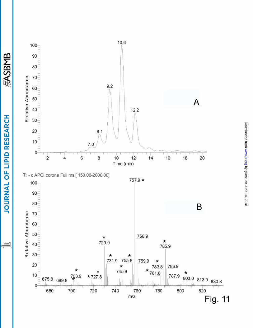
Very long chain (O-acyl)-ω-hydroxy fatty acids.
Another group of WE-related, but poorly characterized, compounds was identified in
meibum. These compounds were detected in the negative ion mode as a series of HPLC
peaks with RT between 8 and 12 min with ions m/z 729.9, 757.9, and 785.9 being the major
ones (Fig. 11). A range of related compounds with varying degree of unsaturation, chain
lengths, and locations of their hydroxy groups were detected, as well. As these compounds
were visible as anions, they either: 1) had an easily ionizable acidic group(s) in their
structures; 2) formed adducts with acidic components of the HPLC eluent (i.e. formate), or,
3) formed inorganic anion adducts (most likely, Cl-). However, the last two possibilities were
ruled out as the signals were detected in various tested solvents using various MS ion sources
by guest, on June 14, 2018w
ww
.jlr.orgD
ownloaded from

12
(APCI, electrospray ionization, and photoionization). Thus, the most likely explanation was
the acidic nature of the compounds.
To determine their structures, the compounds m/z/ 729, 757, and 785 were fragmented in
MS2 experiments (Fig. 12). All three major compounds produced a strong signal of a product
ion m/z 281. The other major product ions had m/z values of 465, 493, and 521 for
compounds m/z 729, 757, and 785, correspondingly. Interestingly, compound m/z 729
showed another pair of product ions, 253 and 493, which apparently added up to the same
parent ion as 281 and 465 did. This was a clear sign of the presence of at least two isobaric
compounds in the mixture. We hypothesized that these compounds might be related to the
poorly characterized families of di- and triesters of various kinds postulated by Nicolaides
and Santos (19), specifically (O-acyl)-ω-hydroxy FA family (OAHFA). To test this
hypothesis, we needed a standard of such compound to be analyzed by HPLC and
fragmented the same way the meibum compounds were. Unfortunately, to the best of our
knowledge, no such standard was/is available from any manufacturer. Thus, we synthesized a
related, albeit shorter, compound – (O-oleoyl)-16-hydroxypalmitic acid, in-house. Under the
conditions of RP-HPLC analysis, it’s RT was an expectedly short ~4.2-4.5 min, which
reflected its lower hydrophobicity compared with its meibum counterparts. However, in an
negative ion mode MS2 experiment (O-oleoyl)-16-hydroxypalmitic acid fragmented exactly
like the meibum compounds did producing just two major fragments m/z 281 and 271 (Fig.
13). These product anions were clearly those of oleic acid and 16-hydroxypalmitic acid. This
finding allowed us to assign the following structures to the meibum compounds – (O-oleoyl)-
30-hydroxy-triacontenoic and (O-palmitoleoyl)-32-hydroxy-dotriacontenoic acid (both m/z
729), (O-oleoyl)-32-hydroxy-dotriacontenoic acid and (O-palmitoleoyl)-34-hydroxy-
tetratriacontenoic acid (both m/z 757), and (O-oleoyl)-34-hydroxy-tetratriacontenoic acid
(m/z 785) (Scheme 4). The other members of the OAHFA family varied by the carbon chain
length, the number of double bonds, and, possibly, their location (Table 2). Experiments are
in progress to elucidate their structure in more details.
Discussion.
Earlier, we presented our initial results on the characterization of the major nonpolar lipid
species found in human meibum (9-11, 15) and AT (15). The results confirmed independent
by guest, on June 14, 2018w
ww
.jlr.orgD
ownloaded from

13
observations of a number of research laboratories on the presence of large amounts of CE
and WE in human meibum (18-23). However, we could not concur on the reported findings
on DAG (12, 13), oleamide (24), Cer and other typical polar lipids (25, 26). If the
compounds in question are in fact present in normal meibum, their molar ratio to other
meibum lipids (primarily, WE and CE) is extremely small, and it seems to be unlikely that
they could possibly play any structural role in the normal TF. Recently, we discussed in
details CE found in human meibum (11). Thus, the focus of this manuscript is on the second
major group of lipids in meibum, namely WE and related compounds.
To characterize WE, we chose RP HPLC. Unlike NP HPLC technique used to
characterize lipid classes (Fig. 1 and (9, 10), RP HPLC was to separate the individual
members of the WE class based on their hydrophobicity, i.e. chain length, unsaturation,
degree of oxidation, etc. Indeed, RP HPLC had provided separation of various CE species
(11), and was effective in separating WE, too (Fig. 2). A commendable separation of WE
species achieved in these experiments, ability to simultaneously monitor all of the
compounds of interest using their characteristic m/z values and plot them as individual
chromatograms retrospectively, and the ability to compare the RT of meibum compounds
with the RT of authentic lipid standards (where available) (Figs. 2-5, 7), plus the MSn
capabilities of the ion trap mass spectrometer (Figs. 6, 9, 10) made it possible to characterize
WE and related compounds to a much better degree than before. It became clear that WE are
predominantly based on a series of C18:n-based FA (n=1-4), with FAl moiety ranging from
~C17:0 to C32:0 (Table 1). Note that WE can be grouped in sub-families in different ways
based on either their chain length, or unsaturation. Figs. 1-5 clearly demonstrate the
astounding complexity of this class of compounds alone. However, three more factors need
to be discussed – the locations of the double bounds, their cis, trans geometry, and
branching. The location of the double bonds could not be evaluated in our current
experiments and would require chemical modification of the samples as described by
Thomas et al. (27), or sample hydrolysis or transesterification with subsequent silylation and
ozonation of the hydrolyzate, followed by gas-liquid chromatography (GLC) or gas
chromatography-mass spectrometry (GC-MS) (28). The experiments are in progress to
address these issues. In any case, it seems likely that a change in the location of a double
bond in a molecule would have some impact on the RT of the isomeric compound. The
by guest, on June 14, 2018w
ww
.jlr.orgD
ownloaded from

14
cis,trans geometry of the double bonds was also shown to have a noticeable effect on the RT
of the corresponding geometrical isomers (29). Thus, we analyzed the chromatographic
patterns of selected ion families (as an example, ions m/z 641, 643, 645, and 647 were
considered; other tested families of ions followed the trend) and found that the number of
detected HPLC peaks for a given ion was proportional to the degree of unsaturation of the
corresponding compound (Fig. 7): compounds with 1, 2, 3, and 4 double bonds produced,
correspondingly, 1, 2, 3, and 4 HPLC peaks. The number of double bonds in a molecule, and
its cis/trans isomers ratio both influence the physiochemical properties of the compounds: the
higher they are, the lower the melting point of the mixture. With the increase of the degree of
unsaturation, the fragmentation pattern of the compounds also changes indicating lesser
stability of the more unsaturated compounds (Fig. 6; only two extreme cases for 1 and 4
double bonds are presented). The mechanism for the increase of the number of isoforms of
the same compound remains unclear and could include peculiarities of their biosynthesis
and/or post-biosynthetic isomerization.
Theoretically, the presence of one, or more, double bounds in their structure may make
lipids susceptible to enzymatic and/or nonenzymatic (per)oxidation. One of the possible
types of oxidation products reported earlier is the formation of lipid epoxides (18). Epoxides
can be formed in vivo enzymatically or nonenzymatically (30). Under normal conditions,
direct attachment of oxygen to the double bonds is unlikely (31). However, their enzymatic
epoxidation through, for example, cytochrome P450 is a possibility. CYP2B12 transcripts
were found in the mouse skin and meibomian glands (32, 33) studied, among the others, the
cytochrome P450 gene regulation by 17β-estradiol in mouse meibomian glands (33). Its
CYP1B1 variant was localized in various human ocular structures, including cornea (34).
Yet, our experiments did not produce any evidence of the epoxides of free FA and WE being
a noticeable part of human meibum (Table 1 and Fig. 10). This was in contrast with earlier
findings of Shine et al (18) on the meibum epoxides of unclear origin, and those of Sullivan
et al (12) who reported epoxides of DAG in human meibum. However, very little information
on how the putative epoxides of DAG were identified and analyzed is presented in (12), and
no actual chemical structures were proposed in either of the reports. Moreover, we have not
detected any appreciable amounts of regular DAG in human meibum (9, 10, 15), which
makes the existence of their epoxides even less likely. Shine et al (18) reported epoxides of
by guest, on June 14, 2018w
ww
.jlr.orgD
ownloaded from
15
C18 and C20 acids as prominent meibum lipids. They, however, analyzed heavily chemically
manipulated samples which could have undergone inadvertent spontaneous post-extraction
epoxidation, while our current findings are based on the direct analyses of unmanipulated
intact lipid samples, which greatly reduces the chances of their oxidation and isomerization.
Lastly, an interesting, but poorly characterized, group of lipids, namely very long chain
OAHFA, is of special interest. Their existence was originally postulated by Nicolaides (19)
25 years ago. However, the relatively high molecular weights of OAHFA made it impossible
to analyze them directly by GLC-MS or GC-MS, the earlier standard analytical techniques)
without hydrolyzing them first. Unfortunately, by doing so, all the information on the intact
compounds is lost, and the structures of the starting intact compounds (i.e. the exact
combinations of their detected hydrolyzed fragments) cannot be reconstructed. The
demonstrated diversity of these compounds (see Scheme 4 and Fig. 11) made the earlier
attempts a difficult, but courageous, adventure. Earlier, we reported the presence of the then-
unidentified species in meibum (10). The absence of chemical standards did not allow us to
positively identify the compounds. Once a related compound, (O-oleoyl)-16-hydroxypalmitic
acid, had been synthesized in our laboratory, and its fragmentation pattern had been
determined, OAHFA identification became possible. More than 20 related lipid species were
observed in direct experiments (Fig. 11 and Table 2). However, this number could possibly
much greater if one considers the (quite possible) existence of multiple isobaric compounds
(Scheme 4) of the same nature.
What physiological significance these compounds may have? In skin, they a part of a
family of very long chain (O-acyl)-Cer (35). Recently, the latter were linked to a vital
function of skin permeability (35, 36). However, neither (O-acyl)-Cer, nor regular Cer were
found in our studies as the major meibum components. Thus, OAHFA should play some
other role in meibum and TFLL. A striking difference between them and the meibum lipids
of regular type (WE, CE, and TAGs) described in our earlier publications is that OAHFA are
carrying an overall negative charge due to their free carboxyl groups, while the other lipids
are electroneutral and very hydrophobic. Indeed, the carboxyl group of a typical aliphatic
compound has a pKa value of around 4.5, which may slightly change one way or the other
depending on the chain length and the solvent. However, under the conditions of
physiological pH (~7.5 for tears (37)) a large part of these groups is ionized thus providing
by guest, on June 14, 2018w
ww
.jlr.orgD
ownloaded from

16
them amphiphilic and surfactant properties. With the absence of appreciable amounts of
phospholipids, Cer, MAG and DAG in meibum (9-11, 15), OAHFA are perfect candidates
for fulfilling the role of a somewhat elusive amphiphilic barrier between the postulated very
thick nonpolar TF lipid sub-layer formed of WE, CE, TAG etc., and the underlaying aqueous
layer which is in contact with the cornea (38). Thus, a finely tuned balance between OAHFA
and other, less polar, components of the human TFLL could play a role in its stabilization.
Future work will address this hypothesis.
by guest, on June 14, 2018w
ww
.jlr.orgD
ownloaded from

17
Table 1. Major intact WE of human meibum and their epoxidized derivatives detected in the
positive ion mode*
Intact wax esters found in meibum,
general formula CnH2n-2O2 [11]
Epoxidized wax esters,
general formula CnH2n-2O3
n Compound, FA:FAl
Molecular formula
m/z (H+ adduct)
Compound, epFA:FAl
Molecular formula
m/z (H+ adduct)
Detected in meibum, Yes/No
35 C18:1:C17:0 C35H68O2 521.53 epC18:0:C17:0 C35H68O3 537.52 no
36 C18:1:C18:0 C36H70O2 535.54 epC18:0:C18:0 C36H70O3 551.54 no
37 C18:1:C19:0 C37H72O2 549.56 epC18:0:C19:0 C37H72O3 565.55 no
38 C18:1:C20:0 C38H74O3 563.58 epC18:0:C20:0 C38H74O3 579.57 no
39 C18:1:C21:0 C39H76O2 577.59 epC18:0:C21:0 C39H76O3 593.59 no
40 C18:1:C22:0 C40H78O2 591.61 epC18:0:C22:0 C40H78O3 607.60 no
41 C18:1:C23:0 C41H80O2 605.62 epC18:0:C23:0 C41H80O3 621.62 no
42 C18:1:C24:0 C42H82O2 619.64 epC18:0:C24:0 C42H82O3 635.63 no
43 C18:1:C25:0 C43H84O2 633.65 epC18:0:C25:0 C43H84O3 649.65 no
44 C18:1:C26:0 C44H86O2 647.67 epC18:0:C26:0 C44H86O3 663.66 no
45 C18:1:C27:0 C45H88O2 661.69 epC18:0:C27:0 C45H88O3 677.68 no
46 C18:1:C28:0 C46H90O2 675.70 epC18:0:C28:0 C46H90O3 691.70 no
47 C18:1:C29:0 C47H92O2 689.72 epC18:0:C29:0 C47H92O3 705.71 no
48 C18:1:C30:0 C48H94O2 703.73 epC18:0:C30:0 C48H94O3 719.73 no
49 C18:1:C31:0 C49H96O2 717.75 epC18:0:C31:0 C49H96O3 733.74 no
50 C18:1:C32:0 C50H98O2 731.76 epC18:0:C32:0 C50H98O3 747.76 no
* Only 9,10-epoxy-C18:0-based WE are shown.
by guest, on June 14, 2018w
ww
.jlr.orgD
ownloaded from

18
Table 2. Ten major (O-acyl)-ω-hydroxy FA of human meibum detected in the negative ion
mode
Detected m/z,
(M-H)-
Relative intensity of MS
signals
OAHFA anion, (M-H)-,
CnH2n-7O4
Proposed structures, (O-FA1)-
(ω−hydroxy-FA2)*
Theoretical m/z, (M-H)-
701.8 0.057454 C46H85O4 701.6
703.9 0.062461 C46H87O4 703.7
729.8 0.387235 C48H89O4 (O-C16:1)-ωC32:1
(O-C18:1)-ωC30:1 729.7
743.8 0.066614 C49H91O4 743.7
745.8 0.10733 C49H93O4 745.7
755.7 0.213779 C50H91O4 755.7
757.8 1** C50H93O4 (O-C16:1)-ωC34:1
(O-C18:1)-ωC32:1 757.7
781.7 0.091433 C52H93O4 781.7
783.8 0.154135 C52H95O4 783.7
785.8 0.355308 C52H97O4 (O-C18:1)-ωC34:1 785.7
* FA1 – a shorter chain FA; FA2 – a very long chain ω-hydroxy FA; structures confirmed by HPLC and MS/MS analyses ** Most intense signal
by guest, on June 14, 2018w
ww
.jlr.orgD
ownloaded from

19
Legends to the Figures. Scheme 1. Chemical structures of intact and epoxidized WE Scheme 2. Fragmentation pattern of 9,10-epoxy-stearic acid methyl ester Scheme 3. A family of related intact and epoxidized wax esters Scheme 4. A family of major very long chain (O-acyl)-ω-hydroxy FA detected inhuman meibum Figure 1. Normal phase chromatograms and MS spectrum of nonpolar lipids present in human meibum. Positive ion mode. Panel A. Total ion chromatogram (TIC) and selected ion monitoring (SIM) traces of wax esters ions m/z 563, 619, and 673 extracted from the TIC. Panel B. Mass spectrum of the HPLC peak with the retention time of 3.6 min. Positive ion mode. Figure 2. Reversed phase HPLC-MS analysis of human meibum in the positive ion mode. Panel A. TIC of the whole meibum sample. Panel B. SIM of ion m/z 563 Panel C. SIM of ion m/z 619 Panel D. SIM of ion m/z 673 Figure 3. Direct HPLC-MS comparison of selected authentic lipid standards and the corresponding wax esters of human meibum. Positive ion mode. Panel A. Authentic stearyl oleate, m/z 535 Panel B. Meibum compound m/z 535 Panel C. Authentic arachidyl oleate, m/z 563 Panel D. Meibum compound m/z 563 Panel E. Authentic behenyl oleate, m/z 591 Panel F. Meibum compound m/z 591 Figure 4. Direct HPLC-MS comparison of synthesized in-house very long chain wax esters and the corresponding compounds of human meibum. Positive ion mode. Panel A. RP chromatogram of synthetic hexacosanyl oleate, m/z 647 Panel B. RP chromatogram of synthetic octacosanyl oleate, m/z 675 Panel C. RP chromatogram of a meibum compound m/z 647 Panel D. RP chromatogram of a meibum compound m/z 675 Panel E. MS spectrum of synthetic hexacosanyl oleate, peak RT 14.3 min Panel F. MS spectrum of synthetic octacosanyl oleate, peak RT 16.4 min Panel G. MS spectrum of meibum compound m/z 647, peak RT 14.2 min Panel H. MS spectrum of meibum compound m/z 675, peak RT 16.2 min
by guest, on June 14, 2018w
ww
.jlr.orgD
ownloaded from

20
Figure 5. Expanded MS spectrum of human meibum wax esters (Panel A) and SIM RP chromatograms of a family of homologous wax esters differing in the degree of their unsaturation (Panels B through E). Ion m/z 627 – C18:4 (stearidonic acid)-based ester of C25:0 (pentacosanol) alcohol Ion m/z 629 – C18:3 (linolenic acid)-based ester of pentacosanol Ion m/z 631 – C18:2 (linoleic acid)-based ester of pentacosanol Ion m/z 633 – C18:1 (oleic acid)-based ester of pentacosanol Figure 6. Effects of the degree of unsaturation on the fragmentation spectra of wax esters. Panel A. Fragmentation spectrum of ion m/z 645 (linoleic acid-based ester of hexacosanol; positive ion mode). Note the formation of a product ion m/z 281 (a proton adduct of linoleic acid), and its dehydration products 263 and 245. Indicative is the absence of prominent ions in the range of m/z 400 to 600. Panel B. Fragmentation spectrum of ion m/z 641 (stearidonic acid-based ester of hexacosanol; positive ion mode). Note the formation of a product ion m/z 277 (a proton adduct of stearidonic acid), and its dehydration products 259 and 241. Indicative is the presence of a large number of prominent ions in the range of m/z 400 to 600 caused by the lesser stability of polyunsaturated FA under the conditions of MS/MS. Figure 7. The number of double bonds in the series of homologous wax esters correlates with the number of the detected HPLC peaks. Panel A. SIM RP HPLC trace of ion m/z 641 (positive ion mode; four isomers detected; all have similar fragmentation patterns) Panel B. SIM RP HPLC trace of ion m/z 643 (three isomers detected) Panel C. SIM RP HPLC trace of ion m/z 645 (one major, and two minor isomers detected) Panel D. SIM RP HPLC trace of ion m/z 647 one major isomer detected) Figure 8. RP HPLC-MS analysis of cis- and trans-9,10-epoxy stearic acids in the positive ion mode. Panel A. Chromatograms of the compounds demonstrated no difference in the RT Panel B. Mass spectrum of cis-9,10-epoxy stearic acid Panel C. Mass spectrum of trans-9,10-epoxy stearic acid Figure 9. Fragmentation spectrum of cis-9,10-epoxy stearic acid taken in the positive ion mode (Panel A) and the TIC of the compound (Panel B). Figure 10. Human meibum before (Panel A) and after (Panel B) epoxidation with peroxyacetic acid. Figure 11. RP HPLC-MS analysis of human meibum in the negative ion mode. Panel A. TIC chromatogram Panel B. Expanded MS spectrum of the peaks shown in Panel A. Major compounds are labeled with asterisk.
by guest, on June 14, 2018w
ww
.jlr.orgD
ownloaded from

21
Figure 12. Structural analysis of compound m/z 757 in the negative ion mode. Panel A. SIM of ion m/z 757 showed the presence of one major, and one minor RP HPLC peak. Panel B. Fragmentation pattern of ion m/z 757, its proposed structure, and identified fragments. Figure 13. RP HPLC-MS analysis of an authentic (O-oleoyl)-16-hydroxypalmitic acid. Panel A. SIM of ion m/z 535 showed the presence of one major RP HPLC peak (RT ~4.4 min). Panel B. Zoom scan MS spectrum of the compound Panel C. Fragmentation pattern of ion m/z 535, its proposed structure, and identified fragments: m/z 281 (oleate) and 271 (16-hydroxypalmitate)
by guest, on June 14, 2018w
ww
.jlr.orgD
ownloaded from

22
References.
1. Tiffany, J. M. 2008. The normal tear film. Dev Ophthalmol 41: 1-20. 2. Bron, A. J., J. M. Tiffany, S. M. Gouveia, N. Yokoi, and L. W. Voon. 2004. Functional aspects of the tear film lipid layer. Exp Eye Res 78: 347-360. 3. Bron, A. J., and J. M. Tiffany. 1998. The meibomian glands and tear film lipids - Structure, function, and control. Lacrimal Gland, Tear Film, and Dry Eye Syndromes 2 438: 281-295. 4. Sullivan, D. A., H. Yamagami, M. Liu, R. J. Steagall, F. Schirra, T. Suzuki, K. L. Krenzer, J. M. Cermak, R. M. Sullivan, S. M. Richards, D. A. Schaumberg, M. R. Dana, and B. D. Sullivan. 2002. Sex steroids, the meibomian gland and evaporative dry eye. Adv Exp Med Biol 506: 389-399 5. Ohashi, Y., M. Dogru, and K. Tsubota. 2006. Laboratory findings in tear fluid analysis. Clin Chim Acta 369: 17-28. 6. Foulks, G. N. 2007. The correlation between the tear film lipid layer and dry eye disease. Surv Ophthalmol 52: 369-374. 7. McCulley, J. P., and W. E. Shine. 2001. The lipid layer: the outer surface of the ocular surface tear film. Biosci Rep 21: 407-418. 8. Mathers, W. D., and J. A. Lane. 1998. Meibomian gland lipids, evaporation, and tear film stability. Adv Exp Med Biol 438: 349-360. 9. Butovich, I. A., E. Uchiyama, M. A. Di Pascuale, and J. P. McCulley. 2007. Liquid chromatography-mass spectrometric analysis of lipids present in human meibomian gland secretions. Lipids 42: 765-776. 10. Butovich, I. A., E. Uchiyama, and J. P. McCulley. 2007. Lipids of human meibum: mass-spectrometric analysis and structural elucidation. J Lipid Res 48: 2220-2235. 11. Butovich, I. A. 2009. Cholesteryl esters as a depot for very long chain fatty acids in human meibum. J Lipid Res 50: 501-513. 12. Sullivan, B. D., J. E. Evans, K. L. Krenzer, M. Reza Dana, and D. A. Sullivan. 2000. Impact of antiandrogen treatment on the fatty acid profile of neutral lipids in human meibomian gland secretions. J Clin Endocrinol Metab 85: 4866-4873. 13. Krenzer, K. L., M. R. Dana, M. D. Ullman, J. M. Cermak, D. B. Tolls, J. E. Evans, and D. A. Sullivan. 2000. Effect of androgen deficiency on the human meibomian gland and ocular surface. J Clin Endocrinol Metab 85: 4874-4882. 14. Borchman, D., G. N. Foulks, M. C. Yappert, D. Tang, and D. V. Ho. 2007. Spectroscopic evaluation of human tear lipids. Chem Phys Lipids 147: 87-102. 15. Butovich, I. A. 2008. On the lipid composition of human meibum and tears: comparative analysis of nonpolar lipids. Invest Ophthalmol Vis Sci 49: 3779-3789. 16. Smith, K. R., and D. M. Thiboutot. 2008. Thematic review series: skin lipids. Sebaceous gland lipids: friend or foe? J Lipid Res 49: 271-281. 17. Feingold, K. R. 2007. The importance of lipids in cutaneous function. J Lipid Res 48: 2529-2530. 18. Shine, W. E., and J. P. McCulley. 1993. Role of wax ester fatty alcohols in chronic blepharitis. Invest Ophthalmol Vis Sci 34: 3515-3521. 19. Nicolaides, N., and E. C. Santos. 1985. The di- and triesters of the lipids of steer and human meibomian glands. Lipids 20: 454-467.
by guest, on June 14, 2018w
ww
.jlr.orgD
ownloaded from

23
20. Nicolaides, N., J. K. Kaitaranta, T. N. Rawdah, J. I. Macy, F. M. Boswell, 3rd, and R. E. Smith. 1981. Meibomian gland studies: comparison of steer and human lipids. Invest Ophthalmol Vis Sci 20: 522-536. 21. Shine, W. E., and J. P. McCulley. 1991. The role of cholesterol in chronic blepharitis. Invest Ophthalmol Vis Sci 32: 2272-2280. 22. Shine, W. E., and J. P. McCulley. 2000. Association of meibum oleic acid with meibomian seborrhea. Cornea 19: 72-74. 23. Joffre, C., M. Souchier, S. Gregoire, S. Viau, L. Bretillon, N. Acar, A. M. Bron, and C. Creuzot-Garcher. 2008. Differences in meibomian fatty acid composition in patients with meibomian gland dysfunction and aqueous-deficient dry eye. Br J Ophthalmol 92: 116-119. 24. Nichols, K. K., B. M. Ham, J. J. Nichols, C. Ziegler, and K. B. Green-Church. 2007. Identification of fatty acids and fatty acid amides in human meibomian gland secretions. Invest Ophthalmol Vis Sci 48: 34-39. 25. Shine, W. E., and J. P. McCulley. 2003. Polar lipids in human meibomian gland secretions. Curr Eye Res 26: 89-94. 26. Shine, W. E., and J. P. McCulley. 2004. Meibomianitis: polar lipid abnormalities. Cornea 23: 781-783. 27. Thomas, M. C., T. W. Mitchell, D. G. Harman, J. M. Deeley, J. R. Nealon, and S. J. Blanksby. 2008. Ozone-induced dissociation: elucidation of double bond position within mass-selected lipid ions. Anal Chem 80: 303-311. 28. Butovich, I. A., M. Hamberg, and O. Radmark. 2005. Novel oxylipins formed from docosahexaenoic acid by potato lipoxygenase - 10(S)-hydroxydocosahexaenoic acid and 10,20-dihydroxydocosahexaenoic acid. Lipids 40: 249-257. 29. Butovich, I. A., and C. C. Reddy. 2001. Enzyme-catalyzed and enzyme-triggered pathways in dioxygenation of 1-monolinoleoyl-rac-glycerol by potato tuber lipoxygenase. Biochim Biophys Acta 1546: 379-398. 30. Schneider, C., W. E. Boeglin, H. Yin, N. A. Porter, and A. R. Brash. 2008. Intermolecular peroxyl radical reactions during autoxidation of hydroxy and hydroperoxy arachidonic acids generate a novel series of epoxidized products. Chem Res Toxicol 21: 895-903. 31. Yamada, T., T. Takai, O. Rhode, and T. Mukaiyama. 1991. Direct Epoxidation of Olefins Catalyzed by Nickel(Ii) Complexes with Molecular-Oxygen and Aldehydes. Bulletin of the Chemical Society of Japan 64: 2109-2117. 32. Keeney, D. S., C. Skinner, S. Wei, T. Friedberg, and M. R. Waterman. 1998. A keratinocyte-specific epoxygenase, CYP2B12, metabolizes arachidonic acid with unusual selectivity, producing a single major epoxyeicosatrienoic acid. J Biol Chem 273: 9279-9284. 33. Suzuki, T., F. Schirra, S. M. Richards, R. V. Jensen, and D. A. Sullivan. 2008. Estrogen and progesterone control of gene expression in the mouse meibomian gland. Invest Ophthalmol Vis Sci 49: 1797-1808. 34. Doshi, M., C. Marcus, B. A. Bejjani, and D. P. Edward. 2006. Immunolocalization of CYP1B1 in normal, human, fetal and adult eyes. Exp Eye Res 82: 24-32. 35. McMahon, A., I. A. Butovich, N. L. Mata, M. Klein, R. Ritter, 3rd, J. Richardson, D. G. Birch, A. O. Edwards, and W. Kedzierski. 2007. Retinal pathology and skin barrier defect in mice carrying a Stargardt disease-3 mutation in elongase of very long chain fatty acids-4. Mol Vis 13: 258-272. 36. Zuo, Y., D. Z. Zhuang, R. Han, G. Isaac, J. J. Tobin, M. McKee, R. Welti, J. L. Brissette, M. L. Fitzgerald, and M. W. Freeman. 2008. ABCA12 maintains the epidermal lipid
by guest, on June 14, 2018w
ww
.jlr.orgD
ownloaded from

24
permeability barrier by facilitating formation of ceramide linoleic esters. J Biol Chem 283: 36624-36635. 37. Fischer, F. H., and M. Wiederholt. 1982. Human precorneal tear film pH measured by microelectrodes. Graefes Arch Clin Exp Ophthalmol 218: 168-170. 38. McCulley, J. P., and W. Shine. 1997. A compositional based model for the tear film lipid layer. Trans Am Ophthalmol Soc 95: 79-88; discussion 88-93.
by guest, on June 14, 2018w
ww
.jlr.orgD
ownloaded from

O
O
C18
R
C18
O
O R
O
R= CmH2m+1; m=17-32intact wax esters
epoxidized wax esters
Scheme 1
by guest, on June 14, 2018w
ww
.jlr.orgD
ownloaded from

O
OO
H+
O
OHO
H+O
O
H+
OOH
+
O
OH
H+
OO
H+
O H+
Isotopic mass =313.27Molecular Formula =C19H37O3
Isotopic mass = 299.26Molecular Formula =C18H35O3
Isotopic mass = 295.26Molecular Formula =C19H35O2
Isotopic mass = 281.25Molecular Formula =C18H33O2
Isotopic mass = 263.24Molecular Formula =C18H31O
Isotopic mass (M+1) = 245.23Molecular Formula =C18H28
Scheme 2
by guest, on June 14, 2018 www.jlr.org Downloaded from

C18
O
OO
H+
C25
C18
O
O
H+
C26
C18
O
OO
H+
C25
O
O
H+
C26
C18
Isotopic mass = 649.65Molecular Formula =C43H85O3
Isotopic mass = 647.67Molecular Formula =C44H87O2
Isotopic mass = 647.63Molecular Formula =C43H83O3
Isotopic mass = 647.67Molecular Formula =C44H87O2
compound 1
compound 2
compound 3
compound 4
Scheme 3
by guest, on June 14, 2018w
ww
.jlr.orgD
ownloaded from

C16O
O C32
O
O
C18O
O
O
O
C30
C18O
O
O
O
C32
C16O
O
O
O
C34
C18O
O
O
O
C34
Isotopic mass = 729.68Molecular Formula =C48H89O4
Isotopic mass = 757.71Molecular Formula =C50H93O4
Isotopic mass = 785.74Molecular Formula =C52H97O4
Scheme 4
by guest, on June 14, 2018w
ww
.jlr.orgD
ownloaded from