Human Skeletal Anatomy
20
Eder, et al.: Laboratory Atlas of Anatomy and Physiology, Third Edition 2. Human Skeletal Anatomy Text © The McGraw-Hill Companies, 2001 45 2 Human Skeletal Anatomy Detail of Compact Bone C H A P T E R
Transcript of Human Skeletal Anatomy
Eder, et al.: Laboratory Atlas of Anatomy and Physiology, Third Edition
2. Human Skeletal Anatomy Text © The McGraw−Hill Companies, 2001
45
2Human Skeletal Anatomy
Detail of Compact Bone
C H A P T E R
Eder, et al.: Laboratory Atlas of Anatomy and Physiology, Third Edition
2. Human Skeletal Anatomy Text © The McGraw−Hill Companies, 2001
46 C H A P T E R 2
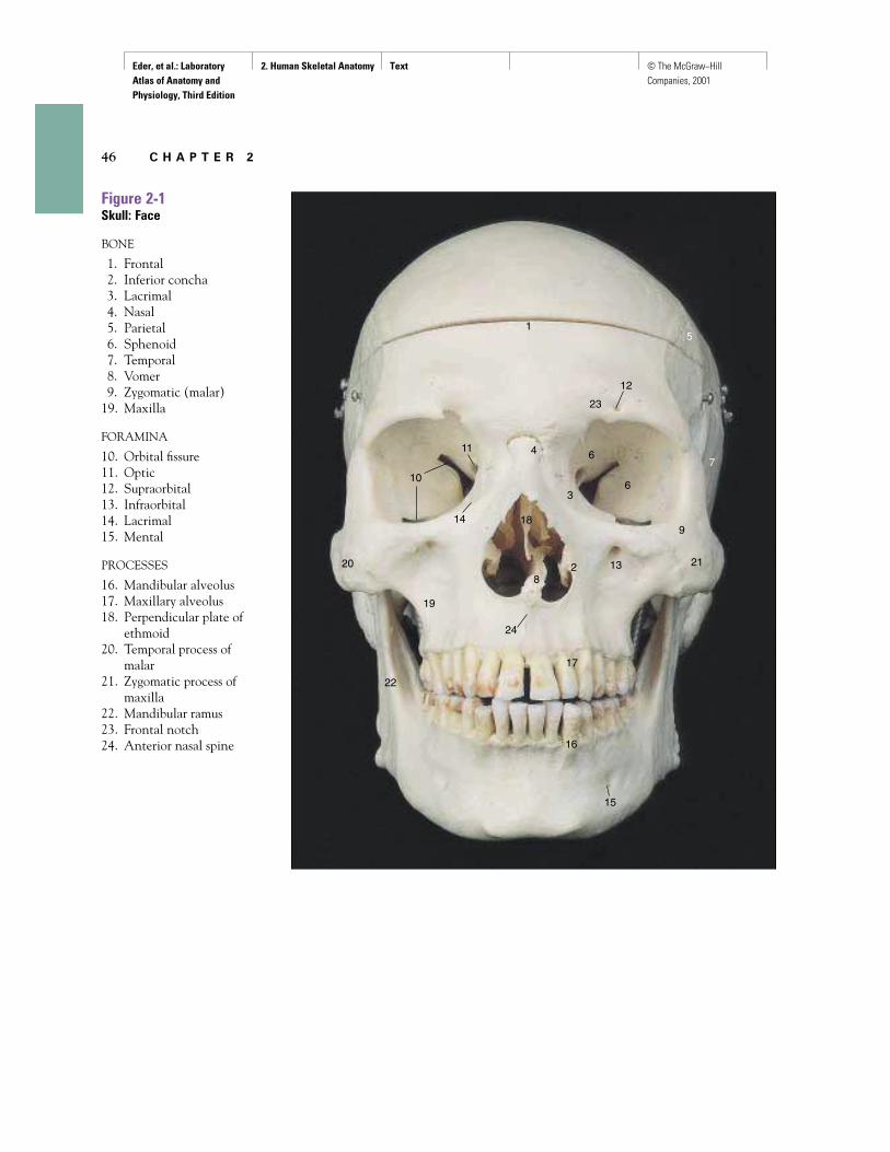
Figure 2-1Skull: Face
BONE
1. Frontal2. Inferior concha3. Lacrimal4. Nasal5. Parietal6. Sphenoid7. Temporal8. Vomer9. Zygomatic (malar)
19. Maxilla
FORAMINA
10. Orbital fissure11. Optic12. Supraorbital13. Infraorbital14. Lacrimal15. Mental
PROCESSES
16. Mandibular alveolus17. Maxillary alveolus18. Perpendicular plate of
ethmoid20. Temporal process of
malar21. Zygomatic process of
maxilla22. Mandibular ramus23. Frontal notch24. Anterior nasal spine
1
3
2
4
5
6
67
21
22
23
24
11
10
12
13
14
15
16
17
8
918
19
20
Eder, et al.: Laboratory Atlas of Anatomy and Physiology, Third Edition
2. Human Skeletal Anatomy Text © The McGraw−Hill Companies, 2001
H u m a n S k e l e t a l A n a t o m y 47
3
4
5
6
7
8
9
10
23
24
25
26
27
28
29
30
13
14
15
16
17
18
19
33
20
1
2
21 22
11
12
31
32
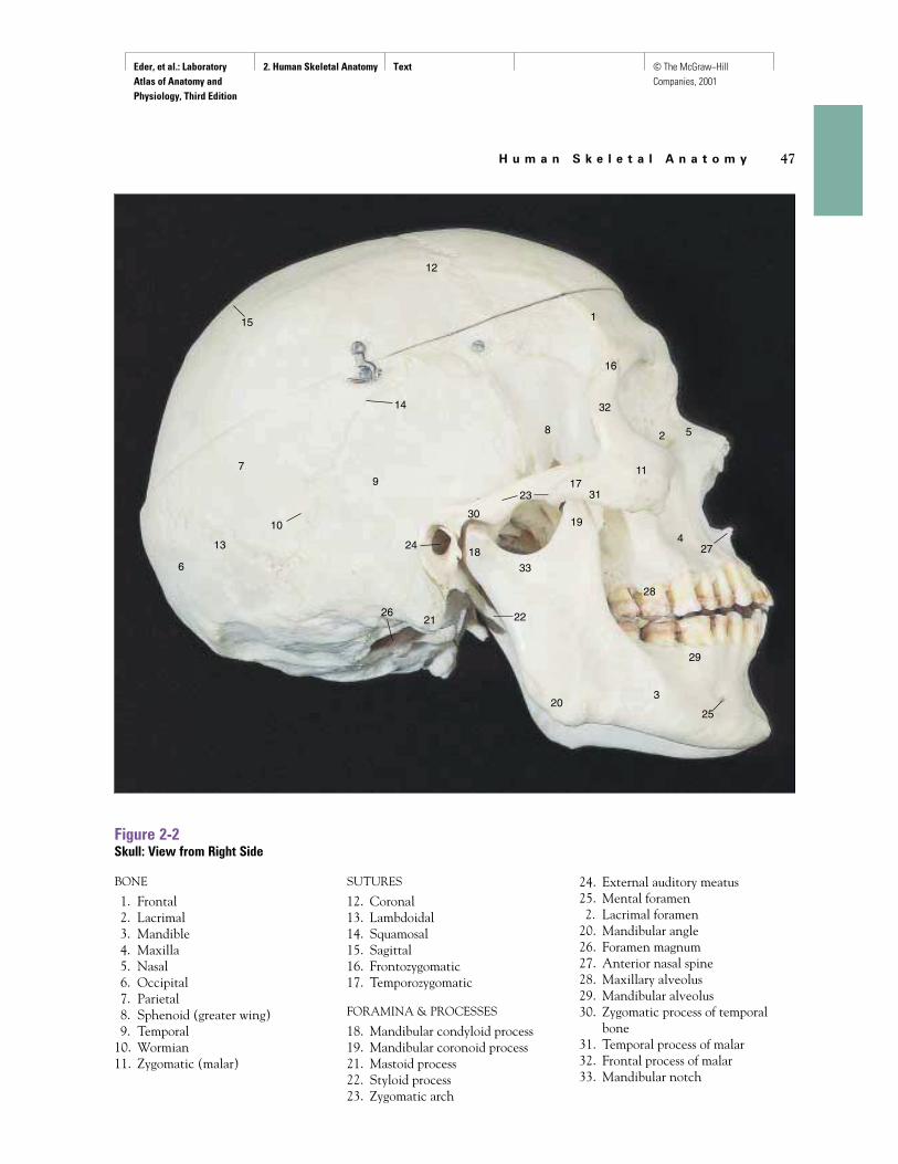
Figure 2-2Skull: View from Right Side
BONE
1. Frontal2. Lacrimal3. Mandible4. Maxilla5. Nasal6. Occipital7. Parietal8. Sphenoid (greater wing)9. Temporal
10. Wormian11. Zygomatic (malar)
SUTURES
12. Coronal13. Lambdoidal14. Squamosal15. Sagittal16. Frontozygomatic17. Temporozygomatic
FORAMINA & PROCESSES
18. Mandibular condyloid process19. Mandibular coronoid process21. Mastoid process22. Styloid process23. Zygomatic arch
24. External auditory meatus25. Mental foramen2. Lacrimal foramen
20. Mandibular angle26. Foramen magnum27. Anterior nasal spine28. Maxillary alveolus29. Mandibular alveolus30. Zygomatic process of temporal
bone31. Temporal process of malar32. Frontal process of malar33. Mandibular notch
Eder, et al.: Laboratory Atlas of Anatomy and Physiology, Third Edition
2. Human Skeletal Anatomy Text © The McGraw−Hill Companies, 2001
48 C H A P T E R 2
1
2
3
4
5
6
7
8
1
2
3
4
5
6
7
8 910 21
22
23
24
1112
13
14
15
15
15
16
17
18
19
20
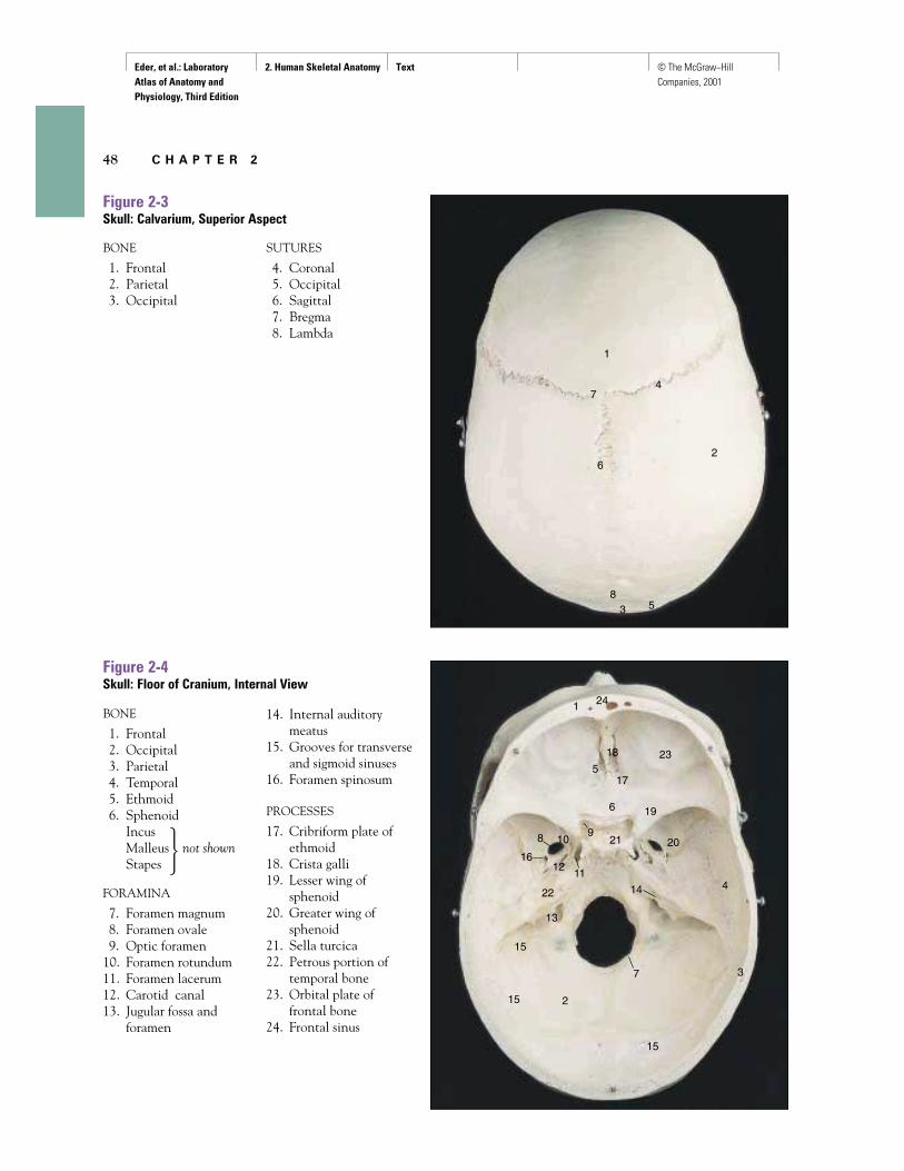
Figure 2-3Skull: Calvarium, Superior Aspect
Figure 2-4Skull: Floor of Cranium, Internal View
BONE
1. Frontal2. Occipital3. Parietal4. Temporal5. Ethmoid6. Sphenoid
IncusMalleus not shownStapes
FORAMINA
7. Foramen magnum8. Foramen ovale9. Optic foramen
10. Foramen rotundum11. Foramen lacerum12. Carotid canal13. Jugular fossa and
foramen
14. Internal auditorymeatus
15. Grooves for transverseand sigmoid sinuses
16. Foramen spinosum
PROCESSES
17. Cribriform plate ofethmoid
18. Crista galli19. Lesser wing of
sphenoid20. Greater wing of
sphenoid21. Sella turcica22. Petrous portion of
temporal bone23. Orbital plate of
frontal bone24. Frontal sinus
BONE
1. Frontal2. Parietal3. Occipital
SUTURES
4. Coronal5. Occipital6. Sagittal7. Bregma8. Lambda
�
Eder, et al.: Laboratory Atlas of Anatomy and Physiology, Third Edition
2. Human Skeletal Anatomy Text © The McGraw−Hill Companies, 2001
H u m a n S k e l e t a l A n a t o m y 49
1
2
2
3
4
4
5
6
7
8
9
10
2122
23
24
25
26
2728
11
12
13
141516
17
1818
20
31
32
33
34
12
34
5
6
7 8 9
10
11
12
13
14
30
19
29
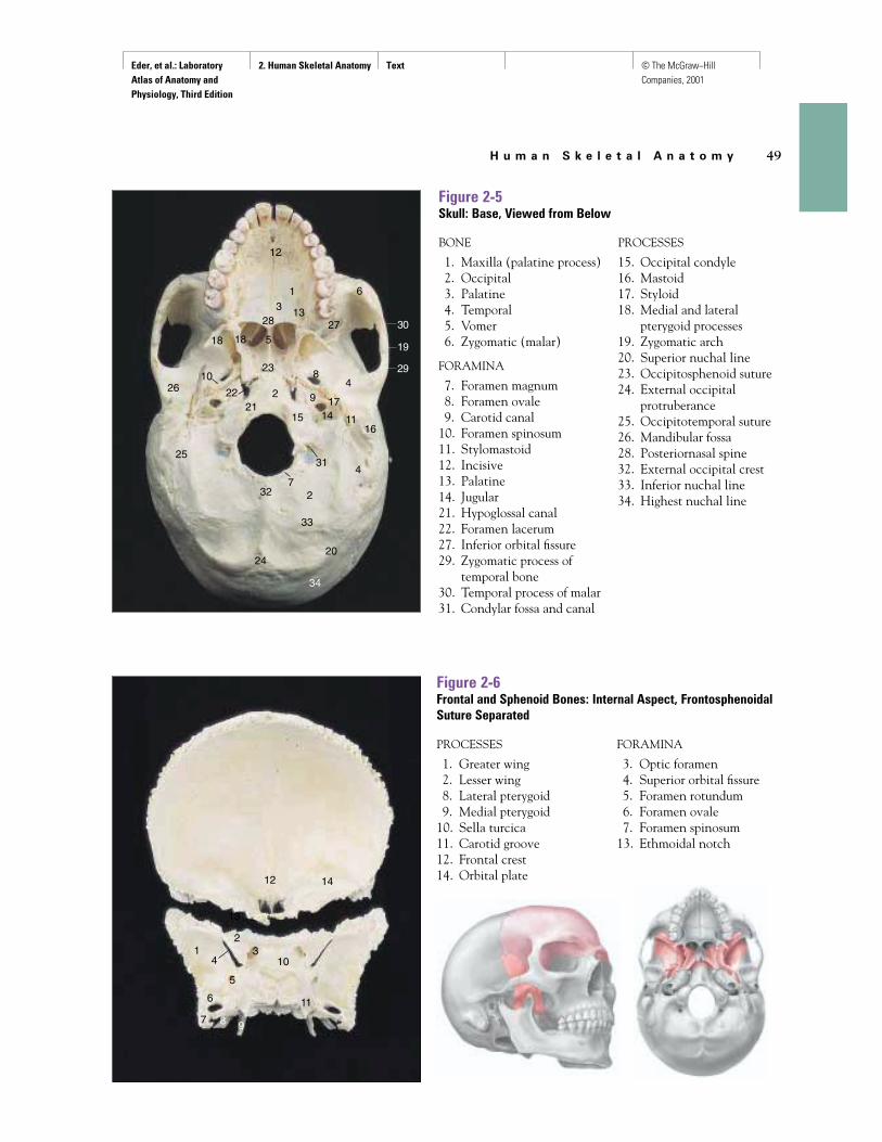
Figure 2-5Skull: Base, Viewed from Below
Figure 2-6Frontal and Sphenoid Bones: Internal Aspect, FrontosphenoidalSuture Separated
BONE
1. Maxilla (palatine process)2. Occipital3. Palatine4. Temporal5. Vomer6. Zygomatic (malar)
FORAMINA
7. Foramen magnum8. Foramen ovale9. Carotid canal
10. Foramen spinosum11. Stylomastoid12. Incisive13. Palatine14. Jugular21. Hypoglossal canal22. Foramen lacerum27. Inferior orbital fissure29. Zygomatic process of
temporal bone30. Temporal process of malar31. Condylar fossa and canal
PROCESSES
15. Occipital condyle16. Mastoid17. Styloid18. Medial and lateral
pterygoid processes19. Zygomatic arch20. Superior nuchal line23. Occipitosphenoid suture24. External occipital
protruberance25. Occipitotemporal suture26. Mandibular fossa28. Posteriornasal spine32. External occipital crest33. Inferior nuchal line34. Highest nuchal line
PROCESSES
1. Greater wing2. Lesser wing8. Lateral pterygoid9. Medial pterygoid
10. Sella turcica11. Carotid groove12. Frontal crest14. Orbital plate
FORAMINA
3. Optic foramen4. Superior orbital fissure5. Foramen rotundum6. Foramen ovale7. Foramen spinosum
13. Ethmoidal notch
Eder, et al.: Laboratory Atlas of Anatomy and Physiology, Third Edition
2. Human Skeletal Anatomy Text © The McGraw−Hill Companies, 2001
50 C H A P T E R 2
1
2
34
5
6
78 9
10
11
1
2 3
4
5
6
7
8
9
10
11
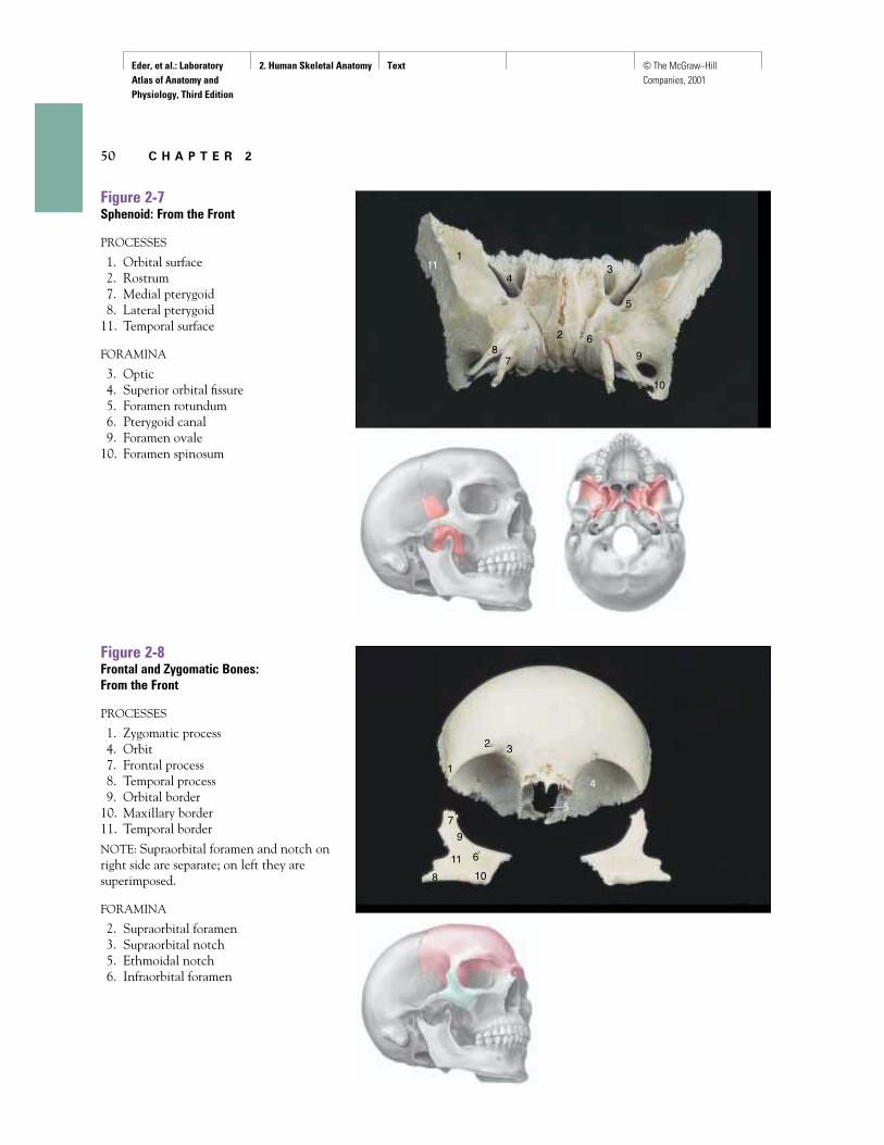
Figure 2-7Sphenoid: From the Front
PROCESSES
1. Orbital surface2. Rostrum7. Medial pterygoid8. Lateral pterygoid
11. Temporal surface
FORAMINA
3. Optic4. Superior orbital fissure5. Foramen rotundum6. Pterygoid canal9. Foramen ovale
10. Foramen spinosum
Figure 2-8Frontal and Zygomatic Bones: From the Front
PROCESSES
1. Zygomatic process4. Orbit7. Frontal process8. Temporal process9. Orbital border
10. Maxillary border11. Temporal border
NOTE: Supraorbital foramen and notch onright side are separate; on left they aresuperimposed.
FORAMINA
2. Supraorbital foramen3. Supraorbital notch5. Ethmoidal notch6. Infraorbital foramen
Eder, et al.: Laboratory Atlas of Anatomy and Physiology, Third Edition
2. Human Skeletal Anatomy Text © The McGraw−Hill Companies, 2001
H u m a n S k e l e t a l A n a t o m y 51
1
2
34
5
6
7
89
10
1
2 34
5
6
7
7
89
10
1112
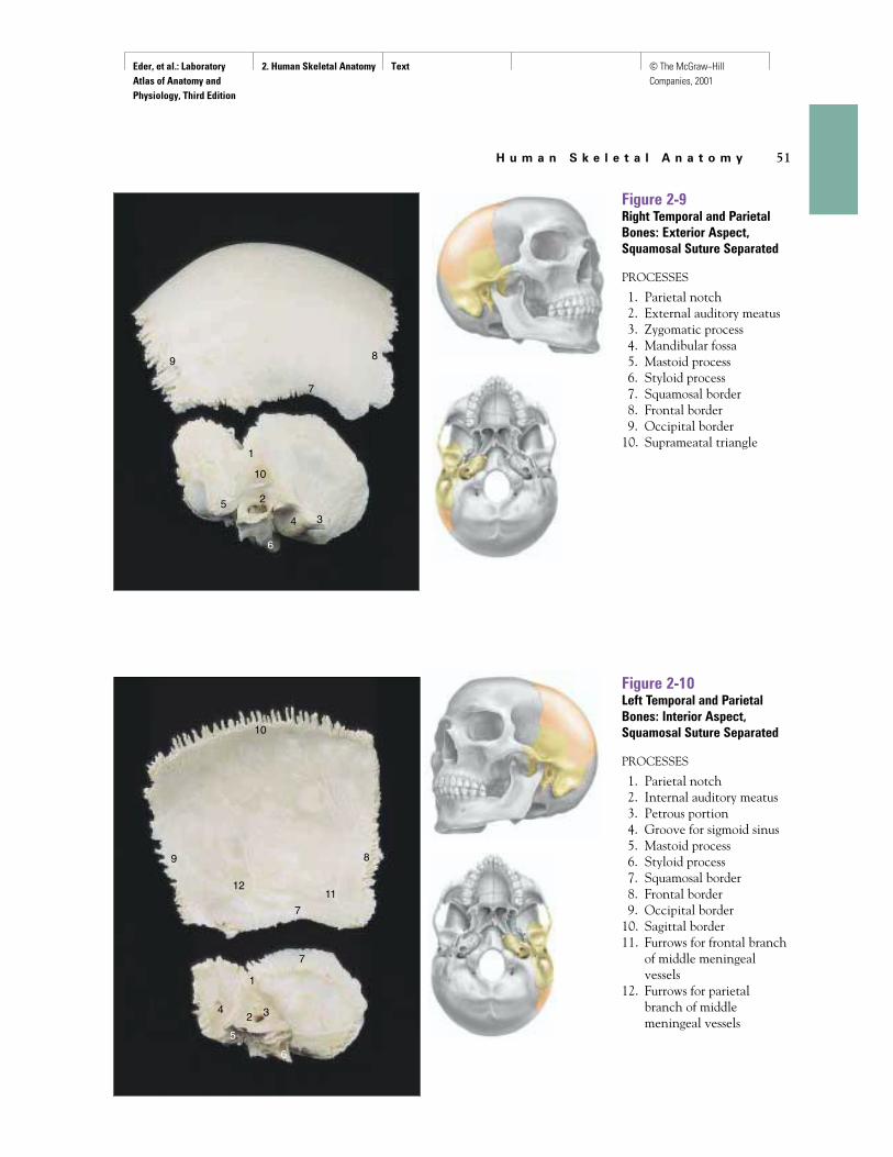
Figure 2-9Right Temporal and ParietalBones: Exterior Aspect,Squamosal Suture Separated
PROCESSES
1. Parietal notch2. External auditory meatus3. Zygomatic process4. Mandibular fossa5. Mastoid process6. Styloid process7. Squamosal border8. Frontal border9. Occipital border
10. Suprameatal triangle
Figure 2-10Left Temporal and ParietalBones: Interior Aspect,Squamosal Suture Separated
PROCESSES
1. Parietal notch2. Internal auditory meatus3. Petrous portion4. Groove for sigmoid sinus5. Mastoid process6. Styloid process7. Squamosal border8. Frontal border9. Occipital border
10. Sagittal border11. Furrows for frontal branch
of middle meningealvessels
12. Furrows for parietalbranch of middlemeningeal vessels
Eder, et al.: Laboratory Atlas of Anatomy and Physiology, Third Edition
2. Human Skeletal Anatomy Text © The McGraw−Hill Companies, 2001
52 C H A P T E R 2
1
2
3
45
6
7
89
10
11
12
13
1
A B
2 3
4
5
7
12
4
68
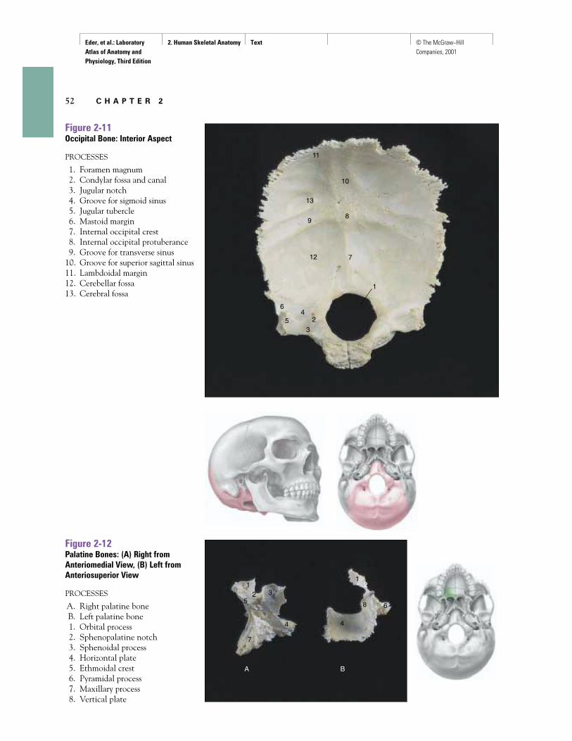
Figure 2-11Occipital Bone: Interior Aspect
PROCESSES
1. Foramen magnum2. Condylar fossa and canal3. Jugular notch4. Groove for sigmoid sinus5. Jugular tubercle6. Mastoid margin7. Internal occipital crest8. Internal occipital protuberance9. Groove for transverse sinus
10. Groove for superior sagittal sinus11. Lambdoidal margin12. Cerebellar fossa13. Cerebral fossa
Figure 2-12Palatine Bones: (A) Right fromAnteriomedial View, (B) Left fromAnteriosuperior View
PROCESSES
A. Right palatine boneB. Left palatine bone1. Orbital process2. Sphenopalatine notch3. Sphenoidal process4. Horizontal plate5. Ethmoidal crest6. Pyramidal process7. Maxillary process8. Vertical plate
Eder, et al.: Laboratory Atlas of Anatomy and Physiology, Third Edition
2. Human Skeletal Anatomy Text © The McGraw−Hill Companies, 2001
H u m a n S k e l e t a l A n a t o m y 53
A B
1
22
3
4
5
678
99
10
11 12
13
14
1516
17
18
19
1
23
4
5
6
78
3
9
10
11
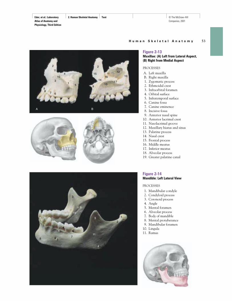
Figure 2-13Maxillae: (A) Left from Lateral Aspect, (B) Right from Medial Aspect
PROCESSES
A. Left maxillaB. Right maxilla1. Zygomatic process2. Ethmoidal crest3. Infraorbital foramen4. Orbital surface5. Infratemporal surface6. Canine fossa7. Canine eminence8. Incisive fossa9. Anterior nasal spine
10. Anterior lacrimal crest11. Nasolacrimal groove12. Maxillary hiatus and sinus13. Palatine process14. Nasal crest15. Frontal process16. Middle meatus17. Inferior meatus18. Alveolar process19. Greater palatine canal
Figure 2-14Mandible: Left Lateral View
PROCESSES
1. Mandibular condyle2. Condyloid process3. Coronoid process4. Angle5. Mental foramen6. Alveolar process7. Body of mandible8. Mental protuberance9. Mandibular foramen
10. Lingula11. Ramus
Eder, et al.: Laboratory Atlas of Anatomy and Physiology, Third Edition
2. Human Skeletal Anatomy Text © The McGraw−Hill Companies, 2001
54 C H A P T E R 2
1
2
3
4
4 5
6
6
A
B
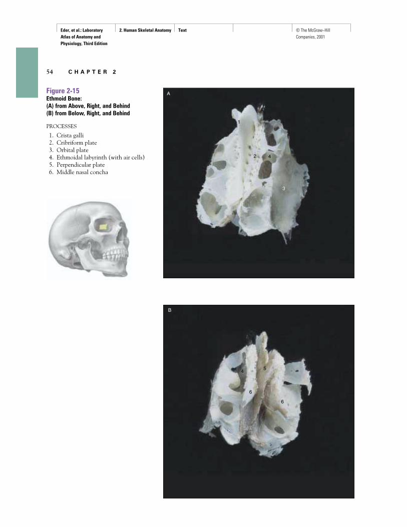
Figure 2-15Ethmoid Bone: (A) from Above, Right, and Behind(B) from Below, Right, and Behind
PROCESSES
1. Crista galli2. Cribriform plate3. Orbital plate4. Ethmoidal labyrinth (with air cells)5. Perpendicular plate6. Middle nasal concha
Eder, et al.: Laboratory Atlas of Anatomy and Physiology, Third Edition
2. Human Skeletal Anatomy Text © The McGraw−Hill Companies, 2001
H u m a n S k e l e t a l A n a t o m y 55
I
H
J
J
J
K
K
F
G
H
I
I
A
B
C
D
E
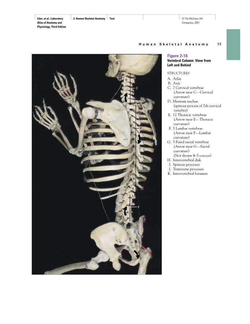
Figure 2-16Vertebral Column: View from Left and Behind
STRUCTURES
A. AtlasB. AxisC. 7 Cervical vertebrae
(Arrow near C—Cervicalcurvature)
D. Mentum nuchae (spinous process of 7th cervicalvertebra)
E. 12 Thoracic vertebrae (Arrow near E—Thoraciccurvature)
F. 5 Lumbar vertebrae (Arrow near F—Lumbarcurvature)
G. 5 Fused sacral vertebrae (Arrow near G—Sacralcurvature)(Not shown 4–5 coccyx)
H. Intervertebral diskI. Spinous processesJ. Transverse processes
K. Intervertebral foramen
Eder, et al.: Laboratory Atlas of Anatomy and Physiology, Third Edition
2. Human Skeletal Anatomy Text © The McGraw−Hill Companies, 2001
56 C H A P T E R 2
E
C
A
F
D
B
1
1
1
1
1
2
3
3
33
4
4
5
5
5
6
7
7
7
7
7
8
8
9
9
9
10
11
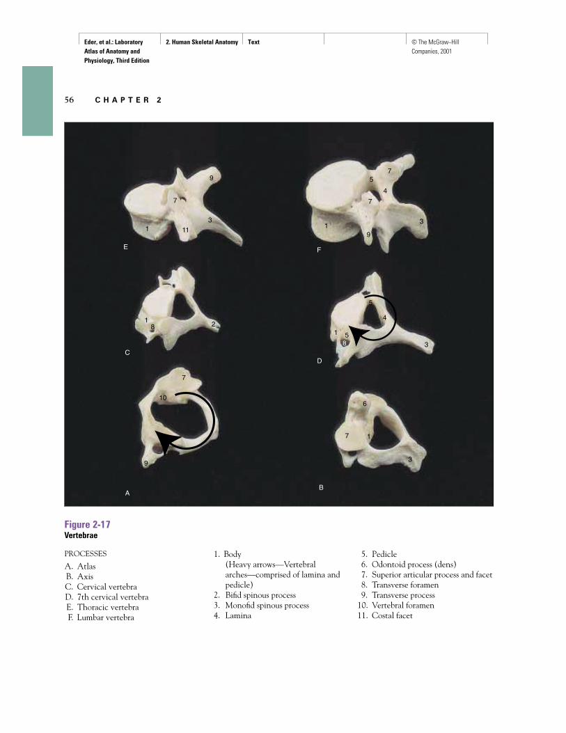
Figure 2-17Vertebrae
PROCESSES
A. AtlasB. AxisC. Cervical vertebraD. 7th cervical vertebraE. Thoracic vertebraF. Lumbar vertebra
1. Body(Heavy arrows—Vertebralarches—comprised of lamina andpedicle)
2. Bifid spinous process3. Monofid spinous process4. Lamina
5. Pedicle6. Odontoid process (dens)7. Superior articular process and facet8. Transverse foramen9. Transverse process
10. Vertebral foramen11. Costal facet
Eder, et al.: Laboratory Atlas of Anatomy and Physiology, Third Edition
2. Human Skeletal Anatomy Text © The McGraw−Hill Companies, 2001
H u m a n S k e l e t a l A n a t o m y 57
1
A
B2
3
4
4
5
6
6
7
8
9
1
2
3
4
5
67
78
8
1
2
2
39
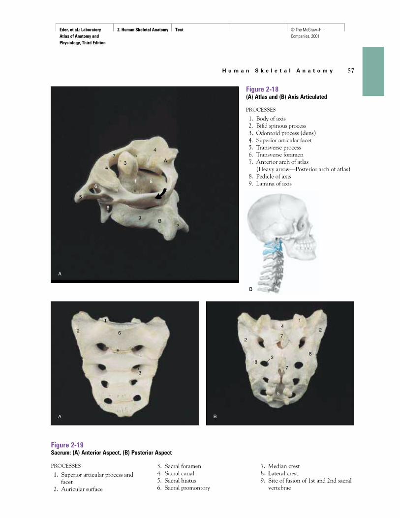
Figure 2-18(A) Atlas and (B) Axis Articulated
PROCESSES
1. Body of axis2. Bifid spinous process3. Odontoid process (dens)4. Superior articular facet5. Transverse process6. Transverse foramen7. Anterior arch of atlas
(Heavy arrow—Posterior arch of atlas)8. Pedicle of axis9. Lamina of axis
Figure 2-19Sacrum: (A) Anterior Aspect, (B) Posterior Aspect
PROCESSES
1. Superior articular process and facet
2. Auricular surface
3. Sacral foramen4. Sacral canal5. Sacral hiatus6. Sacral promontory
7. Median crest8. Lateral crest9. Site of fusion of 1st and 2nd sacral
vertebrae
A
B
A B
Eder, et al.: Laboratory Atlas of Anatomy and Physiology, Third Edition
2. Human Skeletal Anatomy Text © The McGraw−Hill Companies, 2001
58 C H A P T E R 2
1
A
B
C
2345
8
10
12
1234
5
67
8
9
11
12 3 4
5
8
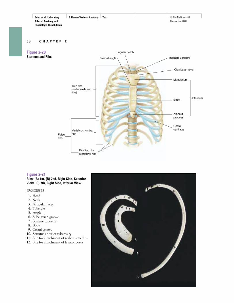
Figure 2-20Sternum and Ribs
Figure 2-21Ribs: (A) 1st, (B) 2nd, Right Side, SuperiorView, (C) 7th, Right Side, Inferior View
PROCESSES
1. Head2. Neck3. Articular facet4. Tubercle5. Angle6. Subclavian groove7. Scalene tubercle8. Body9. Costal groove
10. Serratus anterior tuberosity11. Site for attachment of scalenus medius12. Site for attachment of levator costa
True ribs(vertebrosternal ribs)
Falseribs
Floating ribs(vertebral ribs)
Vertebrochondralribs
Sternal angle
Jugular notch
Thoracic vertebra
Clavicular notch
Sternum
Manubrium
Body
Xiphoidprocess
Costalcartilage
Eder, et al.: Laboratory Atlas of Anatomy and Physiology, Third Edition
2. Human Skeletal Anatomy Text © The McGraw−Hill Companies, 2001
H u m a n S k e l e t a l A n a t o m y 59
A
B
C
D
E
12
3
3
4
5
67
10
89
10
A B
12
4
5
6
7
8
111213
14
1
23
6
7
8
9
10
12
13
14
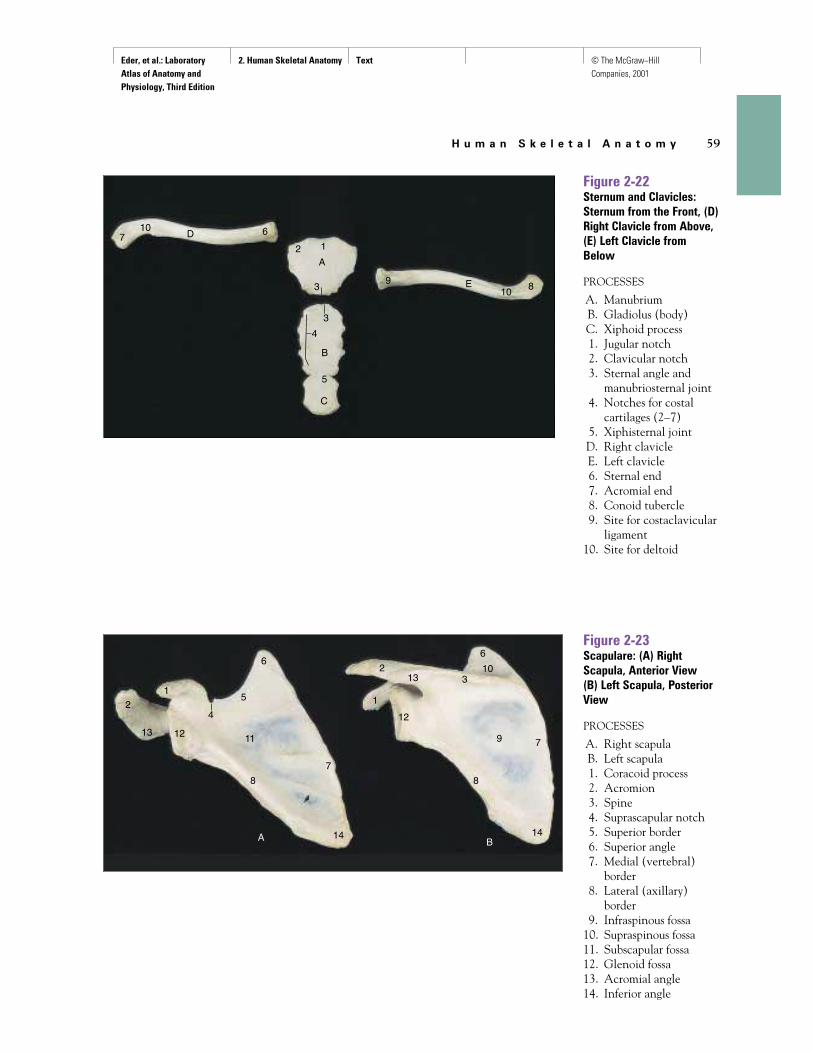
Figure 2-22Sternum and Clavicles:Sternum from the Front, (D)Right Clavicle from Above,(E) Left Clavicle fromBelow
PROCESSES
A. ManubriumB. Gladiolus (body)C. Xiphoid process1. Jugular notch2. Clavicular notch3. Sternal angle and
manubriosternal joint4. Notches for costal
cartilages (2–7)5. Xiphisternal jointD. Right clavicleE. Left clavicle6. Sternal end7. Acromial end8. Conoid tubercle9. Site for costaclavicular
ligament10. Site for deltoid
Figure 2-23Scapulare: (A) RightScapula, Anterior View(B) Left Scapula, PosteriorView
PROCESSES
A. Right scapulaB. Left scapula1. Coracoid process2. Acromion3. Spine4. Suprascapular notch5. Superior border6. Superior angle7. Medial (vertebral)
border8. Lateral (axillary)
border9. Infraspinous fossa
10. Supraspinous fossa11. Subscapular fossa12. Glenoid fossa13. Acromial angle14. Inferior angle
Eder, et al.: Laboratory Atlas of Anatomy and Physiology, Third Edition
2. Human Skeletal Anatomy Text © The McGraw−Hill Companies, 2001
60 C H A P T E R 2
A B
12
3
4
6
8
9
10
11 13 141617
12
3
4
56
7
8
9 10
11
12 13 14
15
17
A B C D
12
3
4
5
6
5
89 10
11
13
14
14
15 1
2
3
5
5
67
8
1213
14
1413
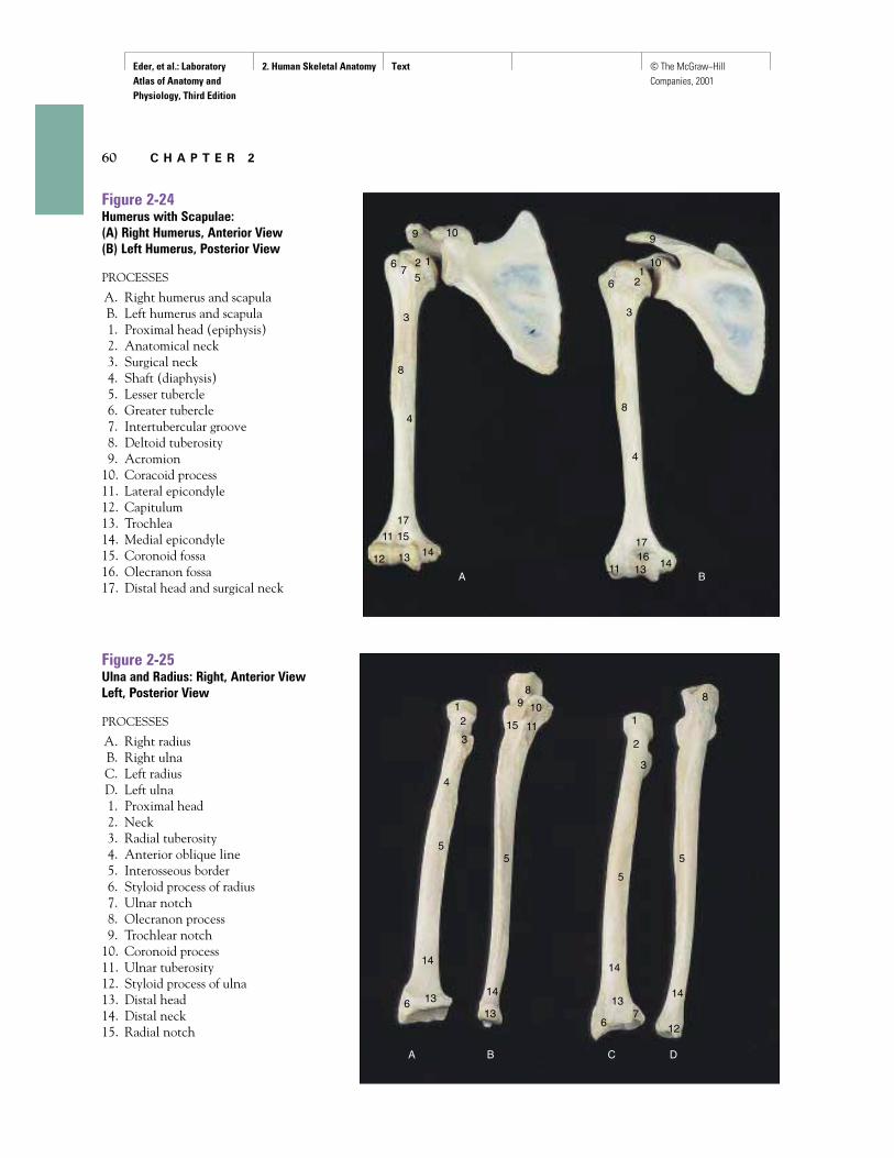
Figure 2-24Humerus with Scapulae: (A) Right Humerus, Anterior View(B) Left Humerus, Posterior View
PROCESSES
A. Right humerus and scapulaB. Left humerus and scapula1. Proximal head (epiphysis)2. Anatomical neck3. Surgical neck4. Shaft (diaphysis)5. Lesser tubercle6. Greater tubercle7. Intertubercular groove8. Deltoid tuberosity9. Acromion
10. Coracoid process11. Lateral epicondyle12. Capitulum13. Trochlea14. Medial epicondyle15. Coronoid fossa16. Olecranon fossa17. Distal head and surgical neck
Figure 2-25Ulna and Radius: Right, Anterior ViewLeft, Posterior View
PROCESSES
A. Right radiusB. Right ulnaC. Left radiusD. Left ulna1. Proximal head2. Neck3. Radial tuberosity4. Anterior oblique line5. Interosseous border6. Styloid process of radius7. Ulnar notch8. Olecranon process9. Trochlear notch
10. Coronoid process11. Ulnar tuberosity12. Styloid process of ulna13. Distal head14. Distal neck15. Radial notch
Eder, et al.: Laboratory Atlas of Anatomy and Physiology, Third Edition
2. Human Skeletal Anatomy Text © The McGraw−Hill Companies, 2001
H u m a n S k e l e t a l A n a t o m y 61
12
34 5
678
9
9 9 9
9
10
11
12
12
12 12
13
13
1313
14
14
1414
1
2
3
4
5
6
16
15
20
7
8
9
10
11
12
13
14
17
18
19
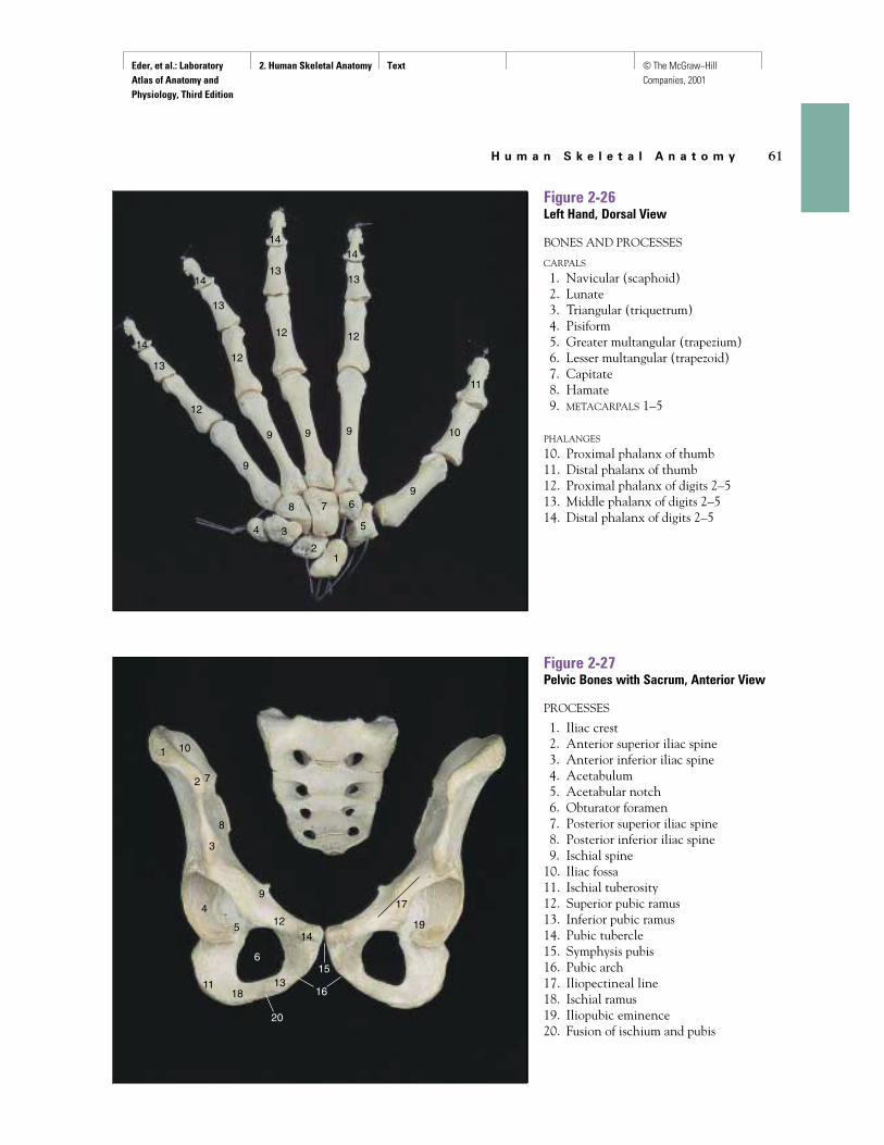
Figure 2-26Left Hand, Dorsal View
BONES AND PROCESSES
CARPALS
1. Navicular (scaphoid)2. Lunate3. Triangular (triquetrum)4. Pisiform5. Greater multangular (trapezium)6. Lesser multangular (trapezoid)7. Capitate8. Hamate9. METACARPALS 1–5
PHALANGES
10. Proximal phalanx of thumb11. Distal phalanx of thumb12. Proximal phalanx of digits 2–513. Middle phalanx of digits 2–514. Distal phalanx of digits 2–5
Figure 2-27Pelvic Bones with Sacrum, Anterior View
PROCESSES
1. Iliac crest2. Anterior superior iliac spine3. Anterior inferior iliac spine4. Acetabulum5. Acetabular notch6. Obturator foramen7. Posterior superior iliac spine8. Posterior inferior iliac spine9. Ischial spine
10. Iliac fossa11. Ischial tuberosity12. Superior pubic ramus13. Inferior pubic ramus14. Pubic tubercle15. Symphysis pubis16. Pubic arch17. Iliopectineal line18. Ischial ramus19. Iliopubic eminence20. Fusion of ischium and pubis
Eder, et al.: Laboratory Atlas of Anatomy and Physiology, Third Edition
2. Human Skeletal Anatomy Text © The McGraw−Hill Companies, 2001
62 C H A P T E R 2
A
A B
B
1
2
3
4
5
6
6
7
8
921
11
12
13
16
16
18
19
20
20
2
3
19
7
8
9
10
21
12
13
14
15
17
18
12
34
5
10 1114
15
16
1718
12
34
5
6
7
8
9
1011
12
13
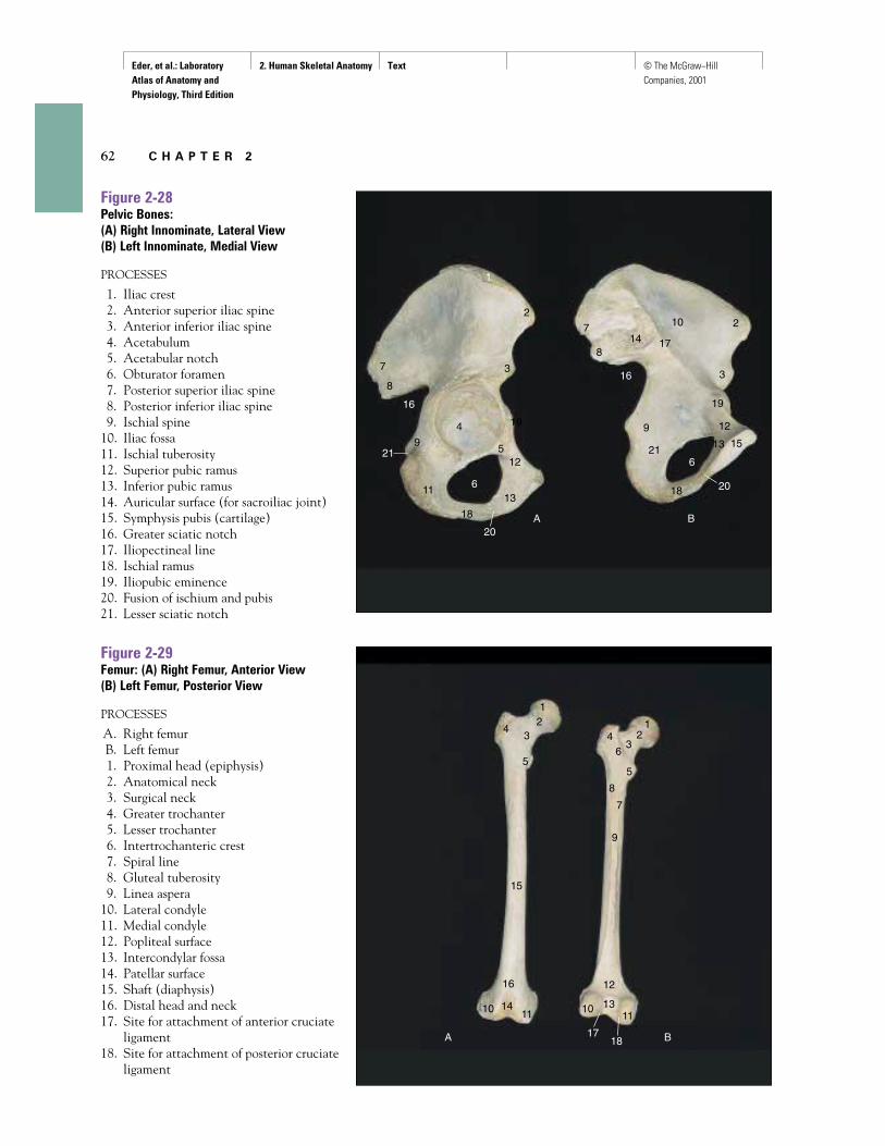
Figure 2-28Pelvic Bones:(A) Right Innominate, Lateral View(B) Left Innominate, Medial View
PROCESSES
1. Iliac crest2. Anterior superior iliac spine3. Anterior inferior iliac spine4. Acetabulum5. Acetabular notch6. Obturator foramen7. Posterior superior iliac spine8. Posterior inferior iliac spine9. Ischial spine
10. Iliac fossa11. Ischial tuberosity12. Superior pubic ramus13. Inferior pubic ramus14. Auricular surface (for sacroiliac joint)15. Symphysis pubis (cartilage)16. Greater sciatic notch17. Iliopectineal line18. Ischial ramus19. Iliopubic eminence20. Fusion of ischium and pubis21. Lesser sciatic notch
Figure 2-29Femur: (A) Right Femur, Anterior View(B) Left Femur, Posterior View
PROCESSES
A. Right femurB. Left femur1. Proximal head (epiphysis)2. Anatomical neck3. Surgical neck4. Greater trochanter5. Lesser trochanter6. Intertrochanteric crest7. Spiral line8. Gluteal tuberosity9. Linea aspera
10. Lateral condyle11. Medial condyle12. Popliteal surface13. Intercondylar fossa14. Patellar surface15. Shaft (diaphysis)16. Distal head and neck17. Site for attachment of anterior cruciate
ligament18. Site for attachment of posterior cruciate
ligament
Eder, et al.: Laboratory Atlas of Anatomy and Physiology, Third Edition
2. Human Skeletal Anatomy Text © The McGraw−Hill Companies, 2001
H u m a n S k e l e t a l A n a t o m y 63
A B
1
2
3 45
2
A B C D
11
2
34
9
12
1516
1 13
4
55 6
78
1011
1314
16
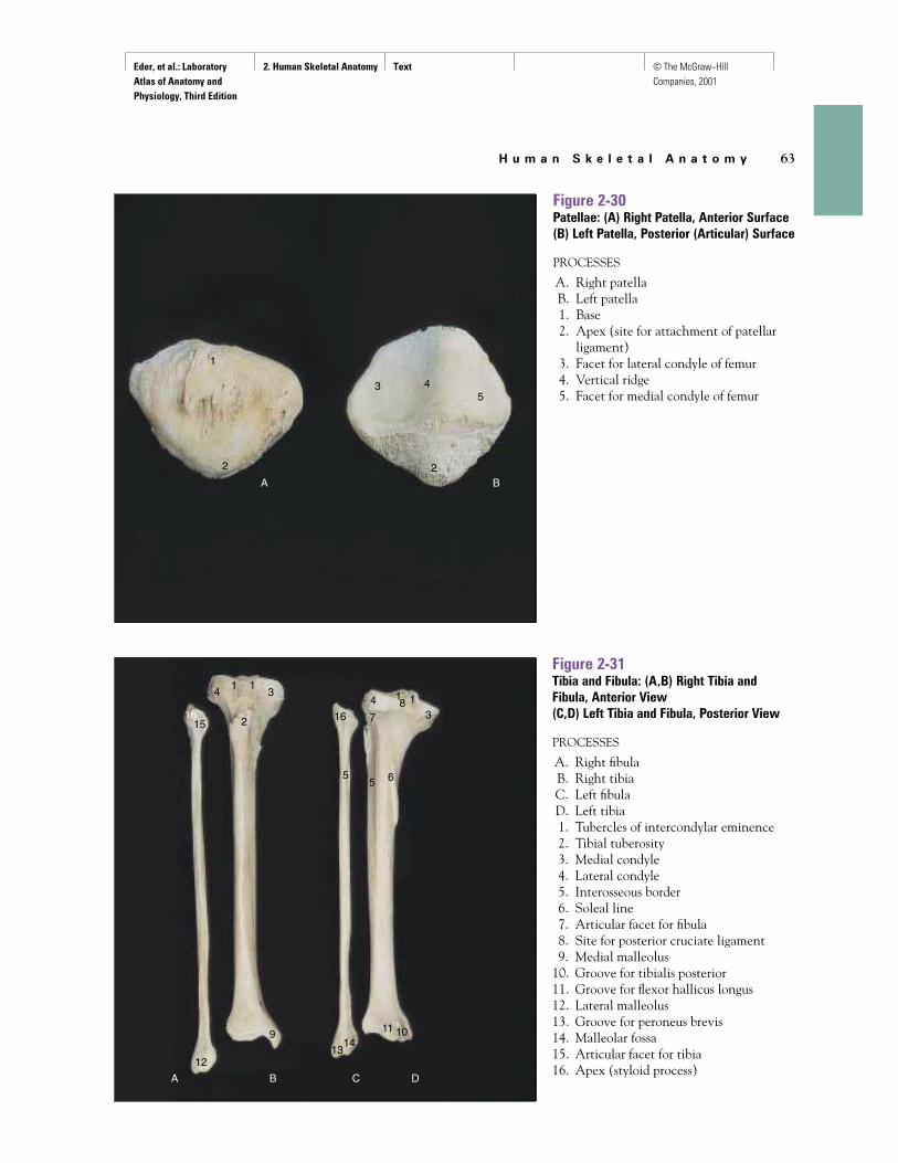
Figure 2-30Patellae: (A) Right Patella, Anterior Surface(B) Left Patella, Posterior (Articular) Surface
PROCESSES
A. Right patellaB. Left patella1. Base2. Apex (site for attachment of patellar
ligament)3. Facet for lateral condyle of femur4. Vertical ridge5. Facet for medial condyle of femur
Figure 2-31Tibia and Fibula: (A,B) Right Tibia andFibula, Anterior View(C,D) Left Tibia and Fibula, Posterior View
PROCESSES
A. Right fibulaB. Right tibiaC. Left fibulaD. Left tibia1. Tubercles of intercondylar eminence2. Tibial tuberosity3. Medial condyle4. Lateral condyle5. Interosseous border6. Soleal line7. Articular facet for fibula8. Site for posterior cruciate ligament9. Medial malleolus
10. Groove for tibialis posterior11. Groove for flexor hallicus longus12. Lateral malleolus13. Groove for peroneus brevis14. Malleolar fossa15. Articular facet for tibia16. Apex (styloid process)
Eder, et al.: Laboratory Atlas of Anatomy and Physiology, Third Edition
2. Human Skeletal Anatomy Text © The McGraw−Hill Companies, 2001
64 C H A P T E R 2
1
2
3
45
67
89
10
1112
13
1414
1414
14
15
16
17
1717
17
18
1818
1819
1919
19
20
1
2
2
3
3
4
5
6
6
7
8
8
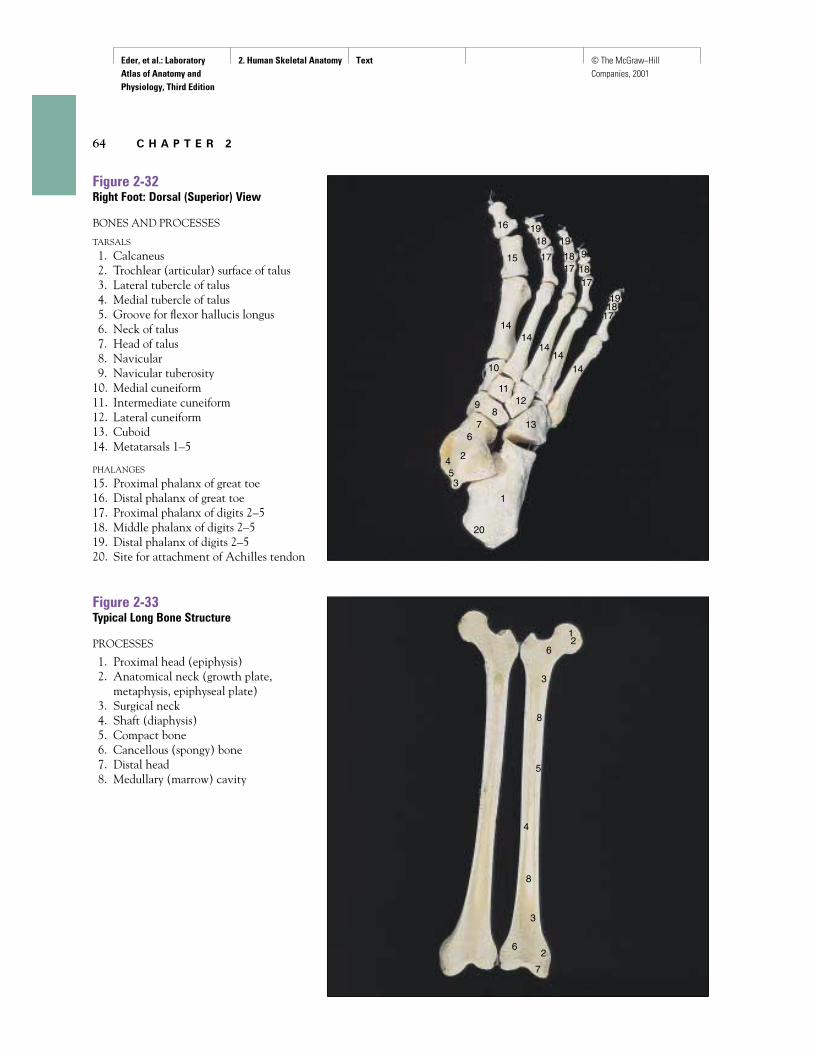
Figure 2-32Right Foot: Dorsal (Superior) View
BONES AND PROCESSES
TARSALS
1. Calcaneus2. Trochlear (articular) surface of talus3. Lateral tubercle of talus4. Medial tubercle of talus5. Groove for flexor hallucis longus6. Neck of talus7. Head of talus8. Navicular9. Navicular tuberosity
10. Medial cuneiform11. Intermediate cuneiform12. Lateral cuneiform13. Cuboid14. Metatarsals 1–5
PHALANGES
15. Proximal phalanx of great toe16. Distal phalanx of great toe17. Proximal phalanx of digits 2–518. Middle phalanx of digits 2–519. Distal phalanx of digits 2–520. Site for attachment of Achilles tendon
Figure 2-33Typical Long Bone Structure
PROCESSES
1. Proximal head (epiphysis)2. Anatomical neck (growth plate,
metaphysis, epiphyseal plate)3. Surgical neck4. Shaft (diaphysis)5. Compact bone6. Cancellous (spongy) bone7. Distal head8. Medullary (marrow) cavity