Human Papillomaviruses and Their Association with …
57
Thesis for doctoral degree (Ph.D.) 2009 Human Papillomaviruses and Their Association with Squamous Cell Carcinoma of The Conjunctiva Charles Ateenyi Agaba Thesis for doctoral degree (Ph.D.) 2009 Charles Ateenyi Agaba Human Papillomaviruses and Their Association with Squamous Cell Carcinoma of The Conjunctiva Makerere University Karolinska Institutet SE-171 77 Stockholm • Sweden Makerere University P.O. Box 7062 • Kampala • Uganda This thesis is the basis for a joint degree of Doctor of Philosophy (PhD) between Karolinska Institutet and Makerere University.
Transcript of Human Papillomaviruses and Their Association with …

Thesis for doctoral degree (Ph.D.)2009
Human Papillomaviruses and TheirAssociation with Squamous Cell Carcinoma of The Conjunctiva
Charles Ateenyi Agaba
Thesis fo
r do
ctoral d
egree (Ph.D
.) 2009C
harles A
teenyi A
gaba
Hu
man
Papillomaviru
ses and T
heir A
ssociation w
ith Squ
amou
s Cell C
arcinom
a of Th
e Con
jun
ctiva
FIXED-DOSE CHLOROQUINE AND
SULFADOXINE/PYRIMETHAMINE
TREATMENT OF MALARIA:
OUTCOME AND
PHARMACOKINETIC ASPECTS
Celestino Obua
Karolinska Institutet
Makerere University
Thesis for doctoral degree (PhD)
Kampala and Stockholm 2007
Karolinska InstitutetSE-171 77 Stockholm • Sweden
Makerere UniversityP.O. Box 7062 • Kampala • Uganda
This thesis is the basis for a joint degree of Doctor of Philosophy (PhD) between Karolinska Institutet and Makerere University.

Makerere University
Department of Ophthalmology, School of Medicine, Makerere University Department of Medical Epidemiology and Biostatistics,
Karolinska Institutet
Human papillomairuses and their association with squamous cell carcinoma of the conjunctiva
ACADEMIC THESIS
The public defence for the degree of Doctor of Philosophy at Karolinska Institutet and Makerere University will be held at Makerere University, Davis Lecture Theatre, Mulago, Kampala,
Uganda. Wednesday 28th October 2009 at 9.00 am.
AKADEMISK AVHANDLING som för avläggande av gemensam medicine doktorsexamen (Doctor of Philosophy, PhD) vid Karolinska Institutet och Makerere University offentligen försvaras på det engelska språket i
Makerere University, Davis Lecture Theatre, Mulago, Kampala, Uganda, onsdagen den 28 oktober, 2009, kl 9.00
Charles Ateenyi Agaba
Supervisors: Elisabete Weiderpass, Associate Professor Department of Medical Epidemiology and Biostatistics, Karolinska Institutet, Stockholm Fred Wabwire-Mangen, Associate Professor Department of Epidemiology and Biostatistics, School of Public Health, College of Health Sciences, Makerere University, Kampala, Uganda :
Faculty opponent: Lars Vatten, Professor Norwegian University of Science and Technology, Norway Examination Board: Bjorn Hagmar, Professor Department of Pathology, Faculty of Medicine, Oslo University Charles Amon Sunday Karamagi, PhD Clinical Epidemiology Unit School of Medicine, College of Health Sciences, Makerere University, Kampala Uganda Fred Nuwaha Ntoni, Associate professor School of Public Health College of Health Sciences Makerere University, Kampala Uganda
Kampala and Stockholm 2009

ABSTRACT Squamous cell carcinoma of the conjunctiva (SCCC), hitherto a rare cancer, has increased manyfold since the advent of HIV/AIDS. Solar ultra violet radiation (UV) may also be a risk factor for the disease. The increased incidence has led to the hypothesis of an infective agent as a risk factor, especially infections with human papillomaviruses (HPV) types 16 and 18. The main purpose of this thesis was to study the association between these factors and SCCC. Pilot studies I and II. Aims: to assess the feasibility and test methods for a larger case control study in Uganda; to investigate the presence of HPV in conjunctival tissue using broad spectrum PCR; to investigate the presence of UV induced mutations in the TP53 gene in SCCC samples. Methods: 21 SCCC cases and 22 control patients were tested for the presence of HPV in conjunctival tissue; the DNA from the samples was further analyzed for the presence of somatic mutations in the T53 gene. Results: Cutaneous HPV were found in 86% cases and 36% controls, suggesting a role of cutaneous HPV in the aetiology of SCCC. Seven of the mutations were CC to TT transitions, which are characteristic of solar UV light DNA damage. Study III. Aims: to assess the presence of HPV in lesion-free conjunctiva and assess whether there is an excess of HPV infection in concurrent HIV/AIDS disease. Methods: 136 lesion-free frozen conjunctival samples from autopsies performed at Mulago hospital, Kampala, Uganda, were analyzed for the presence of HPV using broad spectrum PCR methods. Results: 14.6% samples tested positive for cutaneous HPV, and no mucosal HPV infection was detected; no excess of HPV infection was found in individuals who had died of HIV/AIDS-related causes as compared to those who had died of other diseases. Conclusion: HPV infection occurs in the conjunctiva: there was no excess of HPV infection in HIV/AIDS patients as compared to other patients, though we cannot rule out the possibility of the misclassification of HIV/AIDS patients.
Study IV. Aims: to compare the prevalence of HPV infection in SCCC patients patients with other eye diseases. Methods: Hospital-based case control study in Mulago and Jinja Hospitals, Uganda, involving 94 cases of SCCC and 285 controls with other eye diseases. We compared the prevalence of HPV infection in 94 biopsies of SCCC patients to 285 biopsies from hospital control patients with other eye diseases. Highly sensitive broad spectrum PCR tests that detect up to 75 types of HPV were used to analyze the frozen tissue biopsies. Adjusted odds ratios (OR) and 95% confidence intervals (CI) were computed, adjusting for age, sex and HIV status. Results: cutaneous HPV were detected in 44.7% of the SCCC cases and 10.5% in controls (OR = 6.22; 95% CI = 3.60-10.72). The strength of the association of cutaneous HPV with SCCC was stronger in multiple infections than single infections, (OR = 74.2; 95% CI = 23.4-235.7) and (OR = 12.8; 95% C = 5.5-29.6) respectively. Mucosal types were detected in 6.4% SCCC and 3.5% controls (OR = 1.0; 95% CI = 0.-2.9). HPV5, 8 and 24 were most common in SCCC. The association of cutaneous HPV with SCCC was stronger in SCCC patients with HIV (OR = 17.0; 95% CI = 5.5-52.5). HIV infection was detected in 85.1% SCCC and 44.9% controls, (OR = 7.3; 95% CI = 4.2-12.4). There was no significant association of SCCC with age, sex, educational level, smoking habits, indoor or outdoor occupation. Conclusion: Cutaneous HPV and HIV are significantly associated with SCCC, while mucosal HPV are not significantly associated. Key words: Squamous cell carcinoma of the conjunctiva, human papillomavirus, cutaneous HPV, Mucosal HPV, Broad spectrum PCR tests, HIV, LiPa, SPF, Uganda.

From DEPARTMENT OF OPHTHALMOLOGY Makerere University, Kampala, Uganda
and DEPARTMENT OF MEDICAL EPIDEMIOLOGY AND
BIOSTATISTICS Karolinska Institutet, Stockholm, Sweden
HUMAN PAPILLOMAVIRUSES AND THEIR ASSOCIATION WITH SQUAMOUS CELL CARCINOMA OF
THE CONJUNCTIVA
CHARLES ATEENYI AGABA
Kampala/Stockholm 2009
MAKERERE UNIVERSITY

All previously published papers were reproduced with permission from the publisher. Published by Karolinska Institutet and Makerere University. Printed by Universitetsservice AB (Stockholm, Sweden) and Makerere University Printery (Kampala, Uganda) Cover photos by Bo Lambert. © Ateenyi Agaba C, 2009 ISBN 978-91-7409-9


1
ABSTRACT Squamous cell carcinoma of the conjunctiva (SCCC), hitherto a rare cancer, has increased manyfold since the advent of HIV/AIDS. Solar ultra violet radiation (UV) may also be a risk factor for the disease. The increased incidence has led to the hypothesis of an infective agent as a risk factor, especially infections with human papillomaviruses (HPV) types 16 and 18. The main purpose of this thesis was to study the association between these factors and SCCC.
Pilot studies I and II. Aims: to assess the feasibility and test methods for a larger case control study in Uganda; to investigate the presence of HPV in conjunctival tissue using broad spectrum PCR; to investigate the presence of UV induced mutations in the TP53 gene in SCCC samples. Methods: 21 SCCC cases and 22 control patients were tested for the presence of HPV in conjunctival tissue; the DNA from the samples was further analyzed for the presence of somatic mutations in the T53 gene. Results: Cutaneous HPV were found in 86% cases and 36% controls, suggesting a role of cutaneous HPV in the aetiology of SCCC. Seven of the mutations were CC to TT transitions, which are characteristic of solar UV light DNA damage. Study III. Aims: to assess the presence of HPV in lesion-free conjunctiva and assess whether there is an excess of HPV infection in concurrent HIV/AIDS disease. Methods: 136 lesion-free frozen conjunctival samples from autopsies performed at Mulago hospital, Kampala, Uganda, were analyzed for the presence of HPV using broad spectrum PCR methods. Results: 14.6% samples tested positive for cutaneous HPV, and no mucosal HPV infection was detected; no excess of HPV infection was found in individuals who had died of HIV/AIDS-related causes as compared to those who had died of other diseases. Conclusion: HPV infection occurs in the conjunctiva: there was no excess of HPV infection in HIV/AIDS patients as compared to other patients, though we cannot rule out the possibility of the misclassification of HIV/AIDS patients. Study IV. Aims: to compare the prevalence of HPV infection in SCCC patients patients with other eye diseases. Methods: Hospital-based case control study in Mulago and Jinja Hospitals, Uganda, involving 94 cases of SCCC and 285 controls with other eye diseases. We compared the prevalence of HPV infection in 94 biopsies of SCCC patients to 285 biopsies from hospital control patients with other eye diseases. Highly sensitive broad spectrum PCR tests that detect up to 75 types of HPV were used to analyze the frozen tissue biopsies. Adjusted odds ratios (OR) and 95% confidence intervals (CI) were computed, adjusting for age, sex and HIV status. Results: cutaneous HPV were detected in 44.7% of the SCCC cases and 10.5% in controls (OR = 6.22; 95% CI = 3.60-10.72). The strength of the association of cutaneous HPV with SCCC was stronger in multiple infections than single infections, (OR = 74.2; 95% CI = 23.4-235.7) and (OR = 12.8; 95% C = 5.5-29.6) respectively. Mucosal types were detected in 6.4% SCCC and 3.5% controls (OR = 1.0; 95% CI = 0.-2.9). HPV5, 8 and 24 were most common in SCCC. The association of cutaneous HPV with SCCC was stronger in SCCC patients with HIV (OR = 17.0; 95% CI = 5.5-52.5). HIV infection was detected in 85.1% SCCC and 44.9% controls, (OR = 7.3; 95% CI = 4.2-12.4). There was no significant association of SCCC with age, sex, educational level, smoking habits, indoor or outdoor occupation. Conclusion: Cutaneous HPV and HIV are significantly associated with SCCC, while mucosal HPV are not significantly associated. Key words: Squamous cell carcinoma of the conjunctiva, human papillomavirus, cutaneous HPV, Mucosal HPV, Broad spectrum PCR tests, HIV, LiPa, SPF, Uganda.
2
LIST OF PUBLICATIONS
I. Ateenyi-Agaba C, Weiderpass E, Smet A, Dong W, Dai M, Kahwa B,
Wabinga H, Katongole-Mbidde E, Franceschi S, Tommasino M.
Epidermodysplasia Verruciformis human papillomavirus types and
carcinoma of the conjunctiva: a pilot study. Br J Cancer 2004;90:
1777-9.
II. Ateenyi-Agaba C, Dai M, Le Calvez F, Katongole-Mbidde E, Smet
A, Tommasino M, Franceschi S, Hainaut P, Weiderpass E.TP53
mutations in squamous-cell carcinoma of the conjunctiva: evidence
for UV-induced mutagenesis. Mutagenesis 2004;19:399-401.
III. Ateenyi- Agaba C, Weiderpass E, Tomassimo M, Smet A, Arslan A,
Dai M Katongole-Mbidde E, Hainaut P, Snijders PJF, Franceschi S.
Papilloma virus infection in the conjunctiva of individuals with and
without AIDS: An autopsy series from Uganda. Cancer Letters
2006;239:98-102.
IV. Ateenyi-Agaba C, Franceschi S, Wabwire-Mangen Fred, Arslan A,
Othieno E, Binta-Kahwa J, van Doorn LJ, Kleter B, Quint W,
Weiderpass E. Human Papilloma virus infection and squamous cell
carcinoma of the conjunctiva. (Submitted for Publication 2009)

3
CONTENTS PAGE
1. INTRODUCTION ..................................................................................... 5
2. BACKGROUND ....................................................................................... 7
3.AIMS.......................................................................................................... 18
4.SUBJECTS, MATERIALS AND METHODS ............................................. 19
4.1. Paper I and II ........................................................................................... 19
4.2. Paper III .................................................................................................. 21
4.3. Paper IV .................................................................................................. 22
4.4. STATISTICAL ANALYSIS .................................................................... 27
4.5. ETHICAL CONSIDERATIONS ............................................................. 27
5. RESULTS .................................................................................................. 29
5.1. Paper I..................................................................................................... 29
5.2. Paper II ................................................................................................... 29
5.3. Paper III .................................................................................................. 30
5.4. Paper IV .................................................................................................. 31
6. GENERAL DISCUSSION ......................................................................... 33
6.1. Methodological considerations ................................................................. 33
6.2. Interpretations of findings and Implications .............................................. 36
6.2.4. Future studies ....................................................................................... 38
7. CONCLUSIONS........................................................................................ 40
8. ACKNOWLEDGEMENTS ........................................................................ 41
9. REFERENCES .......................................................................................... 42

4
LIST OF ABBREVIATIONS
AIDS
CI
CIN
DNA
ELISA
HIV
HPV
LiPA
OR
ORF
PCR
SCCC
URR
UV
SPF
Acquired Immunodeficiency Syndrome
Confidence Interval
Cervical Intraepithelial Neoplasia
Deoxyribonucleic Acid
Enzyme-Linked Immunosorbent Assay
Human Immunodeficiency Virus
Human Papilloma Virus
Line Probe Assay
Odds Ratio
Open Reading Frame
Polymerase Chain Reaction
Squamous Cell Carcinoma of the Conjunctiva
Upper Regulatory Region
Ultraviolet
Short PCR Fragment

1 INTRODUCTION Squamous cell carcinoma of the conjunctiva (SCCC) is the major cancer that arises from the
conjunctival membrane of the eye. This cancer has been comparatively more common in Africa
than in Europe or America (Newton et al 1996). Before the 1980s, the cancer was a slow-
growing tumour of middle-aged to elderly people.
Since the advent of HIV/AIDS, the cancer has become more frequent and has assumed a more
aggressive clinical course. Subsequent case control studies have found significant associations
with the human immunodeficiency virus infection, HIV, and the acquired immunodeficiency
syndrome, AIDS (Goedert and Cote, 1995; Ateenyi-Agaba, 1995; Waddell et al, 1996;
Newton and Beral, 1999; Ateenyi-Agaba and Newton, 1999; Newton et al, 2001; Guech-
Ongey et al, 2008). The exact incidence of this cancer is still not known in Uganda, since the
functional cancer registry in the country, the Kyadondo Cancer Registry, covers only
Kampala, the capital city, and the immediate surrounding area. However, there is evidence
that the incidence of SCCC has increased more than tenfold since the HIV/AIDS pandemic
began in the 1980s (Parkin et al, 1999; Wabinga et al, 2000). An increased incidence of this
cancer has also been noted in Rwanda (Kestelyn, 1990), Malawi and Zimbabe (Parkin et al,
2003).
Solar ultra violet radiation has long been hypothesized as carcinogenic to the conjunctiva,
given the extraordinary geographic distribution of the SCCC being relatively more common
in areas close to the equator.
In Iceland, at the fringes of the Arctic Circle, there was not a single recorded case of SCCC in
the cancer registry as of 1996 (Newton et al, 1996). On the other hand, the tumour is
relatively more common in Uganda, which is at the equator, with incidence rates estimated at
over 12 per million population per year (Ateenyi-Agaba, 1995; Wabinga et al, 2000; Parkin
et al, 1999).
Due to the increased incidence concurrent with the HIV/AIDS epidemic, the location of the
tumours (in exposed areas of the conjunctiva) and the anatomical similarities of the
conjunctiva (epithelial surface) with the cervix uteri, where cancer of the cervix (also of
epithelial origin) arises, an infective aetiology of SCCC has been hypothesized.
Human papillomaviruses (HPV), being known oncogenic viruses with a tropism for infecting
epithelial dividing cells, has been hypothesized as a possible aetiological factor. Mucosal
5

6
HPV types 16 and 18 have particularly been studied in the aetiology of SCCC, given their
roles in cancers of the anogenital region.
However, relatively little research on SCCC has been done so far, possibly because the
tumour has been relatively rare. The increase in the incidence of SCCC in the last three
decades has set the stage for the timely investigation of the risk factors for the disease.

7
2 BACKGROUND 2.1 THE CONJUNCTIVA The eyeball consists of the following layers from inside: the retina is the light sensitive
innermost layer consisting of light sensitive cells the rods and cones. The retina is nourished by
the pigmented choroid; the pigment reduces internal reflections within the eye. The choroid is
covered by the tough white fibrous sclera, whose function is mainly mechanical protection and
providing a template for insertion of the ocular muscles that are responsible for eye movements
(Bron et al, 1997).
The conjunctiva consists of the loose translucent mucous membrane, of the stratified non-
keratinizing epithelium, which overlies the loose connective tissue that joins the eyeball to the
lids, and hence the name (Bron et al, 1997). In the loose connective tissue are found vessels,
nerves and dormant melanocytes that tend to be reactivated in response to a chemical or
physical irritation leading to the production of melanin that is deposited as a brown pigment in
the ocular tissues.
The conjunctiva is divided into:
bulbar conjunctiva which covers the sclera;
tarsal conjunctiva which covers the inside of the lids;
fornicial conjunctiva which lines the upper and lower fornices of the eye.
The cornea is the transparent front portion of the eye and is a continuation of the scleral fibres,
which are arranged perfectly at 900 to each other to ensure transparency.
The conjunctiva fuses with the loose connective tissue at the junction of the sclera and the
cornea but is loosely attached to the rest of the eyeball and the fornices (Bron et al, 1997). The
epithelium of the conjunctiva is continuous with the corneal epithelium, and diseases of the
cornea can extend to the conjunctiva and vice versa.
The junction of the conjunctiva and the cornea is called the limbus, and this is the
transformation zone with active mitotic stem cells that replace the corneal epithelium.
The space between the lower lid and the upper lid is called the interpalpebral space. This space
exposes the cornea and conjunctiva to the external environmental agents.
The eye has protective mechanisms against chemical and biologic attacks. The tear film offers
physical, chemical and biologic mechanisms of protection. Tears dilute toxins and physically
cleanse the eye, while the enzyme lysozyme and immunoglobulins G and E offer the
8
biochemical protection. The blinking reflex offers physical protection by spreading the tear
film, hence preventing desiccation of the cornea and conjunctiva. During sleep, the eyelids do
not close perfectly on the medial or nasal aspect. This renders the medial aspect of the eye prone
to desiccation (Crawford, 1995).
The conjunctiva has goblet cells which produce a lipid that protects the tear film from breaking
up. A lack of the lipid fraction in tears, as occurs in conjunctival diseases like xerophthalmia
(lack of vitamin A), acid burns or drug reactions, lead to drying of the conjunctiva and cornea,
which predisposes the eye to infections.
Tumours of the conjunctiva are mainly benign. The only important malignant tumour in
Uganda, and generally in areas near the equator, is SCCC.
Epithelial tumours may be:
epithelial cysts; may be infective or inclusion cysts.
carcinomas; SCCC is the most common
benign hyperplasia
dysplasias and carcinoma in situ which are precancerous tumors
Degenerative growths
Connective tissue may give rise to degenerative growths. Sub-epithelial connective tissue may
also give rise to degenerative growths, which consist mainly of sub-epithelial collagen
degeneration. When the small yellowish raised degenerative growth does not involve the cornea
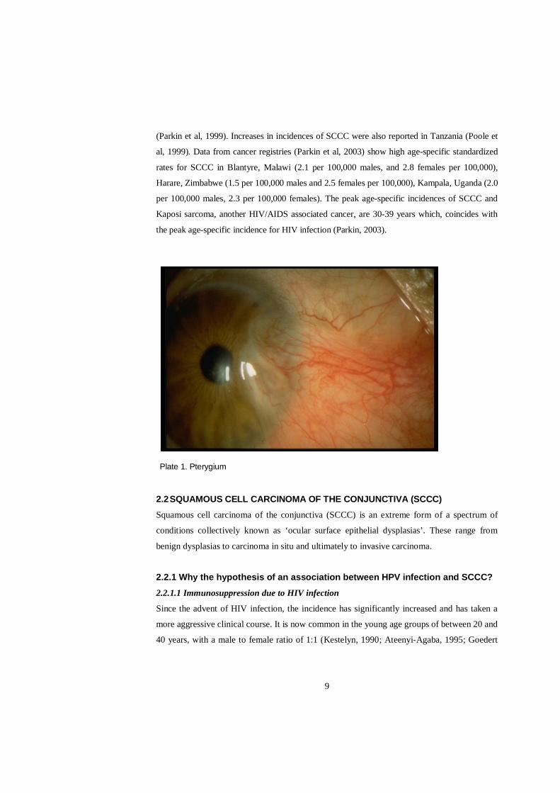
it is called a pingueculum. When the wing-shaped degeneration grows towards the limbus and
involves the cornea, it is called a pterygium, and this may grow to involve the visual axis (Plate
1).
Both pterygium and pingueculum have been reported to be associated with HPV infection
(Detorakis et al, 2001; Gallagher et al, 2001).
Melanocytes in the conjunctiva may give rise to benign tumours like naevi and conjunctival
melanosis or to malignant melanoma, which is rare in blacks (Wabinga et al, 2000).
Generally, tumours of the conjunctiva are clinically treated by surgical excisions. The prognosis
in malignancies largely depends on the stage of the tumour and the degree of differentiation.
Eye Cancers
According to figures from Kyadondo Cancer Registry, eye cancers have become frequent,
which is explained by the increasing incidence of SCCC (Wabinga et al, 2000). The increase in
incidence was reported to be almost tenfold in the interval between 1960-1971 to 1995-1997
9
(Parkin et al, 1999). Increases in incidences of SCCC were also reported in Tanzania (Poole et
al, 1999). Data from cancer registries (Parkin et al, 2003) show high age-specific standardized
rates for SCCC in Blantyre, Malawi (2.1 per 100,000 males, and 2.8 females per 100,000),
Harare, Zimbabwe (1.5 per 100,000 males and 2.5 females per 100,000), Kampala, Uganda (2.0
per 100,000 males, 2.3 per 100,000 females). The peak age-specific incidences of SCCC and
Kaposi sarcoma, another HIV/AIDS associated cancer, are 30-39 years which, coincides with
the peak age-specific incidence for HIV infection (Parkin, 2003).
Plate 1. Pterygium 2.2 SQUAMOUS CELL CARCINOMA OF THE CONJUNCTIVA (SCCC) Squamous cell carcinoma of the conjunctiva (SCCC) is an extreme form of a spectrum of
conditions collectively known as ‘ocular surface epithelial dysplasias’. These range from
benign dysplasias to carcinoma in situ and ultimately to invasive carcinoma.
2.2.1 Why the hypothesis of an association between HPV infection and SCCC?
2.2.1.1 Immunosuppression due to HIV infection
Since the advent of HIV infection, the incidence has significantly increased and has taken a
more aggressive clinical course. It is now common in the young age groups of between 20 and
40 years, with a male to female ratio of 1:1 (Kestelyn, 1990; Ateenyi-Agaba, 1995; Goedert

10
and Cote, 1995; Waddell et al, 1996; Sun et al, 1997; Beral et al, 1998; Ateenyi-Agaba and
Newton, 1999; Newton and Beral, 1999; Newton et al, 2001; Guech-Ongey et al, 2008).
Immunosuppression appears to lead to the selective increase in cancer incidence that is
thought to be associated with viral infections such as Kaposi`s sarcoma and non-Hodgkins
lymphomas (Beral et al, 1998). The identification of additional HIV-associated cancers has
important public health implications, because tumours associated with infections are at least
theoretically preventable by early treatment of the infection or by prevention by vaccination,
for example, an effective vaccine against Hepatitis B, which is the principal cause of liver
cancer in the developing countries.
2.2.1.2 Patients treated with immunosuppressive drugs
Renal transplant patients who are on immunosuppressive treatment have developed tumours
that are frequently associated with HPV infection Patients (Barr et al, 1989; Bavinck et al,
1993; Queille et al, 2007).
2.2.1.3 Solar UV radiation
Solar UV radiation has long been hypothesized as causally linked to the aetiology of SCCC,
given the extraordinary geographic distribution of the disease, being relatively more common
as you approach the equator line.
In areas far from the equator, SCCC is extremely rare (Newton et al 1996). On the other hand
the tumour is relatively common in Uganda, which is at the equator, with incidence rates
estimated at over 12 per million population per year (Ateenyi-Agaba, 1995; Wabinga et al,
2000; Parkin et al, 1999).
However, there is no evidence that solar UV radiation increased with the advent of HIV/AIDS
in Uganda to independently explain the sudden increase of SCCC. This may be explained by
solar UV radiation being a synergistic factor for the increased incidence of SCCC. Solar DNA
damage may render the cells permissive to HPV infection, as evidenced by the increased
occurrence of HPV DNA in keratotic skin lesions and skin cancer in renal transplant patients.
HPV DNA was detected in 78% of the skin lesions (Quielle et al, 2007).
More evidence of the association with UV radiation comes from the study of patients with
Xeroderma Pigmentosum, XP, a rare autosomal recessive skin disease, characterized by the
inability to repair DNA damaged by ultra violet radiation. Patients with XP have a
significantly higher risk of SCCC than the general population, and EV HPV have been
implicated in the aetiology (Daya-Grosjean and Sarasin, 1995).

11
2.2.1.4 Site of tumour
Almost all SCCC cases occur at the limbus, which is the transition zone at the squamo-
columnar junction (Plates 2 and 3).
The interpalpebral aperture is the exposed portion of conjunctiva which is prone to exposure
to ultraviolet radiation, and this may explain the frequency of these tumours at the
interpalpebral space (Waddell et al, 2006).
Moreover, HPV have a tropism for mitotically active squamous epithelial cells, conditions
which are adequately provided for by the ocular inter-palpebral space.
Plate 2. Stage I (Early SCCC) Plate 3. Stage II (Ocular muscle involvement)
2.2.2 Studies evaluating human papillomaviruses and squamous cell
carcinoma of the conjunctiva Few studies have been conducted to find an association between HPV infection and
conjunctival tumours and dysplasias.
Prior to 2002, PCR detection methods of evaluating HPV infection in conjunctival formaline
fixed tissue biopsies were based on type-specific HPV probes against HPV 16 and 18. These
case control studies consistently yielded inconclusive results, partly owing to their small size
(Scott et al, 2002; Palazzi et al, 2000; Dushku et al, 1999; Tabrizi et al, 1997; Karcioglu and
Issa, 1997; Adachi et al, 1995; Waddell et al, 1996; McDonnel et al, 1989) and partly due to
using archival formaline samples. The main role in DNA degradation is the formaline fixation
time. The longer tissues are fixed in formaline, the more the DNA is degraded. Degradation of
DNA starts in the first three hours, even in phosphate buffered formaline. DNA is degraded into
smaller fragments that need optimal methods for detection (Yagi et al, 1996), methods which
were not yet available.
Broad spectrum PCR assays that are more sensitive and can detect a wider spectrum of HPV
have been introduced in recent years, namely the (LipA), Line probe Asssay (Kleter et al,
1999) and the SPF, short PCR fragment (Kleter et al, 1998). Three case control studies have

12
been published so far, using these newer PCR techniques; they have not shown any significant
association of mucosal HPV and conjunctival squamous neoplasia. The study by Tornesello
et al, 2005, tested only for HPV 16 and 18. They isolated only one HPV type six in the
controls. Tulvatana et al, 2003, tested for multiple HPV, both mucosal and cutaneous, but did
not isolate any HPV. De Koning et al, 2008, tested for multiple HPV types and found no
significant association in mucosal HPV types. The association of cutaneous HPV and SCCC
had a high odds ratio, but the yield was low 22% (Tulvatana et al, 2003, de Koning et al,
2008).
Table 1. Case control studies using broad PCR methods for cutaneous HPV Study
Detection
method
Tissue
preparation
Cases
(HPV positive)
Controls
(HPV positive)
Odds ratio
*Tulvatana et
al, 2003
PCR Formaline fixed 0/28 (0%) 0/23 (0%) 0
Tornesello et
al, 2005
PCR Formaline fixed 15/86 (17%) 0/63 (0%) -
de Koning et
al, 2008
PCR Formaline fixed 18/81 (22%) 1/29 (3%) 8.0 (1.0-168.5)
*40% formaline used for tissue fixation
2.2.3 Limitations of earlier studies The limitations of the earlier studies include:
The small samples sizes, probably because of the rarity of SCCC.
The use of archival material fixed in formaline. Formaline is a known PCR inhibitor,
as it degrades the protein in the tissues. All studies have used formaline fixed tissues
to evaluate the presence of human papillomavirus DNA using polymerase chain
reaction (PCR) assays. To our knowledge, no study has utilized fresh frozen tissues for
evaluating the presence of HPV DNA.
The lack of broad spectrum highly sensitive PCR tests with the ability to test multiple
human papillomaviruses hampered the efforts of researchers to test for a wide variety
of HPV.
2.3 THE ROLE OF INFECTIONS IN HUMAN CANCERS
Infections are an important cause of human cancers, and in 2002 it was estimated that cancers
due to infections comprised 18% of all cancers globally, being higher at approximately 25%
in developing countries (Parkin et al, 2006). Among the cancers highly associated with
13
infections are liver cancer (hepatocellular carcinoma) caused by hepatitis B and C viruses,
gastric cancer caused by Helicobacter Pylori, Burkit’s lymphoma and nasopharyngeal
carcinoma caused by Epstein Bar virus, and, particularly in immuno-compromised
individuals, Kaposi sarcoma has been strongly associated with human herpes virus type 8.
Cancer of the cervix, causally linked to HPV infections, will be given more emphasis in the
following paragraphs, since it is a tumour of epithelial origin, like SCCC, and hence the
hypothesis that SCCC may have a related aetiology. Furthermore, most of the HPV
knowledge so far emanates from studies in the cervix uteri.
2.3.1 Cancer of the cervix
The recognition that morphological abnormalities that constitute cervical dysplasia were the
cytopathic effects of HPV infection was the first evidence suggesting a role of HPV in
cervical cancer (Meisels and Fortin, 1976). Subsequent work already in the 1970s produced
more evidence of the cytopathic effects of HPV in the cervical epithelium (Purola and Savia,
1977; Laverty et al, 1978).
The inability to propagate papillomaviruses in cell cultures hampered research on the
oncogenic potential of HPV until the advent of the molecular cloning of viral DNA in the
early 1980s, which allowed labeled viral DNA probes to be used to investigate viral DNA
presence in various epithelial tumours. This development further led to the cloning and
characterization of novel HPV types (Howley, 1991).
HPV 18 was found to be associated more with glandular differentiation cancers, while HPV
16 appeared to be associated more with squamous differentiation cancers (Wilczynski et al,
1988), though it has now been established that both are responsible for a vast majority of
cervical cancer worldwide.
More recent genetic studies have confirmed the integration of HPV DNA in the human
genome, which is an important step towards neoplastic transformation by disrupting the E1
and E2 viral genes leading to an over-expression of the E6 and E7 oncoproteins (Cricca et al,
2009). While some investigators have found integration of HPV DNA almost exclusively in
high-grade lesions and invasive cancers (Klaes et al, 1999; Tonon et al, 2001; Hudelist et al,
2004), others have found integration occurring earlier in precancerous lesions or even in
asymptomatic patients (Peitsaro et al, 2002; Gallo et al, 2003; Andersson et al, 2005; Kulmala
et al, 2006).

14
Evidence from molecular epidemiological studies were evaluated twice by the International
Agency for Research on Cancer (IARC) monograph programme, which concluded that there
was strong evidence of an association of cervical cancer and HPV infection (IARC
Monograph, 1995; IARC Monograph, 2007). By 2002, PCR tests were detecting high risk
HPV types in almost 100 % cervical cancer biopsies (Parkin et al, 2006).
Based on their frequent presence, either in precancerous lesions or invasive cancers, the HPV
were segregated into low-risk and high-risk types (Richart et al, 1998) but have recently been
reclassified into three groups, taking into account a third group that has a potential to cause
cancer (Munoz et al, 2003). This reclassification was inevitable, as HPV are a large family of
viruses, and novel HPV are being added onto the list every year as the sensitivity and
specificity of PCR methods improve. Over 100 HPV types have been identified so far, and the
epidemiological classification currently includes three groups namely:
The high-risk types including; 16, 18, 31, 33, 35, 45, 51, 52, 56, 58, 59, 68, 73 and 82;
Probable risk include; 26, 53 and 66;
Low-risk types; 6, 11, 40, 42, 43, 44, 54, 61, 70, 72, 81, and CP6108.
It is not uncommon for an epithelium to be infected by more than one HPV type, particularly
in low-grade lesions (Lungu et al, 1992). The multiple infecfions have been hypothesized to
be a risk factor for progression to invasive cervical cancer, but evidence is still contradictory.
Some investigators have found an association with progression to invasive cancer (Fife et al,
2001; Sasagawa et al, 2001; van der Graaf et al, 2002; Chaturvedi et al, 2005; Herrero et al,
2005), while others have found no association with progression (Rolon et al 2000, Levi et al,
2002; Bosch FX et al, 2002; Cuschieri et al, 2004).
Viral loads, especially for HPV 16, have a predictive value for the development of cervical
cancer (Gravit et al, 2007).
2.3.2 Human papillomaviruses The papillomaviridae family of HPV was, up until 2004, grouped into one family called the
papovaviridae, a large family of viruses that includes the Simian virus 40 (SV40) and the
polyomavirus (Bernard et al, 2006). All these viruses are obligatory intranuclear DNA
viruses. They are all tumour viruses and appear to transform normal cells into neoplastic cells.
After 2004, the HPV were recognized as a family of their own since pylogenetic studies had
shown that the viruses have been genetically stable for a long time within their mammalian
and bird species, since they do not recombine and do not change host species (de Villiers et al,
2004; Bernard et al, 2006).

15
Figure 1. Classification of human papillomaviruses (de Villiers et al, 2004).
Reproduced with permission, source: de Villiers et al, 2004.
The HPV have been reclassified into genera with each genus having a specific tissue tropism
(de Villiers et al, 2004). The Alpha genera have a tropism for anogenital mucous membranes
and were formerly known as mucosal HPV, to which HPV 16 and 18 belong. The alpha
genera is further classified into high-risk, probable-risk and low-risk groups, depending on
their oncogenic potential (Munoz et al, 2003).
The Beta genera have a tropism for cutaneous tissue, and this large group includes the
Epidermodysplasia Verruciformis (EV) viruses formerly known as the cutaneous HPV (de
Villiers et al, 2004). See figure 1 for related genera.
Recently, de Villiers et al, 2004 have classified HPV into genera. Genus papillomaviruses
consist of tightly coiled, circular, double-stranded DNA with about 6,800-8000 base pairs,
depending on the HPV type, in their genome with 8 or 9 open reading frames (zur Hausen et
al, 2002; Doorbar et al, 2005). The complete virion is the infectious unit and consists of a
DNA core surrounded by an icosahedral-shaped protein coat, the capsid, with a diameter of
45-55 nm. The capsid has 360 copies of the L1 protein (Doorbar et al, 2005; Lowe et al,
2008).

16
Papillomaviruses are widely distributed in the animal kingdom, producing infections in
multiple mammalian and avian species. They tend to be highly species-specific (Antonsson
and Hanson, 2002; Antonsson et al, 2003; Bernard et al, 2006).
HPV frequently infects humans, and various papillomavirus types have a predilection for
surface epithelial cells, including cutaneous or mucous membranes. They can infect mucous
membranes of the anogenital regions, oesophagus, and pharynx (Doorbar, 2005).
2.3.3 Human papillomavirus genome There are three functional domains in the HPV viral genome. They are known as the upstream
regulatory region (URR), the early region and the late region (Doorbar et al 2005). Both the
early and late regions contain linear sequences without stop codons that are potentially
transcribable into proteins and are known as `open reading frames` (ORFs). The E2 ORF is
often disrupted during nitration into the host cell genome, leading to uncontrolled expression
of E6 and E7 oncoproteins (zur Hausen et al, 2002; Doorbar et al, 2006; Cricca et al, 2009).
The early region is the only papillomavirus protein with enzymatic activity and is associated
with the DNA-dependent ATPase and DNA helicase, enzymes which are useful in DNA
replication (Wilson et al, 2002).
The late region consists of L1 and L2 proteins. The L1 protein is a major component of the
capsid protein, while the L2 is the minor component. The L1 protein is highly immunogenic
and plays a key role in viral entry into epithelial cells (Kirnbrauer et al, 1992; Day et al,
2006). The immunogenic property has been utilized in making the first generation vaccines
against HPV.
L2 is a bigger protein but a minor component of the capsid. It facilitates entry into the basal
cells and is responsible for virion assembly in vivo (Pereira et al, 2009). It has immunogenic
surface antigens conserved across HPV types and is a candidate for broad spectrum HPV
vaccines (Lowe et al, 2008).
2.3.4 Natural history of HPV infection Infection of epithelia is thought to begin through minor wounds and is facilitated by L1and L2
proteins. The micro-abrasions expose the basal layer, which is preferred by the HPV. L1 binds
to cell surface receptors with the help of L2 proteins (Day et al 2006; Joyce et al, 1999). Cell
tropism has been found to be associated with the total net charge of the virion, which differs
between genera (Mistry et al, 2008). Replication takes place in the differentiating cells
(Longworth and Laimins, 2004), and once the cells reach the supra-basal layer, the late viral

17
proteins are produced to form the complete virion, which is the infectious unit (Graham et al,
2006; Longworth and Laiminis, 2004).
The majority of infections of the cervix appear to clear spontaneously without therapeutic
intervention, probably as a result of interaction with the host immune system (Morrison et al,
1991). Persistent infection with the high-risk HPV types is thought to be a precursor to
neoplastic transformation (Munoz et al, 2003).
2.4 TP53 MUTATIONS AND SOLAR ULTRAVIOLET RADIATION The p53 protein encoded by the TP53 gene is an inhibitor protein that is involved in the cell
cycle to regulate cell multiplication. p53 protein acting with the retinoblastoma (Rb) gene is
responsible for programmed cell death or apoptosis (Cricca et al, 2009). Together with
terminal maturation regulated by telomeres with the aid of the tolemerase enzyme, these cell
proteins are able to regulate cell multiplication and prevent the cell from entering an
uncontrolled phase of multiplication in instances of genomic attacks (Sarasin, 1999; Daya-
Grosjean and Sarasin, 2005 ).
In cancer of the cervix, the E6 protein has been shown to inactivate p53 protein by binding to
it (Band et al, 1993; Crook et al, 1991; Scheffner, 1990; Weirness et al, 1990; Narisawa-Saito
and Kiyono, 2007). Similarly, E7 has been shown to inactivate the Rb gene by binding to it
(Dyson et al, 1989; Gonzalez et al, 2001). Inactivation of the p53 and the retinoblastoma,
pRb, inhibitor proteins lead to uncontrolled cell division, which is the beginning of neoplastic
transformation (Nairisawa-Saito and Kiyono, 2007).
Solar UV radiation is thought to be a significant risk factor, as evidenced by the occurrence of
these UV related tumours at increasing levels of ambient solar radiation (Newton et al, 1996).
Molecular evidence, especially in non-melanoma skin tumours and Xeroderma pigmentosum
associated skin cancers, has established a strong causal link, as observed by the very high
frequencies of mutations in the TP53 gene (Sarasin, 1999; Daya-Grosjean and Sarasin, 2005).
The C to T or tandem CC to TT mutations, which are a molecular signature of UV DNA
damage, have been observed in 50% of patients with this disease who develop skin tumours
(Daya-Grosjean, 1995; Daya-Grosjean and Sarasin, 2005).

18
3 AIMS
The overall aim of this thesis was to investigate the association between human papillomavirus
infection and squamous cell carcinoma of the conjunctiva.
The specific objectives were:
Study I
To ascertain the presence of HPV DNA and HPV types in tissues with SCCC and in
conjunctival tissues with other eye conditions.
(Paper I published)
Study II
To evaluate the presence of p53 mutations in SCCC tissues as a more objective way of
assessing the role of UV radiation in the tumour aetiology.
(Paper II Published)
Study III
To establish whether there was any HPV DNA in the normal conjunctiva and assess whether
there was an excess of HPV infection in AIDS patients.
(Paper III Published)
Study IV
To ascertain the presence of HPV infection and HPV types in tissues with SCCC and
in conjunctival tissues with other eye conditions.
To compare the socio-demographic characteristics of patients with SCCC to those
patients with other eye conditions.
To compare the HIV sero-status of patients with SCCC and those patients with other
eye conditions.
(Paper IV submitted)
19
4 SUBJECTS, MATERIALS AND METHODS
4.1 PAPERS I AND II Study site. The study was conducted at New Mulago, which is the national referral hospital
serving a population of about 30 million people. The eye clinic at Mulago registers an average
of fifty patients per day who are referred from the whole country.
Participants. These included patients with eye disorders seeking treatment from the hospital.
Study design and Procedure. A hospital based pilot case control study was conducted
between March 2000 and January 2001 aimed at testing methods and assessing the feasibility of
a wider case control study to follow at the eye department in New Mulago Hospital, Kampala,
Uganda. All patients who presented with eye tumours clinically suspected to be SCCC had
excision biopsies taken, after informed consent, as part of the routine clinical management of
the patients. The biopsy was divided into two pieces. One piece for histology was placed in a
10% formaline, while the piece for HPV DNA was stored at -400 C with no added preservative.
Patients who presented with other operable conjunctival diseases, which included pterygia,
pingueculua and conjunctival naevi, also had excision biopsies as part of the routine
management of the patients. The biopsies of the controls were treated the same as for the
patients suspected to have SCCC. Histology was performed at the Department of pathology at
Makerere University by two pathologists. All those patients who had histologically confirmed
SCCC were recruited as cases and other patients as controls. We frequency matched cases to
controls by age and sex. Overall, 21 cases of squamous cell carcinoma of the conjunctiva and
22 controls were recruited.
A questionnaire was completed to capture the socio-demographic characteristics.
Controls included benign conjunctival lesions, which included 10 pterygia, 7 pinguecula, 4 solar
keratoses and 1 pigmented naevus. Tissues for HPV analysis were deep frozen to -400 C before
being transported in ice flasks filled with ice and flown overnight to the International Agency
for Research on Cancer (IARC) in Lyon, France, for HPV testing and mutational analysis.
4.1.1 DNA extraction DNA was extracted from each sample in the IARC laboratory that is exclusively used for this
purpose. To monitor contamination between the different specimens during DNA extraction,
tubes containing distilled water (negative controls) were included for every 10 specimens.
20
4.1.2 HPV detection
HPV detection was performed by means of polymerase chain reaction (PCR) assays using
different sets of primers (Chen et al, 1994; Berkhout et al, 1995; Jacobs et al, 1995; Forslund et
al, 1999; Harwood et al, 1999; Caldeira et al, 2003)
Laboratory personnel were blinded as to the histological diagnosis of each patient.
The PCR mixture was prepared in a special laboratory in sterile conditions while the PCR
analysis of the products was analyzed in different rooms. Negative controls were included in the
different PCRs. Products obtained with type specific primers were also tested by southern
blotting. To further enhance the quality control, all products were tested in duplicate using
consecutive sections of the frozen specimens.
4.1.3 Assessment of TP53 mutations DNA was extracted, and Exons 5-9, including splice junctions, were screened for thepresence
of somatic mutations by denaturing high performance liquid chromatography (DHPLC) using
primers and conditions described (Dai et al, 2004).
Exons with abnormal DHPLC were further analyzed by automated dideoxysequencing as
described (Taniere et al, 2000) for TP53 mutations at hot spots for UV light damage.
4.1.4 Statistical analysis Paper I. Age adjusted odds ratios were calculated to compare the cases and controls with
respect to educational level, outdoor and indoor occupation and sun exposure. Odds ratios for
the association of Epidermodysplasia Verruciformis (EV), HPV DNA and SCCC were
computed after adjustment for age, and then they were computed after adjusting for occupation
and hours of exposure to sunlight
Paper II. Percentages of UV-induced mutations in cases and controls were calculated, and the
percentage of mutations in cases with positive HPV DNA samples were compared to mutations
in controls with positive HPV DNA tests.
4.1.5 Ethical issues The study was approved by the Faculty of Medicine Higher Degrees, Research and Ethical
Committee (the ethical review committee) and the National Council for Science and
Technology (the national regulatory committee).
21
4.2 PAPER III 4.2.1 Materials
Setting. The study was conducted in the mortuary of Mulago Hospital, Kampala Uganda. This
morgue admits only patients who have died in the hospital. All other cases who have died
before being admitted to hospital are admitted to the police mortuary. All patients who die in
the hospital are supposed to have a postmortem examination done to confirm the cause of death.
Subjects. All patients who died at Mulago hospital and had a postmortem examination,
between the 1st and 31st of August 2002.
Study design and procedure. This was a cross-sectional study involving one hundred and
thirty six subjects. Autopsy specimens from the conjunctiva were collected within 24 hours
from individuals who had died. The subjects were classified into three categories namely; AIDS
(for those fulfilling the AIDS criteria as per the 1993 Center for Disease control (CDC)
classification), other infectious diseases and chronic diseases or trauma. Individuals with
macroscopic conjunctival diseases were excluded. A minimal quantity of saline solution was
injected close to the limbus, and a biopsy was taken from the medial aspect of the conjunctiva in
the inter-palpebral region. Biopsies were immediately transferred to a -800 C freezer and then
shipped to the IARC laboratory overnight in flasks filled with Ice. Information was collected on
the subjects´ socio-demographic characteristics and cause of death from the hospital files.
4.2.2 DNA extraction DNA was extracted using Biorobot (Qiagen). Beta globulin was tested for, and all the 136
samples were Beta globulin positive.
To reduce the risk of contamination during DNA extraction, the preparation of the PCR mix
and PCR was performed in different rooms. One negative control, consisting of tubes only
with distilled water, in every 10 samples was included.
HPV typing. Testing was done at the IARC laboratory using a newly developed general
primer PCR system for the detection of beta- and gamma cutaneous papillomaviruses (BGC-
PCR).
Typing was done by reverse line blotting for genus beta HPV types 5, 8, 9, 12, 14, 15, 17, 19,
20-25, 36-38, 47 and 49, and genus gamma types 4, 48, 50, 60 and 65. Testing for genus alpha
HPV types including 6, 11, 16, 18, 26, 31, 33-35, 39, 40, 42-45, 51-59, 61, 66, 68, 70, 71, 72,
73, 81, 83, 84 and CP8308 was performed at the laboratory of Molecular Pathology at Virje
University Medical center, Amsterdam, using general primer-mediated GP5+/6+PCR, and by
hybridization of PCR products in an enzyme immunoassay, using two oligo cocktails.

22
4.2.3 Statistical analysis
The association of HPV infection and other subject characteristics were computed using
univariate and multivariate odds ratios (OR). Multivariate odds ratios were computed using
the multiple logistic regression equations, including terms for age, sex and cause of death.
4.2.4 Ethical issues The study was approved by the Faculty of Medicine Higher Degrees and Research Ethical
Committee (the ethical review committee) and the National Council for Science and
Technology (the national regulatory committee).
4.2.5 Study limitations
HIV testing was not consistently available in the medical records, and therefore we used a
clinical classification. The lack of HIV results could have introduced significant bias in the
classification of subjects. Those subjects who had HIV/AIDS related chronic diseases or
infectious diseases were considered by us in a sub-analysis as potentially HIV/AIDS.
4.3 PAPER IV 4.3.1 Setting The study was conducted at New Mulago Hospital, Kampala and Jinja Hospital, Jinja,
Uganda. As explained above, New Mulago Hospital is the national referral hospital serving a
population of about 30 million people in Uganda. The eye clinic at Mulago registers an
average of fifty patients per day who are referred from the whole country. Jinja Hospital is a
regional referral hospital, mainly treating patients from the eastern region of Uganda. Patients
are ordinarily referred to a regional hospital and from the regional hospital to the National
referral hospital.
4.3.2 Participants
These consisted of patients seeking medical treatment from New Mulago and Jinja eye clinics
during the study period.
4.3.3 Study design and procedure The study was a hospital-based case control study. A total of 142 patients who presented with
tumours clinically suspected to be squamous cell carcinoma of the conjunctiva were recruited.

23
Specimens were obtained from Mulago and Jinja Hospitals from January 2004 to June 2007.
All patients presenting with incident conjunctival tumours that were clinically suspected to be
malignant had excision biopsies of the tumours, and all those tumours that were histologically
confirmed to be SCCC were taken as cases. Two pathologists read the slides, and where there
was a disagreement, a third pathologist reviewed the slides. A consensus was achieved on the
presence of SCCC in 100 patients. A diagnosis of dysplasia was made in the remaining 42
patients. Of the 100 cases, 10 were from Jinja, and of the 42 dysplasias, 6 were from Jinja. Five
SCCC cases were excluded, due to missing biopsy samples, empty tubes or mislabelled tubes.
One SCCC sample from Jinja was excluded, due to a negative beta-globulin result. Beta
globulin presence in the tumour samples is a way of ascertaining whether the tissue is still fresh.
Beta globulin negative samples indicate that the tissues have undergone protein degeneration,
which affects the PCR results.
Three patients eligible to be recruited as controls, and one patient who fulfilled the eligibility
criteria for a case declined to participate in the study because they feared to have their blood
tested for HIV.
Controls were those patients presenting to the same clinics during the study period with eye
diseases other than squamous cell carcinoma of the conjunctiva, who required surgical
intervention as part of routine treatment. Pterygia and pingueculae were excluded, as these have
been reported to be associated with human papillomaviruses (Detorakis et al, 2001; Gallagher et
al, 2001). Controls included, among others; ruptured eyes, cataracts, perforated corneal ulcers
and chalazia. Both cases and controls had biopsies, and a sample of blood for HIV serology was
taken. Tissue specimens were deep frozen at -800 C and shipped to Delft Laboratories,
Netherlands, for Polymerase Chain reaction (PCR) to detect and type HPV DNA.
4.3.4 Case definition A case was defined as any patient presenting to Mulago or Jinja Hospital eye clinic, during
the study period, with histologically confirmed (SCCC).
4.3.5 Controls Controls were all those patients presenting to New Mulago or Jinja Hospital eye clinic during
the same study period, with eye diseases other than SCCC.

24
4.3.6 Exclusion criteria i) All patients presenting with recurrent SCCC who had been on radiotherapy or
chemotherapy were excluded as cases, since treatment could have an effect on the PCR
results.
ii) Pterygia and pingueculae were excluded as controls, as relatively recent reports
(Detorakis et al, 2001; Gallagher et al, 2001) have suggested a possible association with HPV
infection. SCCC has also been reported to arise from a pterygium (Sevel and Rossal, 1965).
4.3.7 Power calculation The desired parameter of interest is the prevalence of HPV DNA in SCCC biopsies. However,
data on the prevalence of HPV DNA in comparable populations is scanty. The only study
conducted in Uganda and Malawi showed a prevalence of 35% in a small sample size of 20
cases of SCCC. Only HPV 16 and 18 were tested for, and since we were to test for all types,
both mucosal and cutaneous HPV, this was an underestimatation, and a prevalence of 50%
was used to calculate the minimum sample size. For a minimum acceptable odds ratio of 2, a
control to case ratio of 3 allowing a 5% error, the minimum number of cases is 88 and 264
controls for a study with 80% power using the Schelsselman SE formula for case control
studies design and analysis (Schelsselman, 1982).
4.3.8 Information and biological materials A questionnaire was administrated to all study subjects. It contained demographic and
lifestyle questions as well as questions about previous eye pathologies. Interviewing of
patients was done by selected nurses in the clinics, who were specially trained to conduct the
study interview.
4.3.8.1 Procedure for Recruitment of cases and controls
All patients presenting to New Mulago Hospital, and Jinja Hospital out-patient eye clinics
with suspicious conjunctival growths had excision biopsies taken under sterile surgical
conditions. This is part of the usual therapeutic and diagnostic care for suspicious conjunctival
growths. Patients were asked for their informed consent to be included in the study. The
biopsy was divided into two pieces. One piece was fixed in 10% formaline for
histopathological examination, and the other piece was immediately placed in a sterile
container sealed and stored immediately in a deep freezer at temperatures of –800 C for viral
HPV DNA studies. For patients in Jinja Hospital without -800 C freezers, the biopsies for viral
studies were placed in Liquid Nitrogen until they were transported to Mulago for storage.

25
All patients had their samples analyzed by 2 pathologists, and where there was a
disagreement, a third reviewer was engaged. A 10 ml blood sample was collected at the time
of surgery for HIV testing.
All patients who presented to the same clinics during the period of the study with lesions
other than pterygia, pingueculae, carcinoma in situ or squamous cell carcinoma of the
conjunctiva that required surgery as part of the usual clinical management, were recruited as
controls. These included patients who presented with cataracts, painful blind eyes, scleral
tears, eyes removed due to severe eye injuries and corneal tears. These biopsies were obtained
and processed in the same manner as for the biopsies of the cases. The biopsies were divided
into two pieces: one piece was fixed in formaline 10% and sent for histology, and another
piece was immediately stored in a sterile container at a temperature of –800 C for viral DNA
studies. Formaline processing of biopsies was done after every 15 patients for those patients
who did not have obvious pathology of the conjunctiva, for instance patients with cataracts
and corneal tears.
A 10 ml blood sample was taken from consenting patients for HIV serology, following the
same procedures as above described for cases, including pre- and post test counseling for HIV
testing.
Patients had a detailed medical history taken and were subjected to a general physical
examination to assess any signs and symptoms of immunosuppressive conditions which were
noted on a clinical form. A detailed ocular examination was carried out using diffuse light and
slit lamp bio-microscopy. The findings of both the examination and history were recorded on
a data sheet that had both the Identification number of the patient and the patient’s particulars.
This data sheet was only accessible to the principal investigator/treating physician.
Patients were booked for surgical theatre after informed consent. In the surgical theatre, the
growths were excised by the principal investigator/ treating physician under sterile conditions.
The excised growth was divided into two equal pieces. One piece, for viral DNA tests, was
marked with an identification number and placed in a sterile container, without any additives,
sealed and stored in a deep freezer at –800 C.
The second piece of the excised growth was fixed in 10% formaline solution for
histopathology. Histopathological examinations of fixed tissues were done at the Department
of Pathology of Makerere University. Slides were read by a senior pathologist.
The blood sample was marked with an identification number corresponding to the
identification number of the biopsies. Samples were blinded as to their case/control status.
Samples from controls were processed using the same procedures as samples from cases.

26
4.3.8.2 Laboratory Procedures
Histopathology
The diagnosis of SCCC was established at the Department of Pathology of Makerere University
Kampala, Uganda. Three slides were prepared per patient with a clinically malignant growth.
The slides were reviewed by a pathologist in the Department of Pathology at Makerere
University. For quality assurance, the slides were reviewed by another pathologist at the Delft
Laboratory in the Netherlands. The slides for which there was disagreement between the two
pathologists were reviewed by a third pathologist at Kampala. All those patients who had a
histopathological diagnosis of SCCC from at least two pathologists were included in the
analysis as cases.
HIV testing
HIV testing was done at the Nakasero Blood Bank in Uganda, using an enzyme-Linked
immunosorbet assay (ELISA). A 10ml sample of blood was taken from the patients and put in a
vacutainer containing Ethyl DiamineTetraAcetic Acid (EDTA). The blood was centrifuged at
3000 rpm and serum separated from the cells. The serum was stored at 40 C before being taken
for the Elisa test. The Elisa test utilized the Murex HIV-1.2.O from the ABBOT Diagnostics
Division in the United Kingdom with a specificity of 99.91% and sensitivity of 100%. The tests
were done by one technician following the manufactureer’s instructions. All those patients that
were positive had confirmation with the Western Blot method.
HPV typing
Tissue biopsies were frozen at -80 C and shipped to the Delft Diagnostic Laboratory (DDL) for
HPV testing. DNA was extracted by proteinase K treatment. Five SCCC cases, 2 conjunctival
dysplasia cases, and 19 controls had no biopsies because of losses or empty or mislabelled
tubes, and hence were excluded. For each 10 tissue samples, one DNA isolation-negative
control was included. Isolated DNA was tested using three different PCR-based assays, one
targeting beta-globin (for DNA quality control), one targeting mucosal, and one targeting
cutaneous HPV types. Beta-globin-negative biopsies (1 SCCC, 1 conjunctival dysplasia, and 5
controls) were also excluded from the present study.
For each 10 tissue samples, one negative DNA isolation control was included. Isolated DNA
was tested by 3 different PCRs, beta-globin PCR, genital HPV PCR and beta HPV PCR. The
beta-globin PCR was applied for controlling the quality of isolated DNA. The genital HPV
genotyping PCR was carried out using the short PCR fragment, SPF (Kleter et al 1998), the line
Probe assay, LiPA system (Kleter et al, 1999), and HPV LiPA, version 1; manufactured by
Labo Bio-Medical Products, Rijswijk, Netherlands as described previously. Briefly, the broad
27
spectrum SPF PCR amplifies a 65-base pair fragment from the L1 region of the HPV genome.
By using biotinylated reverse primers the amplimers could be captured onto streptavidin-coated
microtiter plates. After denaturation of the PCR products by alkaline treatment, a defined
cocktail of digoxigenin-labeled probes was used to detect HPV positive samples. This method,
designated as the HPV DNA Enzyme Immunoassay (DEIA), provides an optical density value
and is able to detect more than 50 HPV types. Amplimers from positive samples were used for
subsequent genotyping of twenty-five individual genital HPV genotypes (high-risk HPV: 16,
18, 31, 33, 35, 39, 45, 51, 52, 56, 58, 59, 66, 68, 70, and low-risk HPV: 6, 11, 34, 40, 42-44, 53,
54, 74) simultaneously in a reverse hybridization assay (RHA). Beta HPV genotyping was
performed with the PM-PCR RHA method (The skin (beta) HPV prototype research assay;
Diassay BV, Rijswijk, Netherlands). It consists of a broad spectrum PCR specific for the
amplification of the beta HPV genus and targets a fragment of 117 bp from the E1 region of the
HPV genome. Combined with the RHA, it was possible to identify 25 beta HPV types (i.e.,
HPV type 5, 8, 9, 12, 14, 15, 17, 19-25, 36-38, 47, 49, 75, 76, 80, 92, 93 and 96). As no DEIA
was developed for this assay, all amplimers were directly analyzed by RHA.
4.4 Statistical analysis After data cleaning, data were entered into Excel and analyzed using the STATA version 10.0
programme. The association between HPV infection and the various study variables was
evaluated using unconditional multiple logistic regressions. Odds ratios (OR) and the
corresponding 95% confidence intervals were calculated after adjusting for age, sex and HIV
status as appropriate. The association between HIV and SCCC was evaluated using
unconditional logistic regression after adjusting for age, sex and HPV infection. Odds ratios for
co-infection with HPV were calculated after adjusting for age and sex. Odds ratios and
confidence intervals were computed to evaluate the association between smoking habits,
educational level and indoor or outdoor occupation.
4.5 Ethical considerations Each potential study participant was approached in the eye clinics by the treating doctor, after
the preliminary exam of the eye lesion. The treating doctor informed the subject about the
study, and explained what participation implied, namely answering a short questionnaire,
allowing biopsies to be used for HPV testing, and donation of a blood sample. The physician
also informed the subject about the possibility of having an HIV test, which included pre- and
post-testing counseling. The subjects were clearly informed that their participation in the

28
study was voluntary, and that it had no consequences for the usual clinical treatment. Subjects
were also clearly informed that they could opt to donate a blood sample but not to know the
HIV results, if they so wished. They were also informed of the potential benefit of improved
patient management, depending on the findings of the study. The risks involved in surgery
and taking a blood sample were explained to the participants before requesting their consent.
An informed consent form was signed by subjects willing to participate in the study. The
consent forms were available in the most common languages used in the country, namely
Luganda and English.
Strict confidentiality was followed, and no data was to be made public unless the patient’s
approval was sought or identification removed. All specimens were marked using an
identification number. The corresponding names were kept by the investigators only. During
the HIV pre-counseling session, the patients had an education session on the cause of AIDS
and its various complications and the way to cope with the disease in case they were found to
be positive for HIV. In the post- and pre- counseling sessions, the patients were taught how to
live safely, emphasizing the social and emotional after-effects. A trained counselor was
engaged especially for those patients who were found to be HIV positive. Ethical approval
was sought from the Faculty of Medicine Higher Degrees, Research and ethical Committee
(the Review Committee) and the National Council for Science and Technology (the national
regulatory committee).
4.6 Study Limitations We were unable to perform CD4 counts, mainly due to financial constraints. This would have
given us a clearer picture of the level of immunosuppression of patients with SCCC at first
time of presentation, which is important in patient management.

29
5 RESULTS
5.1 PAPER I 5.1.1 HPV Typing General primers (GP) 5+/6+, GP1/GP2 and specific primers for HP16 and 18 and 45 were
used to screen for mucosal types. HPV types that were isolated from the SCCC cases were
exclusively cutaneous types belonging to the Epidermodysplasia verruciformis group, as
opposed to the long hypothesized mucosal HPV types.
No mucosal types were found in either cases or controls. In one case, a novel HPV type
closely related to EV HPV type RTRX7 was detected using GP1/GP2 primers.
Highly sensitive PCR protocols for detecting EV HPV types, namely CP62/69-EN3F/EN3R;
CP62/69-EN1F/EN1R and CP66/CP69-CP65/CP70 (Berkhout et al, 1995; Harwood et al,
1999), revealed HPV types in six cases and one control patient. In the majority of HPV
positive patients, the PCR product was directly sequenced, and HPV type 5, HPV14, HPV24,
HPV36, HPV37 and HPV38 were isolated. Two novel HPV types related to HPV8 and
HPV12 were also isolated. In two SCCC cases and one control patient, multiple HPV
infections were isolated. PCR analysis followed by southern blotting using specific primers
for the EV HPV type 38 (Caldeira et al, 2003), which are 50-100 fold more sensitive than CP
primers in detecting HPV 38, showed a positive signal in 17 SCCC cases and 8 controls.
SCCC cases and 20 controls who had a questionnaire including socio-demographic and
lifestyle information were compared by means of age adjusted odds ratios (OR) and
corresponding confidence intervals (CI). The OR for the presence of EV HPV was 12.0; 95%
CI = 1.7-84.9). The prevalence of EV HPV did not vary by age group in either cases or
controls. The educational level was inversely associated with SCCC risk, whereas for outdoor
occupation and sun exposure there were indications of associations, but the confidence
intervals were wide, and the values included unity due to the small sample size.
The association of EV HPV with SCCC was strengthened by adjustment for sun exposure
(OR = 22.7; 95% CI = 1.7-312).
5.2 PAPER II 5.2.1 TP53 mutations
Eleven (52.4%) cases had TP53 mutations in Exons 5-9, including one sample with two
independent mutations. TP53 mutations were also detected in three controls 13.6%: two

30
pterygia and one pingueculum. Seven mutations in SCCC cases (sample 1 with 2 mutations,
2, 5, 7, 10 and 11) and one pterygium sample were CC TT mutations. Several CC TT
mutations spanned adjacent codons and had complex consequences, as in sample 1 containing
two distinct CC TT mutations. The first spans codon 195-196 and induces a silent mutation
at codon 195 and a nonsense mutation at codon 196.
The majority of SCC cases (18/21, 86%) tested positive for EV HPV types, mainly HPV38
TP53 mutations were detected in 10 (56%) of the 18 EV HPV positive samples and one of the
three HPV negative samples. Nine (41%) of the 22 controls were EV HPV positive. TP53 was
mutated in one of the controls.
5.3 PAPER III Individuals who had died of AIDS were significantly younger (median age = 30 years range
15-58 years) than either those who had died of infectious diseases other than AIDS (median
age = 40 years; range 17-85 years X2 for trend = 14.6; p < 0.001) or chronic diseases or
trauma (median age = 44years; range 15-94 years; X2 for trend = 8.0; p = 0.005). Women
represented 51.5% of the individuals who had died of AIDS, but 35.6% (p = 0.16) and 27.6%
(p = 0.02) respectively of those who had died of other infectious diseases and chronic diseases
or trauma.
Conjunctival infection with genera beta/gamma HPV types was found in 14.7% of
individuals.
Univariate analysis showed a slightly increased OR for HPV infection for age greater than 25,
female gender, indoor occupation and AIDS related death but all 95% confidence intervals
were broad and included unity.
In multivariate analysis that included age, gender and cause of death, there was no significant
difference in HPV prevalence by cause of death among individuals who had died of AIDS and
from other infectious diseases versus individuals who had died of trauma or chronic diseases(
OR = 1.3; 95% CI = 0.4-4.8 and OR = 0.9; 95% CI = 0.3-2.9) respectively.
Eleven genus beta and two genus gamma HPV types were detected in either single HPV
infections representing 80% (n = 16) or multiple type HPV infections representing 20% (n =
4). The most frequently found types were HPV22 (n = 7), HPV8 (n = 6), HPV23 (n = 3) and
HPV17 and 19 (n = 2 each). Of the individuals who had died of AIDS, six had a single HPV
type infection and one had multiple HPV infections. HPV23 was found in three of these
individuals and HPV8, 17, 19, 20, and 22 were all found in the one individual. All 136
biopsies tested negative for genus alpha HPV types.

31
5.4 PAPER IV A total of 94 cases of SCCC (mean age 36.7 years; range 15-80 years), 39 cases of dysplasia
(mean age 32.8 years; range 11-50 years), and 285 controls (mean age 34.0 years; range 15-80
years) were included in the present study. Education, occupation, and cigarette smoking were
not significantly associated with SCCC, whereas a strong association was seen with HPV
infection (OR = 6.22; 95% CI = 3.60-10.72) and HIV infection (OR = 7.3; 95% CI = 4.2-12.4).
Signs of severe immunosuppression such as the presence of cytomegalovirus retinitis,
cryptococcal meningitis, or skin rash with weight loss, were observed in 13 cases and 3
controls. There was a significant association between severe immunosuppression and
SCCC/dysplasia (OR = 5.6; 95% CI = 1.5-20.3).
Mucosal HPV types were detected in 6.4, 7.7, and 3.5%, respectively, of SCCC, dysplasia, and
controls. The majority were of uncharacterized HPV types. In contrast, cutaneous HPV types
were found more often in cases of SCCC (44.7%) and dysplasia (41.0%) than in controls
(10.5%), and uncharacterized types were few. Multiple-type infections were much more
frequently detected in SCCC (57.1% of cutaneous HPV-positive SCCC cases) and dysplasia
(75.0%) than in controls (13.3%). The most common types among SCCC cases were HPV5 (n
= 15), HPV8 (n = 16) and HPV24 (n=9). HPV5, 14, and 17 were each found in at least 5
dysplasia cases. HPV5, 15, and 24 were each found in at least 6 controls and only 4 controls had
multiple infections.
No association emerged between infection with mucosal HPV types and either SCCC or
dysplasia (OR = 1.0; 95% CI = 0.4-2.7). In contrast, positivity for cutaneous HPV types was
associated with significantly increased risks for SCCC (OR = 2.3; 95% CI = 1.2-4.4) for single-
and (OR = 18.3; 95% CI = 6.2-54.4) for multiple-type infections. Significantly elevated ORs of
SCCC/dysplasia were found for HPV5, 8, 14 and 23. The OR was also of borderline statistical
significance for HPV24. In general, ORs for individual cutaneous types were similar for SCCC
and dysplasia, except for HPV8, which showed a stronger association with SCCC (OR = 39.0;
95% CI = 4.9-310) than with dysplasia (OR = 11.3; 95% CI = 1.0-134). The difference between
the two ORs was not, however, statistically significant.
When cases with well-differentiated and poorly-differentiated SCCC were evaluated separately,
no differences in the positivity for cutaneous HPV types emerged. However, SCCC is largely a
well-differentiated tumour, and only 2 cases out of 9 poorly- differentiated tumours were found
to have cutaneous HPV infection.

32
The association between HPV infection and SCCC among HIV negative patients was not
significant (OR = 2.5; 95% CI = 0.6-10.0). HIV infection was significantly associated with
SCCC in the absence of HPV infection (OR = 4.8; 95% CI = 2.6-8.8). Among HIV positive
patients, compared to double-negative patients, ORs with single- and multiple-type cutaneous
HPV infections were (OR = 12.8; 95% CI = 5.5-29.6) and (OR = 74.2; 95% CI = 23.4-236)
respectively.
33
6 GENERAL DISCUSSION
6.1 METHODOLOGICAL CONSIDERATIONS 6.1.1 Study design SCCC is a rare cancer and has not been studied much, and the available literature is rather
limited as compared to other cancers. However, with the advent of HIV/AIDS, the cancer
assumed epidemic proportions in some countries, in particular those countries close to the
geographical equator, such as Uganda and Rwanda. The increased incidence of SCCC parallel
to the spread of HIV/AIDS, strengthened our hypothesis of an infectious agent as a risk factor
for this malignancy. Mucosal HPV types, being known oncogenic viruses, and given their role
in cancer of the cervix were the first hypothesized causal agents, but this was not confirmed in
our studies.
A case control study was the most cost effective way of studying SCCC, given its relative
rarity, even in Uganda. At the time of designing the study, it was unknown whether HPV
actually infected the normal human conjunctiva. In case HPV infected normal human
conjunctiva the types of HPV were unknown, as past studies had concentrated on
investigating the mucosal HPV types with negative or inconclusive results.
Furthermore, Solar UV radiation had long been hypothesized as causally linked to SCCC, but
epidemiological studies failed to establish a clear association. The populations living around
the geographical equator in Africa, where SCCC is relatively common, generally inhabit rural
areas, work outdoors in agricultural fields, and are all heavily exposed to UV radiation. Thus,
variation in UV exposure is limited and difficult to assess. Furthermore, estimations of the
length of exposure to the sun are prone to recall bias, since SCCC cases will recall more
accurately the time than the control patients with less threatening diseases.
To address the above shortfalls, we performed a pilot study to assess the feasibility of the
larger case control study. Indeed, from the autopsy and pilot case control study, we found that
cutaneous HPV types, rather than the mucosal HPV types, were more prevalent in the
conjunctiva of patients with SCCC as well as those without obvious lesions in the
conjunctiva. Our studies were the first in medical literature to report such findings. These
studies formed the basis of the design of our main case control study using broad spectrum
methods for HPV detection.
Similar findings, though with lower yields, were later reported using broad spectrum PCR
assays on archival material (Tornesello et al, 2005; de Koninget al, 2008).

34
To address the issues of recall bias and the heavy exposure to solar UV radiation by the
general population, we performed a molecular study investigating genetic mutations in the
TP53 gene at hot spots for UV DNA. This was a more objective method of assessing the role
of UV radiation in SCCC compared to a questionnaire- based study design only.
Important strengths of our present study include the large number of cases and controls, the
exclusion from the control group of any condition suspected to be associated with HPV
infection, the availability of frozen biopsies (shown to be better for the detection of cutaneous
HPV types than paraffin biopsies (Pfister, 1987) and the use of highly sensitive and specific
PCR assays, which were able to recognize a wide range of mucosal and cutaneous HPV types.
In fact, compared to previous studies (Tornesello et al, 2005; de Koning et al, 2008) we
found a higher proportion of SCCC positive for cutaneous HPV types and fewer
uncharacterized cutaneous HPV types which we hope to study further.
6.1.2 Internal validity
To evaluate an epidemiological study requires consideration of the three potential sources of
error, namely chance, bias and confounding.
6.1.2.1 Chance
Since effects measurements in epidemiological studies are derived from samples, random
error or chance is a recognized source of wrong estimates. To address this concern statistical
methods have been derived to speculate the probabilities of observed findings being due to
chance. The p-value is the value which shows the probability of having the test statistics as
extreme or more extreme than the observed value if there is no association between the
exposure and outcome, assuming there is no bias in the study. A more useful measure is the
confidence interval which gives a range of values with a particular certainty and includes the
true measure of the association.
Error due to chance may be reduced by increasing the sample size. Based on published studies
on SCCC, our case control study on SCCC is the largest. Our results of the association
between HPV and SCCC were overall highly significant with narrow confidence intervals.
6.1.2.2 Bias
Misclassification
This is always a significant source of error, especially when relying on laboratory testing, for
estimation of the exposure. For the histology of SCCC, we had three different pathologists to
review the slides. This greatly minimized the error of misclassification, as at least two
pathologists had to agree on the lesion’s diagnosis for it to be included in the study. For HPV

35
testing, great care was taken to avoid contamination by utilizing negative controls. However,
due to the continuous evolvement of the PCR tests, we may not completely rule out some
degree of misclassification. However, since all laboratory workers were blinded as to the case
control status of each sample, any misclassification, if it occurred, would necessarily be non-
differential and thus probably shift the results towards the null. Similarly, there is a possibility
of contamination at the time of collecting the surgical specimens from cutaenous HPV from
the skin of the patient. Again, the basic surgical techniques employed to collect biopsies for
SCCC cases and control patients were basically identical. Contamination, if it occurred,
would also be non-differential and would also probably shift the results towards the null. But
considering the almost similar results between the prevalence of HPV in the postmortem
samples (14%) harvested from a completely different setting and the prevalence of HPV in
the controls (11%) harvested from sterile theatre conditions, it is very unlikely that
contamination could have played significant role.
For PCR tests, different laboratories were used. The pilot samples were analyzed in Lyon,
France, and the laboratory analysis for the main study was done at the Delft Diagnostic
Laboratories (DDL), in the Netherlands. Despite the differences in laboratory methods used,
the results were almost similar regarding the HPV types, which is reassuring. This is another
strong bit of evidence that the observed results are reproducible and hence likely to be
accurate.
For the autopsy study, there was very likely misclassification of the AIDS patients, especially
for patients who had died of chronic diseases. Many of the chronic diseases patients could
have actually been suffering from AIDS, but since we did not have HIV results, we could not
have known.
In our main case control study, HIV testing was performed using Western blot confirmation
of Elisa, a combination of tests that yields highly sensitive and specific results, and therefore
the misclassification of patients regarding their HIV status is unlikely.
Confounding
A confounder must be associated with the exposure of interest, or be a risk factor for the
disease in question, and must not be an intermediate factor from exposure to outcome of
interest. Age is a potential confounder in SCCC. Important potential confounders we took
take care of and adjusted for in the analysis were age, gender and HIV infection.

36
6.1.3. External validity Being a hospital based study, our case control study has the disadvantage that it may not be
generalizable to a large population. However, given the necessity of using invasive methods
to test for HPV, it would be difficult, for ethical reasons, to conduct it in patients other than
those used in our study, namely those, seeking eye care for conditions that required surgery.
Furthermore, our study is among the first few studies on this disease. More studies in different
localities need to be conducted to assess the risk factors in the different ethnicities and
geographical regions where SCCC is an emerging public health concern, such as Rwanda,
Malawi, Zimbabwe and South Africa.
6.2 INTERPRETATIONS AND IMPLICATIONS OF THE FINDINGS 6.2.1 Role of cutaneous human papillomaviruses in squamous cell carcinoma of the conjunctiva Our present study, so far the largest on HPV infection and risk of SCCC, lends further support
to the hypothesis that cutaneous but not mucosal HPV types are involved in the aetiology of this
relatively rare malignancy. Our study also helps elucidate the impact of co-infection with
cutaneous HPV types and HIV in the onset of SCCC. The association of cutaneous HPV types
with SCCC is unlikely to derive from confounding from HIV infection, because a significantly
increased risk was found in HIV positive and cutaneous HPV positive patients compared to
those who had HIV infection and no HPV infection. Some association with cutaneous HPV was
also observed among HIV negative individuals but, because SCCC in HIV negative patients
was rare, it could not be firmly established, and the numbers of HIV negative SCCC patients
were small.
The increased risk of SCCC among people infected with HIV, but not with cutaneous HPV
types, must be interpreted cautiously because of the limitations in the sensitivity of the current
tests for cutaneous HPV and secondly, due to the small numbers of HIV negative SCCC.
Therefore, the statistical power is limited.
Our studies do not allow firm conclusions on the aetiological role of specific cutaneous HPV
types. Cutaneous HPV infections were found in about half of SCCC cases but, when positive,
the majority of cases had multiple types. The risk of SCCC seemed to be substantially higher in
the presence of multiple than single-type infections, even after controlling for HIV status.
Individually, HPV5 and HPV8 were the most frequently detected types in SCCC cases,
followed by HPV14, 23 and 24.

37
HPV5, 8, 14 and 24 belong to the beta 1-species and are commonly associated with skin lesions
in Epidermodysplasia Verruciformis and common in immunodeficient individuals. They are
mainly associated with benign lesions but have also been detected in malignant lesions both in
immunosuppressed and immunocompetent individuals. Subsequent investigators Tornesselo et
al 2005 and de Koning et al, 2008 also identified HPV5, HPV8, HPV14, and HPV24 in SCCC
biopsies.
We found a tendency towards the prevalence of infection with HPV8 to increase with the
severity of conjunctival lesions, varying from 0.4% among controls (with diseases unrelated to
SCCC), to 5.1% among dysplasias (precancerous stage of SCCC), and 17.0% among SCCC
cases, respectively. This trend raises for the first time the possibility that a cutaneous type,
HPV8, may be a “high-risk” cutaneous HPV type, at least at the conjunctiva. The classification
of types into high- and low-risk types, based on the change in their prevalence from less to more
severe neoplastic lesions, has been essential to understand the role of mucosal HPV infection in
cervical cancer. A similar classification would greatly help elucidate the role of cutaneous HPV
types in SCCC and skin carcinoma. HPV8 early genes have been shown to be expressed in the
epidermis and to induce spontaneous benign and malignant skin lesions in transgenic mouse
models (Schaper et al, 2005).
The lack of association of SCCC risk with mucosal HPV types in our present study is in
agreement with another recent study (de Koning et al, 2008). Notably, most of the few
infections with mucosal HPV types in our study could not be assigned to any of the 42 most
common mucosal HPV types for which we performed genotyping. We hope to characterize
them in future.
6.2.2 HPV detection The SPF PCR we used for mucosal HPV types includes broad-spectrum primers that
preferentially amplify mucosal types but also allows amplifications of some cutaneous HPV
types. It is, therefore, possible that uncharacterized mucosal HPV types were actually low viral
copy cutaneous HPV infections. In fact, all SCCC and dysplasia cases, and 3 out of 7 controls
with uncharacterized mucosal HPV types also harboured cutaneous HPV types.
Cutaneous types have been detected significantly in at least two studies, though owing to their
small sample sizes, a causal association was not possible to evaluate. It is worthy to note that
the yield of cutaneous HPV using the broad spectrum HPV is almost fourfold when fresh
frozen tissues are used as compared to archival formaline fixed material (Table1). Tulvatana
et al, 2003, used 40% formaline for tissue fixation, and the high concentration of formaline

38
(usually 10% is used) may explain the complete failure to detect any HPV in their samples.
Other studies have utilized serology for assessing human papillomavirus infection (Newton et
al, 2001; Waddell et al, 2003) but the results are prone to interpretation problems, given the
multifocal nature of HPV infection in the human body. HPV infection is widely distributed in
the body.
6.2.3 Role of solar ultraviolet radiation The conjunctiva is the only site in black Africans that is not protected from UV radiation by
heavy pigmentation (Waddell et al, 2006). A causal role of heavy UV radiation exposure in
SCCC onset is strongly supported by the geographical distribution of the disease (Guech-Ongey
et al, 2008) and by the preferential onset of the malignancy in the interpalpebral zone, which is
the part of the conjunctiva most heavily exposed to ultraviolet radiation. In addition, the TP53
mutation pattern in SCCC (i.e. high prevalence of CC-TT transitions which are a molecular
signature of UV DNA damage) we demonstrated lends further support to the causal role of solar
radiation. Our findings resemble the findings in skin carcinomas of individuals with Xeroderma
Pigmentosum, a rare DNA repair deficiency syndrome characterized by hypersensitivity to
ultraviolet light (Giglia-Mari and Sarasin, 2003). Eye cancer is second to skin cancer in
individuals with Xeroderma Pigmentosum in Africa (Jayk, 1999). All Ugandans can probably
be considered heavily exposed to UV radiation, and it is therefore not surprising that no clear
association was found between outdoor occupation and SCCC risk, due to a lack of exposure
variability. Education level and cigarette smoking were also unrelated to SCCC risk in our
present study.
6.2.4 Future studies A single epidemiological study does not suffice to satisfy the requirements for causation (Hill,
1965). There is a need to carry out more epidemiological studies in areas where HIV infection
rates are relatively high, especially in Africa. The median CD4 levels of patients with SCCC
have been reported to be 111 cells/ul in one study (Waddell et al, 2006). Unfortunately we were
unable to assess the association between CD4 levels and SCC in our study. More studies need to
be conducted in order to establish tumour response to antiretroviral therapy as well as the
minimum CD4 levels at which protection of the eyes against UV radiation should routinely be
recommended for HIV infected patients.
Molecular studies to detect the presence of HPV capsid proteins within tumour cells would add
more support to the causal role of HPV infection, and we intend to pursue this line in the near

39
future. This is important to dispel the possibility of HPV being an innocent bystander.
Molecular studies would also help to elucidate the most likely HPV type in the aetiology of
SCCC, as suspicion now points to HPV 5 and HPV8 as being the likely candidates.

40
7 CONCLUSIONS
Study I
Infection with cutaneous HPV was detected in SCCC cases and controls and there was a
significant association of HPV with SCCC. No mucosal HPV was found in both SCCC
and controls.
Study II
Characteristic Solar U-V induced genetic mutations are frequent in SCCC suggesting an
aetiological role.
Study III
Only infection with cutaneous HPV (beta and gamma) was detected in lesion free-
conjunctiva. No mucosal HPV (alpha) HPV were found.
Study IV
Infection with cutaneous HPV was significantly associated with SCCC and often in
multiple types, HPV 5 and 8 being the most common. Infection with mucosal HPV was
not significantly associated with SCCC.
There was no significant association of SCCC with age, gender, education level,
smoking habits, indoor or outdoor occupation.
HIV infection is significantly associated with SCCC and concurrent infection with HPV
markedly increases the strength of the association.

41
8 ACKNOWLEDGEMENTS I would like to thank the Chief of the Department of Medical epidemiology at Karolinska
Institute, Henrik Gronberg, and his predecessors Nancy Pederson and Hans-Olov Adami, for
welcoming us to the department.
I thank Makerere University staff, in particular Prof. Sewankambo and staff at the Graduate
school, and staff at Karolinska Institute/IHACAR, in particular Dr. Stefan Peterson, for their
tireless efforts to ensure the collaboration especially the joint degree programme succeeds.
I would like to acknowledge my supervisors, Elisabete Weiderpass and Fred Wabwire-
Mangen, for the time they devoted to helping me with the research proposal design, data
collection and data analysis. Their devotion made this thesis possible. I will remain indebted
to Elisabete Weiderpass, my principal supervisor, for her total commitment, devotion and her
inexhaustible energy.
The staff of the Departments of Ophthalmology at Jinja and Mulago Hospitals was very
supportive. I also extend my gratitude to the pathology lab assistants at the Department of
pathology, Makerere University, who prepared and fixed the tissues.
The staff at Nakasero Blood Bank was available whenever I needed assistance. I would like to
thank the staff at the Department of Microbiology who ensured that the specimens were kept
frozen.
Professor Bo Lambert my mentor was always available to assist.
The efforts of Professor Katabira who invested all his energies to start off the programme are
very commendable.
This work was not possible without the generous financial support of Makerere University
SIDA/SAREC collaboration.
Lastly to my wife and children your support and encouragement was the key to completing this
thesis.

42
9 REFERENCES Andersson S, Safari H, Mints M, Lewenson-Fuchs I, Gyllensten U, Johansson B.
Typedistribution, viral load and integration status of high risk human papillomaviruses in pre-
stages of cervical cancer (CIN). Br J Cancer 2005;92:2195–200.
Antonsson A, Hanson BG. Healthy skin of many animal species harbors papillomaviruses
which are closely related to their human counterparts. J Virol 2002;76:12537-42.
Antonsson A, Erfurt C, Hazard K, Holmgren V, Simon M, Kataoka A, et al. Prevalence and
type spectrum of human papilloma viruses in healthy skin samples collected in three countries.
J Gen Virol 2003;84:1881-6.
Adachi W, Nishida A, Shimizu K, Soma H, Yokoi N, Kinoshita S, et al. Human papilloma virus
of the conjunctiva in ocular surface diseases. Jpn J Clin Ophthalmol 1995;49:439-42.
Ateenyi-Agaba, C. Conjunctival squamous cell carcinoma associated with HIV infection,
Kampala, Uganda. Lancet 1995;345;695-6.
Ateenyi-Agaba, Newton R. Squamous cell carcinoma of the conjunctiva: a HIV associated
cancer. Bulletin of the Royal society of Tropical Health 1999;7;3-4.
Band V, Dalal S, Delmolimo L, Androphy EJ. Enhanced degradation of p53 protein in HPV-16
and BPV-1 E6 immortalized human mammary epithelial cells. EMBO J 1993;12;1847-52.
Barr BB, Benton EC, McLaren K, Bunney MH, Smith IW, Blessing K, et al. Human papilloma
virus infection and skin cancer in renal allograft recipients. Lancet 1989;1:124-9.
Bavinck JN, De Boer A, Vermeer BJ, Hartevelt MM, van der Woude FJ, Claas FH,
et al. Sunlight, keratotic skin lesions and skin cancer in renal transplant recipients. Br J
Dermatol 1993;129:242-9.
Beral V, Newton R. Overview of the epidemiology of immunodeficiency associated cancers.
Monogr Natl Cancer Inst 1998;23:1-6.
Berkhout RJ, Teiben LM, Smits HL, Bavink JN, Vermeer BJ, ter Schegget. Nested PCR
approach for the detection and typing of Epidermodysplasia Verruciformis-associated human
papilloma types in cutaneous cancers from renal transplant recipients. J Clin Microbiol
1995;33:690-5.

43
Bernard HU. Phylogeny and Taxonomy of Papillomaviruses. In: Papillomaviruses research:
From natural history to vaccines and beyond. Saveria Campo M, ed. Norfolk, England: Caister
Academic Press, 2006. pp 11-17.
Bosch FX, Lorincz A, Munoz N, Meijer CJ, Shah KV. The causal relation between human
papillomavirus and cervical cancer. J Clin Pathol 2002;55:244–65.
Bron JA, Ramesh C, Tripathi, Tripathi JB. The ocular appendages: eye lids, conjunctiva and
lacrimal apparatus. In: Wolff’s Anatomy. 8th Edition. London: Chapman and Hall, 1997. pp 30-
72.
Caldeira S, Zehbe I, Accardi R, Malanchi I, Dong W, Giarre M, et al.. The E6 and E7 proteins
of the cutaneous human papillomaviruses type 38 display transforming properties. J Virol
2003;209:2195-206.
Chaturvedi AK, Myers L, Hammons AF, Clark RA, Dunlap K, Kissinger PJ, et al. Prevalence
and clustering patterns of human papillomavirus genotypes in multiple infections. Cancer
Epidemiol Biomarkers Prev 2005;14:2439–45.
Chen B, Yin H, Dhurandhar N. Detection of human papilloma DNA in esophageal squamous
cell carcinoma by polymerase chain reaction using general consesus primers. Hum Pathol
1994;25:920-3.
Crawford BJ. Conjunctival tumours. In: Duane’s Clinical ophthalmology. Tasman EW, Jaeger
AE, eds. Vol 4. Lippincot, 1995. pp 1-10
Cricca M, Venturoli S, Leo E, Costa S, Musiani M, Zerbini M. Disruption of HPV 16 E1 and
E2 genes in precancerous cervical lesion. J Virol Methods 2009;158:180-3.
Crook T, Tidy JA, Vousden KH. Degradation of p53 can be targeted by HPV E6 sequencies
distinctict from those required for p53 binding and trans-activation. Cell 1991;67:547-56.
Cuschieri KS, Cubie HA, Whitley MW, Seagar AL, Arends MJ, Moor C, et al. Multiple high
risk HPV infections are common in cervical neoplasia and young women in a cervical screening
population. J Clin Pathol 2004;57:68–72.
Dai M, Clifford GM, le Calvez F, Castellsague X, Snijders PJ, Pawlita M, et al. Human
papillomavirus type16 and TP53 mutation in oral cancer: matched analysis of the IARC
multicenter study. Cancer Res 2004;64:468-71.

44
Daya-Grosjean L, N Dumaz, A Sarasin. The specificity of p53 mutation spectra in sunlight
induced skin cancers. J Photochem Photobiol 1995;28:115-24.
Daya-Grosjean L, Sarasin A.The role of UV-induced lesions in skin carcinomas: an overview of
oncogene and tumour suppressor gene modification in Xeroderma pigmentosun skin tumours.
Muta Res 2005;571:43-56.
Day PM, Schiller JT.Early events in the papilloma life cycle. In: Papillomavirus research:
From natural history to vaccines and beyond. Saveia Campo M, ed. Norfolk, England: Caister
Academic Press, 2006. pp 175-92.
de Koning MN, Waddell K, Magyezi J, Purdie K, Proby C, Harwood C, et al.. Genital and
cutaneous human papilloma types in relation to conjunctival squamous neoplasia: A case
control study in Uganda. Infect Agent Cancer 2008;3:12.
de Villiers EM, Fauquet C, Broker TR, Bernard HU, Zur Hausen H. Classification of
papillomaviruses. Virology 2004;324:17-27.
Detorakis E, Sourvinos G, Spandidos DA. Detection of hepes simplex virus and human
papilloma virus in ophthalmic pterygium Cornea 2001;20:164-7.
Doorbar J. The papilloma life cycle. J Gen Virol 2005;32(Suppl 1):7-1.
Dushku N, Hatcher SL, Albert DM, Reid TW. P53 expression and relation to Human
papillomavirus infection in pingueculae, pterygia and limbal tumours. Arch ophthalmol
1999;117:1593-9.
Dyson N, PM Howley, K Munger, E Harlow. The human papillomavirus 16 E7 oncprotein is
able to bind to the retinoblastoma gene product. Science 1989;17:934-7.
Fife KH, Cramer HM, Schroeder JM, Brown DR. Detection of multiple human papillomavirus
types in the lower genital tract correlates with cervical dysplasia. J Med Virol 2001;64:550–9.
Forslund O, Antonson A, Nordin P, Stenquist B, Hansson BG. A method suitable for analysis of
cutaneous tumours and normal skin. J Gen Virol 1999;80:2437-43.
Gallagher MJ, Giannoudis A, Herrington CS, Hiscott P. Human papillomavirus in pterygium.
Br J Ophthalmol 2001;85:782-4.
Gallo G, Bibbo M,Bagella L, Zamparelli A, Sanseverino F, Giovagnoli MR, et al.. Study of
viral integration of HPV-16 in young patients with LSIL. J Clin Pathol2003;56:5326.
Giglia-Mari G, Sarasin A. TP53 mutations in human skin cancers. Hum Mutat 2003;21:217-28.

45
Goedert JJ, Cote TR. Conjunctival malignant disease with AIDS in USA. Lancet 1995;346:257-
8.
Gonzalez SL, Stremlau M, He X, Basile JR, Munger K. Degradaton of the retinoblastoma
tumor suppressor by the human paillomavirus type 16 E7 oncoprotein is important for
functional inactivation and is separable from protasomal degradation of E7. J Virol
2001;75:583-91.
Guech-Ongey M, Engels E, Goedert JJ, Biggar RJ, Mbulaiteye SM. Elevated risk for squamous
cell carcinoma of the conjunctiva among adults with AIDS in the USA. Int J Cancer
2008;122:2590-3.
Graham SV. Late events in the life cycle of human papillomavirus. In: Papillomavirus
research: from natural history to vaccines and beyond, Saveria Campo M, ed. Norfolk,
England: Caister Academic Press, 2006. pp 193-211.
Gravitt PE, Kovacic MB, Herrero R, Schiffman M, Bratti C, Hildesheim A, et al. High load for
most high risk human papillomavirus genotypes is associated with prevalent cervical cancer
precursors but only HPV16 load predicts the development of incident disease. Int J Cancer
2007;121:2787-93.
Harwood CA, Spink PJ, Surentheran T, Leigh IM, de Villiers, McGregor JM, et al.. Degenerate
and nested PCR: a highly sensitive and specific method of human papillomavirus infection in
cutaneous warts. J Clin Microbiol 1999;37:3545-55.
Herrero R, Castle PE, Schiffman M, Bratti MC, Hildesheim A, Morales J, et al. Epidemiologic
profile of type-specific human papillomavirus infection and cervical neoplasia in Guanacaste,
Costa Rica. J Infect Dis 2005;191:1796–807
Hill A. Environment and disease: association or causation. Proc R Soc Med 1965; 58:295-300.
Howley PM. Role of the human papillomaviruses in human cancer. Cancer Res
1991;51(suppl):5019-22.
Hudelist G, Manavi M, Pischinger KID, Watkins-Riedel T, Singer CF, Kubista E. Physical state
and expression of HPV DNA in benign and dysplastic cervical tissue: different levels of viral
integration are correlated with lesion grade. Gynecol Oncol 2004;92:873–8.
IARC monographs on the evaluation of carcinogenic risks to humans. Human papillomaviruses.
Vol 64. Lyon: IARC, 1995.

46
IARC monographs on the evaluation of carcinogenic risks to humans. Human papillomaviruses.
Vol 90. Lyon: IARC, 2007.
Jacobs MV, de RodaHusman AM, van de Brule AJ, Snijders PJ, Meijer Cj, et al. Group specific
differentiatiation between high risk and low risk human papillomavirus genotypes by general
primer mediated PCR and two oligonucleotide probes. J Clin Microbiol 1995;33:901-5.
Jayk WK. Xeroderma pigmentosum in black South Africans. Int J Dermatol 1999;38:511-4.
Joyce JG, Tung JS, Przysiecki CT, Cook JC, Lehman ED, Sands JA, et al. The L1 major capsid
protein of human papillomavirus type 11 recombinant virus-like particles interact with heparin
and cell-surface glycosaminoglycans on human keratinocytes. J Biol Chem 1999;274:5810-22.
Karcioglu ZA, Issa THuman papillomavirus in neoplastic and non-neoplastic conditions of the
external eye. Br J Ophthalmol 1997;81:595-8.
Kestelyn P. Ocular problems in AIDS. Int Ophthalmol 1990;14:165-72.
Kirnbauer R, Booy F, Cheng N, Lowy DR, Schiller JT. Papillomavirus L1 major capsid protein
self-assembles into virus-like particles that are higly immunogenic. Proc Natl Acad Sci USA
1992;89:12180-4.
Klaes R, Woerner SM, Ridder R, Wentzensen N, Duerst M, Schneider A, et al. Detection of
high-risk cervical intraepithelial neoplasia and cervical cancer by amplification of transcripts
derived from integrated papillomavirus oncogenes, Cancer Res 2004;59:6132–6.
Kleter B, van Doorn, ter Schegget, Schrauwen L, van Kripen, ter Harmsel et al. Novel Short-
Fragment Assay for highly sensitive broadspectrum detection of anogenital human
papillomaviruses. Am J Pathol 1998;153:1731-9.
Kleter B, van Doorn LJ, Schrauwen L, Molijin A, Sastrowijoto S et al. Deveolpment and
clinical evaluation of a highly sensitive PCR-Reverse Hybridisation Line Probe Assay for
dection and identification of Anogenital Human Papillomavirus. J Clin Microbiol
1999;37:2508-17.
Kulmala SM, Syrjänen SM, Gyllesten UB, Shabalova IP, Petrovichev N, Tosi P, et al. Early
integration of high copy HPV 16 detectable in women with normal and low grade cervical
cytology and histology, J. Clin Pathol 2006;59: 513–7.

47
Laverty CR, Russell P, Hills E, Booth N. The significance of noncondylomatous wart virus
infection of the cervical transformation zone. A review with discussion of two illustrative cases.
Acta cytol 1978;22:195-201.
Levi JE, Kleter B, Quint WG, Fink MC, Canto CL, Matsubara R, et al. High prevalence of
human papillomavirus (HPV) infections and high frequency of multiple HPV genotypes in
human immunodeficiency virus-infected women in Brazil. J Clin Microbiol 2002;40:3341–5.
Longworth MS, Laimins LA. Pathogenesis of human papillomavirus in differentiating epithelia.
Microbiol Mol Biol Rev 2004;68:362-72.
Lowe J, Panda D, Rose S, Jensen T, Hughes WA, Tso FY, et al. Evolutionary and structural
analyses of alpha-papillomavirus capsid proteins yields novel insights into L2 structure and
interaction with L1. Virol J 2008;5:150.
Lungu O, Sun XW, Felix J, Richart RM, Silverstein S, Wright TC JR. Relationship of human
papillomavirus type to grade of cervical intraepithelial neoplasia. JAMA 1992;267:2493-6.
McDonnel JM, Mayr AJ, Martin WJ. DNA of human papilloma type 16 in dysplastic and
malignant lesions of the conjunctiva and cornea. N Eng J Med 1989;320:1442-6.
Meisels A, Fortin R. Condylomatous lesions of the cervix and vagina. Cytologic patterns. Acta
Cytol 1976;20:505-9.
Mistry N, Wibom C Evander M. Cutaneous and mucosal human papillomaviruses differ in net
surface charge, potent impact on tropism. Virol J 2008;5:118.
Morrison EA, Ho GY, Vermund SH, Goldberg GL, Kadish AS, Kelly KF, et al. Human
papillomavirus infection and other risk factors for cervical neoplasia: a case-control study. Int J
Cancer 1991;49:6-13.
Munoz N, Bosch FX, de Sanjose S, Herero R, Castellsague X, Shah KV, Snijders PJ, et al.
Epidemiologic classification of human papillomavirus types associated with cervical cancer. N
England J Med 2003;348:518-27.
Narisawa-Saito M, Kiyono T. Basic mechanisms of high-risk human papillomavirus –induced
carcigenesis: roles of E6 and E7 proteis. Cancer Sci 2007; 98:1505-11.
Newton R, Ferlay J, Reeves G, Beral V, Parkin DM. Effect of ambient solar ultraviolet
radiation on incidence of squamous-cell carcinoma of the eye. Lancet 1996;347:1450-1.

48
Newton R, Beral V. Human immunodeficiency virus infection and cancer. Vol 33. Oxford: Cold
Spring Harbor Laboratory Press, 1999.
Newton R, Ziegler J, Beral V, Mbidde E, Carpenter L, Wabinga H, et al. A case-control study
of human immunodeficiency virus infection and cancer in adults and children residing in
Kampala, Uganda. Int J Cancer 2001;92:622-7.
Palazzi MA, Erwenne CM, Villa LL. Detection of human papillomavirus in epithelial lesions of
the conjunctiva. Sao Paolo Med J 2000;118:125-30.
Parkin DM, Wabinga H Wabwire-Mangen, Nambooze. AIDS related cancers in Africa:
maturation of the epidemic. AIDS 1999;13:2563-70.
Parkin DM, Ferlay J, Hamdi-Chérif M, Sitas F, Thomas JO, Wabinga H, Whelan SL, eds.
Cancer in Africa. Epidemiology and prevention. IARC Scientific Publication No. 153. Lyon:
IARC Press, 2003.
Parkin DM. The global health burden of infection-associated cancers in the year 2002, Int J
Cancer 2006;118:3030–44.
Pater MM, Dunne J, Hogan G, Ghatage P, Pater A. Human papillomavirus types 16 and 18
sequences in early cervical neoplasia. Virology 1986;155:13-8.
Pereira R, Hitzeroth II, Rybicki EP. Insights into the role and function of L2, the minor capsid
protein of papillomaviruses. Arch Virol 2009;154:187-97.
Peitsaro P, Johansson B, Syrjänen S. Integrated human papillomavirus type 16 is frequently
found in cervical cancer precursors as demonstrated by a novel quantitative real-time PCR
technique, J Clin Microbiol 2002;40:886–91.
Pfister H. Human Papillomaviruses and genital cancer. Adv Cancer Res 1987;48:113-47.
Poole TR. Conjunctival squamous cell carcinoma in Tanzania. Br J Ophthalmol 1999;83:177-
9.
Purola E, Savia E. Cytology of gynaecologic condylomat acuminatum. Acta Cytol 1977;21:26-
31.
Queille S, Luron L, Spatz A, Avril MF, Ribrag V, Duvillard P, et al. Analysis of skin cancer
risk factors in immunosuppressed renal transplant patients shows high levels of UV specific
tandem CC to TT mutations of p53 gene. Carcinogenesis 2007;28:724-31.

49
Richart MR, Masood S, Syrjanen KJ, Vassilakos P, Kaufman RH, Meisels A, et al. Human
papillomavirus. International Academy of Cytology Task Force summary. Diagnostic Cytology
Towards the 21st Century: An International Expert Conference and Tutorial. Acta Cytol
1998;42:50-8.
Rolon PA, Smith JS, Munoz N, Klug JS, Herrero R, Basch X, et al. Human papillomavirus
infection and invasive cervical cancer in Paraguay. Int J Cancer 2000;92:225–31.
Rowson KE, Mahy BW. Human papova (wart) virus. Bacteriol Rev 1967;31:110-31.
Sarasin A. The molecular Pathways of ultraviolet-induced carcinogenesis. Mutat Res
1999;428:5-10.
Sasagawa T, Basha W, Yamazaki H, Inoue M. High-risk and multiple human papillomavirus
infections associated with cervical abnormalities in Japanese women Cancer Epidemiol
Biomarkers Prev 2001;10:45–52.
Schaper ID, Marcuzzi GP, Weissenborn SJ, Kasper HU, Dries V, Smyth N, et al. Development
of skin tumours in mice transgenic for early human papillomavirus type 8. Cancer Res
2005;65:1394-400.
Scheffner M, Werness BA, Huibregtse JM, Levine AJ, Howley PM. The E6 oncoprotein
encoded by human papillomavirus types 16 and 18 promotes the degradation of p53. Cell
1990;63:1129-36.
Schlesselmann P, Fritz H, Lehmann J, Uchiyama J, Brewer CF, Hehre EJ. Factors determining
steric course of enzymic glycosylation reactions: glycosyl transfer products formed from 2,6-
anhydro-1-deoxy-D-gluco-hept-1-enitol by alpha-glucosidases and an inverting exo-alpha-
glucanase. Biochemistry 1982;21:6606-14.
Schneider A. (1990). Latent and subclinical genital infections. Papilloma Virus Rep , 1, 2.
Scott IU, Karp CL, Nuovo GJ. Human papillomavirus 16 and 18 expression in conjunctival
intraepithelial neoplasia. Ophthalmology 2002;109:542-7.
Sevel D, Rossal S. Pterygia and carcinoma of the conjunctiva. Trans Ophthalmol Soc
1965;25:567-72.
Sun EC, Fears TR, Goedert JJ. Epidemiology of squamous cell conjunctival cancer. Cancer
Epidemiol Biomarkers Prev 1997;6:73-7.

50
Tabrizi SN, MacCurrach FE, Drewe RH, Borg AJ, Garland SM, Taylor HR. Human
papillomavirus infection in corneal and conjunctival carcinoma. Aust N Z J Ophthalmol
1997;25:211-5.
Taniere P, Martel-Plache G, Puttawibul P, Casson A, Montessano R, Chanvitan A, et al. T53
Mutations and MDM2 gene amplification in squamous cell carcinoma of the esophagus in
South Thailand. Int J Cancer 2000;88:223-7.
Tonon SA, Picconi MA, Bos PD, Zinovich JB, Galuppo J, Alonio LV, et al. Physical status of
the E2 human papillomavirus 16 viral gene in cervical preneoplastic and neoplastic lesions. J
Clin Virol 2001;21:12934.
Tornesello ML, Waddell K, Duraturo ML, Biryahwaho B, Downing ML, Lucas SB, et al. TP53
codon 72 polymorphism and risk of conjunctival squamous cell carcinoma in Uganda. Cancer
Detect Prev 2005;29:501-8.
Tulvatana W, Bhattarakosol P, Sansopha L, Sipiyarak W, Kowitdamrong E, Paisuntornsug T, et
al. Risk factors for conjunctival squamous cell neoplasia: a matched case-control study. Br J
Ophthalmol 2003;87:396-8.
van der Graaf Y, Molijn A, Doornewaard H, Quint W, van Doorn LJ, van den Tweel J. Human
papillomavirus and the long-term risk of cervical neoplasia. Am J Epidemiol 2002;156:158–64.
Wabinga H, Parkin DM,Wabwire-Mangen F, Nambooze S. Trends in cancer incidence in
Kyadondo county Uganda, 1960-1997. Br J Cancer 2000;82:1585-92.
Waddell KM, Lewallen S, Lucas SB, Ateenyi-Agaba C, Herrington CS, Liomba G. carcinoma
of the conjunctiva and HIV infection in uganda and Malawi. Br J Ophthalmol 1996;80:503-38.
Waddell K, Magyezi J, Bousarghin L, Coursaget P, Lucas S, Downing R. Antibodies against
human papillomavirus type 16(HPV-16) and conjunctival squamous cell neoplasia in Uganda.
Br J Cancer 2003;88:2002-3.
Waddell KM, Downing RG, Lucas SB, Newton R. Corneo-conjunctival carcinoma in Uganda.
Eye 2006;20:893-9.
Weirness BA, Levine AJ, Howley P. Association of human papillomavirus type 16 and 18
proteins with p53. Science 1990;248:76-9.

51
Wilczynski SP, Bergen S, Walker J, Liao SY, Pearlman LF. Human papillomaviruses and
cervical cancer: analysis of histopathologic features associated with different viral types.
Human Pathol 1988;19:697-704.
Wilson VG, West M, Woytek K, Rangasamy D. Papillomavirus E1 proteins: form, function and
features. Virus Genes 2002;24:275-90.
Yagi N, Satonaka K, Horio M, Shimogaki H, Tokuda H, Maeda S. The role of DNase and
EDTA on DNA degradation in formaldehyde fixed tissues. Biotech Histochem 1996;71:123–9.
zur Hausen H. Papillomaviruses and cancer: from basic studies to clinical application. Nat Rev
Cancer 2002;2:342-50.