HUMAN MODELS OF CENTRAL SENSITIZATION - VBN
71
Aalborg Universitet Human models of central sensitization assessed by nociceptive withdrawal reflexes and reflex receptive fields Manresa, José Alberto Biurrun Publication date: 2011 Document Version Publisher's PDF, also known as Version of record Link to publication from Aalborg University Citation for published version (APA): Biurrun Manresa, J. (2011). Human models of central sensitization assessed by nociceptive withdrawal reflexes and reflex receptive fields. Center for Sensory-Motor Interaction (SMI), Department of Health Science and Technology, Aalborg University. General rights Copyright and moral rights for the publications made accessible in the public portal are retained by the authors and/or other copyright owners and it is a condition of accessing publications that users recognise and abide by the legal requirements associated with these rights. ? Users may download and print one copy of any publication from the public portal for the purpose of private study or research. ? You may not further distribute the material or use it for any profit-making activity or commercial gain ? You may freely distribute the URL identifying the publication in the public portal ? Take down policy If you believe that this document breaches copyright please contact us at [email protected] providing details, and we will remove access to the work immediately and investigate your claim. Downloaded from vbn.aau.dk on: januar 03, 2019
Transcript of HUMAN MODELS OF CENTRAL SENSITIZATION - VBN
Aalborg Universitet
Human models of central sensitization assessed by nociceptive withdrawal reflexesand reflex receptive fieldsManresa, José Alberto Biurrun
Publication date:2011
Document VersionPublisher's PDF, also known as Version of record
Link to publication from Aalborg University
Citation for published version (APA):Biurrun Manresa, J. (2011). Human models of central sensitization assessed by nociceptive withdrawal reflexesand reflex receptive fields. Center for Sensory-Motor Interaction (SMI), Department of Health Science andTechnology, Aalborg University.
General rightsCopyright and moral rights for the publications made accessible in the public portal are retained by the authors and/or other copyright ownersand it is a condition of accessing publications that users recognise and abide by the legal requirements associated with these rights.
? Users may download and print one copy of any publication from the public portal for the purpose of private study or research. ? You may not further distribute the material or use it for any profit-making activity or commercial gain ? You may freely distribute the URL identifying the publication in the public portal ?
Take down policyIf you believe that this document breaches copyright please contact us at [email protected] providing details, and we will remove access tothe work immediately and investigate your claim.
Downloaded from vbn.aau.dk on: januar 03, 2019

HUMAN MODELS OF CENTRAL SENSITIZATION ASSESSED BY
NOCICEPTIVE WITHDRAWAL REFLEXES AND REFLEX
RECEPTIVE FIELDS
PH.D. THESIS
JOSÉ A. BIURRUN MANRESA
Integrative Neuroscience Group
Center for Sensory-Motor Interaction
Department of Health Science and Technology
Faculty of Medicine
Aalborg University
2011


ISBN 978-87-7094-114-3


Human models of central sensitization
I
Preface
This Ph.D. thesis is the result of work carried out between October 2007 and October 2010 at the Center
for Sensory-Motor Interaction, Aalborg University (Denmark), supported by The Danish Research
Council for Technology and Production Sciences (FTP). Five months of this period, between September
2009 and February 2010, were carried out at the University Hospital of Bern, Inselspital (Switzerland) as
part of an ongoing collaboration between these two institutions. This stay abroad was supported through
an EliteForsk travel stipend, granted by the Danish Ministry of Science, Technology and Innovation.
This Ph.D. dissertation is a contribution to the understanding of the mechanisms underlying central
sensitization of spinal nociception in humans. The aims of this Ph.D. project were to explore different
models of central sensitization in humans and to assess them objectively using nociceptive withdrawal
reflexes and reflex receptive fields.
The thesis contains four chapters. The first chapter presents the necessary background knowledge on
central sensitization, the aim of the project and an overview of the dissertation. The second chapter
depicts the methodology used for objective assessment of central sensitization, using nociceptive
withdrawal reflexes and reflex receptive fields. The third chapter describes the human models for central
sensitization studied during this project, and the thesis is completed with a fourth chapter with a brief
conclusion and future perspectives.
The core of this dissertation is based on four original papers that were either published or submitted to
international peer-reviewed journals. In addition, two peer-reviewed journal papers, two peer-reviewed
conference papers and several conference abstracts complement the scientific work conducted in this
project.

Human models of central sensitization
II

Human models of central sensitization
III
Acknowledgments
This Ph.D. thesis has only been possible thanks to the help and support of many people to whom I am
deeply grateful. In particular, I would like to express my most sincere gratitude to Ole K. Andersen,
because I could not have expected or imagined a better professional and personal relationship with my
supervisor before coming to Denmark, in addition to the great scientific environment he provided. I
deeply appreciate his constant guidance and support, his confidence in me and his patience to bear with
my strange working schedule. I am also most grateful to Carsten D. Mørch for his excellent
predisposition to help me at all times (especially when Ole was away) and for all the fruitful scientific
discussions, valuable comments and suggestions, and his generous assistance in so many things (as for
example, the Danish translation of the summary in this thesis). Thanks to John Hansen as well, for
sharing with me his passion for teaching and technology.
I would also like to thank all the people who helped me with the studies that constitute this thesis, from
those who discussed hypothesis and ideas with me, to those who participated through long (and painful)
hours during my experiments. I would especially like to acknowledge Michele Curatolo, Alban Neziri
and Martina Dickenmann for their kindness and help during our stay in Bern, Emanuel van der Broeke
and Oliver Wilder Smith for my short but very pleasant stay in Nijmegen, Michel B. Jensen for all the
hours we shared planning, discussing and carrying out experiments and Lars Arendt-Nielsen for his
insightful reviews of our papers. I also appreciate the kindness and good predisposition of all the
administrative and technical and staff at SMI, especially from Susanne Nielsen, Lone S. Andersen,
Birgitte K. Hansen, Peter Thonning and Jan Stavnshøj.
My sincere gratitude to all my friends here at SMI: Kersting Jung, Sara Finocchietti, Carolina Vila-chã,
Silvia Muceli, Rogerio Hirata, Dejan Ristic, Jonas Emborg, Thomas N. Nielsen, Thomas Lorrain, Afshin
Samani, Francesco Negro, Héctor Caltenco, Steffen Frahm, Mogens Nielsen, Shilpa Razdan, Jovana
Kojovic and all the others that are elusive to my poor memory: I am really fortunate to have spent so
many delightful hours amongst you, that certainly made work and life in Denmark an incredible
experience. In this regard, I am deeply in debt with my Argentinian friends in Aalborg, who were truly a
family for me during all this time and especially when I was just arrived, alone and far away from all the
things that I cherished. It was your kindness, affection and encouragement that helped me through those
difficult periods of transition during the first months here.

Human models of central sensitization
IV
I also wish to express my gratitude and appreciation to my family in Argentina: my mom Adriana, my
Dad José, my sister Jimena and my brother Juan Manuel for always managing to stay close to my heart
despite the distance. Finally, my deepest love to my wife Marta and our future baby, who give meaning to
everything in my life. This thesis is dedicated to them.

Human models of central sensitization
V
English summary
Central sensitization is believed to be one of the key mechanisms that are responsible for many of the
temporal, spatial and threshold changes in pain sensitivity in acute and chronic clinical pain settings.
Uncovering the mechanisms that initiate and maintain central sensitization is of utmost importance in
order to develop more effective treatments against painful conditions. However, clinical trials in pain
patients are usually a costly and time-consuming process, and they always involve a degree of
heterogeneity in regards to the factors that could potentially interact with the mechanisms under
evaluation. Thus, prior evaluation of the efficacy of new drugs or alternative methods for pain relief in
human surrogate models of central sensitization in healthy volunteers may serve as an initial proof of
concept and may also help improving study designs and defining relevant efficacy parameters in
subsequent clinical trials.
Within this context, the aims of this Ph.D. project were to explore different models of central sensitization
in humans and to assess these models objectively using nociceptive withdrawal reflexes (NWR) and
reflex receptive fields (RRF). To this end, four studies were carried out, referred to as Study I to IV. In
Studies I and II, a number of methodological aspects about the NWR and the RRF were addressed in
healthy volunteers and chronic pain patients, in order to find the best parameters for NWR and RRF
quantification in relation to spinal nociception. In Studies III and IV, the NWR and the RRF were used as
objective assessment methods for human surrogate models of central sensitization. Such models were
based on conditioning electrical stimulation on the skin and chemical irritation induced by intramuscular
injection of capsaicin.
The results from Studies I and II indicated that the NWR and the RRF are robust and reliable measures of
spinal nociception in healthy volunteers as well as in chronic pain patients. Moreover, the results from
Study III and IV showed that central sensitization models could be established using two different types
of nociceptive activation, and the outcome of these models was successfully assessed using the NWR and
the RRF. In conclusion, the NWR and RRF are valid alternatives for objective assessment in experimental
and clinical pain research towards a better understanding of the mechanisms behind acute and chronic
pain conditions.

Human models of central sensitization
VI

Human models of central sensitization
VII
Dansk sammenfatning
Central sensibilisering menes at være en af de vigtigste mekanismer, der er ansvarlige for mange af de
tidslige, rumlige og tærskel ændringer under smerte sensibilitet i akutte sammenhænge og kroniske
smerteklinikker. At afdække de mekanismer, der starter og opretholder central sensibilisering er yderst
vigtigt med henblik på at udvikle mere effektive behandlinger mod smerte. Men kliniske smerteforsøg i er
normalt en dyr og tidskrævende proces. Forudgående vurdering af effekten af nye lægemidler eller
alternative metoder til smertelindring i menneskelige surrogatmodeller af central sensibilisering hos raske
forsøgspersoner kan derfor tjene som en første "proof of concept”, og kan også bidrage til at forbedre
studiedesign og definere relevante effektparametre i de efterfølgende kliniske forsøg.
I denne forbindelse sigtede dette Ph.D. projektet på at udvikle pålidelige modeller for central
sensibilisering hos mennesker og til at vurdere disse modeller objektivt ved hjælp af nociceptive
afværgereflekser (NWR) og refleks-receptive felter (RRF). Til dette formål, blev fire undersøgelser, der
omtales som Studie I-IV, udført. I Studie I og II blev en række metodiske aspekter af NWR og RRF
behandlet i raske frivillige forsøgspersoner og i kroniske smertepatienter, for at finde de bedste parametre
til at kvantificere NWR og RRF i forbindelse med spinal nociception. I Studie III og IV, blev NWR og
RRF testet som objektive metoder til vurdering af humane surrogatmodeller af central sensibilisering.
Disse modeller er baseret på konditionerende elektrisk stimulation på huden og kemiske irritation
fremkaldt ved intramuskulær injektion af capsaicin.
Resultaterne fra Studie I og II indikerede at NWR og RRF er robuste og pålidelige mål for spinal
nociception hos raske forsøgspersoner såvel som hos kroniske smertepatienter. Desuden viste resultaterne
fra Studie III og IV viste, at central sensibilisering modeller kan etableres ved hjælp af to forskellige typer
af nociceptive aktivering, og at effekten af disse modeller kunne vurderes ved hjælp af NWR og RRF.
Konklusionen er, at NWR og RRF er brugbare alternativer for objektiv vurdering i eksperimentel og
klinisk smerteforskning sigtende på en bedre forståelse af mekanismerne bag akutte og kroniske
smertetilstande.

Human models of central sensitization
VIII

Human models of central sensitization
IX
List of abbreviations
ASI adjusted stimulation intensity
CR coefficient of repeatability
CV coefficient of variation
EMG electromyography
EP-T electrical pain threshold
FSI fixed stimulation intensity
HFS high-frequency stimulation
ICC intraclass correlation coefficient
LA limits of agreement
LFS low-frequency stimulation
NI non-injured
NMDA N-methyl-D-aspartate
NWR nociceptive withdrawal reflex
NWR-T nociceptive withdrawal reflex threshold
QST quantitative sensory test
RMS root-mean-square
ROC receiver operating characteristic
RRF reflex receptive field
SCI spinal cord injured
SEM standard error of the mean
SOL soleus
TA tibialis anterior
TKEO Teager-Kaiser energy operator
WDR wide-dynamic-range

Human models of central sensitization
X

Human models of central sensitization
XI
Table of contents
Preface ........................................................................................................................................................... I
Acknowledgments ....................................................................................................................................... III
English summary .......................................................................................................................................... V
Dansk sammenfatning ............................................................................................................................... VII
List of abbreviations ................................................................................................................................... IX
Table of contents ......................................................................................................................................... XI
1. Introduction ................................................................................................................................................ 1
1.2 Aims of the Ph.D. project .................................................................................................................... 2
1.3 Dissertation overview .......................................................................................................................... 4
2. Objective assessment of central sensitization ............................................................................................ 7
2.1 The nociceptive withdrawal reflex ...................................................................................................... 7
2.1.1 Stimulation and recording of the NWR ........................................................................................ 7
2.1.2 Detection and quantification of the NWR .................................................................................... 8
2.2 The reflex receptive field ................................................................................................................... 10
2.2.1 Influence of stimulation paradigm in RRF assessment .............................................................. 12
2.2.2 Influence of stimulation sites in RRF assessment ...................................................................... 14
2.2.3 Influence of temporal summation in RRF assessment ............................................................... 15
2.3 Reliability of the NWR and RRF ....................................................................................................... 16
2.3.1 Methodological aspects of reliability assessment ....................................................................... 16
2.3.2 Reliability of the NWR ............................................................................................................... 18
2.3.2 Reliability of the RRF ................................................................................................................. 20
2.4 Assessment of central sensitization using the NWR and RRF .......................................................... 22
3. Human models of central sensitization .................................................................................................... 25
3.1 Conditioning electrical stimulation model ......................................................................................... 25
Human models of central sensitization
XII
3.1.1 Neural mechanisms of central sensitization ............................................................................... 26
3.1.2 Conditioning paradigm ............................................................................................................... 28
3.2 Capsaicin model ................................................................................................................................. 30
3.2.1 Differences in RRF assessment between SCI and NI volunteers ............................................... 32
3.2.2 Central sensitization effects on RRF .......................................................................................... 36
3.2.3 Temporal summation effects on RRF ......................................................................................... 37
4. Conclusion ............................................................................................................................................... 39
4.1 Future perspectives ............................................................................................................................ 39
References .................................................................................................................................................... 41

Human models of central sensitization
1
1. Introduction
Long-lasting, activity-dependent synaptic plasticity in the nociceptive system was first documented by
Woolf in 1983; the description corresponding to an immediate-onset increase in the excitability of
neurons in the dorsal horn of the spinal cord after brief, intense nociceptive input. Since this effect could
not be caused solely by peripheral mechanisms (Woolf, 1983; Woolf and Wall, 1986), this phenomenon
was initially termed central sensitization. Nowadays, this concept describes an enhanced responsiveness
of nociceptive neurons in the central nervous system to their normal and/or sub-threshold afferent input
(Loeser and Treede, 2008), as well as the enlargement of neuronal receptive fields (Cook et al., 1987;
Latremoliere and Woolf, 2009). Therefore, most of the forms of synaptic plasticity that occur in the spinal
cord in response to noxious stimuli, from short-term effects that only persist for a few seconds like wind-
up (Herrero et al., 2000), to more long-lasting phenomena, such as activity-dependent central sensitization
(Woolf, 1983) and spinal long-term potentiation (Woolf and Salter, 2000), are encompassed into this
wider definition (Ji et al., 2003).
Over the years, increasing evidence has been found linking central sensitization with pathological pain
states. Indeed, central sensitization is responsible for many of the temporal, spatial and threshold changes
in pain sensitivity in acute and chronic clinical pain settings, exemplifying the fundamental contribution
of the central nervous system to the generation of pain hypersensitivity (Latremoliere and Woolf, 2009).
Therefore, uncovering the mechanisms that initiate central sensitization is of utmost importance in order
to develop more effective treatments against painful conditions. However, regulatory guidelines for the
conduct of clinical trials in pain patients usually recommend long study periods, in addition to the several
weeks of medication adjustments that are often necessary to reach steady-state conditions, making clinical
trials in this indication a costly and time-consuming process (Klein et al., 2008). Thus, prior evaluation of
the efficacy of new drugs or alternative methods for pain relief in human surrogate models in healthy
volunteers may serve as an initial proof of concept, while they may also help to improve the study design
and to define relevant efficacy parameters in subsequent clinical trials.
Several forms of nociceptive activation have been used in experimental models of sensitization in
humans, among which electrical stimulation and chemical irritation using a variety of substances (e.g.
capsaicin, mustard oil) have frequently been used (LaMotte et al., 1991; Koltzenburg et al., 1992;
Torebjork et al., 1992; Grönroos and Pertovaara, 1993; Magerl et al., 1998; Koppert et al., 2001; Klein et

Human models of central sensitization
2
al., 2004). The assessment of central sensitization effects produced by these models is usually carried out
using psychophysical measures, based on a subjective evaluation performed by the volunteers (Hansen et
al., 2007). However, the nociceptive withdrawal reflex (NWR) appears as an excellent alternative for the
assessment of central sensitization within this context. It is an objective, electrophysiological measure of
spinal nociception, highly correlated to pain in healthy volunteers and in several pain syndromes in
patients arc a- arrea and a g re, andr n et a , . Moreover, derived measures such as
the reflex receptive fields (RRF) can provide additional information about functional characteristics of the
NWR under different conditions (Andersen, 2007).
1.2 Aims of the Ph.D. project
The aims of this Ph.D. project were: 1) to explore different models of central sensitization in humans and
2) to assess these models objectively using the NWR and the RRF.
Specifically, the research questions addressed in this project were:
1. Is it possible to improve the assessment of NWR and RRF in humans?
2. How reliable are the NWR and RRF as objective measures of spinal nociception?
3. What are the parameters that influence the induction and establishment of human surrogate
models of central sensitization?
4. Are the NWR and RRF able to assess the effects of central sensitization models in humans?
These questions are addressed throughout eight peer-reviewed articles, divided in four main studies (from
now on referred to as Study I to IV), and four supplementary papers (referred to as SP I to IV).
The four main studies are:
Study I
Biurrun Manresa JA, Jensen MB, Andersen OK (2011) Introducing the reflex probability maps in the
quantification of nociceptive withdrawal reflex receptive fields in humans. J Electromyogr Kines 21:67-
76. DOI:10.1016/j.jelekin.2010.09.003
Study II
Biurrun Manresa JA, Neziri AY, Curatolo M, Arendt-Nielsen L, Andersen OK (2010) Test-retest
reliability of the nociceptive withdrawal reflex and electrical pain thresholds after single and repeated

Human models of central sensitization
3
stimulation in patients with chronic low back pain. Eur J Appl Physiol 111:83-92. DOI: 10.1007/s00421-
010-1634-0
Study III
Biurrun Manresa JA, Mørch CD, Andersen OK (2010) Long-term facilitation of nociceptive withdrawal
reflexes following low-frequency conditioning electrical stimulation: A new model for central
sensitization in humans. Eur J Pain 14:822-831. DOI: 10.1016/j.ejpain.2009.12.008
Study IV
Biurrun Manresa JA, Finnerup NB, Johannesen IL, Biering-Sørensen F, Jensen TS, Arendt-Nielsen L,
Andersen OK (2011) Expansion of nociceptive withdrawal reflex receptive fields in complete spinal cord
injured patients and healthy volunteers during capsaicin-induced central sensitization. Submitted to J
Neurosci.
The four supplementary papers are:
SP I
Biurrun Manresa JA, Hansen J, Andersen OK (2010) Development of a data acquisition and analysis
system for nociceptive withdrawal reflex and reflex receptive fields in humans. Proc 32nd Annual
International Conference of the IEEE Engineering in Medicine and Biology Society IEEE EMBS 2010.
Buenos Aires, Argentina, August 31 - September 4, 2010. ©IEEE, pp. 6619-6624.
SP II
Biurrun Manresa JA, Mørch CD, Andersen OK (2010) Teager-Kaiser energy operator improves the
detection and quantification of nociceptive withdrawal reflexes from surface electromyography. Proc 18th
European Signal Processing Conference EUSIPCO 2010. Aalborg, Denmark, 23-27 August. ©EURASIP
ISBN 2076-1465, pp. 910-913.
SP III
Neziri AY, Haesler S, Petersen-Felix S, Müller M, Arendt-Nielsen L, Biurrun Manresa JA, Andersen
OK, Curatolo M (2010) Generalized expansion of nociceptive reflex receptive fields in chronic pain
patients. Pain 151:798-805. DOI: 10.1016/j.pain.2010.09.017
SP IV
Van Den Broeke EN, Van Rijn CM, Biurrun Manresa JA, Andersen OK, Arendt-Nielsen L, Wilder-
Smith OHG (2010) Neurophysiological correlates of nociceptive heterosynaptic long-term potentiation in
humans. J Neurophysiol 103:2107-2113. DOI: 10.1152/jn.00979.2009

Human models of central sensitization
4
1.3 Dissertation overview
This thesis describes the methodology for the induction and establishment of human models of central
sensitization and the assessments of the effects of these models using the NWR and RRF, as reported in
the studies mentioned before. The link between these studies can be seen in fig. 1.1.
Fig. 1.1: Dissertation overview.
The assessment of the NWR and RRF in humans (question no. 1) is addressed in Study I, SP I and SP II.
The reliability of these methods (question no. 2) is established in Study I and Study II. The parameters
that influence the induction and establishment of human surrogate models of central sensitization
(question no. 3) are investigated in Study III, Study IV and SP IV. Finally, the possibility of using the

Human models of central sensitization
5
NWR and RRF as assessment methods for different models of central sensitization (question no. 4) is
addressed in Study III, Study IV and SP III.

Human models of central sensitization
6

Human models of central sensitization
7
2. Objective assessment of central sensitization
The assessment of the effects of central sensitization in experiments involving human participants is a
challenging task. A widely used option is to examine specific somatosensory changes in pain perception
after conditioning stimulation, assessing the state of the entire nociceptive system using methods based on
subjective responses (Hansen et al., 2007; Lang et al., 2007; Arendt-Nielsen and Yarnitsky, 2009). The
NWR, on the other hand, is an objective electrophysiological measure commonly used to assess spinal
processing of nociception in animal (Le Bars et al., 2001) and human experiments, where it has been
extensively applied in studies involving healthy volunteers as wells as in the research of chronic pain
conditions and other painful disorders (Sandrini et al., 2005; Andersen, 2007).
2.1 The nociceptive withdrawal reflex
The NWR is a typical defense reaction observed in almost all living species, with the purpose of
withdrawing the extremities from potential damaging stimuli. Sherrington first described this response in
animals at the beginning of the 20th
century, running a series of experiments where noxious electrical
stimulation of the limbs caused a stereotyped flexion of the stimulated limb to withdraw it from the
stimulus, associated with an extension of the contralateral limb to preserve balance (Sherrington, 1910).
He named this pattern flexion reflex, although later research showed that an extension reflex could also be
elicited depending on the site where the stimulus was applied (Hagbarth, 1960), thus expanding the
concept to the more general term withdrawal reflex.
2.1.1 Stimulation and recording of the NWR
A NWR can be elicited by natural and artificial stimuli. Examples of natural stimuli are heat and
mechanical punctuate stimuli, which activate specific pain receptors in the skin (Willer et al., 1979b;
Schouenborg and Kalliomäki, 1990; Mørch et al., 2007). Although the NWR elicited by these stimuli
could be easily associated with responses in natural conditions (e.g. stepping on a sharp object or
touching a hot plate), they present a few methodological disadvantages, such as the impossibility to rely
on accurate timing from the onset of the stimulus until the response is measured or potential tissue
damage after repeated stimulation. On the other hand, electrical stimulation is the most widely used
artificial method for eliciting the NWR, since it is easier to control and deliver (Tørring et al., 1981).
Moreover, this kind of stimulus bypasses the skin receptor and generates a synchronous action potential
directly in the sensory nerve, resulting in highly reproducible reflexes in comparison with other methods,
such as radiant heat (Mørch et al., 2007).

Human models of central sensitization
8
The afferent barrage eliciting the NWR depends on the anatomical structures being stimulated:
stimulation of a nerve trunk / bundle will likely produce an afferent barrage consisting of cutaneous
component from the stimulated skin, plus components from afferents innervating distal skin,
proprioceptors, muscles, joint capsules and deep structures (Meinck et al., 1981). Localized stimulation of
the skin likely depolarizes thin myelinated and unmyelinated fibers, although components from other
structures cannot be discarded. In both cases, the terms RII and RIII were introduced to characterize
ref exes evoked by gro p II Aβ and gro p III Aδ f bers respect ve y, s a y d fferent ated by the
reflex onset latency (Hugon, 1973). In all the studies presented in this thesis, the volunteers described the
electrical stimulus as a sharp, pricking, and well localized sensation, most likely reflecting Aδ afferent
inflow (Gardner et al., 2000).
In any case, electromyography (EMG) is commonly used to record the NWR response from the muscles
herr ngton, agbarth, arc a- arrea and a g re, andr n et a , . There are
two different recording strategies for EMG: invasive, in which a direct measurement of muscle fibre
activity is obtained by intra-muscular needle electrodes, and non-invasive, where integrated potentials are
acquired by surface electrodes placed on the skin. In humans, surface EMG recording is generally
preferred. The most important advantage of surface EMG is that it is not necessary to insert needles into
the muscle, avoiding damage the muscle tissue during a contraction and risk of infection. Moreover, the
insertion of needles can change the sensory inflow to the spinal cord and therefore affect the spinal
control. However, surface EMG has the disadvantage of possible contamination by noise, e.g., ambient
and transducer noise, artefacts and unwanted signals from other muscles in close proximity to the muscle
fibres of interest, namely myoelectric cross-talk (De Luca and Merletti, 1988). For more details refer to
SP I, which presents a description of a data acquisition and analysis system for NWR in humans.
2.1.2 Detection and quantification of the NWR
Several methods for detection and quantification of the NWR in surface EMG recordings have been
introduced, e.g., integrated and mean EMG amplitude (Campbell et al., 1991), area under the curve
(Ellrich and Treede, 1998), maximal peak to peak amplitude (Koceja et al., 1991), and root-mean-square
(RMS) amplitude (Andersen et al., 2005), among others. In particular, it is worth mentioning the efforts
of Rhudy, France and colleagues towards a standard definition of the NWR threshold using the best
possible scoring criteria for NWR detection (Rhudy and France, 2007; France et al., 2009). Nevertheless,
the performance of all detection and quantification methods is negatively affected when the surface EMG

Human models of central sensitization
9
signal is contaminated with noise. Most of the methods developed to overcome this difficulty are complex
and computationally intense, and often a priori knowledge of the properties of the surface EMG signals is
required (Li et al., 2007).
In SP II, a fast and simple method to improve the characterization of the NWR was proposed. It consisted
on pre-processing the surface EMG signals with the Teager-Kaiser energy operator (TKEO) prior to the
detection and quantification stage. The algorithm is based on a nonlinear operator that tracks the energy
of the system that prod ces a s gna nstead of the s gna ’s energy itself (Kaiser, 1990). A subset of NWR
data from 300 healthy volunteers, recorded from tibialis anterior (TA) and soleus (SOL) muscles, was
used to evaluate several methods for reflex detection and quantification, compared with and without
TKEO pre-processing. Receiver operating characteristic (ROC) analysis was carried out to determine the
performance of each method while detecting the NWR, by comparison to NWR detection performed by
an expert. The results showed a significant improvement on NWR detection when the TKEO operator
was used to pre-process the EMG signals.
Fig. 2.1 Surface EMG (sEMG) signals before and after pre-processing (R: reflex, CT: cross-talk).
Human models of central sensitization
10
ROC analysis showed a good performance of all methods in the detection of the NWR, in agreement with
previous studies (Rhudy and France, 2007). Methods involving peak values performed best, with areas
under the ROC curve greater than 0.92. There is a noticeable difference between performances in TA
recordings compared to SOL recordings: NWR detection in TA is in average 5% better than in SOL. This
is to be expected because SOL signals are more affected by cross-talk and noise than TA signals, due to
the fact that the most common withdrawal pattern is dorsiflexion of the ankle, which mostly involves TA
activity (Andersen, 2007). Nevertheless, this difference disappears when TKEO pre-processing is applied
(with improvements up to 12% in some cases), and all methods accomplish areas under the ROC curve
greater than 0.95, therefore becoming reliable for NWR detection task.
Since there is not an objective pattern to measure the accuracy of quantification for any method, a
comparison cannot be established. Previous work using both simulated surface EMG models and
experimental data showed that the frequency content of the signal recorded alone cannot give any
indication on cross-talk, and as a consequence, cross-talk reduction cannot be achieved by temporal high-
pass filtering only (Farina et al., 2004; Jensen et al., 2010). In the light of these results, it could be argued
that if the detection improves after pre-processing the recordings with the TKEO (taking into account
both amplitude and frequency content), it must be due to a reduction in the effect of noise and cross-talk
over the signals, that is, an enhancement in the signal-to-noise ratio (as can be seen on fig. 2.1). Thus, if
the signal-to-noise ratio improves, then the quantification process should be more accurate, leading to a
better characterization of the NWR.
2.2 The reflex receptive field
Studies in both animals (Schouenborg and Kalliomäki, 1990; Weng and Schouenborg, 1996; Garwicz et
al., 1998; Clarke and Harris, 2004) and humans (Andersen et al., 1999; Sonnenborg et al., 2001) have
demonstrated a modular organization in the spinal control of the nociceptive withdrawal reflexes,
meaning that each muscle or group of synergistic muscles has a well-defined and unique cutaneous reflex
receptive field (RRF). Noxious stimulation of the skin within the RRF may cause a reflex response
involving the related muscles, whereas stimulation outside the RRF may have no effect or may even
inhibit activity in the same muscles (Weng and Schouenborg, 1996; Sonnenborg et al., 2000). The RRF is
hence defined as the skin area from which a reflex can be evoked, which generally adheres to the
biomechanical function of the related group of muscles ensuring adequate withdrawal (Schouenborg and
Kalliomäki, 1990; Weng and Schouenborg, 1996; Clarke and Harris, 2004).

Human models of central sensitization
11
Several studies assessed the RRF of lower limb muscles in humans using electrical stimulation, the first
one published more than a decade ago (Andersen et al., 1999). From that starting point, many aspects of
the RRF have been studied: the modular organization of excitatory and inhibitory receptive fields
(Sonnenborg et al., 2000), the sensory convergence of painful and non-painful inputs (Andersen et al.,
2001) and the modulation of RRF by several parameters, such as ongoing motor programme and
stimulation site, phase and frequency, among others (Andersen et al., 2003; Spaich et al., 2004; Andersen
et al., 2005; Spaich et al., 2009). In time, this led to the development of a method for quantification of the
RRF based on bidimensional interpolation and extrapolation of EMG amplitudes (fig. 2.2). A set of
derived features describing the size and location of the RRF can be derived for each muscle (Neziri et al.,
2009), from which the RRF area appears to be the most representative parameter (Neziri et al., 2010). A
thorough description of a data acquisition and analysis system for RRF in humans is also presented in SP
I.
Fig. 2.2 General method for obtaining RRF. A NWR responses are evoked by distributed electrical stimulation on the sole of
the foot using surface electrodes at distinct locations. The reflex responses are recorded by surface EMG. This could be at any
muscle biomechanically involved in the NWR response. B Stimuli are delivered at all sites in randomised sequence, and the
EMG signals are averaged for every stimulation site. The reflex size is quantified in the 60–180 ms time interval (indicated by
the vertical lines). C The NWR size detected at each electrodes is interpolated and extrapolated. D The two-dimensional
interpolation map is then superimposed onto a map of the foot for depicting the NWR sensitivity in a particular muscle
(modified from Neziri et al., 2009).

Human models of central sensitization
12
Since it is a recently developed technique, variations in the stimulation parameters needed to be explored
in order to investigate their influence on several variables that can affect RRF assessment, e.g.,
stimulation intensity, subjective pain perception, stimulus repetition and electrode location, among others.
An optimal combination of these parameters could provide a more reliable assessment, reducing the effect
of factors that typically increase the variability of the measurements, such as habituation and changes in
vigilance. In addition, only reflex size and, in a lesser degree, onset latencies and joint angles have been
used as quantification variables. However, these are not the only measurable factors in NWR analysis; as
an example, recent studies have demonstrated that not only the size, but also probability of reflex
occurrence could be modulated after conditioning electrical stimulation (Serrao et al., 2006; Don et al.,
2008). The advantage of an approach based on probabilities would be that they can be readily obtained
and quantified, and the outcome measurements are intrinsically normalized across subjects, resulting in a
more general applicability. Thus, a quantification method based on the probability of occurrence, i.e. the
RRF probability maps, could provide additional insight into the processes underlying the NWR at a spinal
level.
In Study I, repeated electrical stimulation was applied to elicit the NWR in healthy volunteers in order to
determine the best parameters for optimal RRF quantification in humans. During two different sessions,
fixed (FSI) and adjusted (ASI) stimulation intensities were applied on non-uniformly distributed sites on
the foot sole, and pain intensity ratings along with EMG responses were recorded. RRF sensitivity and
probability maps were derived using two-dimensional interpolation, and RRF areas were calculated for
these maps. The FSI paradigm kept the stimulation intensities constant, but the pain ratings dropped
significantly after ten repetitions (fig. 2.3). In contrast, ASI maintained the pain ratings stable, but the
stimulation intensities increased significantly after five and ten repetitions (fig. 2.3). However, none of
the paradigms altered the RRF areas in a significant way.
2.2.1 Influence of stimulation paradigm in RRF assessment
The RRF reflects the reflex responsiveness as a function of the stimulation site, and it is often interpreted
as the sensitivity of the spinal reflex pathways. This assumption implies that there is no spatial
dependency in the sensitivity related to the stimulation site; thus, factors like variations in skin thickness
and nerve innervation density must be considered carefully in order to select appropriate stimulation
intensities (Andersen, 2007). One possible way to accomplish this is to titrate the stimulus intensity to the
pain threshold at every electrode site, which can be done in different ways. The FSI paradigm resembles

Human models of central sensitization
13
procedures previously used in several studies (Sonnenborg et al., 2000; Andersen et al., 2005; Spaich et
al., 2005; Neziri et al., 2009). It assumes linearity of the stimulus-response functions for the various
stimulation sites, i.e., that multiplication of the intensity corresponding to the pain threshold by a fixed
factor entails uniform pain intensity. This is an indirect method for accomplishing equal afferent input, as
t ass mes eq a sens t v ty n the ascend ng sensory pathways and n the reflex encod ng pathways and
ignores peripheral stimulus-response differences between sites (Andersen, 2007).
The ASI paradigm is not based on the linearity assumption; instead, the pain threshold is determined at a
single site (e.g. arch of the foot), a multiplication factor is applied and only afterwards the rest of the sites
are assessed until homogeneous pain intensity level is obtained across sites. Additionally, the intensities
were reassessed after five and ten repetitions were completed at each site, in order to counterbalance
central changes that can provoke diminished reflex size and lower pain intensity ratings (e.g. changes in
descending activity, habituation). As a result, subjective pain ratings showed a strong relationship with
the stimulation paradigm: using the FSI paradigm, the ratings dropped significantly with time, whereas in
the ASI paradigm the stimulation intensities had to be steadily increased in order to maintain the pain
intensity ratings at a constant level.
Fig. 2.3 a Mean stimulation intensities across sites as a function of time. Intensities in ASI session at time 1 were significantly
lower than intensities at any other session – time combination (*** p < 0.001). Intensities in ASI session at time 2 were
significantly lower than intensities at time 3 (** p < 0.01). b Mean pain ratings across sites as a function of time. Pain ratings
in FSI session at time 3 were significantly lower than pain ratings at any other any other session – time combination (* p <
0.05). Mean + SEM values across 15 volunteers are shown.

Human models of central sensitization
14
FSI throughout the experiment causes decreasing pain intensity sensation with time, probably due to
habituation of subjective pain perception to repetitive stimulation (Thompson and Spencer, 1966; Milne
et al., 1991). This becomes an issue when the subjective pain ratings are used to determine the initial
reflex stimulation parameters or when they become the quantifiable outcome variable in human pain
models. In the first case, if the pain threshold is used as a reference value, stimulation paradigms that
were initially painful might become non-painful within a variable interval of time. In the second case,
additional experimental considerations (e.g. supplementary control conditions) must be taken so
habituation does not mask the underlying phenomenon under investigation (Klein et al., 2008; Rottmann
et al., 2008).
Interestingly, variations due to stimulation paradigms were not observed for RRF measurements. The
results in Study I showed the RRF areas were not significantly affected by the stimulation paradigms, and
remained stable over time during the course of the experiment. Similar results were already reported in
previous reflex studies, where it was discovered that a proper selection of the stimulation parameters, e.g.
random inter-stimulus intervals, stimulation of different sites and varying stimulation intensities, can
prevent reflexes from habituating (Arendt-Nielsen et al., 2000; Dincklage et al., 2009) or can even
dishabituate them if habituation already occurred (D m tr jev ć et a , 7 ranat et a , .
2.2.2 Influence of stimulation sites in RRF assessment
Site dependency of the EMG and kinematic responses of the NWR in humans have been reported for
stimulation in sitting position (Andersen et al., 1999), during gait (Spaich et al., 2004), for repetitive
stimulation during sitting and standing (Andersen et al., 2005), and as a consequence of pathological
conditions (Schmit et al., 2003), among others. In general terms, ankle flexor muscles (primarily tibialis
anterior) are activated after stimulation of the medial and distal regions of the sole of the foot, while ankle
extensor muscles (mainly soleus and gastrocnemius medialis) are activated after stimulation of the
proximal region of the sole of the foot (Andersen, 2007). However, these studies did not investigate in
detail the effects of the spatial resolution of stimulated area, and consequently a fixed number of locations
(ranging from three to sixteen) was chosen and non-uniformly distributed across the sole of the foot
(Sonnenborg et al., 2001; Andersen et al., 2004; Emborg et al., 2009; Spaich et al., 2009).
In Study I, sixteen electrodes were placed in such a way that they covered the entire sole of the foot. In
addition to RRF maps, a cluster analysis was performed in order to group the stimulation sites according

Human models of central sensitization
15
to two factors depicting similarity: size and probability of occurrence of the reflex. The results
consistently remarked a higher sensitivity in the medial region that was singled out in all groupings for
both factors. Studies in animals point out that there is no evidence for differences in nociceptor density in
the sole of the foot (Leem et al., 1993; Cain et al., 2001), so these differences might be primarily due to
variations in skin thickness, since below a certain depth primarily thick myelinated fibres are activated
and therefore it is difficult to obtain the same amount of thin fiber activation at the heel / central pads
(Mørch et al., 2009). Another outcome of this analysis was the fact that the proximal region entails a
significant redundancy regarding information about size or probability: sites at the heel area were always
grouped together. There were differences in the groups according to the criterion that was used,
reinforcing the idea that size and probability, although still correlated, might convey different information
(i.e. they are complementary rather than mutually exclusive measures). Although these results might help
in the selection of the number and location of electrodes (e.g., suggesting a higher density of stimulation
electrodes in the arc and a smaller density at the heel or variations in innervation depth) in the final
decision there are other important factors to weigh, among others the expected or required motor
response, the relative level of discomfort and the total duration of the experiment.
2.2.3 Influence of temporal summation in RRF assessment
When comparing single vs. repeated stimulation, it could be noted that larger RRF sensitivity areas were
elicited by the 2nd
stimulus compared to the 1st stimulus in both paradigms. This observation supports
previous findings of temporal summation, indicating graded sensitivity of the NWR (Arendt-Nielsen et
al., 1994; Arendt-Nielsen et al., 2000) and the RRF (Andersen et al., 2005; Andersen, 2007). Another
factor to be considered is the state of vigilance or awareness (Liebermann and Defrin, 2009), since the 1st
stimulus acts as a warning for the 2nd
one; several studies have shown that this anticipation of strong pain
(Willer et al., 1979a; Boureau et al., 1991; Defrin et al., 2007) or the introduction of a warning signal
(Boureau et al., 1991; Floeter et al., 1998) induce facilitation of the NWR. RRF probability areas,
however, were not significantly affected by the number of stimuli. This does not necessarily mean that the
probability of occurrence of the NWR is not affected by temporal summation or anticipation; instead, a
likely explanation for these results can be found in the particular choice of threshold for the RRF
probability maps or the fact that the frequency of occurrence of the NWR is affected by temporal
summation and other central mechanisms on a lesser degree than reflex size (which can be also observed
in Study III).
Human models of central sensitization
16
2.3 Reliability of the NWR and RRF
Reliability can be defined as the consistency of measurements on a test (Safrit and Wood, 1989). It could
be considered as the amount of measurement error that has been deemed acceptable for the effective
practical use of a measurement tool. Reliability is essential if a pain test is used for detecting differences
between healthy and diseased patients, to follow-up the progression of a given disease in patients, and to
investigate the effect of pharmacological interventions, among others. As such, reliability has to be
analyzed prior to any other experimental hypothesis, since their validity could be questioned if such test is
not adequately consistent in whatever value it indicates from repeated measurements (Atkinson and
Nevill, 1998).
2.3.1 Methodological aspects of reliability assessment
Two types of reliability can be derived: within-session reliability, also called internal consistency, and
between-session reliability, also referred to as stability over time (Baumgarter, 1989). The former assesses
the reliability of measures that are applied repeatedly during the course of a single session usually within
the same day (e.g. before-after experimental designs). The latter evaluates the reliability of measures
when repeated experimental sessions are carried out in different days. Both types can be assessed using
several methods described below:
- Intraclass correlation coefficient (ICC): it measures the relative homogeneity within sessions in relation
to the total observed variation between sessions. ICC values above 0.75 are indicative of good reliability
(Portney and Watkins, 2009).
- Coefficient of variation (CV): it represents the standard error of measurement expressed as a percentage
of the volunteer’s average thresho d The CV can be nterpreted as the percentage of dev at on from the
average threshold below which 68% of the differences between sessions may be expected to lie (Atkinson
and Nevill, 1998).
- Bland-Altman agreement analysis: it is based on the analysis of the average vs. the difference of the
thresholds between two given sessions, from which the so called limits of agreement (LA) can be derived,
as the average difference ± 1.96 times the standard deviation of the differences. The LA delimit the range
within which 95% of the differences between thresholds in two single sessions may be expected to lie. In
close relation to this definition, the coefficient of repeatability (CR) is defined as the value below which
95% of the absolute differences between thresholds in two single sessions may be expected to lie (Bland
and Altman, 1999). A graphical interpretation of some of these reliability measures can be seen in fig.
2.4.

Human models of central sensitization
17
Average NWR-T (mA)
0 5 10 15 20
Dif
fere
nc
e (
mA
)
-10
-8
-6
-4
-2
0
2
4
6
8
10
+1.96 SD
-1.96 SD
Mean difference
Session 1 vs. Session 2
+CV
-CV
Fig. 2.4 Graphical illustration of reliability measures. The Bland-Altman plot depicts the relationship between the difference
and the average of a particular measure (in this case, the nociceptive withdrawal reflex threshold, NWR-T) assessed at two
different time points (in this case, two sessions that were one week apart in time). The dashed line represents the mean
difference (that should be close to zero if there is no bias between sessions); the dotted line represents the coefficient of
variability (CV) and the dashed-dotted line represents the limits of agreement (LA).
The assessment of test-retest reliability and the comparisons of results from different studies should be
done cautiously, depending on the type of parameter used to measure reliability (Atkinson and Nevill,
1998). The ICC has an advantage over other correlation methods s ch as Pearson’s corre at on
coefficient), because it can be used when more than one retest is performed. However, these methods
depend on the sample heterogeneity (Bland and Altman, 1990), and thus are considered measures of
relative reliability, since the more homogeneous a population is, the lower measurement error is needed in
order to detect differences between individuals in a population (Atkinson and Nevill, 1998). In contrast,
measures of absolute reliability (such as standard error of measurement, CV and LA) are not affected by
the range of measurements in use. The standard error of measurement and the LA are reported in the same
dimension (i.e. units) of the test, whereas the CV is a dimensionless statistic and thus it is useful to
compare the reliability among studies using different methodologies (Feltz and Miller, 1996).
Human models of central sensitization
18
2.3.2 Reliability of the NWR
Although several parameters can be employed to describe the NWR (e.g. amplitude, latency, RMS), one
of the most frequently used ones is the NWR threshold (NWR-T), defined as the smallest stimulation
intensity that elicits a reflex. Moreover, the NWR-T is usually assessed in connection to the electrical
pain threshold (EP-T), i.e., the smallest stimulation intensity that elicits a painful sensation. Previous
studies addressed the reliability between the NWR-T and EP-T mainly in populations of healthy
volunteers. Dincklage et al. (2009) reported that the variability between test and retest of the NWR-T
after single stimulation, measured as the standard deviation of the differences between measurements,
was approximately 4.4 mA when the sessions were approximately 16 weeks apart. Micalos et al. (2009)
reported that the reliability analysis of the NWR-T after single stimulation showed in average a CV of
16.9% and an ICC of 0.82, whereas for EP-T, also after single stimulation, the values were in average a
CV of 16.1% and an ICC of 0.88, when the sessions were separated approximately by 4 days. Similar
conclusions were also reached by Lund et al. (2005) in a study involving healthy volunteers and pain
patients; however only sensory and pain thresholds to electrocutaneous stimulation were tested, and a
custom-designed device with an ordinal scale was used, thus making it difficult to compare these results
against similar studies. Both studies concluded that the NWR-T and EP-T are reliable measurements in
healthy volunteers, and therefore can be applied as tools in experimental pain studies.
In connection with the assessment of central sensitization, it was also necessary to assess population
groups that display pain hypersensitivity, in order to confirm that these methods are still reliable in such
situations. In Study II, the aims were to determine the test-retest reliability of the NWR-T and EP-T after
single and repeated (temporal summation) electrical stimulation in a group of patients with chronic low
back pain, and to investigate the association between the NWR-T and the EP-T. Three identical sessions
were carried out, separated in average by one week, where the NWR-T and EP-T after single and repeated
stimulation were assessed. The results showed that the NWR-T was significantly higher than the EP-T
and that the thresholds obtained after single stimulation were significantly higher than those obtained
after repeated stimulation, but no significant differences (bias) were found between sessions. Both NWR-
T and EP-T presented good to excellent test-retest reliability, as can be seen in table 2.I. After repeated
stimulation, the reliability values were similar for NWR-T and EP-T and generally lower when compared
to the results obtained after single stimulation. Threshold reliability was highest when the assessment was
done between the second and third sessions and lowest between the first and the last sessions. Finally, the
association between the NWR-T and EP-T was better for repeated stimulation than for single stimulation.

Human models of central sensitization
19
Intraclass correlation (ICC)
NWR threshold (NWR-T) Electrical pain threshold (EP-T)
Sessions 1-2 Sessions 2-3 Sessions 1-3 Sessions 1-2 Sessions 2-3 Sessions 1-3
Single stimulation 0.82 ** 0.85 *** 0.71 0.91 *** 0.94 *** 0.84 **
Repeated stimulation 0.80 ** 0.84 ** 0.62 0.81 ** 0.85 ** 0.68
Coefficient of variation (CV)
NWR threshold (NWR-T) Electrical pain threshold (EP-T)
Sessions 1-2 Sessions 2-3 Sessions 1-3 Sessions 1-2 Sessions 2-3 Sessions 1-3
Single stimulation 16.8% 14.4% 22.0% 11.4% 9.4% 15.2%
Repeated stimulation 14.8% 13.4% 22.4% 12.7% 12.5% 18.8%
Table 2.I Detailed analysis for ICC and CV (* p < 0.05; ** p < 0.01; *** p < 0.001 in F test for ICC with hypothesized value
of 0.5). For Bland-Altman analysis results, refer to Study II in the appendix.
The results in Study II rendered similar reliability values in comparison with studies involving healthy
volunteers. In particular, the EP-T appears to have slightly better reliability than the reflex threshold after
single stimulation. A possible explanation lies in the fact that the nociceptive input that ultimately elicits
the NWR is largely processed in the spinal cord subjected to descending modulation from supraspinal
structures (Andersen, 2007), whereas report of a painful sensation is subjected to further processing in the
brain, that integrates this nociceptive input with additional cognitive and perceptual information (Price,
2000; Price, 2002). Thus, several other variables play an important role in pain perception, and some of
them (e.g. habituation to electrical stimulation, attention, memory of the ratings of previous stimulations)
can affect it in such a way that the overall variability of the pain ratings is decreased, resulting in an
increase of the repeatability (e.g. volunteers tend to repeat the same scores if many ratings are requested).
Interestingly, this effect is not so remarkable for temporal summation, probably due to the fact that
repeated stimulation provides a more stable, long-lasting nociceptive input that might allow a more
reliable reflex response and a better assessment of the pain sensation.
It should be noted that the differences in reliability among the different tests were in general modest.
Therefore, it cannot be ruled out that at least some of these differences were the result of chance. In
general, the reliability was good to excellent for all tests. Lastly, and although there is no systematic bias
in the average NWR-T and EP-T between sessions, the reliability is best for the last two sessions and
worst when the first and the last sessions are used for the assessment, possibly suggesting a learning
effect (Schouenborg, 2004) or gradually lower vigilance despite the initial familiarization with the
experimental procedures. Thus, it is expected that the estimated reliability of the NWR-T and EP-T will
improve with an increasing number of sessions and a smaller interval of time between sessions (for

Human models of central sensitization
20
instance, in crossover studies). Finally, special caution should be taken when follow-up reliability studies
are planned involving long periods of time between sessions.
The reliability of the NWR-T and EP-T obtained in studies involving healthy volunteers appear to be
comparable to those presented in Study II for patients with chronic pain. Moreover, a number of studies
have addressed the reliability of other tests that are also used to assess somatosensory function (including
cutaneous and deep pain sensitivity), such as the quantitative sensory test, QST (Rolke et al., 2006;
Arendt-Nielsen and Yarnitsky, 2009; Magerl et al., 2010). QST test have been widely used to test for
sensory differences in a variety of human pain syndromes, such as low back pain (O'Neill et al., 2007),
whiplash (Sterling et al., 2003), irritable bowel syndrome (Wilder-Smith et al., 2004), endometriosis
(Bajaj et al., 2003), and other pain states (Curatolo et al., 2006). In a recent review, Chong and Cros
(2004) presented a meta-analysis of the reproducibility of several QST methods (vibration perception
threshold, heat-electrical pain threshold, cold perception threshold, and warm perception threshold) in
healthy volunteers as well as in patients suffering from pathological conditions (diabetic patients with or
without neuropathy), concluding that these tests appeared to be sufficiently reproducible during short-
term studies (intervals ranging from 1 to 8 weeks). In comparison to the values exhibited by these
methods, the reliability of the NWR-T and EP-T reported here is similar or even better, therefore making
them suitable for clinical use.
2.3.2 Reliability of the RRF
The reliability of the RRF methodology was addressed for the first time in Study I. The results showed
that RRF area measurements presented high within-session reliability for all assessment methodologies,
ranging from good to excellent depending on the specific stimulation paradigm being used. Moreover, the
RRF area estimation error also showed acceptable values when five or more repetitions were used for the
estimation. In all cases, the error remained under 10% after five repetitions, and under 5% after ten
repetitions (fig. 2.5). In a large study set out to establish normative values for NWR and RRF in a
population of 300 healthy volunteers, the standard deviation of the RRF area was found to be 17% (Neziri
et al., 2010). The estimation error in this study is well below that number, and it decreases significantly
when an increasing number of repetitions are used. Furthermore, the number of repetitions can be selected
in the light of these results, to match a specific requirement of precision in the estimation for a particular
purpose.

Human models of central sensitization
21
Fig. 2.5 top RRF sensitivity area estimation error as a function of the number of repetitions. Estimation error for FSI session is
significantly smaller than for ASI session (** p < 0.01) bottom RRF probability area estimation error as a function of the
number of repetitions. Estimation error for FSI session is significantly smaller than for ASI session (** p < 0.01). Estimation
error for the 2nd stimulus is significantly smaller than for the 1st stimulus (* p < 0.05). Mean + SEM values across 15
volunteers are shown.
A remarkable finding in Study I was that the RRF areas obtained after the 2nd
stimulus (temporal
summation) are more reliable than those obtained after the 1st stimulus, especially when fixed stimulation
intensities were used throughout the experiment, most likely due to the fact that the fixed intensities
introduce less variability in the RRF assessment, and thus a higher consistency can be achieved in area
estimations when focusing on the 2nd
stimulus. Since repetitive stimulation does not significantly increase
the time required to finish the experiment and does not requires additional considerations either, repeated
stimulation appears to be a good way to provide more complete, stable and reliable assessment of RRF

Human models of central sensitization
22
parameters without compromising any other aspects of the experiment. Special care must be taken,
however, on the selection of the intensities to be used: high stimulation intensities in addition to temporal
summation might lead to a decreased range of measurement that could result in saturation, i.e., RRF areas
covering the entire sole of the foot (Andersen et al., 2004; Andersen et al., 2005) and potentially induce
unacceptable discomfort for the volunteer.
2.4 Assessment of central sensitization using the NWR and RRF
Most of the methods used to assess sensitization in humans rely on the volunteer’s pa n reports after
sensory stimulation, which are subjective in nature. A paradigmatic case involves patients suffering from
whiplash or fibromyalgia, who present exaggerated pain responses after minimal, undetectable tissue
damage following sensory stimulation (Price et al., 2002; Curatolo et al., 2004). Nowadays, there is
increasing evidence that objective methods, such as the NWR, can detect pain hypersensitivity without
the setbacks usually associated to subjective assessments. Indeed, studies involving several patient groups
showed that they present lower NWR-T compared to control groups of healthy volunteers (Desmeules et
al., 2003; Banic et al., 2004; Perrotta et al., 2010; Sterling, 2010), which can be interpreted as
electrophysiological evidence for hypersensitivity of spinal cord neurons in these patients.
In an attempt to corroborate these observations and generalize them to other patient populations, the
hypothesis that patients with chronic pelvic pain due to endometriosis display enlarged RRF and lower
reflex and pain thresholds compared to pain-free volunteers was tested in SP III. Twenty chronic pain
patients and twenty five healthy volunteers participated in the study, in which repeated electrical
stimulation was applied on ten sites on the sole of the foot. EMG responses from TA muscle were
recorded, from which RRF sensitivity maps were obtained. Additionally, electrical stimulation was
applied caudal to the lateral malleolus at the innervation area of the sural nerve, in order to assess NWR-T
and EP-T to single and repeated stimulation. The results showed that RRF areas were larger (fig. 2.6) and
that NWR-T and EP-T were significantly lower in chronic pain patients compared to healthy volunteers.

Human models of central sensitization
23
Fig. 2.6 Mean reflex receptive fields (RRF) for healthy volunteers (left) and chronic pelvic pain patients (right). The white dots
indicate the stimulation sites. The black line represents the contour of the RRF area.
These results provide evidence for widespread expansion of spinal neuronal RRF in chronic pain
conditions in humans. It is then clear that the NWR and RRF are valuable tools aiming at elucidating the
mechanisms that are involved in central sensitization in chronic pain. With that in mind, it is necessary to
test if the same conclusion can be achieved using human surrogate models of central sensitization in
healthy volunteers, in order to rely on these models for clinical testing, for instance, in the development of
new drugs or alternative methods that could potentially alleviate hypersensitivity effects after central
sensitization is induced.

Human models of central sensitization
24

Human models of central sensitization
25
3. Human models of central sensitization
Several forms of nociceptive activation can evoke central sensitization, as for example heat or
inflammation. However, commonly used human models for sensitization involve topical or intradermal
chemical irritation or conditioning electrical stimulation onto the skin (Treede et al., 1992; Kidd and
Urban, 2001; Klein et al., 2005; Klein et al., 2006; Geber et al., 2007). Indeed, perceptual correlates of
central sensitization have been identified after topical or intradermal administration of capsaicin or
repetitive conditioning electrical stimulation (Magerl et al., 1998; Koppert et al., 2001; Klein et al., 2004).
3.1 Conditioning electrical stimulation model
Focusing on conditioning electrical stimulation models, two different paradigms, high- and low-frequency
stimulation (HFS and LFS respectively) are often employed, intending to resemble the firing pattern of
primary afferent fibers under different pathophysiological conditions. Several in vitro and animal in vivo
experiments have previously demonstrated that these paradigms are effective in eliciting sensitization in
spinal nociception (Sandkühler, 2000; Ikeda et al., 2006; Drdla and Sandkühler, 2008). Recent studies in
humans using both HFS and LFS delivered through a special electrode, designed to target nociceptive
afferents using high current densities, were able to show perceptual correlates of central sensitization
(Klein et al., 2004).
Study III reports an attempt to establish a model for central sensitization in humans, in which high- and
low-frequency conditioning electrical stimulation were applied to the dorsum of the foot of healthy
volunteers. Blood flow scans were acquired and perceptual intensity ratings to mechanical stimuli were
assessed in the conditioned area and surroundings. In addition, the NWR was elicited within the same
innervation area at graded stimulation intensities, in order to obtain an objective correlate of long-term
changes in central nociception. Following LFS, a significant long-lasting facilitation of the NWR was
observed for all stimulation intensities used, with an increase of 31% in the reflex RMS amplitudes (fig.
3.1), an increase of 22% in the number of reflexes elicited (fig. 3.2) and a decrease of 2% in the reflex
latencies. Coincidentally, the blood flow increased up to 80% in the 10 min after conditioning stimulation
(fig. 3.3), differing significantly from HFS and Control sessions. No changes in reflex response were
observed after HFS or in the Control session, and no significant difference in the blood flow was
observed between these two sessions either.

Human models of central sensitization
26
Fig. 3.1 Reflex RMS amplitude. a Time course of normalized RMS amplitude before and after conditioning electrical
stimulation. b Mean values of post-conditioning changes of RMS amplitude across time, in the 0 – 60 min interval; asterisks
on top of the bars indicate significant differences (*** p < 0.001) on the contrast analysis between pre- and post-conditioning
values in each session; asterisks between bars indicate significant differences (* p < 0.05) on post-hoc Student-Newman-Keuls
tests following RM ANOVA between sessions. c Stimulus-response functions for RMS amplitudes, before and after
conditioning. Dotted lines indicate mean level of baseline period. Mean ± SEM values across 13 volunteers are shown.
3.1.1 Neural mechanisms of central sensitization
Reflex facilitation with that electrode positioning was likely to be heterotopic, because the conditioning
site was different from the test site. However, it is important to note that the conditioning electrode was
located within the innervation territory of the superficial peroneal nerve and the reflexes were evoked by

Human models of central sensitization
27
compound action potentials of the nerve trunk proximal to the conditioned site, so some fibers could be
activated during both conditioning and reflex testing. The design of the conditioning electrode with very
small contact surfaces favors activation of nocicept ve Aδ and C f bers (Nilsson and Schouenborg, 1999;
Inui et al., 2002), which is assumed to be a prerequisite for induction of central sensitization in this
experimental model. The conditioning stimulation intensity (10 times the detection threshold) suggests
that Aδ f bers (McCarthy and Lawson, 1989) and C fibers (Klein et al., 2004) would be simultaneously
activated. This was corroborated by a significant increase in blood flow after LFS (fig. 3.3), since
spreading vasodilatation is correlated to the activation of peptidergic afferents (Brain and Williams,
1988). The electrical test stimulus for evoking NWR, on the other hand, is known to reflect A-fiber
activation, as reflected by its onset latencies (Andersen, 2007). Considering the stimulation intensities and
stimulation site used in this experiment, probab y Aδ f bers were strongly involved. Therefore, the
observations in Study III suggest that sensitization is not mediated by an exclusive pathway and also that
it is not restricted to a single synapse (Latremoliere and Woolf, 2009).
Although some homosynaptic phenomena, such as long-term potentiation (LTP), are considered partially
responsible for central sensitization (Klein et al., 2007; Sandkühler, 2007), the essential mechanisms
underlying plasticity of somatosensory perception require heterosynaptic interactions of different
pathways (Prescott, 1998; Bailey et al., 2000; Klein et al., 2004; Klein et al., 2008). Several hypotheses
have been proposed to explain such interactions in relation to pain processing in the spinal cord, although
they are probably complementary rather than mutually exclusive mechanisms (Klein et al., 2008). One of
these seems particularly suited to account for the effects observed in Study III; the convergence of A- and
C-fiber input onto central nociceptive neurons in the dorsal horn. This convergence has been observed in
previous animal (Schouenborg and Sjolund, 1983) and human reflex studies (Andersen et al., 1994), and
it is likely to occur in wide-dynamic-range (WDR) neurons located in the deep dorsal horn, which in time
are capable of expressing long-term facilitation of synaptic transmission (Svendsen et al., 1999; Rygh et
al., 2002). It cannot be ruled out, however, that a very large potentiation exclusively at the fibers that have
undergone conditioning stimulation may produce the NWR facilitation observed during the experiment,
since the conditioning stimulation affected an area within the territory of the nerve stimulated to elicit the
NWR.

Human models of central sensitization
28
Fig. 3.2 Reflex count. a Time course of normalized reflex count before and after conditioning electrical stimulation. b Mean
values of post-conditioning changes of reflex count across time, in the 0 – 60 min interval; asterisks on top of the bars indicate
significant differences (** p < 0.01) on the contrast analysis between pre- and post-conditioning values in each session;
asterisks between bars indicate significant differences (* p < 0.05) on post-hoc Student-Newman-Keuls tests following RM
ANOVA between sessions. c Stimulus-response functions for reflex count, before and after conditioning. Dotted lines indicate
mean level of baseline period. Mean ± SEM values across 13 volunteers are shown.
3.1.2 Conditioning paradigm
In the past, primarily HFS was shown to evoke LTP in the spinal cord and ć et a , and
Sandkühler, 1997; Ji et al., 2003). Moreover, perceptual correlates of spinal long-term potentiation (a
particular type of central sensitization) were obtained in experiments involving human participants (Klein

Human models of central sensitization
29
et al., 2004; Klein et al., 2006; Klein et al., 2008). In SP IV, the HFS paradigm was tested on healthy
volunteers using the same electrode and stimulation parameters as in Study III in order to induce central
sensitization, the only difference being the site of application (forearm vs. dorsum of the foot). The results
in SP IV showed that HFS is capable of producing central sensitization, and that it can be measured not
only using subjective behavioral correlates, but also through objective electrophysiological measures like
event-related potentials.
LFS, on the other hand, had previously been used mainly for eliciting long-term depression (LTD) of
synaptic transmission in the hippocampus (Dudek and Bear, 1992; Mulkey and Malenka, 1992), although
this paradigm was later shown to cause a similar effect in spinal cord synapses (Sandkühler et al., 1997).
More recently, however, sustained LFS has successfully been used as a model for electrically-evoked
pain and hyperalgesia in human skin (Koppert et al., 2001; Chizh et al., 2007). Moreover, it has been
shown that a low-frequency afferent barrage at C-fiber intensity, (similar to those produced in
inflammation or ectopic beats from neuromas) could also induce LTP at superficial (Ikeda et al., 2006;
Drdla and Sandkühler, 2008) and deep (Haugan et al., 2008) dorsal horn neurons.
Fig. 3.3 Vascular response. a Time course of normalized perfusion before and after conditioning electrical stimulation. b Mean
values of post-conditioning changes of blood flow across time, in the 0 – 60 min interval; asterisks on top of the bars indicate
significant differences (* p < 0.05, ** p < 0.01) on the contrast analysis between pre- and post-conditioning values. Dotted
lines indicate mean level of baseline period. Mean ± SEM values across 13 volunteers are shown.

Human models of central sensitization
30
In line with these observations, the results in Study III showed that LFS at sufficient intensity activated
thin nociceptive afferents, including C fibers as judged by the increase in cutaneous blood flow (Jänig and
Lisney, 1989; Schmelz et al., 2000; Dusch et al., 2007). The NWR latencies reported there (~ 80 ms) also
prov de ev dence for a sp na mechan sm nvo v ng Aδ- and C-fiber pathways (Andersen, 2007).
Therefore, the findings in Study III support a model of central sensitization where low-frequency
activation of primary afferents could induce heterosynaptic activity-dependent amplification in
nociceptive processing, most likely due to changes in synaptic transmission within the dorsal horn.
A different situation was observed after HFS; relatively high pain intensity scores were detected during
conditioning stimulation (probably due to an affective response triggered by the stimulation), but the
vasodilatation was not significantly different from the Control session and significantly smaller compared
to that evoked by LFS, and no modulation of the reflex responses or perceptual measurements was
detected. One factor that could influence the outcome of the experiment is the conditioning site; the
sensitivity on the dorsum of the foot is probably lower than on the forearm (Liu et al., 1998), which is the
site chosen for most of the trials involving conditioning electrical stimulation and central sensitization
performed in humans until now. Another possible explanation for those observations could be that
descending inhibition was triggered after high-frequency conditioning stimulation. Although many
studies have successfully induced long-lasting facilitation using HFS, others have shown that strong
nociceptive input may trigger enhanced descending inhibition, which may overshadow the quantification
of long-term facilitation (Sandkühler and Liu, 1998; Gjerstad et al., 2000; Gjerstad et al., 2001).
This disparity might also be linked to the parameters of the input to the spinal cord (e.g. stimulation
intensity and frequency, electrode location and configuration) and the relationships between them, which
could explain the different effects observed when using a similar model. Since potentiation and
depression of synaptic transmission have been elicited with a variety of stimulation paradigms, it has been
suggested that the thresholds for induction of these mechanisms are narrowly tuned and slight changes in
experimental conditions can influence the occurrence and polarity of the resulting phenomena
(Sandkühler, 2007).
3.2 Capsaicin model
The injection of capsaicin provides a unique model to study the mechanisms of central sensitization in
humans via a neurogenic inflammation, i.e. capsaicin activates the nerve via the TRPV1 receptor, causing

Human models of central sensitization
31
strong firing at the central synapse but also depletion of vasoactive agents in the periphery following
antidromic activity. Thus, it resembles the effects of an actual nerve injury (e.g., hyperalgesia, allodynia,
enlargement of receptive fields) without any evident tissue damage (Treede et al., 1992; Ziegler et al.,
1999; Magerl et al., 2001; Klein et al., 2005). Moreover, such effects are evident just seconds after the
administration of the substance and may last up to a couple of hours, depending on the delivery method,
the dosage and the site of application (Simone et al., 1989; Liu et al., 1998).
Variations in RRF were previously shown to reflect changes in central processing of nociceptive activity,
for instance after repetitive painful stimulation (Andersen et al., 2005), increased excitability in the
nociceptive system (SP III) and alterations in descending control (Andersen et al., 2004). Since the
responses through the reflex pathways are facilitated by sensitization, and that this phenomenon is
depending on the site of injury and the degree of descending control (Harris and Clarke, 2003), it is
hypothesized that the descending modulation may affect the RRF control following strong nociceptive
input. In this regard, patients with complete spinal cord injury constitute the best human experimental
model to test these mechanisms.
In Study IV, the NWR and the RRF were used to investigate the role of descending control on temporal
summation and central sensitization (as elicited by capsaicin injection) in humans. Fifteen volunteers with
complete spinal cord injury (SCI) and fourteen non-injured (NI) volunteers participated in a single
experimental session, where the RRF were assessed before, 1 min after and 60 min after intramuscular
injection of capsaicin used to induce central sensitization. In order to elicit temporal summation of the
NWR, repeated electrical stimulation was applied on eight sites on the foot sole, and EMG responses
were recorded. RRF sensitivity and probability maps were obtained from the EMG using two-
dimensional interpolation, and RRF derived measures (area, volume, average probability) were calculated
from these maps. The results showed that RRF measures were significantly larger in SCI volunteers
compared to NI volunteers, especially during temporal summation of the NWR. Moreover, both groups
presented expansion of the RRF immediately after capsaicin injection, as reflected in the enlargement of
RRF sensitivity areas and the increase in RRF probability averages (fig. 3.4 and 3.5).

Human models of central sensitization
32
Fig. 3.4 Reflex sensitivity maps for each of the eight stimuli in the train applied to SCI and NI volunteers before, 1 min and 60
min after the capsaicin injection. The black line delimits the RRF sensitivity area. The white cross marks the injection site.
Mean RMS amplitudes across all volunteers are shown.
3.2.1 Differences in RRF assessment between SCI and NI volunteers
The NWR has been extensively used to investigate differences in spinal nociception between SCI and NI
volunteers, often related to the influence of supraspinal control (Hornby et al., 2003). After spinal cord
transection, the NWR becomes larger and turns into a stereotyped flexor pattern with flexion of all joints
r mby, b D m tr jev ć and Nathan, 8 . Moreover, in accordance with previous evidence
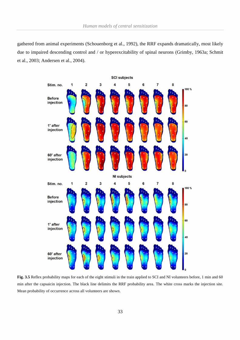
Human models of central sensitization
33
gathered from animal experiments (Schouenborg et al., 1992), the RRF expands dramatically, most likely
due to impaired descending control and / or hyperexcitability of spinal neurons (Grimby, 1963a; Schmit
et al., 2003; Andersen et al., 2004).
Fig. 3.5 Reflex probability maps for each of the eight stimuli in the train applied to SCI and NI volunteers before, 1 min and 60
min after the capsaicin injection. The black line delimits the RRF probability area. The white cross marks the injection site.
Mean probability of occurrence across all volunteers are shown.

Human models of central sensitization
34
In Study IV, NWR thresholds were higher in SCI volunteers compared to NI volunteers regardless of the
stimulation site, in agreement with previous studies (Hiersemenzel et al., 2000; Andersen et al., 2004).
Moreover, the arch of the foot presented the highest thresholds in SCI volunteers and the lowest
thresholds in NI volunteers. The RRF also showed the same difference, in which the topography of the
sensitivity and probability maps displayed a striking contrast. The most sensitive area in NI volunteers is
the arch of the foot, whereas this is completely reversed for SCI volunteers (fig 3.6). The sensitivity of the
RRF is shaped by excitatory and inhibitory spinal neuronal circuits under supraspinal influence, among
other factors (Schouenborg, 2002). In chronic SCI volunteers, the loss of descending control and
appropriate peripheral input causes a predominance of inhibitory influences on the spinal circuitry (Dietz,
2010), that eventually leads to abnormal RRF configurations.
A marked decrease was noted in NWR amplitudes 60 min after the capsaicin injection compared to the
baseline measurements, reflected by the RRF sensitivity volume. This effect could also be noticed right
after the capsaicin injection, but the decrease in NWR amplitude was compensated by the enlargement of
the RRF sensitivity areas, and therefore the resulting RRF sensitivity volume did not present a significant
variation in relation to baseline. Similar phenomena have been described before, in relation to the strong
habituation to electrical stimulation that SCI volunteers exhibit during NWR experiments (Andersen et
al., 2004), or in locomotion experiments (Dietz and Müller, 2004), probably indicating a diminished
capacity to recruit flexor motoneurons. Antispasticity medication interacting with the GABA system can
also be responsible for decreased excitability, yielding smaller reflexes in general (Andersen et al., 2004).
However, it was unlikely to cause this trend within the time course of the experiment (the measurements
at every time point should be affected equally), together with the fact that the values in SCI were still
significantly larger than in NI volunteers. Interestingly, RRF sensitivity areas and RRF probability
measures did not exhibit this behavior, from which it can be hypothesized that these methods might be
more robust against habituation.

Human models of central sensitization
35
Fig. 3.6: Functional organization of the nociceptive withdrawal reflex (NWR) pathways. In healthy volunteers, the reflex
receptive field (RRF) of the tibialis anterior (TA) muscle is characterized by a high sensitivity (+Sens) in the medial, distal
region, resulting in inversion (Inv) and dorsiflexion (DorFl) of the foot when these sites are activated. Functionally antagonist
muscles (not shown) have RRF that evoke plantarflexion (PlanFl) or eversion (Eve). The RRF is likely shaped by excitatory
(Excit) and inhibitory (Inhib) descending control input coming from supraspinal structures (SupCtrl). SupCtrl may act
presynaptically (not shown) or postsynaptically on one or more interneurons (In) and on reflex encoders (RE) in the NWR
pathways modifying their excitability (color-coded similarly to RRF), adjusting the weight of afferent information from
noc cept ve np t The net o tp t s trans ated by α-motone rons α-Mn) into efferent signals that evoke a proper contraction in
the target muscle. b After an injury to the spinal cord, SupCtrl is partially or totally lost, so In that were subjected to tonic
inhibitory descending signals increase their excitability due to disinhibition and vice versa, resulting in abnormal RRF maps.
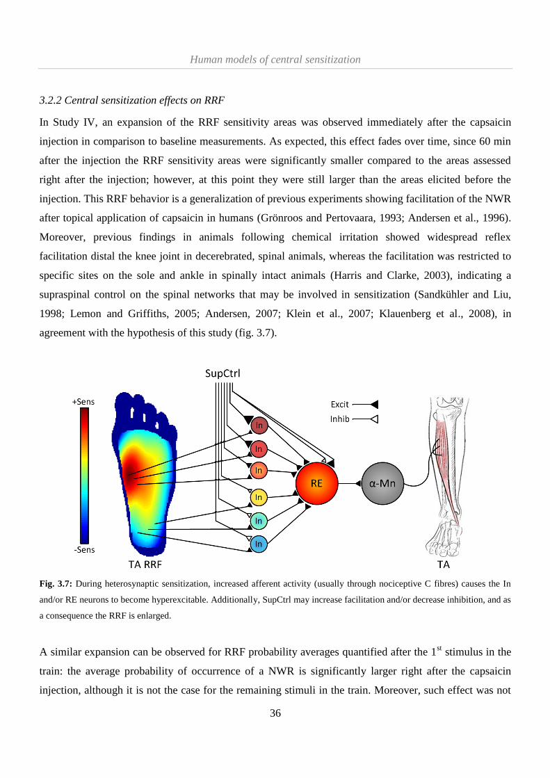
Human models of central sensitization
36
3.2.2 Central sensitization effects on RRF
In Study IV, an expansion of the RRF sensitivity areas was observed immediately after the capsaicin
injection in comparison to baseline measurements. As expected, this effect fades over time, since 60 min
after the injection the RRF sensitivity areas were significantly smaller compared to the areas assessed
right after the injection; however, at this point they were still larger than the areas elicited before the
injection. This RRF behavior is a generalization of previous experiments showing facilitation of the NWR
after topical application of capsaicin in humans (Grönroos and Pertovaara, 1993; Andersen et al., 1996).
Moreover, previous findings in animals following chemical irritation showed widespread reflex
facilitation distal the knee joint in decerebrated, spinal animals, whereas the facilitation was restricted to
specific sites on the sole and ankle in spinally intact animals (Harris and Clarke, 2003), indicating a
supraspinal control on the spinal networks that may be involved in sensitization (Sandkühler and Liu,
1998; Lemon and Griffiths, 2005; Andersen, 2007; Klein et al., 2007; Klauenberg et al., 2008), in
agreement with the hypothesis of this study (fig. 3.7).
Fig. 3.7: During heterosynaptic sensitization, increased afferent activity (usually through nociceptive C fibres) causes the In
and/or RE neurons to become hyperexcitable. Additionally, SupCtrl may increase facilitation and/or decrease inhibition, and as
a consequence the RRF is enlarged.
A similar expansion can be observed for RRF probability averages quantified after the 1st stimulus in the
train: the average probability of occurrence of a NWR is significantly larger right after the capsaicin
injection, although it is not the case for the remaining stimuli in the train. Moreover, such effect was not

Human models of central sensitization
37
observed at all for RRF probability areas, which encompass the higher range of probabilities (60-100%
occurrence). Together, these results indicated that it is easier to modulate the occurrence of the NWR in
the lower range of probabilities (below 50%); it is likely that the descending inhibitory mechanisms play
a more important role above this threshold, as can be observed by the much larger RRF probability areas
and averages in SCI compared to NI volunteers during temporal summation. Finally, it is worth
mentioning that RRF sensitivity and probability maps exhibit complementary (rather than redundant)
information, as it is also observed in Study II and III. This could be an indication that the size and
occurrence of the NWR are regulated by different neural mechanisms, which in time are strongly
modulated by the influence of descending control (Willis Jr., 1988).
3.2.3 Temporal summation effects on RRF
Temporal summation can be described as successive increases in perceived pain intensity to repeated
stimuli. As a physiological correlate, the NWR has proven to be a particularly useful tool (Arendt-Nielsen
et al., 1994; Arendt-Nielsen et al., 2000; Guirimand et al., 2000; Serrao et al., 2004). In healthy
volunteers, it is characterized by a gradual increase in size and duration of the NWR for a few seconds
following repetitive stimulation depending on stimulus intensity and frequency (Arendt-Nielsen et al.,
2000), after which the NWR size reaches a plateau or even decreases, probably due to descending
inhibitory control (Bajaj et al., 2005). Temporal summation can be associated to the early part of the
wind-up process that involves summation of excitatory post-synaptic potentials (Randic, 1996), and when
assessed by the NWR, is a strong measure of the sensitivity of spinal nociceptive integration, involving
N-methyl-D-aspartate (NMDA) receptor pathways that are not detected by single stimuli (Arendt-Nielsen
et al., 1995). Moreover, the NMDA pathways are likely involved in the regulation of descending control
(Clarke et al., 2002) and the induction and maintenance of central sensitization (Woolf and Thompson,
1991).
The findings in Study IV showed that RRF sensitivity areas were larger in SCI than in NI volunteers, and
this effect was clearly more pronounced during temporal summation. These results are consistent with
previous experiments showing enlarged RRF in SCI compared to NI volunteers at different stimulation
intensities but using a single electrical stimulus (Andersen et al., 2004), as well as gradual enlargement of
the RRF in response to repetitive stimulation in NI volunteers (Andersen et al., 2005). An interesting
finding was that electrical stimulation consistently elicited larger RRF probability areas and RRF
probability averages in SCI compared to NI volunteers only during temporal summation (2nd
to 8th
stimuli

Human models of central sensitization
38
in the train); the 1st stimuli in the train, however, did not elicit significant differences between groups.
Indeed, spinal facilitatory effects (including temporal summation and central sensitization) can be masked
or even completely overridden by supraspinal inhibitory processes triggered after high intensity
stimulation (Gozariu et al., 1997; Bajaj et al., 2005), as demonstrated by the differential effect between NI
and SCI volunteers.

Human models of central sensitization
39
4. Conclusion
The results from Study I and II showed that the NWR and the RRF are robust and reliable tools in
experimental and clinical pain research. In particular, the parameters that can be derived from RRF
sensitivity and probability maps (areas, volumes, averages) are not significantly affected by factors like
habituation to single and repeated electrical stimulation and small variations in stimulation intensities.
With that in mind, the NWR and the RRF were used in Study III and IV in the assessment of human
surrogate models of central sensitization.
In Study III, a central sensitization model using noxious conditioning electrical stimulation was tested.
Persistent facilitation of the NWR was observed following low-frequency stimulation, probably mediated
by thin primary afferents. Although supraspinal interactions cannot be completely ruled out, the most
likely neuronal mechanism that could explain these observations involves heterosynaptic interactions
within the spinal dorsal horn. In Study IV, an intramuscular injection of capsaicin was used to induce
central sensitization in both healthy and complete spinal cord injured volunteers. The results showed that
RRF were significantly modulated after the injection in both groups, and that this modulation was under
strong influence of descending control, as demonstrated by the differential effects in size and shape of the
RRF observed in spinal cord injured volunteers compared to healthy volunteers.
In summary, the studies presented in this thesis have hopefully contributed to a better understanding of
human models of central sensitization, as well as the establishment of the NWR and RRF as viable
alternatives for objective assessment of central changes in spinal nociception.
4.1 Future perspectives
Several methodological aspects of the NWR and RRF assessment can still be addresseed in order to
further improve the robustness and reliability of these tools towards widespread clinical application.
Among these it is worth mentioning further standardization of NWR and RRF recordings, new methods
to reduce cross-talk in EMG signals based (for instance based on double differential recordings) and
technological improvements on electrode in order to minimize differences arising from skin thickness /
impedance on the sole of the foot.

Human models of central sensitization
40
Regarding human models of central sensitization, the main goal for future research should focus on the
investigation of which are the optimal experimental conditions (e.g., type and dose of algogenic
substances, conditioning electrical stimulation parameters) that are able to elicit stable, reproducible
effects over time, while still closely resembling the mechanisms behind clinical pathophysiological
conditions. Once that is accomplished, further research can be directed into testing new drugs or
alternative methods to modulate these effects, in order to develop better alternatives for pain relief.

Human models of central sensitization
41
References
Andersen OK (2007) Studies of the organization of the human nociceptive withdrawal reflex. focus on
sensory convergence and stimulation site dependency. Acta Physiol 189:1-35.
Andersen OK, Felsby S, Nicolaisen L, Bjerring P, Jensen TS, Arendt-Nielsen L (1996) The effect of
ketamine on stimulation of primary and secondary hyperalgesic areas induced by capsaicin - A double-
blind, placebo-controlled, human experimental study. Pain 66:51-62.
Andersen OK, Finnerup NB, Spaich EG, Jensen TS, Arendt-Nielsen L (2004) Expansion of nociceptive
withdrawal reflex receptive fields in spinal cord injured humans. Clin Neurophysiol 115:2798-2810.
Andersen OK, Jensen LM, Brennum J, Arendt-Nielsen L (1994) Evidence for central summation of C and
A[delta] nociceptive activity in man. Pain 59:273-280.
Andersen OK, Sonnenborg FA, Arendt-Nielsen L (2001) Reflex receptive fields for human withdrawal
reflexes elicited by non-painful and painful electrical stimulation of the foot sole. Clin Neurophysiol
112:641-649.
Andersen OK, Sonnenborg FA, Arendt-Nielsen L (1999) Modular organization of human leg withdrawal
reflexes elicited by electrical stimulation of the foot sole. Muscle Nerve 22:1520-1530.
Andersen OK, Sonnenborg FA, Matjacic Z, Arendt-Nielsen L (2003) Foot-sole reflex receptive fields for
human withdrawal reflexes in symmetrical standing position. Exp Brain Res 152:434-443.
Andersen OK, Spaich EG, Madeleine P, Arendt-Nielsen L (2005) Gradual enlargement of human
withdrawal reflex receptive fields following repetitive painful stimulation. Brain Res 1042:194-204.
Arendt-Nielsen L, Brennum J, Sindrup S, Bak P (1994) Electrophysiological and psychophysical
quantification of temporal summation in the human nociceptive system. Eur J Appl Physiol Occup
Physiol 68:266-273.
Arendt-Nielsen L, Petersen-Felix S, Fischer M, Bak P, Bjerring P, Zbinden AM (1995) The effect of N-
methyl-D-aspartate antagonist (ketamine) on single and repeated nociceptive stimuli: A placebo-
controlled experimental human study. Anesth Analg 81:63-68.
Arendt-Nielsen L, Sonnenborg FA, Andersen OK (2000) Facilitation of the withdrawal reflex by repeated
transcutaneous electrical stimulation: An experimental study on central integration in humans. Eur J Appl
Physiol Occup Physiol 81:165-173.

Human models of central sensitization
42
Arendt-Nielsen L, Yarnitsky D (2009) Experimental and clinical applications of quantitative sensory
testing applied to skin, muscles and viscera. J Pain 10:556-572.
Atkinson G, Nevill AM (1998) Statistical methods for assessing measurement error (reliability) in
variables relevant to sports medicine. Sports Med 26:217-238.
Bailey CH, Giustetto M, Huang Y-, Hawkins RD, Kandel ER (2000) Is heterosynaptic modulation
essential for stabilizing hebbian plasticity and memory? Nat Rev Neurosci 1:11-20.
Bajaj P, Arendt-Nielsen L, Andersen OK (2005) Facilitation and inhibition of withdrawal reflexes
following repetitive stimulation: Electro- and psychophysiological evidence for activation of noxious
inhibitory controls in humans. Eur J Pain 9:25-31.
Bajaj P, Bajaj P, Madsen H, Arendt-Nielsen L (2003) Endometriosis is associated with central
sensitization: A psychophysical controlled study. J Pain 4:372-380.
Banic B, Petersen-Felix S, Andersen OK, Radanov BP, Villiger PM, Arendt-Nielsen L, Curatolo M
(2004) Evidence for spinal cord hypersensitivity in chronic pain after whiplash injury and in
fibromyalgia. Pain 107:7-15.
Baumgarter TA (1989) Norm-referenced measurement: Reliability. In: Measurement concepts in physical
education and exercise science (Safrit MJ, Wood TM, eds), pp45-72. Champaigne (IL), USA: Human
Kinetics.
Bland JM, Altman DG (1999) Measuring agreement in method comparison studies. Stat Methods Med
Res 8:135-160.
Bland JM, Altman DG (1990) A note on the use of the intraclass correlation coefficient in the evaluation
of agreement between two methods of measurement. Comput Biol Med 20:337-340.
Boureau F, Luu M, Doubrere JF (1991) Study of experimental pain measures and nociceptive reflex in
chronic pain patients and normal subjects. Pain 44:131-138.
Brain SD, Williams TJ (1988) Substance P regulates the vasodilator activity of calcitonin gene-related
peptide. Nature 335:73-75.
Cain DM, Khasabov SG, Simone DA (2001) Response properties of mechanoreceptors and nociceptors in
mouse glabrous skin: An in vivo study. J Neurophysiol 85:1561-1574.

Human models of central sensitization
43
Campbell IG, Carstens E, Watkins LR (1991) Comparison of human pain sensation and flexion
withdrawal evoked by noxious radiant heat. Pain 45:259-268.
Ch h A, hr ng , Tr ster A, artey , chme , oppert 7 ffects of ora pregaba n
and aprepitant on pain and central sensitization in the electrical hyperalgesia model in human volunteers.
Br J Anaesth 98:246-254.
Chong PST, Cros DP (2004) Technology literature review: Quantitative sensory testing. Muscle Nerve
29:734-747.
Clarke RW, Eves S, Harris J, Peachey JE, Stuart E (2002) Interactions between cutaneous afferent inputs
to a withdrawal reflex in the decerebrated rabbit and their control by descending and segmental systems.
Neuroscience 112:555-571.
Clarke RW, Harris J (2004) The organization of motor responses to noxious stimuli. Brain Res Rev
46:163-172.
Cook AJ, Woolf CJ, Wall PD, McMahon SB (1987) Dynamic receptive field plasticity in rat spinal cord
dorsal horn following C-primary afferent input. Nature 325:151-153.
Curatolo M, Arendt-Nielsen L, Petersen-Felix S (2006) Central hypersensitivity in chronic pain:
Mechanisms and clinical implications. Phys Med Rehabil Clin North Am 17:287-302.
Curatolo M, Arendt-Nielsen L, Petersen-Felix S (2004) Evidence, mechanisms, and clinical implications
of central hypersensitivity in chronic pain after whiplash injury. Clin J Pain 20:469-476.
De Luca CJ, Merletti R (1988) Surface myoelectric signal cross-talk among muscles of the leg.
Electroencephalogr Clin Neurophysiol 69:568-575.
Defrin R, Peleg S, Weingarden H, Heruti R, Urca G (2007) Differential effect of supraspinal modulation
on the nociceptive withdrawal reflex and pain sensation. Clin Neurophysiol 118:427-437.
Desmeules JA, Cedraschi C, Rapiti E, Baumgartner E, Finckh A, Cohen P, Dayer P, Vischer TL (2003)
Neurophysiologic evidence for a central sensitization in patients with fibromyalgia. Arthritis Rheum
48:1420-1429.
Dietz V (2010) Behavior of spinal neurons deprived of supraspinal input. Nat Rev Neurol 6:167-174.
Dietz V, Müller R (2004) Degradation of neuronal function following a spinal cord injury: Mechanisms
and countermeasures. Brain 127:2221-2231.
Human models of central sensitization
44
D m tr jev ć , Fagane J, regor ć , Nathan P , Tronte j J 7 ab t at on: ffects of reg ar
and stochastic stimulation. J Neurol Neurosurg Psychiatry 35:234-242.
D m tr jev ć , Nathan P 8 t d es of spast c ty n man: ana ys s of ref ex act v ty evoked by
noxious cutaneous stimulation. Brain 91:349-368.
Dincklage Fv, Hackbarth M, Schneider M, Baars JH, Rehberg B (2009) Introduction of a continual RIII
reflex threshold tracking algorithm. Brain Res 1260:24-29.
Don R, Pierelli F, Ranavolo A, Serrao M, Mangone M, Paoloni M, Cacchio A, Sandrini G, Santilli V
(2008) Modulation of spinal inhibitory reflex responses to cutaneous nociceptive stimuli during upper
limb movement. Eur J Neurosci 28:559-568.
Drdla R, Sandkühler J (2008) Long-term potentiation at C-fibre synapses by low-level presynaptic
activity in vivo. Mol Pain 4:.
Dudek SM, Bear MF (1992) Homosynaptic long-term depression in area CA1 of hippocampus and effects
of N-methyl-D-aspartate receptor blockade. Proc Natl Acad Sci USA 89:4363-4367.
Dusch M, Schley M, Rukwied R, Schmelz M (2007) Rapid flare development evoked by current
frequency-dependent stimulation analyzed by full-field laser perfusion imaging. Neuroreport 18:1101-
1105.
Ellrich J, Treede R- (1998) Convergence of nociceptive and non-nociceptive inputs onto spinal reflex
pathways to the tibialis anterior muscle in humans. Acta Physiol Scand 163:391-401.
Emborg J, Spaich EG, Andersen OK (2009) Withdrawal reflexes examined during human gait by ground
reaction forces: Site and gait phase dependency. Med Biol Eng Comput 47:29-39.
Farina D, Merletti R, Indino B, Graven-Nielsen T (2004) Surface EMG crosstalk evaluated from
experimental recordings and simulated signals. reflections on crosstalk interpretation, quantification and
reduction. Methods Inf Med 43:30-35.
Feltz CJ, Miller GE (1996) An asymptotic test for the equality of coefficients of variation from k
populations. Stat Med 15:647-658.
Floeter MK, Gerloff C, Kouri J, Hallett M (1998) Cutaneous withdrawal reflexes of the upper extremity.
Muscle Nerve 21:591-598.

Human models of central sensitization
45
France CR, Rhudy JL, McGlone S (2009) Using normalized EMG to define the nociceptive flexion reflex
(NFR) threshold: Further evaluation of standardized NFR scoring criteria. Pain 145:211-218.
arc a- arrea , a g re F ectrophys o og ca assessment of noc cept on n norma s and
patients: The use of nociceptive reflexes. Electroencephalogr Clin Neurophysiol Suppl 41:102-118.
Gardner EP, Martin JH, Jesell TM (2000) The bodily senses. In: Principles of neural science (Kandell ER,
Schwartz JH, Jesell TM, eds), pp430-450. New York (USA): McGraw-Hill.
Garwicz M, Jörntell H, Ekerot C- (1998) Cutaneous receptive fields and topography of mossy fibres and
climbing fibres projecting to cat cerebellar C3 zone. J Physiol 512:277-293.
eber C, Fonde , r mer , o ke , Treede -, Sommer C, Birklein F (2007) Psychophysics, flare,
and neurosecretory function in human pain models: Capsaicin versus electrically evoked pain. J.Pain
8:503-514.
Gjerstad J, Tjølsen A, Hole K (2001) Induction of long-term potentiation of single wide dynamic range
neurones in the dorsal horn is inhibited by descending pathways. Pain 91:263-268.
Gjerstad J, Tjølsen A, Svendsen F, Hole K (2000) Inhibition of spinal nociceptive responses after
intramuscular injection of capsaicin involves activation of noradrenergic and opioid systems. Brain Res
859:132-136.
Gozariu M, Bragard D, Willer J-, Le Bars D (1997) Temporal summation of C-fiber afferent inputs:
Competition between facilitatory and inhibitory effects on C-fiber reflex in the rat. J Neurophysiol
78:3165-3179.
Granat MH, Nicol DJ, Baxendale RH, Andrews BJ (1991) Dishabituation of the flexion reflex in spinal
cord-injured man and its application in the restoration of gait. Brain Res 559:344-346.
Grimby L (1963a) Normal plantar response: Integration of flexor and extensor reflex components. J
Neurol Neurosurg Psychiatr 26:39-50.
Grimby L (1963b) Pathological plantar response: Disturbances of the normal integration of flexor and
extensor reflex components. J Neurol Neurosurg Psychiatr 26:314-321.
Grönroos M, Pertovaara A (1993) Capsaicin-induced central facilitation of a nociceptive flexion reflex in
humans. Neurosci Lett 159:215-218.

Human models of central sensitization
46
Guirimand F, Dupont X, Brasseur L, Chauvin M, Bouhassira D (2000) The effects of ketamine on the
temporal summation (wind-up) of the R(III) nociceptive flexion reflex and pain in humans. Anesth Analg
90:408-414.
Hagbarth KE (1960) Spinal withdrawal reflexes in the human lower limbs. J Neurol Neurosurg Psychiatry
23:222-227.
Hansen N, Klein T, Magerl W, Treede R- (2007) Psychophysical evidence for long-term potentiation of
C-fiber and Aδ-fiber pathways in humans by analysis of pain descriptors. J Neurophysiol 97:2559-2563.
Harris J, Clarke RW (2003) Organisation of sensitisation of hind limb withdrawal reflexes from acute
noxious stimuli in the rabbit. J Physiol (Lond ) 546:251-265.
Haugan F, Wibrand K, Fiskå A, Bramham CR, Tjølsen A (2008) Stability of long term facilitation and
expression of zif268 and arc in the spinal cord dorsal horn is modulated by conditioning stimulation
within the physiological frequency range of primary afferent fibers. Neurosci 154:1568-1575.
Herrero JF, Laird JMA, Lopez-Garcia JA (2000) Wind-up of spinal cord neurones and pain sensation:
Much ado about something? Prog Neurobiol 61:169-203.
Hiersemenzel L-, Curt A, Dietz V (2000) From spinal shock to spasticity: Neuronal adaptations to a
spinal cord injury. Neurology 54:1574-1582.
Hornby TG, Rymer WZ, Benz EN, Schmit BD (2003) Windup of flexion reflexes in chronic human
spinal cord injury: A marker for neuronal plateau potentials? J Neurophysiol 89:416-426.
Hugon M (1973) Exteroceptive reflexes to stimulation of sural nerve in man. In: New developments in
electromyography and clinical neurophysiology (Desmedt JE, ed), pp. 713-729. Basel (Switzerland):
Kargel.
Ikeda H, Stark J, Fischer H, Wagner M, Drdla R, Jäger T, Sandkühler J (2006) Synaptic amplifier of
inflammatory pain in the spinal dorsal horn. Science 312:1659-1662.
Inui K, Tran TD, Hoshiyama M, Kakigi R (2002) Preferential stimulation of Aδ fibers by intra-epidermal
needle electrode in humans. Pain 96:247-252.
Jänig W, Lisney SJW (1989) Small diameter myelinated afferents produce vasodilatation but not plasma
extravasation in rat skin. J Physiol (Lond ) 415:477-486.

Human models of central sensitization
47
Jensen MB, Biurrun Manresa JA, Andersen OK (2010) Optimal setup for quantification of nociceptive
withdrawal reflex receptive fields in humans. Abstracts of the XVIII Congress of the International Society
of Electrophysiology and Kinesiology, ISEK 2010 (Aalborg, Denmark) .
Ji RR, Kohno T, Moore KA, Woolf CJ (2003) Central sensitization and LTP: Do pain and memory share
similar mechanisms? Trends Neurosci 26:696-705.
Kaiser JF (1990) On a simple algorithm to calculate the 'energy' of a signal. International Conference on
Acoustics, Speech, and Signal Processing 381-384.
Kidd BL, Urban LA (2001) Mechanisms of inflammatory pain. Br J Anaesth 87:3-11.
Klauenberg S, Maier C, Assion H-, Hoffmann A, Krumova EK, Magerl W, Scherens A, Treede R-,
Juckel G (2008) Depression and changed pain perception: Hints for a central disinhibition mechanism.
Pain 140:332-343.
Klein T, Magerl W, Hanschmann A, Althaus M, Treede R- (2008) Antihyperalgesic and analgesic
properties of the N-methyl-d-aspartate (NMDA) receptor antagonist neramexane in a human surrogate
model of neurogenic hyperalgesia. Eur J Pain 12:17-29.
Klein T, Magerl W, Hopf HC, Sandkühler J, Treede R- (2004) Perceptual correlates of nociceptive long-
term potentiation and long-term depression in humans. J Neurosci 24:964-971.
Klein T, Magerl W, Nickel U, Hopf H-, Sandkühler J, Treede R- (2007) Effects of the NMDA-receptor
antagonist ketamine on perceptual correlates of long-term potentiation within the nociceptive system.
Neuropharmacol 52:655-661.
Klein T, Magerl W, Rolke R, Treede R- (2005) Human surrogate models of neuropathic pain. Pain
115:227-233.
Klein T, Magerl W, Treede R- (2007) Forget about your chronic pain. Pain 132:16-17.
Klein T, Magerl W, Treede R- (2006) Perceptual correlate of nociceptive long-term potentiation (LTP) in
humans shares the time course of early-LTP. J Neurophysiol 96:3551-3555.
Klein T, Stahn S, Magerl W, Treede R- (2008) The role of heterosynaptic facilitation in long-term
potentiation (LTP) of human pain sensation. Pain 139:507-519.
Koceja DM, Bernacki RH, Kamen G (1991) Methodology for the quantitative assessment of human
crossed-spinal reflex pathways. Med Biol Eng Comput 29:603-606.

Human models of central sensitization
48
Koltzenburg M, Lundberg LER, Torebjork HE (1992) Dynamic and static components of mechanical
hyperalgesia in human hairy skin. Pain 51:207-218.
oppert , Dern , tt , A brecht , ch tt er J, chme A new mode of e ectr ca y
evoked pain and hyperalgesia in human skin: The effects of intravenous alfentanil, S(+)-ketamine, and
lidocaine. Anesthesiol 95:395-402.
LaMotte RH, Shain CN, Simone DA, Tsai E-P (1991) Neurogenic hyperalgesia: Psychophysical studies
of underlying mechanisms. J Neurophysiol 66:190-211.
Lang S, Klein T, Magerl W, Treede R- (2007) Modality-specific sensory changes in humans after the
induction of long-term potentiation (LTP) in cutaneous nociceptive pathways. Pain 128:254-263.
Latremoliere A, Woolf CJ (2009) Central sensitization: A generator of pain hypersensitivity by central
neural plasticity. J Pain 10:895-926.
Le Bars D, Gozariu M, Cadden SW (2001) Animal models of nociception. Pharmacol Rev 53:597-652.
Leem JW, Willis WD, Chung JM (1993) Cutaneous sensory receptors in the rat foot. J Neurophysiol
69:1684-1699.
Lemon RN, Griffiths J (2005) Comparing the function of the corticospinal system in different species:
Organizational differences for motor specialization? Muscle Nerve 32:261-279.
Li X, Zhou P, Aruin AS (2007) Teager-kaiser energy operation of surface EMG improves muscle activity
onset detection. Ann Biomed Eng 35:1532-1538.
Liebermann DG, Defrin R (2009) Characteristics of the nociceptive withdrawal response elicited under
aware and unaware conditions. J Electromyogr Kinesiology 19:.
Liu M, Max MB, Robinovitz E, Gracely RH, Bennett GJ (1998) The human capsaicin model of allodynia
and hyperalgesia: Sources of variability and methods for reduction. J Pain Symptom Manag 16:10-20.
Liu X-, Sandkühler J (1997) Characterization of long-term potentiation of C-fiber-evoked potentials in
spinal dorsal horn of adult rat: Essential role of NK1 and NK2 receptors. J Neurophysiol 78:1973-1982.
Loeser JD, Treede R- (2008) The kyoto protocol of IASP basic pain terminology. Pain 137:473-477.
Lund I, Lundeberg T, Kowalski J, Sandberg L, Budh CN, Svensson E (2005) Evaluation of variations in
sensory and pain threshold assessments by electrocutaneous stimulation. Physiother Theory Pract 21:81-
92.
Human models of central sensitization
49
Magerl W, Fuchs PN, Meyer RA, Treede R- (2001) Roles of capsaicin-insensitive nociceptors in
cutaneous pain and secondary hyperalgesia. Brain 124:1754-1764.
Magerl W, Krumova EK, Baron R, Tölle T, Treede R-, Maier C (2010) Reference data for quantitative
sensory testing (QST): Refined stratification for age and a novel method for statistical comparison of
group data. Pain 151:598-605.
Magerl W, Wilk SH, Treede R- (1998) Secondary hyperalgesia and perceptual wind-up following
intradermal injection of capsaicin in humans. Pain 74:257-268.
McCarthy PW, Lawson SN (1989) Cell type and conduction velocity of rat primary sensory neurons with
substance P-like immunoreactivity. Neuroscience 28:745-753.
Meinck H-, Piesiur-Strehlow B, Koehler W (1981) Some principles of flexor reflex generation in human
leg muscles. Electroencephalogr Clin Neurophysiol 52:140-150.
Micalos PS, Drinkwater EJ, Cannon J, Arendt-Nielsen L, Marino FE (2009) Reliability of the nociceptive
flexor reflex (RIII) threshold and association with pain threshold. Eur J Appl Physiol 105:55-62.
Milne RJ, Kay NE, Irwin RJ (1991) Habituation to repeated painful and non-painful cutaneous stimuli: A
quantitative psychophysical study. Exp Brain Res 87:438-444.
Mørch CD, Hennings K, Andersen OK (2009) Modeling preferential activation of Aδ-fibers fibers by
small-area surface electrodes. 39th Annual Meeting of the Society for Neuroscience, Neuroscience 2009
No. 763.19/35.
Mørch CD, Andersen OK, Graven-Nielsen T, Arendt-Nielsen L (2007) Nociceptive withdrawal reflexes
evoked by uniform-temperature laser heat stimulation of large skin areas in humans. J Neurosci Methods
160:85-92.
Mulkey RM, Malenka RC (1992) Mechanisms underlying induction of homosynaptic long-term
depression in area CA1 of the hippocampus. Neuron 9:967-975.
Neziri AY, Andersen OK, Petersen-Felix S, Radanov B, Dickenson AH, Scaramozzino P, Arendt-Nielsen
L, Curatolo M (2010) The nociceptive withdrawal reflex: Normative values of thresholds and reflex
receptive fields. Eur J Pain 14:134-141.
Neziri AY, Curatolo M, Bergadano A, Petersen-Felix S, Dickenson A, Arendt-Nielsen L, Andersen OK
(2009) New method for quantification and statistical analysis of nociceptive reflex receptive fields in
humans. J Neurosci Methods 178:24-30.
Human models of central sensitization
50
Nilsson H-, Schouenborg J (1999) Differential inhibitory effect on human nociceptive skin senses
induced by local stimulation of thin cutaneous fibers. Pain 80:103-112.
O'Neill S, Manniche C, Graven-Nielsen T, Arendt-Nielsen L (2007) Generalized deep-tissue hyperalgesia
in patients with chronic low-back pain. Eur J Pain 11:415-420.
Perrotta A, Sandrini G, Serrao M, Buscone S, Tassorelli C, Tinazzi M, Zangaglia R, Pacchetti C, Bartolo
M, Pierelli F, Martignoni E (2010) Facilitated temporal summation of pain at spinal level in parkinson's
disease. Movement Disorders n/a-n/a.
Portney LG, Watkins MP (2009) Foundations of clinical research : Applications to practice. Upper Saddle
River, N.J.: Pearson/Prentice Hall.
Prescott SA (1998) Interactions between depression and facilitation within neural networks: Updating the
dual-process theory of plasticity. Learn Memory 5:446-466.
Price DD (2002) Central neural mechanisms that interrelate sensory and affective dimensions of pain.
Mol Interv 2:392-403, 339.
Price DD (2000) Psychological and neural mechanisms of the affective dimension of pain. Science
288:1769-1772.
Price DD, Staud R, Robinson ME, Mauderli AP, Cannon R, Vierck CJ (2002) Enhanced temporal
summation of second pain and its central modulation in fibromyalgia patients. Pain 99:49-59.
Randic M (1996) Plasticity of excitatory synaptic transmission in the spinal cord dorsal horn. Prog Brain
Res 113:463-506.
and ć , J ang C, Cerne R (1993) Long-term potentiation and long-term depression of primary
afferent neurotransmission in the rat spinal cord. J Neurosci 13:5228-5241.
Rhudy JL, France CR (2007) Defining the nociceptive flexion reflex (NFR) threshold in human
participants: A comparison of different scoring criteria. Pain 128:244-253.
Rolke R, Magerl W, Campbell KA, Schalber C, Caspari S, Birklein F, Treede R- (2006) Quantitative
sensory testing: A comprehensive protocol for clinical trials. Eur J Pain 10:77-88.
Rottmann S, Jung K, Ellrich J (2008) Electrical low-frequency stimulation induces homotopic long-term
depression of nociception and pain from hand in man. Clin Neurophysiol 119:1895-1904.

Human models of central sensitization
51
Rygh LJ, Tjølsen A, Hole K, Svendsen F (2002) Cellular memory in spinal nociceptive circuitry. Scand J
Psychol 43:153-159.
Safrit MJ, Wood TM (1989) Measurement concepts in physical education and exercise science.
(Champaigne (IL), USA) 400.
Sandkühler J (2007) Understanding LTP in pain pathways. Mol Pain 3:.
Sandkühler J (2000) Learning and memory in pain pathways. Pain 88:113-118.
andk h er J, Chen J , Cheng , and ć 7 ow-frequency stimulation of afferent Aδ-fibers
induces long-term depression at primary afferent synapses with substantia gelatinosa neurons in the rat. J
Neurosci 17:6483-6491.
Sandkühler J, Liu X (1998) Induction of long-term potentiation at spinal synapses by noxious stimulation
or nerve injury. Eur J Neurosci 10:2476-2480.
Sandrini G, Serrao M, Rossi P, Romaniello A, Cruccu G, Willer JC (2005) The lower limb flexion reflex
in humans. Prog Neurobiol 77:353-395.
chme , chae , e dner C, chm dt , Torebj rk , andwerker h ch nerve
fibers mediate the axon reflex flare in human skin? Neuroreport 11:645-648.
Schmit BD, Hornby TG, Tysseling-Mattiace VM, Benz EN (2003) Absence of local sign withdrawal in
chronic human spinal cord injury. J Neurophysiol 90:3232-3241.
Schouenborg J (2004) Learning in sensorimotor circuits. Curr Opin Neurobiol 14:693-697.
Schouenborg J (2002) Modular organisation and spinal somatosensory imprinting. Brain Res Rev 40:80-
91.
Schouenborg J, Holmberg H, Weng HR (1992) Functional organization of the nociceptive withdrawal
reflexes. Exp Brain Res 90:469-478.
Schouenborg J, Kalliomäki J (1990) Functional organization of the nociceptive withdrawal reflexes. Exp
Brain Res 83:67-78.
Schouenborg J, Sjolund BH (1983) Activity evoked by A- and C-afferent fibers in rat dorsal horn neurons
and its relation to a flexion reflex. J Neurophysiol 50:1108-1121.

Human models of central sensitization
52
Serrao M, Pierelli F, Don R, Ranavolo A, Cacchio A, Curra A, Sandrini G, Frascarelli M, Santilli V
(2006) Kinematic and electromyographic study of the nociceptive withdrawal reflex in the upper limbs
during rest and movement. J Neurosci 26:3505-3513.
Serrao M, Rossi P, Sandrini G, Parisi L, Amabile GA, Nappi G, Pierelli F (2004) Effects of diffuse
noxious inhibitory controls on temporal summation of the RIII reflex in humans. Pain 112:353-360.
Sherrington CS (1910) Flexion-reflex of the limb, crossed extension-reflex, and reflex stepping and
standing. J Physiol 40:28-121.
Simone DA, Baumann TK, LaMotte RH (1989) Dose-dependent pain and mechanical hyperalgesia in
humans after intradermal injection of capsaicin. Pain 38:99-107.
Sonnenborg FA, Andersen OK, Arendt-Nielsen L (2000) Modular organization of excitatory and
inhibitory reflex receptive fields elicited by electrical stimulation of the foot sole in man. Clin
Neurophysiol 111:2160-2169.
Sonnenborg FA, Andersen OK, Arendt-Nielsen L, Treede R- (2001) Withdrawal reflex organisation to
electrical stimulation of the dorsal foot in humans. Exp Brain Res 136:303-312.
Spaich EG, Andersen OK, Arendt-Nielsen L (2004) Tibialis anterior and soleus withdrawal reflexes
elicited by electrical stimulation of the sole of the foot during gait. Neuromodulation 7:126-132.
Spaich EG, Arendt-Nielsen L, Andersen OK (2005) Repetitive painful stimulation produces an expansion
of withdrawal reflex receptive fields in humans. Artif Organs 29:224-228.
Spaich EG, Arendt-Nielsen L, Andersen OK (2004) Modulation of lower limb withdrawal reflexes during
gait: A topographical study. J Neurophysiol 91:258-266.
Spaich EG, Emborg J, Collet T, Arendt-Nielsen L, Andersen OK (2009) Withdrawal reflex responses
evoked by repetitive painful stimulation delivered on the sole of the foot during late stance: Site, phase,
and frequency modulation. Exp Brain Res 194:359-368.
Sterling M (2010) Differential development of sensory hypersensitivity and a measure of spinal cord
hyperexcitability following whiplash injury. Pain 150:501-506.
Sterling M, Jull G, Vicenzino B, Kenardy J (2003) Sensory hypersensitivity occurs soon after whiplash
injury and is associated with poor recovery. Pain 104:509-517.

Human models of central sensitization
53
Svendsen F, Tjølsen A, Gjerstad J, Hole K (1999) Long term potentiation of single WDR neurons in
spinalized rats. Brain Res 816:487-492.
Thompson RF, Spencer WA (1966) Habituation: A model phenomenon for the study of neuronal
substrates of behavior. Psychol Rev 73:16-43.
Torebjork HE, Lundberg LER, LaMotte RH (1992) Central changes in processing of mechanoreceptive
input in capsaicin-induced secondary hyperalgesia in humans. J Physiol 448:765-780.
Tørring J, Pedersen E, Klemar B (1981) Standardisation of the electrical elicitation of the human flexor
reflex. J Neurol Neurosurg Psychiatry 44:129-132.
Treede R-, Meyer RA, Raja SN, Campbell JN (1992) Peripheral and central mechanisms of cutaneous
hyperalgesia. Prog Neurobiol 38:397-421.
Weng H-, Schouenborg J (1996) Cutaneous inhibitory receptive fields of withdrawal reflexes in the
decerebrate spinal rat. J PHYSIOL 493:253-265.
Wilder-Smith CH, Schindler D, Lovblad K, Redmond SM, Nirkko A (2004) Brain functional magnetic
resonance imaging of rectal pain and activation of endogenous inhibitory mechanisms in irritable bowel
syndrome patient subgroups and healthy controls. Gut 53:1595-1601.
Willer JC, Boureau F, Albe-Fessard D (1979a) Supraspinal influences on nociceptive flexion reflex and
pain sensation in man. Brain Res 179:61-68.
Willer JC, Boureau F, Berny J (1979b) Nociceptive flexion reflexes elicited by noxious laser radiant heat
in man. Pain 7:15-20.
Willis Jr. WD (1988) Anatomy and physiology of descending control of nociceptive responses of dorsal
horn neurons: Comprehensive review. Prog Brain Res 77:1-29.
Woolf CJ (1983) Evidence for a central component of post-injury pain hypersensitivity. Nature 306:686-
688.
Woolf CJ, Salter MW (2000) Neuronal plasticity: Increasing the gain in pain. Science 288:1765-1768.
Woolf CJ, Thompson SWN (1991) The induction and maintenance of central sensitization is dependent
on N-methyl-D-aspartic acid receptor activation; implications for the treatment of post-injury pain
hypersensitivity states. Pain 44:293-299.
Human models of central sensitization
54
Woolf CJ, Wall PD (1986) Relative effectiveness of C primary afferent fibers of different origins in
evoking a prolonged facilitation of the flexor reflex in the rat. J Neurosci 6:1433-1442.
Ziegler EA, Magerl W, Meyer RA, Treede R- (1999) Secondary hyperalgesia to punctate mechanical
stimuli. central sensitization to A-fibre nociceptor input. Brain 122:2245-2257.