Human facial dysostoses - laserwords.co.in · the molecular bases of ... The characteristic facial...
12
Clin Genet 2013: 83: 499 – 510 Printed in Singapore. All rights reserved © 2013 John Wiley & Sons A/S. Published by Blackwell Publishing Ltd CLINICAL GENETICS doi: 10.1111/cge.12123 Review Human facial dysostoses Wieczorek D. Human facial dysostoses. Clin Genet 2013: 83: 499–510. © John Wiley & Sons A/S. Published by Blackwell Publishing Ltd, 2013 The human facial dysostoses can be subdivided into mandibulofacial dysostoses (MFDs) and acrofacial dysostoses (AFDs). The craniofacial phenotypes of the two groups of patients are similar. Both types are thought to be related to abnormal migration of neural crest cells to the pharyngeal arches and the face. The craniofacial anomalies shared by the two groups consist of downslanting palpebral fissures, coloboma of the lower eyelid, from which the eyelashes medial to the defect may be absent, hypoplasia of the zygomatic complex, micrognathia, and microtia, which is often associated with hearing loss. These facial deformities are associated with limb anomalies in the AFDs. All MFDs present with the typical craniofacial phenotype, but some have additional features that help to distinguish them clinically: intellectual disability, microcephaly, chest deformity, ptosis, cleft lip/palate, macroblepharon, or blepharophimosis. The limb anomalies in the AFDs can be classified into pre-axial, post-axial, and others not fitting into the first two AFD types. Of the pre-axial types, Nager syndrome and of the post-axial types, Miller syndrome are the best-known disorders of their AFD subgroups. Several other AFDs with unknown molecular genetic bases, including lethal ones, have been described. This article reviews the MFDs and AFDs published to date. Conflict of interest The author declares no conflict of interest. D Wieczorek Institut f ¨ ur Humangenetik, Universit ¨ atsklinikum Essen, Universit ¨ at Duisburg-Essen, Essen, Germany Key words: acrofacial dysostosis – AFD – EFTUD2 – mandibulofacial dysostosis – MFD – Miller syndrome – Nager syndrome – Treacher Collins syndrome Corresponding author: Dagmar Wieczorek, Institut f ¨ ur Humangenetik, Universit ¨ atsklinikum Essen, Universit ¨ at Duisburg-Essen, Hufelandstr. 55, 45122 Essen, Germany. Tel.: +49 201 723 4567; fax: +49 201 723 5900; e-mail: [email protected] Received 10 December 2012, revised and accepted for publication 8 February 2013 The development of structures derived from the first and second branchial arches is disturbed in the human facial dysostoses. This heterogeneous group of conditions can be subdivided into those with normal extremities, the mandibulofacial dysostoses (MFDs), and those with limb anomalies, the acrofacial dysostoses (AFDs). For diagnostic convenience, schematic overviews of MFD and AFD are depicted in Figs 1 and 2, respectively. It should be mentioned that this review includes single cases of MFDs and AFDs, although it is still unclear whether each of them truly represents a distinct syndromic condition. Mandibulofacial dysostoses At least eight different types of MFD have been described in the literature (Table 1), and one can expect that their number will rise in the coming years. One has to keep in mind that many of the clinically defined MFDs were described before the causative genes, e.g. Treacher Collins syndrome (TCS), were identified. As the different forms of MFD overlap considerably, some of these patients may need to be reclassified after the molecular bases of their malformations have been determined. As long as molecular data to the contrary are not available, these entities will be listed as distinct MFDs. The craniofacial anomalies observed in the MFDs result from faulty development of the first and second branchial arches. The MFDs are characterized by downslanting palpebral fissures, coloboma of the lower eyelids with or without the absence of eyelashes on the affected lids medial to the defect, hypoplasia of the zygomatic complex, retromicrognathia, and microtia, which is often associated with hearing loss. The craniofacial features show wide intrafamilial and interfamilial variability. All the MFDs described so far have been published independently. To the best of our knowledge, a review of the subclassification of MFDs does not exist. Figure 1 499
Transcript of Human facial dysostoses - laserwords.co.in · the molecular bases of ... The characteristic facial...

Clin Genet 2013: 83: 499–510Printed in Singapore. All rights reserved
© 2013 John Wiley & Sons A/S.Published by Blackwell Publishing Ltd
CLINICAL GENETICSdoi: 10.1111/cge.12123
Review
Human facial dysostoses
Wieczorek D. Human facial dysostoses.Clin Genet 2013: 83: 499–510. © John Wiley & Sons A/S. Published byBlackwell Publishing Ltd, 2013
The human facial dysostoses can be subdivided into mandibulofacialdysostoses (MFDs) and acrofacial dysostoses (AFDs). The craniofacialphenotypes of the two groups of patients are similar. Both types arethought to be related to abnormal migration of neural crest cells to thepharyngeal arches and the face. The craniofacial anomalies shared by thetwo groups consist of downslanting palpebral fissures, coloboma of thelower eyelid, from which the eyelashes medial to the defect may beabsent, hypoplasia of the zygomatic complex, micrognathia, and microtia,which is often associated with hearing loss. These facial deformities areassociated with limb anomalies in the AFDs. All MFDs present with thetypical craniofacial phenotype, but some have additional features that helpto distinguish them clinically: intellectual disability, microcephaly, chestdeformity, ptosis, cleft lip/palate, macroblepharon, or blepharophimosis.The limb anomalies in the AFDs can be classified into pre-axial, post-axial,and others not fitting into the first two AFD types. Of the pre-axial types,Nager syndrome and of the post-axial types, Miller syndrome are thebest-known disorders of their AFD subgroups. Several other AFDs withunknown molecular genetic bases, including lethal ones, have beendescribed. This article reviews the MFDs and AFDs published to date.
Conflict of interest
The author declares no conflict of interest.
D Wieczorek
Institut fur Humangenetik,Universitatsklinikum Essen, UniversitatDuisburg-Essen, Essen, Germany
Key words: acrofacial dysostosis –AFD – EFTUD2 – mandibulofacialdysostosis – MFD – Miller syndrome –Nager syndrome – Treacher Collinssyndrome
Corresponding author: DagmarWieczorek, Institut fur Humangenetik,Universitatsklinikum Essen, UniversitatDuisburg-Essen, Hufelandstr. 55,45122 Essen, Germany.Tel.: +49 201 723 4567;fax: +49 201 723 5900;e-mail: [email protected]
Received 10 December 2012, revisedand accepted for publication 8February 2013
The development of structures derived from the first andsecond branchial arches is disturbed in the human facialdysostoses. This heterogeneous group of conditions canbe subdivided into those with normal extremities, themandibulofacial dysostoses (MFDs), and those withlimb anomalies, the acrofacial dysostoses (AFDs).
For diagnostic convenience, schematic overviewsof MFD and AFD are depicted in Figs 1 and 2,respectively. It should be mentioned that this reviewincludes single cases of MFDs and AFDs, although itis still unclear whether each of them truly represents adistinct syndromic condition.
Mandibulofacial dysostoses
At least eight different types of MFD have beendescribed in the literature (Table 1), and one can expectthat their number will rise in the coming years. Onehas to keep in mind that many of the clinically definedMFDs were described before the causative genes, e.g.
Treacher Collins syndrome (TCS), were identified.As the different forms of MFD overlap considerably,some of these patients may need to be reclassified afterthe molecular bases of their malformations have beendetermined. As long as molecular data to the contraryare not available, these entities will be listed as distinctMFDs.
The craniofacial anomalies observed in the MFDsresult from faulty development of the first and secondbranchial arches. The MFDs are characterized bydownslanting palpebral fissures, coloboma of the lowereyelids with or without the absence of eyelashes onthe affected lids medial to the defect, hypoplasiaof the zygomatic complex, retromicrognathia, andmicrotia, which is often associated with hearing loss.The craniofacial features show wide intrafamilial andinterfamilial variability.
All the MFDs described so far have been publishedindependently. To the best of our knowledge, a reviewof the subclassification of MFDs does not exist. Figure 1
499

Wieczorek
Fig. 1. Classification of mandibulofacial dysostoses (MFDs). Additional clinical findings that are present in the different forms of MFD are shown.
presents an MFD classification based on clinicalfindings and the common craniofacial phenotype.
Treacher Collins syndrome (MIM 154500)
The best-known MFD is the TCS, which was firstdescribed by Thomson in 1846 and Berry in 1889. Itis named after E. Treacher Collins, who delineated thissyndrome in 1900. For an excellent review, see Ref.(1). Two further subtypes of TCS, TCS2 (MIM 613717)and TCS3 (MIM 248390), were described in 2011. Oneof them, TCS3, is inherited in an autosomal recessivemanner (2).
TCS1, an autosomal dominant disorder with reducedpenetrance, is usually characterized by symmetric cran-iofacial anomalies consisting of downslanting palpebralfissures (all of 35), coloboma of the lower eyelids (19 of35) with absent eyelashes medial to the defect, hypopla-sia of the zygomatic bones (34 of 35), variable microtia(25 of 35) often with atresia of the external ear canals(23 of 34), and micrognathia (32 of 35) (3). A patientshowing the typical deformities is shown in Fig. 3.Intellectual ability is usually normal. TCS1 occurs in1:50,000 live births.
Mutations of the TCOF1 gene, which is localized in5q32-q33.1 and encodes the nucleolar phosphoproteinTreacle, are causative of TCS (4). About 60% ofthe mutations have occurred de novo in the indexpatients. The heterozygous, mostly truncating mutationsare scattered throughout the gene. There is no obviousgenotype–phenotype relationship. In a mouse model forTCS, Dixon et al. (5) showed that haploinsufficiencyof Tcof1 leads to defects in migration of neural crestcells, which result in severe craniofacial malformations.It was hypothesized that Treacle regulates proliferationby controlling the production of mature ribosomes.Thus, Treacle is assumed to be a regulator of ribosomebiogenesis.
Very recently, partial TCOF1 gene deletions havebeen identified as being causative of TCS in a subsetof patients (6, 7).
In 2011, two further genes, POLR1D and POLR1C ,were reported to be the causative of TCS2 andTCS3, respectively (2). Those genes encode subunitsof RNA polymerases I and III, and both polymerasesare involved in ribosomal RNA transcription. Twentyheterozygous POLR1D mutations were identified inpatients with TCS with wide clinical variability, andnon-penetrance was observed. In addition, three fami-lies with compound heterozygous POLR1C mutationshave been described. It was assumed that TCS3 followsan autosomal recessive mode of inheritance.
MFD type Hutterite (MIM 248390)
This type of MFD is listed in Online Mendelian Inher-itance in Man (OMIM) as type 3 TCS, although noPOLR1C mutation results have been reported for thisfamily. Lowry et al. (8) described two affected sistersborn to apparently unaffected consanguineous parents.The craniofacial features of the two sisters closelyresemble those of TCS, with malar and mandibularhypoplasia, downslanting of the palpebral fissures,lower eyelid coloboma with lack of eyelashes medialto the defect, dysplastic ears, and conductive hearingloss. The family was described before the causativegenes of TCS were identified. Although the parentsare consanguineous, thus making autosomal recessiveinheritance likely, one cannot exclude that the MFD ofthese sisters follows an autosomal dominant inheritancepattern with reduced penetrance or germline mosaicismin one of the parents.
There is one MFD with intellectual disability (ID)and microcephaly: MFD type Toriello. AFD typeGuion-Almeida, formerly known as an MFD withmicrocephaly or MFD type Guion-Almeida, was reclas-sified as a pre-axial AFD because many of these patientspresented with thumb anomalies.
MFD type Toriello (MIM 301950)
This MFD was described in 1985 (9) as a most probablyX-linked branchial arch syndrome in three boys – two
500

Human facial dysostoses
Fig
.2.
Cla
ssifi
catio
nof
acro
faci
aldy
sost
oses
(AFD
s).
Clin
ical
findi
ngs
char
acte
rizi
ngth
edi
ffer
ent
form
sof
AFD
sar
esh
own.
The
subg
roup
sar
edi
ffer
entia
ted
into
pre-
axia
l(b
lue)
,po
st-a
xial
(ora
nge)
and
othe
r(g
reen
),w
hich
does
not
fitin
toth
efir
sttw
ogr
oups
.ID
,in
telle
ctua
ldi
sabi
lity;
GU
,ge
nito
urin
ary;
CH
D,
cong
enita
lhe
art
defe
cts;
extr
.,ex
trem
ities
;ph
al.,
phal
ange
al;
rect
ovag
.,re
ctov
agin
al;
synd
.,sy
ndac
tyly
;re
t,re
tard
ed.
501

Wieczorek
Table 1. Classification of mandibulofacial dysostoses
Subtype OMIM Inheritance pattern Protein Gene
Treacher Collins syndrome type I 154500 AD Treacle TCOF1Treacher Collins syndrome type II 613717 AD Polymerase (RNA) I polypeptide D, 16 kDa POLR1DTreacher Collins syndrome type III 248390 AR Polymerase (RNA) I polypeptide C, 30 kDa POLR1CMFD type Hutterite 248390 Uncertain – –MFD type Toriello 301950 XR – –MFD type Hedera-Toriello-Petty 608257 AD – –MFD type Bauru 604830 AD – –MFD type Verloes 602562 Uncertain – –MFD type Zhang – Uncertain Duplication 1p36.33 and duplication 1q21.2q22Burn-McKeown syndrome 608572 Uncertain – –
AD, autosomal dominant; AR, autosomal recessive; MFD, mandibulofacial dysostoses; OMIM, Online Mendelian Inheritance in Man;XR, X-chromosomal recessive.
Fig. 3. A girl aged 13 years 6 months presenting with Treacher Collins syndrome due to a TCOF1 mutation [same as patient M17995 in Ref. (3)].(a) The characteristic facial appearance with hypoplasia of the zygomatic bones and mandibula, downslanting palpebral fissures, coloboma of thelower eyelids with absent eyelashes medial to the defect, and large mouth. (b) Side view shows a convex profile of her face with receding foreheadand retromicrognathia, downslanting palpebral fissures and third-grade microtia.
brothers and a maternal cousin were affected. Thecharacteristic clinical findings were ID, microcephaly,downslanting palpebral fissures, highly arched palate,low-set and protruding ears, deafness, and shortstature. Cardiac defects (subvalvular pulmonary steno-sis and mitral regurgitation), cryptorchidism, and tho-racic deformity were also reported, which along withmicrocephaly and short stature are uncommon in TCS.These additional clinical findings might help to distin-guish the different MFDs. Zelante et al. (10), Puri andPhadke (11), and Ensink et al. (12) described furtherpatients who fit into the spectrum of this MFD. Thecausative gene is still unknown.
MFD type Hedera-Toriello-Petty (MIM 608257)
The MFD type Hedera-Toriello-Petty is characterizedby the presence of ptosis. This MFD, described ina single family, followed an autosomal dominantinheritance in four affected generations (13). Thisform of MFD did not show linkage to the TCOF1
gene region in 5q32. The main and variable clinicalfindings were ptosis, hypoplasia of the zygomatic arch,micrognathia, malocclusion, and malformed ears. Theface was asymmetric in some cases, then resemblingoculo-auriculo-vertebral spectrum (OAVS)/Goldenharsyndrome. No causative gene has been described so far.
MFD type Bauru (MIM 604830)
Cleft lip/palate is the discriminating clinical sign inMFD type Bauru. Only one autosomal dominant familyand one single patient with this type of MFD havebeen described (14, 15) by Marcano and Richieri-Costaand by Zechi-Ceide and Guion-Almeida. These patientspresented with malar and mandibular hypoplasia, cleftlip with or without cleft palate, mild upslantingpalpebral fissures, and abnormal ears. The authors statethat this MFD is distinct, differing from the knownMFDs. Mutation analysis of the known MFD genes inthese patients has not been performed so far, and thecausative gene is unknown.
502

Human facial dysostoses
MFD type Verloes (MIM 602562)
Verloes and Lesenfants reported a girl with MFD,macroblepharon and macrostomia (MIM 602562) (16).She had a round, flat face, marked hypertelorism,downslanting palpebral fissures, anteverted nares, smallears, a large mouth, and retrognathia, and had normalintelligence. Macroblepharon helps to distinguish thiscondition from the other MFDs. There is some overlapwith Kabuki syndrome, but the patient was reportedbefore the MLL2 gene, causative of Kabuki syndrome,was identified.
MFD type Zhang
Zhang et al. (17) described a newborn female withMFD, microtia, and limb anomalies without specificlimb defects, but described claw-like hands and clubfeet. The hand or foot anomalies are not clearlyvisible on the photographs. However, she had de novomicroduplications in 1p36.33 (a 722-kb duplicationcontaining 51 genes) and in 1q21.3-q22 (136-kbduplication containing 12 genes). The authors suggestthat two of the duplicated genes, VWA1 and PYGO2 ,are good candidates for causing the disease. VWA1plays an important role in cartilage structure andfunction, and PYGO2 in the Wnt transduction pathway.So far, no further patients have been described with thisphenotype and duplication of these regions. No data ofmutation screening for these genes in MFD patients areavailable. Thus, the significance of the duplicated genesremains unclear.
Burn-McKeown syndrome (MIM 608572)
In 1992, Burn et al. described five children (two pairsof brothers and one isolated female patient) from threefamilies with bilateral choanal atresia, cardiac defects,hearing loss, protruding external ears, and coloboma ofthe eyelids (18). In the female patient, a de novo ringchromosome 18 was identified: 46,XX,r(18)(p14q23).Two subsequent reports of this syndrome (19, 20) didnot confirm chromosome 18 aberrations. However, thecraniofacial phenotype with narrow palpebral fissures,coloboma of the eyelids, high nasal bridge, and anexpressionless face should be an easily recognizableconstellation of Burn-McKeown syndrome that appearsto be recognizable, although there is some overlapwith TCS. In 2006, Hing et al. (21) described a largeconsanguineous Alaskan family with choanal atresia,cleft lip/palate, small cup-shaped ears, mixed hearingloss, and preauricular tags. They named it oculo-oto-facial syndrome (MIM 610332). It is still an opendebate whether Burn-McKeown syndrome and oculo-oto-facial syndrome are different manifestations of thesame disorder as there is considerable clinical overlap(22). This will be resolved as soon as the genetic causeof this condition is identified.
Acrofacial dysostoses
A review of the literature revealed that at least 18different forms of AFDs have been described (Table 2;Fig. 2). One can subdivide these entities into those withpre-axial involvement of the extremities, those withpost-axial involvement, and those with limb anomaliesnot fitting into the first two groups.
Table 2. Classification of acrofacial dysostoses
Subtype OMIM Inheritance pattern Protein Gene
Pre-axialNager syndrome 154400 AD U2SNP SF3B4AFD type Guion-Almeida 610536 AD U5 snRNP-specific protein, 116 kDa EFTUD2AFD type Kennedy-Teebi – AR – –AFD type Kelly – AR/XR – –AFD type Reynolds – AD Microdeletion 16p13.3?
Post-axial (POADS)Miller (Genee-Wiedemann) syndrome 263750 AR Dihydroorotate dehydrogenase DHODHAFD with vertebral defects – Uncertain – –Weyers acrofacial dysostosis 193530 AD EVC2 EVC2AFD type Arens/Tel Aviv – Uncertain – –
OthersAFD type Rodríguez 201170 AR – –AFD severe post-axial type – Uncertain – –AFD type Bates – Uncertain – –AFD type de Macena Sobreira – Uncertain – –AFD type Karaman-Kavecci – Uncertain – –AFD type Patterson-Stevenson 183700 AD Deletion 7q21? DLX5/6?AFD type Catania 101805 AD – –AFD type Richieri-Costa-Pereira 268305 AR – –AFD type Palagonia 601829 AD – –
AD, autosomal dominant; AFD, acrofacial dysostoses; AR, autosomal recessive; OMIM, Online Mendelian Inheritance in Man; POADS,postaxial acrofacial dysostosis syndrome; XR, X-chromosomal recessive.
503

Wieczorek
Fig. 4. A 4-year 5-month-old patient with typical Nager syndromepresenting with slightly downslanting palpebral fissures, severe microg-nathia and tracheostoma. Hands show oligodactyly after pollicization.
Pre-axial AFDs
Nager syndrome (MIM 154400)The best-defined pre-axial dysostosis syndrome is theNager syndrome (23), first described by Nager andDeReynier in 1948 (24). It is characterized by cranio-facial anomalies consisting of downslanting palpebralfissures, malar hypoplasia, micrognathia, external earanomalies leading to conductive deafness, and cleftpalate. The pre-axial limb anomalies are very variableand often asymmetric. The thumb anomalies includehypoplasia or aplasia of the thumb, duplicated thumb,limited movement of the thumb, and symphalangism(Fig. 4). Radial hypoplasia or ahypoplasia is often asso-ciated with proximal radioulnar synostosis. Lower limbinvolvement is usually mild and rare: talipes, hypoplas-tic hallux and other toes, and absence of creases atthe toes. Associated internal malformations are alsorare. The incidence of Nager syndrome appears to below, with an estimate of 3/1,000,000 in Finland (25).Fewer than 100 patients have been described. Convinc-ing autosomal dominant and autosomal recessive fami-lies have been described. Thus, there might be geneticheterogeneity.
In 2012, haploinsufficiency of SF3B4 was shownto be the causative of Nager syndrome in half ofthe patients (20 of 35) fitting this diagnosis by aninternational collaboration using exome sequencing(26). This gene encodes a component of the U2 pre-mRNA spliceosomal complex, which belongs to themajor spliceosome. Bernier et al. (26) described 26mutation-positive patients with a characteristic Nagerphenotype: 15 were sporadic, without a positive familyhistory of Nager syndrome. Downslanting palpebralfissures were present in 20 of 21 (95%) patients, absentlower eyelashes in 6 of 14 (43%), micrognathia in 23 of24 (96%), abnormal palate in 15 of 22 (68%), dysplasticears in 18 of 20 (90%) associated with hearing loss in
19 of 20 (95%), radial ray anomalies in 10 of 17 (59%),abnormal thumbs in all of 22, radioulnar synostoses in15 of 18 (83%), and delayed development in 6 of 12(50%) patients. Bernier et al. stated that the mutation-negative patients had similar clinical findings, makingit impossible to differentiate between mutation-positiveand mutation-negative patients on clinical groundsalone. To the best of our knowledge, only one patientwith Nager syndrome and a deletion comprising thewhole SF3B4 gene has been described (27). No partialgene deletions or duplications have been described sofar. It is assumed that spliceosomes directly regulatedevelopmental genes by the control of splicing or tissuespecificity (26). Thus, Nager syndrome could be causedby the aberrant splicing of genes involved in limb andcraniofacial development. However, it is also knownthat SAP49, the protein encoded by SF3B4 , inhibitsBMP-mediated osteochondral cell differentiation (28).
Our own group investigated 12 further patients withNager syndrome and found truncating SF3B4 mutationsin 7 of them (29). This is in agreement with the datafrom Bernier et al., who also found mutations in halfof the patients. Although gross deletions or duplicationsof SF3B4 were not excluded in the remaining patients,one can assume that Nager syndrome is a heterogeneousdisorder.
There are a few reports in the literature of Nagersyndrome occurring in siblings born to unaffectedparents (30). Either an autosomal recessive form exists,or the disorder shows reduced or non-penetrance, orthere is germline mosaicism in the parents.
Three AFDs with ID have been delineated: AFD typeGuion-Almeida, type Kennedy-Teebi, and type Kelly.
AFD type Guion-Almeida (MIM 610536)In 2006, Guion-Almeida et al. described two new andtwo previously described patients (31) with growthretardation and ID, MFD, microcephaly, and cleftpalate. They proposed that this condition is a newsyndrome (32) (MIM 610536). Three years later,Wieczorek et al. described three unrelated, sporadicpatients with the main clinical findings of MFDconsisting of lower eyelid coloboma, dysplastic ears,micrognathia, cleft palate, and deafness. In addition,the patients had ID, microcephaly, and choanal atresia.They also proposed that the patients represent a new,previously undescribed condition distinct from theknown MFDs (33). Exome sequencing to identify thecausative gene and molecular analyses of the patientsshowed that these patients had the same distinct MFD(Fig. 5). In 2012, the EFTUD2 gene was shown tocause this autosomal dominant condition (34). Themutations occurred throughout the gene; a partialdeletion of the gene and a complex rearrangementwere also identified. The mutations are compatiblewith haploinsufficiency. The EFTUD2 gene (elongationfactor TU GTP-binding domain-containing 2) encodesfor the U5-116 kDa protein, one component of the majorspliceosome. It occupies a central position within theU4/U6-U5 tri-snRNP particle. Considering that splicing
504
Human facial dysostoses
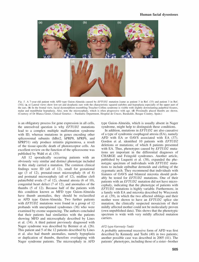
Fig. 5. A 7-year-old patient with AFD type Guion-Almeida caused by EFTUD2 mutation [same as patient 3 in Ref. (33) and patient 3 in Ref.(34)]. (a, c) Lateral views show low-set and dysplastic ears with the characteristic squared earlobes and hypoplasia especially of the upper part ofthe ears. (b) In the frontal view, facial dysmorphism resembling Treacher Collins syndrome is visible with slightly downslanting palpebral fissures,malar and mandibular hypoplasia. Also, note the microcephaly, which is often progressive with age. (d) Proximally placed thumbs are shown.(Courtesy of Dr Blanca Gener, Clinical Genetics – Paediatric Department, Hospital de Cruces, Barakaldo, Basque Country, Spain.)
is an obligatory process for gene expression in all cells,the unresolved question is why EFTUD2 mutationslead to a complex multiple malformation syndromewith ID, whereas mutations in genes encoding otherspliceosomal subunits (hBrr2, hPRP8, hPRP6, andhPRP31) only produce retinitis pigmentosa, a resultof the tissue-specific death of photoreceptor cells. Anexcellent review on the function of the spliceosome waspublished by Wahl et al. (35).
All 12 sporadically occurring patients with anobviously very similar and distinct phenotype includedin this study carried a mutation. The common clinicalfindings were ID (all of 12), small for gestationalage (3 of 12), prenatal-onset microcephaly (6 of 8)and postnatal microcephaly (all of 12), midline cleftpalate/bifid uvula (7 of 12), choanal atresia (6 of 10),congenital heart defect (7 of 12), and anomalies of thethumbs (5 of 12). Because half of the patients withthis condition known as MFD type Guion-Almeidahave thumb anomalies, they should be reclassifiedas AFD type Guion-Almeida. Two further patientswith EFTUD2 mutations were found in a group of 12probands with unexplained syndromes (36) who wereexamined by exome sequencing. The authors concludedthat their patients had similarities with the patientsshowing MFD and microcephaly described by Lineset al. (34). A third patient previously diagnosed withNager syndrome was described by Bernier et al. (26).This patient and 5 of the 12 patients described by Lineset al. also had thumb anomalies, namely hypoplasiaor duplication of thumbs, therefore overlapping withNager syndrome patients. The microcephaly in AFD
type Guion-Almeida, which is usually absent in Nagersyndrome, might help to distinguish these conditions.
In addition, mutations in EFTUD2 are also causativeof a type of syndromic esophageal atresia (EA), namelyAFD with EA or OAVS associated with EA (37).Gordon et al. identified 10 patients with EFTUD2deletions or mutations; of which 8 patients presentedwith EA. Thus, phenotypes caused by EFTUD2 muta-tions are important in the differential diagnoses ofCHARGE and Feingold syndromes. Another article,published by Luquetti et al. (38), expanded the phe-notypic spectrum of individuals with EFTUD2 muta-tions to include epibulbar dermoids and clefting of thezygomatic arch. They recommend that individuals withfeatures of OAVS and bilateral microtia should prob-ably be tested for EFTUD2 mutations. One of theirpatients with an EFTUD2 mutation did not have micro-cephaly, indicating that the phenotype of patients withEFTUD2 mutations is highly variable. Furthermore, ina family with EA and microtia described by Wieczoreket al. (39), in which the two affected siblings and theirmother were shown to have an EFTUD2 splice sitemutation, the clinically suspected mosaicism of theirmildly affected mother could not be molecularly proven(own unpublished data). This shows that the phenotypicspectrum is wide with very mildly affected mutationcarriers.
AFD type Kennedy-TeebiA probably autosomal recessive form of AFD was firstdescribed by Kennedy and Teebi (40) in two patients;another possible case was described in 2005 (41). Thepatients’ phenotypes, including those of a sister–brother
505

Wieczorek
pair, overlapped somewhat with that of Nager syn-drome, but included the following distinguishingfeatures: microcephaly, cleft palate, beaked nose,blepharophimosis, and developmental delay.
AFD type KellyThree male siblings described by Kelly et al. (42) hadmild AFD with hearing loss, mild ID, short stature, andgenitourinary anomalies consisting of hypospadias andundescended testes. The boys had symphalangism ofthe thumbs and the interphalangeal joints of the indexfinger. No further patients with this condition havebeen described. Autosomal recessive inheritance wassuggested, but X-linked inheritance cannot be excluded.
AFD type ReynoldsOne pre-axial AFD without ID has been describedby Reynolds et al., who described an autosomaldominant form of AFD in 1986. Two subsequentarticles confirmed that such an autosomal dominantsubtype of AFD exists (43–45). The patients havemild MFD (prominent forehead, ptosis, downslantingpalpebral fissures, malar hypoplasia, highly archedpalate, and micrognathia) and anomalies of the first ray.They closely resemble patients with Nager syndrome.Dauwerse et al. described an adult patient with clinicalsigns of AFD type Reynolds, who also had tuberoussclerosis and autosomal dominant polycystic kidneydisease (45). He was shown to have a TSC2-PKD1contiguous gene syndrome with a microdeletion in16p13.3. As the patients reported by Reynolds et al. didnot display features of tuberous sclerosis and autosomaldominant polycystic kidney disease, the significance ofthe 16p deletion remains unclear.
Post-axial AFDs
Only four post-axial AFDs have been described untilnow. They are clinically delineated by post-axialinvolvement of limbs and the associated anomalies.Besides the Miller syndrome, which is very distinct, onepost-axial AFD includes vertebral defects, one includesectodermal involvement (AFD type Weyers), and oneincludes syndactyly of the fingers (AFD type Arens).
Miller syndrome (MIM 263750)Miller syndrome [Genee-Wiedemann, Wildervanck-Smith, and postaxial acrofacial dysostosis syndrome(POADS)], the best known and best understood post-axial AFD, was first described by Genee (46) in 1969,Wiedemann (47) in 1973, and Miller et al. (48) in1979. With only about 30 articles having been publishedon this condition, it appears to be rare. Patientsare characterized by craniofacial dysmorphism andsymmetric post-axial limb anomalies with hypoplasia oraplasia of the (fourth and) fifth ray affecting the upperand lower limbs. Hypoplasia of ulna and radius may bepresent. Intelligence is usually normal. This autosomalrecessive condition was the first entity resolved by
exome sequencing (49) in four affected individualsof three independent kindreds. Filtering led to theidentification of a single candidate gene, DHODH ,in each of the four patients. This gene encodes akey enzyme, dihydroorotate dehydrogenase, in thepyrimidine biosynthesis pathway.
Additional clinical, molecular, and detailed func-tional data on DHODH were described recently (50,51). It was shown that those patients with mutationswithin DHODH display a very characteristic pheno-type, whereas those without mutations had involvementof upper or lower limbs only or they had pre-axialanomalies in addition to the post-axial ones. In addition,patients with DHODH mutations presented with cran-iofacial dysmorphism consisting of micrognathia (all of5), orofacial clefts (4 of 5), malar hypoplasia (all of 3),eyelid coloboma (1 of 4) and ear dysplasia (3 of 4).Four of five patients had bilateral absence of the fifthray of the hands and all of 5 of the feet. No muta-tions were identified in the other pyrimidine biosyn-thesis genes (CAD and UMPS ) in those patients withMiller syndrome without DHODH mutations. One typ-ical, mutation-carrying patient with Miller syndrome isdepicted in Fig. 6. No patient with Miller syndrome car-ries homozygous mutations, and truncating mutationsare rare. Thus, the molecular mechanism underlyingMiller syndrome might be atypical (50). Rainger et al.(50) found that two patients with DHODH mutationshad elevated levels of orotic acid but not of dihydrooro-tate in their urine.
Post-axial AFD and vertebral defectsThis condition was described by Medeira and Donnaiin a single male fetus (52). He had MFD with cleftlip and palate and both ears were absent. The fifthdigit was hypoplastic on one hand and aplastic on theother hand. In addition, he had scoliosis and talipesequinovarus. Radiographs showed multiple cervical andthoracic hemivertebrae. The mode of inheritance isunclear.
Weyers AFD (MIM 193530)This autosomal dominant condition was first describedby Weyers in 1952 (53). It is characterized by post-axialpolydactyly, dysplastic nails, oligodontia, and enamelhypoplasia. In the meantime, several patients with thiscondition have been described. The causative genes,EVC and EVC2 , localized in 4p16 (54), are in 5′ to5′ head-to-head orientation close to transcription startsites (55). Weyers AFD is allelic to the autosomalrecessive Ellis van Creveld syndrome. The latter isa skeletal dysplasia with clinical signs that overlapwith those of Weyers AFD but includes additionalfeatures such as disproportionate dwarfism, thoracicdysplasia, and congenital heart disease (54). Both genesare responsible for the basal body of the cilia, leading tothe assumption that Weyers AFD belongs to the groupof ciliary disorders (55). Three heterozygous mutationshave been identified in Weyers AFD, all localized inexon 22 of EVC2 .
506

Human facial dysostoses
Fig. 6. A 3-year 11-month-old patient with Miller syndrome showingthe typical craniofacial and limb phenotype [same patient as family 2 inRef. (50)]. (a, b) The patient has large eyes with lower eyelid coloboma,malar hypoplasia and micrognathia. Her ears are low-set. Absenceof fifth ray is present in foot (c) and also in hands (d). In addition,cutaneous syndactyly of toes I to III on the right and I to II on the leftfoot can be noted. (Courtesy of Dr Blanca Gener, Clinical Genetics –Paediatric Department, Hospital de Cruces, Barakaldo, Basque Country,Spain.)
AFD type Arens or Tel AvivThis condition has been described in a single girlonly (56). The affected girl was born to unaffectedconsanguineous parents. She presented with MFD(hypertelorism, downslanting palpebral fissures, low-setears, micrognathia, and small mouth) and absence of thefifth ray on all extremities. In addition, she displayedsyndactylies on both hands, bilateral congenital hipdislocation, and club feet. The distal phalanges ofthe second and third toes were absent. The mode ofinheritance is unknown.
Other AFDs
This group contains those types not fitting into thepreaxial or postaxial AFDs. Four of these AFDs arelethal: AFD type Rodríguez, severe AFD, AFD typeBates, and AFD de Macena Sobreira. Another typebelonging to this group is AFD type Karaman-Kavecci,characterized by hypoplasia of the ulna, femur, andfibula. In addition, there is a group of post-axial AFDswith ID (AFD type Patterson-Stevenson, Catania, andRichieri-Costa-Pereira) and one type with oligodontia,frizzy hair, and short stature (AFD type Palagonia).
AFD type Rodríguez (MIM201170)In 1990, Rodríguez et al. described three male sibswith a distinct form of AFD and unaffected parents(57). The sibs had facial dysostosis with severe
mandibular hypoplasia leading to neonatal death dueto respiratory complications. The upper limbs wereseverely shortened, with hypoplasia of humerus andsynostotic formation of the lower forearms. The fifthrays were lacking in the hands and/or feet. The handanomalies were variable with additional anomalies ofthe fourth ray, syndactyly of the first and secondfingers, hypoplastic thumbs, and syndactyly of thefourth and fifth rays. In addition, two of the threepatients had congenital heart defects, including atrialseptal defect, ventricular septal defect, pulmonaryatresia, and overriding aorta. Arhinencephaly wasalso reported. Further such cases, including females,have been described, confirming that this may be adistinct autosomal recessive entity(57). The gene is stillunknown. For a detailed review of this disorder, seeRef. (58).
AFD severe post-axial typeIn 1990, Rodríguez and Palacios (59) described a singlefemale stillborn infant as a severe case of POADS. Theinfant had features of MFD with maxillary, mandibular,and malar hypoplasia, low-set and malformed ears,broad nasal bridge, and a coloboma of the lower eyelid.The limb anomalies were severe with shortening of thelimbs, bilateral syndactyly with synostosis of the fourthand fifth fingers, and a hypoplastic left thumb. Addi-tional anomalies included hypoplastic scapulae, shortand broad clavicles, supernumerary cervical vertebrae,absence of the ulnae, and bowed radii. The lower limbswere also short and bowed. There was a short third toeand syndactyly of the fourth and fifth toes.
Further cases were described by Poissonnier et al.(60) and Stephan (61). The genetic cause is stillunknown.
AFD type BatesThis type of severe AFD also appears to be very rare.It was described in a single female fetus (62) born tounaffected consanguineous parents. The fetus showedMFD (hypertelorism, downslanting palpebral fissures,ectropion of lower eyelids, absence of lower eyelashes,micrognathia, cleft lip/palate, and severe microtia). Thelimbs were severely shortened and bowed. There wasoligodactyly of the hands and feet, with three fingersand four toes bilaterally. X-rays showed shorteningof the ulnae, short and bowed radii, and dislocationat the elbow joint. Three metacarpals were present inboth hands. The distal phalanges of the thumbs wereduplicated, as were the distal two phalanges of thethird digits. Tibiae and fibulae were shortened. The feethad four metatarsals. Internal malformations consistedof bilateral renal agenesis and bicornuate uterus. Themode of inheritance is unknown.
AFD type de Macena SobreiraThis type of AFD, associated with frontonasal dyspla-sia, was described in a single stillborn infant only (63).The infant had a prominent forehead, severe hyper-telorism, absent nose, macrostomia with bilateral cleft
507
Wieczorek
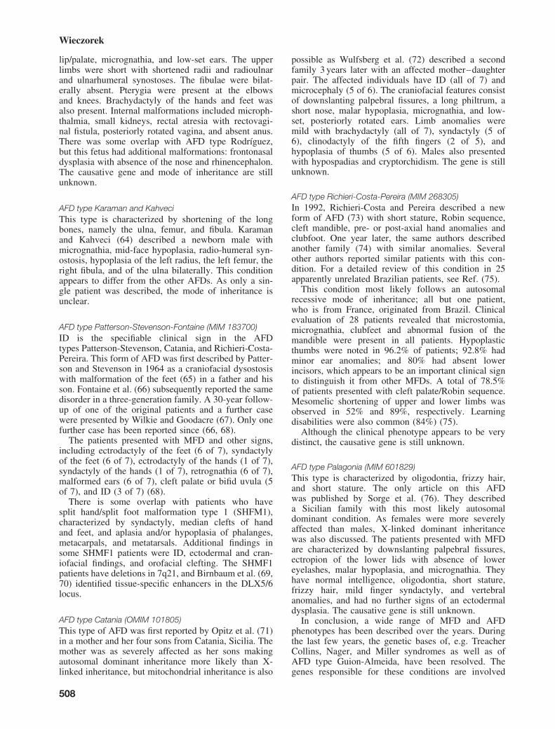
lip/palate, micrognathia, and low-set ears. The upperlimbs were short with shortened radii and radioulnarand ulnarhumeral synostoses. The fibulae were bilat-erally absent. Pterygia were present at the elbowsand knees. Brachydactyly of the hands and feet wasalso present. Internal malformations included microph-thalmia, small kidneys, rectal atresia with rectovagi-nal fistula, posteriorly rotated vagina, and absent anus.There was some overlap with AFD type Rodríguez,but this fetus had additional malformations: frontonasaldysplasia with absence of the nose and rhinencephalon.The causative gene and mode of inheritance are stillunknown.
AFD type Karaman and KahveciThis type is characterized by shortening of the longbones, namely the ulna, femur, and fibula. Karamanand Kahveci (64) described a newborn male withmicrognathia, mid-face hypoplasia, radio-humeral syn-ostosis, hypoplasia of the left radius, the left femur, theright fibula, and of the ulna bilaterally. This conditionappears to differ from the other AFDs. As only a sin-gle patient was described, the mode of inheritance isunclear.
AFD type Patterson-Stevenson-Fontaine (MIM 183700)ID is the specifiable clinical sign in the AFDtypes Patterson-Stevenson, Catania, and Richieri-Costa-Pereira. This form of AFD was first described by Patter-son and Stevenson in 1964 as a craniofacial dysostosiswith malformation of the feet (65) in a father and hisson. Fontaine et al. (66) subsequently reported the samedisorder in a three-generation family. A 30-year follow-up of one of the original patients and a further casewere presented by Wilkie and Goodacre (67). Only onefurther case has been reported since (66, 68).
The patients presented with MFD and other signs,including ectrodactyly of the feet (6 of 7), syndactylyof the feet (6 of 7), ectrodactyly of the hands (1 of 7),syndactyly of the hands (1 of 7), retrognathia (6 of 7),malformed ears (6 of 7), cleft palate or bifid uvula (5of 7), and ID (3 of 7) (68).
There is some overlap with patients who havesplit hand/split foot malformation type 1 (SHFM1),characterized by syndactyly, median clefts of handand feet, and aplasia and/or hypoplasia of phalanges,metacarpals, and metatarsals. Additional findings insome SHMF1 patients were ID, ectodermal and cran-iofacial findings, and orofacial clefting. The SHMF1patients have deletions in 7q21, and Birnbaum et al. (69,70) identified tissue-specific enhancers in the DLX5/6locus.
AFD type Catania (OMIM 101805)This type of AFD was first reported by Opitz et al. (71)in a mother and her four sons from Catania, Sicilia. Themother was as severely affected as her sons makingautosomal dominant inheritance more likely than X-linked inheritance, but mitochondrial inheritance is also
possible as Wulfsberg et al. (72) described a secondfamily 3 years later with an affected mother–daughterpair. The affected individuals have ID (all of 7) andmicrocephaly (5 of 6). The craniofacial features consistof downslanting palpebral fissures, a long philtrum, ashort nose, malar hypoplasia, micrognathia, and low-set, posteriorly rotated ears. Limb anomalies weremild with brachydactyly (all of 7), syndactyly (5 of6), clinodactyly of the fifth fingers (2 of 5), andhypoplasia of thumbs (5 of 6). Males also presentedwith hypospadias and cryptorchidism. The gene is stillunknown.
AFD type Richieri-Costa-Pereira (MIM 268305)In 1992, Richieri-Costa and Pereira described a newform of AFD (73) with short stature, Robin sequence,cleft mandible, pre- or post-axial hand anomalies andclubfoot. One year later, the same authors describedanother family (74) with similar anomalies. Severalother authors reported similar patients with this con-dition. For a detailed review of this condition in 25apparently unrelated Brazilian patients, see Ref. (75).
This condition most likely follows an autosomalrecessive mode of inheritance; all but one patient,who is from France, originated from Brazil. Clinicalevaluation of 28 patients revealed that microstomia,micrognathia, clubfeet and abnormal fusion of themandible were present in all patients. Hypoplasticthumbs were noted in 96.2% of patients; 92.8% hadminor ear anomalies; and 80% had absent lowerincisors, which appears to be an important clinical signto distinguish it from other MFDs. A total of 78.5%of patients presented with cleft palate/Robin sequence.Mesomelic shortening of upper and lower limbs wasobserved in 52% and 89%, respectively. Learningdisabilities were also common (84%) (75).
Although the clinical phenotype appears to be verydistinct, the causative gene is still unknown.
AFD type Palagonia (MIM 601829)This type is characterized by oligodontia, frizzy hair,and short stature. The only article on this AFDwas published by Sorge et al. (76). They describeda Sicilian family with this most likely autosomaldominant condition. As females were more severelyaffected than males, X-linked dominant inheritancewas also discussed. The patients presented with MFDare characterized by downslanting palpebral fissures,ectropion of the lower lids with absence of lowereyelashes, malar hypoplasia, and micrognathia. Theyhave normal intelligence, oligodontia, short stature,frizzy hair, mild finger syndactyly, and vertebralanomalies, and had no further signs of an ectodermaldysplasia. The causative gene is still unknown.
In conclusion, a wide range of MFD and AFDphenotypes has been described over the years. Duringthe last few years, the genetic bases of, e.g. TreacherCollins, Nager, and Miller syndromes as well as ofAFD type Guion-Almeida, have been resolved. Thegenes responsible for these conditions are involved
508

Human facial dysostoses
in ubiquitous processes such as RNA transcriptionand splicing. Future studies are needed to understandwhy such processes lead to specific phenotypes andto discover the genetic bases of the remaining, stillunresolved MFDs and AFDs.
Acknowledgements
Thanks to all individuals and their families for participating inthese studies. I am very grateful to Drs Mary F. Passarge andEberhard Passarge for proofreading this manuscript. This work wassupported by the German Ministry of Education and Research forthe CRANIRARE and the FACE consortium (BMBF 01GM1211Band 01GM1109B).
References1. Trainor PA, Dixon J, Dixon MJ. Treacher Collins syndrome: etiology,
pathogenesis and prevention. Eur J Hum Genet 2009: 17: 275–283.2. Dauwerse JG, Dixon J, Seland S et al. Mutations in genes encoding
subunits of RNA polymerases I and III cause Treacher Collinssyndrome. Nat Genet 2011: 43: 20–22.
3. Teber OA, Gillessen-Kaesbach G, Fischer S et al. Genotyping in 46patients with tentative diagnosis of Treacher Collins syndrome revealedunexpected phenotypic variation. Eur J Hum Genet 2004: 12: 879–890.
4. The Treacher Collins Syndrome Collaborative Group. Positionalcloning of a gene involved in the pathogenesis of Treacher Collinssyndrome. Nat Genet 1996: 12: 130–136.
5. Dixon J, Jones NC, Sandell LL et al. Tcof1/Treacle is required forneural crest cell formation and proliferation deficiencies that causecraniofacial abnormalities. Proc Natl Acad Sci USA 2006: 103:13403–13408.
6. Beygo J, Buiting K, Seland S et al. First report of a single exon deletionin TCOF1 causing Treacher Collins Syndrome. Mol Syndromol 2012:2: 53–59.
7. Bowman M, Oldridge M, Archer C et al. Gross deletions in TCOF1 area cause of Treacher-Collins-Franceschetti syndrome. Eur J Hum Genet2012: 20: 769–777.
8. Lowry RB, Morgan K, Holmes TM, Metcalf PJ. Stauffer GFMandibulo-facial dysostosis in Hutterite sibs: a possible recessive trait. Am J MedGenet 1985: 22: 501–512.
9. Toriello HV, Higgins JV, Abrahamson J, Waterman DF, Moore WD.X-linked syndrome of branchial arch and other defects. Am J MedGenet 1985: 21: 137–142.
10. Zelante L, Vigliaroli L, Mingarelli R, Dallapiccola B. Confirmation ofthe mandibulofacial dysostosis, Toriello type. Am J Med Genet 1993:45: 534–535.
11. Puri RD, Phadke SR. Further delineation of mandibulofacial dysostosis:Toriello type. Clin Dysmorphol 2002: 11: 91–93.
12. Ensink RJ, Brunner HG, Cremers CW. A new type of maxillofacialdysostosis, inherited as an X-linked or autosomal recessive trait. GenetCouns 1997: 8: 285–290.
13. Hedera P, Toriello HV, Petty EM. Novel autosomal dominantmandibulofacial dysostosis with ptosis: clinical description and exclu-sion of TCOF1. J Med Genet 2002: 39: 484–488.
14. Marcano A, Richieri-Costa A. A newly recognized autosomal dominantmandibulofacial dysostosis (Bauru type): report on a Brazilian family.Braz J Dysmorphol Speech-Hear Disord 1998: 1: 37–41.
15. Zechi-Ceide RM, Guion-Almeida ML. Mandibulofacial dysostosisBauru type syndrome: a new case. Am J Med Genet 1999: 86: 199–201.
16. Verloes A, Lesenfants S. A new form of mandibulofacial dysostosiswith macroblepharon and macrostomia. Clin Dysmorphol 1997: 6:21–24.
17. Zhang Y, Dai Y, Liu Y, Ren J. Mandibulofacial dysostosis, microtia,and limb anomalies in a newborn: a new form of acrofacial dysostosissyndrome? Clin Genet 2010: 78: 570–574.
18. Burn J, McKeown C, Wagget J, Bray R, Goodship J. New dysmorphicsyndrome with choanal atresia in siblings. Clin Dysmorphol 1992: 1:137–144.
19. Wieczorek D, Teber OA, Lohmann D, Gillessen-Kaesbach G. Twobrothers with Burn-McKeown syndrome. Clin Dysmorphol 2003: 12:171–174.
20. Toriello HV, Higgins JV. A boy with choanal atresia and cardiac defect:Burn-McKeown syndrome? Clin Dysmorphol 1999: 8: 143–145.
21. Hing AV, Leblond C, Sze RW et al. A novel oculo-oto-facial dysplasiain a native Alaskan community with autosomal recessive inheritance.Am J Med Genet A 2006: 140: 804–812.
22. Opitz JM, Burn J. RE: correspondence from Wieczorek & Gillessen-Kaesbach and Hing & Parisi. Am J Med Genet A 2006: 140: 2385.
23. Nager G. Not available. Pract Otorhinolaryngol (Basel) 1948: 10:184–195.
24. Nager FR, DeReynier JP. Das Gehororgan bei den angeborenenKopfmissbildungen. Pract Otorhinolaryngol (Basel) 1948: 10 (Suppl.2): 1–128.
25. Halonen K, Hukki J, Arte S, Hurmerinta K. Craniofacial structures anddental development in three patients with Nager syndrome. J CraniofacSurg 2006: 17: 1180–1187.
26. Bernier FP, Caluseriu O, Ng S et al. Haploinsufficiency of SF3B4,a component of the pre-mRNA spliceosomal complex, causes Nagersyndrome. Am J Hum Genet 2012: 90: 925–933.
27. Waggoner DJ, Ciske DJ, Dowton SB, Watson MS. Deletion of 1q in apatient with acrofacial dysostosis. Am J Med Genet 1999: 82: 301–304.
28. Watanabe H, Shionyu M, Kimura T, Kimata K, Watanabe H. Splicingfactor 3b subunit 4 binds BMPR-IA and inhibits osteochondral celldifferentiation. J Biol Chem 2007: 282: 20728–20738.
29. Czeschik JC, Voigt C, Alanay Y et al. Clinical and mutation data in12 patients with the clinical diagnosis of Nager syndrome. Hum Genet2013 (in press).
30. Chemke J, Mogilner BM, Ben-Itzhak I, Zurkowski L, Ophir D.Autosomal recessive inheritance of Nager acrofacial dysostosis. J MedGenet 1988: 25: 230–232.
31. Guion-Almeida M, Kokitsu-Nakata N, Richieri-Costa A. Mental andgrowth retardation, microtrigonocephaly, cleft palate and preauricularskin tags: a variant of the C syndrome or a new autosomal recessivesyndrome? Braz J Dysmorphol Speech Hear 2000: 3: 25–29.
32. Guion-Almeida ML, Zechi-Ceide RM, Vendramini S et al. A new syn-drome with growth and mental retardation, mandibulofacial dysostosis,microcephaly, and cleft palate. Clin Dysmorphol 2006: 15: 171–174.
33. Wieczorek D, Gener B, Gonzalez MJ et al. Microcephaly, microtia,preauricular tags, choanal atresia and developmental delay in threeunrelated patients: a mandibulofacial dysostosis distinct from TreacherCollins syndrome. Am J Med Genet A 2009: 149A: 837–843.
34. Lines MA, Huang L, Schwartzentruber J et al. Haploinsufficiency ofa spliceosomal GTPase encoded by EFTUD2 causes mandibulofacialdysostosis with microcephaly. Am J Hum Genet 2012: 90: 369–377.
35. Wahl MC, Will CL, Luhrmann R. The spliceosome: design principlesof a dynamic RNP machine. Cell 2009: 136: 701–718.
36. Need AC, Shashi V, Hitomi Y et al. Clinical application of exomesequencing in undiagnosed genetic conditions. J Med Genet 2012: 49:353–361.
37. Gordon CT, Petit F, Oufadem M et al. EFTUD2 haploinsufficiencyleads to syndromic oesophageal atresia. J Med Genet 2012: 49:737–746.
38. Luquetti DV, Hing AV, Rieder MJ et al. “Mandibulofacial dysostosiswith microcephaly” caused by EFTUD2 mutations: expanding thephenotype. Am J Med Genet A 2013: 161A: 108–113.
39. Wieczorek D, Shaw-Smith C, Kohlhase J et al. Esophageal atresia,hypoplasia of zygomatic complex, microcephaly, cup-shaped ears,congenital heart defect, and mental retardation – new MCA/MRsyndrome in two affected sibs and a mildly affected mother? Am JMed Genet A 2007: 143A: 1135–1142.
40. Kennedy SJ, Teebi AS. Newly recognized autosomal recessiveacrofacial dysostosis syndrome resembling Nager syndrome. Am J MedGenet A 2004: 129A: 73–76.
41. Ruiter M, van Dijken PJ, de Vries BB. Facial characteristics arenot distinctive features for the acrofacial dysostosis syndrome typeKennedy-Teebi. Am J Med Genet A 2005: 135: 344(author reply 345).
42. Kelly TE, Cooke RJ, Kester RW. Acrofacial dysostosis with growth andmental retardation in three males, one with simultaneous Hermansky-Pudlak syndrome. Birth Defects Orig Artic Ser 1977: 13: 45–52.
43. Wessels MW, Den Hollander NS, Cohen-Overbeek TE et al. Prenataldiagnosis and confirmation of the acrofacial dysostosis syndrome typeRodriguez. Am J Med Genet 2002: 113: 97–100.
44. Reynolds JF, Webb MJ, Opitz JM. A new autosomal dominantacrofacial dysostosis syndrome. Am J Med Genet Suppl 1986: 2:143–150.
509

Wieczorek
45. Dauwerse JG, Bouman K, van Essen AJ et al. Acrofacial dysostosisin a patient with the TSC2-PKD1 contiguous gene syndrome. J MedGenet 2002: 39: 136–141.
46. Genee E. An extensive form of mandibulo-facial dysostosis. J GenetHum 1969: 17: 45–52.
47. Wiedemann HR. Malformation-retardation syndrome with bilateralabsence of the 5th rays in both hands and feets, cleft palate, malformedears and eyelids, radioulnar synostosis (author’s transl). Klin Padiatr1973: 185: 181–186.
48. Miller M, Fineman R, Smith DW. Postaxial acrofacial dysostosissyndrome. J Pediatr 1979: 95: 970–975.
49. Ng SB, Buckingham KJ, Lee C et al. Exome sequencing identifies thecause of a Mendelian disorder. Nat Genet 2010: 42: 30–35.
50. Rainger J, Bengani H, Campbell L et al. Miller (Genee-Wiedemann)syndrome represents a clinically and biochemically distinct subgroupof postaxial acrofacial dysostosis associated with partial deficiency ofDHODH. Hum Mol Genet 2012: 21: 3969–3983.
51. Fang J, Uchiumi T, Yagi M et al. Protein instability and functionaldefects caused by mutations of dihydro-orotate dehydrogenase in Millersyndrome patients. Biosci Rep 2012: 32: 631–639.
52. Medeira A, Donnai D. Postaxial acrofacial dysostosis syndrome withvertebral segmentation defects. Clin Dysmorphol 1994: 3: 171–174.
53. Weyers H. Hexadactyly, mandibular fissure and oligodontia, a newsyndrome; dysostosis acrofacialis. Ann Paediatr 1953: 181: 45–60.
54. Ruiz-Perez VL, Ide SE, Strom TM et al. Mutations in a new genein Ellis-van Creveld syndrome and Weyers acrodental dysostosis. NatGenet 2000: 24: 283–286.
55. Huber C, Cormier-Daire V. Ciliary disorder of the skeleton. Am J MedGenet C Semin Med Genet 2012: 160C: 165–174.
56. Arens R, Reichman B, Katznelson MB, Goodman RM. New form ofpostaxial acrofacial dysostosis? Am J Med Genet 1991: 41: 438–443.
57. Rodríguez JI, Palacios J, Urioste M. New acrofacial dysostosissyndrome in 3 sibs. Am J Med Genet 1990: 35: 484–489.
58. Dimitrov B, Balikova I, Jekova N et al. Acrofacial dysostosis typeRodríguez. Am J Med Genet A 2005: 135: 81–85.
59. Rodríguez JI, Palacios J. Severe postaxial acrofacial dysostosis: ananatomic and angiographic study. Am J Med Genet 1990: 35:490–492.
60. Poissonnier M, Neuville V, Petit P, Busuttil R. Lethal mandibulofacialand ulnofibular dysostosis. Ann Pediatr (Paris) 1983: 30: 713–717.
61. Stephan MJ. Autosomal recessive form of mandibular dysostosis. AmJ Med Genet 1990: 35: 493–495.
62. Bates AW, Hall CM, Morgan H, Rosser EM, Scheimberg I. Lethalacrofacial dysostosis, pre- and post-axial defects of the hands, andbilateral renal agenesis. Clin Dysmorphol 2002: 11: 63–66.
63. de Macena Sobreira NL, Alves MT, Alvarez Perez AB et al.Mandibulofacial dysostosis, acral anomalies and frontonasal dysplasia:a new form of acrofacial dysostosis. Clin Dysmorphol 2008: 17:145–148.
64. Karaman A, Kahveci H. Unusual acrofacial dysostosis with severe limbdefects: a new syndrome. Genet Couns 2011: 22: 249–253.
65. Patterson TJ, Stevenson AC. Cranio-facial dysostosis and malforma-tions of feet. J Med Genet 1964: 1: 112–114.
66. Fontaine G, Farriaux JP, Delattre P et al. Familial case of the syndromeof ectrodactyly and mandibulo-facial dysostosis. J Genet Hum 1974:22: 289–307.
67. Wilkie AO, Goodacre TE. Patterson-Stevenson-Fontaine syndrome: 30-year follow-up and clinical details of a further affected case. Am J MedGenet 1997: 69: 433–434.
68. Turnpenny PD, Johnston AW, Dean JC et al. Ectrodactyly-mandibulo-facial dysostosis: case report and delineation of an entity. ClinDysmorphol 1992: 1: 103–109.
69. Birnbaum RY, Clowney EJ, Agamy O et al. Coding exons functionas tissue-specific enhancers of nearby genes. Genome Res 2012: 22:1059–1068.
70. Birnbaum RY, Everman DB, Murphy KK et al. Functional character-ization of tissue-specific enhancers in the DLX5/6 locus. Hum MolGenet 2012: 21: 4930–4938.
71. Opitz JM, Mollica F, Sorge G et al. Acrofacial dysostoses: review andreport of a previously undescribed condition: the autosomal or X-linkeddominant Catania form of acrofacial dysostosis. Am J Med Genet 1993:47: 660–678.
72. Wulfsberg EA, Campbell AB, Lurie IW, Eanet KR. Confirmation ofthe Catania brachydactylous type of acrofacial dysostosis: report of asecond family. Am J Med Genet 1996: 63: 554–557.
73. Richieri-Costa A, Pereira SCS. Short stature, Robin sequence, cleftmandible, pre/postaxial hand anomalies, and clubfoot: a new autosomalrecessive syndrome. Am J Med Genet 1992: 42: 681–687.
74. Richieri-Costa A, Pereira SC. Autosomal recessive short stature, Robinsequence, cleft mandible, pre/postaxial hand anomalies, and clubfeet inmale patients. Am J Med Genet 1993: 47: 707–709.
75. Favaro FP, Zechi-Ceide RM, Alvarez CW et al. Richieri-Costa-Pereirasyndrome: a unique acrofacial dysostosis type. An overview of theBrazilian cases. Am J Med Genet A 2011: 155A: 322–331.
76. Sorge G, Pavone L, Polizzi A et al. Another “new” form, the palagoniatype of acrofacial dysostosis in a Sicilian family. Am J Med Genet1997: 69: 388–394.
510