Human ESC/iPSC-based ‘omics’ and bioinformatics for translational research
10
DRUG DISCOVERY TODAY DISEASE MODELS Human ESC/iPSC-based ‘omics’ and bioinformatics for translational research Gerd A. Mu ¨ller 1 , Kirill V. Tarasov 2 , Rebekah L. Gundry 3 , Kenneth R. Boheler 2,4, * 1 Molecular Oncology, Medical School, University of Leipzig, Leipzig, Germany 2 Molecular Cardiology and Stem Cell Unit, National Institute on Aging, National Institutes of Health, Baltimore, MD 21224, USA 3 The Department of Biochemistry and The Biotechnology and Bioengineering Center, Medical College of Wisconsin, Milwaukee, WI 53226, USA 4 Stem Cell and Regenerative Medicine, LKS Faculty of Medicine, University of Hong Kong, Hong Kong The establishment of human embryonic stem cell lines (hESCs) created the basis for new approaches in regen- erative medicine and drug discovery. Despite the potential of hESCs for cell-based therapies, ethical controversies limit their use. These obstacles could be overcome by induced pluripotent stem cells (iPSCs) that are generated by reprogramming somatic cells. Before iPSCs can be used for clinical applications, however, they must be thoroughly analyzed for aber- rations in the genome, epigenome, transcriptome and proteome. Here, we review how ‘omics’ technologies can be employed for a quantitative and definitive assessment of these cells. Section editor: Ronald Li – LKS Faculty of Medicine, University of Hong Kong, Hong Kong, and Mount Sinai School of Medicine, New York, NY, USA. Introduction Pluripotent stem cells (PSCs) differentiate into all cell types found in the body. The best characterized and standard for PSCs are embryonic stem cells (ESCs) [1], but experimentally derived PSCs, known as induced PSCs (iPSCs), can be gener- ated from almost any type of somatic cell through forced expression of pluripotency-promoting transcription factors or microRNAs (miRs) [2–6]. The ease of generating iPSCs has fostered the idea of immunologically compatible patient- derived cells. iPSCs thus may represent a viable alternative to human ESCs (hESCs) as the primary source of pluripotent cells for regenerative medicine; however, the advantages of iPSCs are counterbalanced by unresolved questions involving differences between the two cell types. Potential iPSC line defects include chromosomal abnormalities, altered gene expression and unanticipated aberrations in the epigenetic landscape and immunogenicity [7]. Taken together, these differences demonstrate that iPSCs must be carefully ana- lyzed on molecular, cellular and functional levels before entering the clinic (Fig. 1). Omics approaches, including genome- and proteome-based, offer platforms for fully char- acterizing and standardizing putative iPSC lines to address these issues of heterogeneity and safety. Applications of ‘omics’ to PSCs The human genome project, which began in 1990, led to major technological advancements that included improved sequencing of the genome and a routine analysis of a cell’s DNA (SNPs, copy number variation, mutations), methylation and histone state (epigenome), RNA abundance (transcrip- tome) or protein content (proteome). Collectively these ana- lyses, among others, have been termed ‘omics’ research Drug Discovery Today: Disease Models Vol. 9, No. 4 2012 Editors-in-Chief Jan Tornell – AstraZeneca, Sweden Andrew McCulloch – University of California, SanDiego, USA Induced pluripotent stem cells *Corresponding author.: K.R. Boheler ([email protected]), ([email protected]) 1740-6757/$ .Published by Elsevier Ltd. DOI: 10.1016/j.ddmod.2012.02.003 e161
Transcript of Human ESC/iPSC-based ‘omics’ and bioinformatics for translational research

DRUG DISCOVERY
TODAY
DISEASEMODELS
Human ESC/iPSC-based ‘omics’and bioinformatics for translationalresearchGerd A. Muller1, Kirill V. Tarasov2, Rebekah L. Gundry3, Kenneth R. Boheler2,4,*1Molecular Oncology, Medical School, University of Leipzig, Leipzig, Germany2Molecular Cardiology and Stem Cell Unit, National Institute on Aging, National Institutes of Health, Baltimore, MD 21224, USA3The Department of Biochemistry and The Biotechnology and Bioengineering Center, Medical College of Wisconsin, Milwaukee, WI 53226, USA4Stem Cell and Regenerative Medicine, LKS Faculty of Medicine, University of Hong Kong, Hong Kong
Drug Discovery Today: Disease Models Vol. 9, No. 4 2012
Editors-in-Chief
Jan Tornell – AstraZeneca, Sweden
Andrew McCulloch – University of California, SanDiego, USA
Induced pluripotent stem cells
The establishment of human embryonic stem cell lines
(hESCs) created the basis for new approaches in regen-
erative medicine and drug discovery. Despite the
potential of hESCs for cell-based therapies, ethical
controversies limit their use. These obstacles could
be overcome by induced pluripotent stem cells (iPSCs)
that are generated by reprogramming somatic cells.
Before iPSCs can be used for clinical applications,
however, they must be thoroughly analyzed for aber-
rations in the genome, epigenome, transcriptome and
proteome. Here, we review how ‘omics’ technologies
can be employed for a quantitative and definitive
assessment of these cells.
Introduction
Pluripotent stem cells (PSCs) differentiate into all cell types
found in the body. The best characterized and standard for
PSCs are embryonic stem cells (ESCs) [1], but experimentally
derived PSCs, known as induced PSCs (iPSCs), can be gener-
ated from almost any type of somatic cell through forced
expression of pluripotency-promoting transcription factors
or microRNAs (miRs) [2–6]. The ease of generating iPSCs has
fostered the idea of immunologically compatible patient-
derived cells. iPSCs thus may represent a viable alternative
*Corresponding author.: K.R. Boheler ([email protected]), ([email protected])
1740-6757/$ .Published by Elsevier Ltd. DOI: 10.1016/j.ddmod.2012.02.003
Section editor:Ronald Li – LKS Faculty of Medicine, University of HongKong, Hong Kong, and Mount Sinai School of Medicine, NewYork, NY, USA.
to human ESCs (hESCs) as the primary source of pluripotent
cells for regenerative medicine; however, the advantages of
iPSCs are counterbalanced by unresolved questions involving
differences between the two cell types. Potential iPSC line
defects include chromosomal abnormalities, altered gene
expression and unanticipated aberrations in the epigenetic
landscape and immunogenicity [7]. Taken together, these
differences demonstrate that iPSCs must be carefully ana-
lyzed on molecular, cellular and functional levels before
entering the clinic (Fig. 1). Omics approaches, including
genome- and proteome-based, offer platforms for fully char-
acterizing and standardizing putative iPSC lines to address
these issues of heterogeneity and safety.
Applications of ‘omics’ to PSCs
The human genome project, which began in 1990, led to
major technological advancements that included improved
sequencing of the genome and a routine analysis of a cell’s
DNA (SNPs, copy number variation, mutations), methylation
and histone state (epigenome), RNA abundance (transcrip-
tome) or protein content (proteome). Collectively these ana-
lyses, among others, have been termed ‘omics’ research
e161

Drug Discovery Today: Disease Models | Induced pluripotent stem cells Vol. 9, No. 4 2012
Putative hiPSCs- ES cell morphology- Self-renewal (unlimited proliferation) (Poor checkpoint controls)- Pluripotency/Pluripotency Transcription Factors- Demethylation of pluripotency genes- Poised (Divalent) Histone Methylation Patterns- Expression of ESC-associated surface markers- Telomerase Activity/No Senescence
Somatic Cells(Human Fbs)- Tissue-specific cell morphology- Pluripotency genes methylated- Monovalent Histone Methylation- Expression of somatic cell markers- Limited proliferation (Active checkpoint controls)- Senescence Susceptibility
Reprogramming Factors(Integrative or Episomal or RNA-mediated)
OCT4, SOX2, KLF4, c-MYCOCT4, SOX2, LlN28, NANOG
OCT4, SOX2, NANOG, KLF4, c-MYC, LlN28, SV40LTMultiple other combinations
miR302/367 +HDAC inhibitors
or
Txn Factors
SurfaceMarkers
DNA StainTOPRO
OCT4
NANOG
SOX2
SSEA4
SSEA3/Tra-1-60
SSEA3/Tra-1-81
Drug Discovery Today: Disease Models
Figure 1. Reprogramming of somatic cells (human fibroblasts (Fbs)) to induced pluripotent stem cells (hiPSCs). Several reprogramming factor combinations
are useful for generating iPSCs, including the 7 factors in episomal constructs used here (in blue). Typical characteristics of starting somatic cells and putative
iPSCs are shown. hiPSC lines should be considered putative until a full analysis of potency is performed. This requires an analysis based on morphology,
expression of pluripotency transcription (Txn) factors (OCT4, NANOG, SOX2), expression of surface markers (SSEA3, SSEA4, Tra-1-60, Tra-1-81) and
teratoma assays. Alternatively ‘omic’ based techniques, as described in the text, may be invaluable to quantitatively assess the quality of these cells.
endeavors that are unique from traditional experimental
designs. This is because ‘omics’ approaches are often large-
scale and data-driven, as opposed to purely hypothesis driven
[8]. Data generated from ‘omics’ approaches, when combined
across platforms, are useful in describing biological relation-
ships related to experimental and cellular fluctuations. Con-
sequently, the integration of multiple ‘omics’ approaches to
understand a cell’s phenotype, permits an ‘integrated system’
that more fully describes a cell’s response to defined variables.
Finally, ‘omics’ approaches require significant statistical and
computational efforts to model dynamic systems that by
their very nature interact on multiple levels within a cell.
Extraction of valuable biological information from ‘omics’
data is challenging, but the results, when properly analyzed,
show great potential in addressing some of the current pro-
blems associated with transplantation and stem cell-based
therapies [9].
A quantitative and definitive assessment of human iPSC
lines should be possible through the use of ‘omics’ techni-
ques. Genome-wide evaluations will be useful for defining the
state of putative iPSC lines, and robust statistical techniques
e162 www.drugdiscoverytoday.com
should be valuable in pin-pointing possible differences/aber-
rations in lines relative to ‘gold-standard’ ESC lines (Fig. 2a).
More specifically, genome-wide DNA sequencing should
uncover any spontaneous DNA mutations that may result
during reprogramming, while microarray analysis of RNA
samples or RNA-Seq experiments can provide insights on
variations in gene expression that may be indicative of resi-
dual epigenetic memory. Chromatin immunoprecipitation
experiments (ChIP-chip or ChIP-Seq) and DNA methylation
studies can reveal variations in chromatin structure and
transcription factor binding. Proteomic studies may also be
valuable in defining variations in protein levels between cells,
but perhaps more importantly, this technique may be of great
value in the development of immunophenotyping techni-
ques that can be employed to isolate and characterize
‘authentic’ iPSC lines. By studying cells at the ‘omics’ level
it should be possible to obtain fingerprints of iPSCs for
comparisons with standard ESC lines and to assess how the
reprogramming process affects biological processes, thus sol-
ving many of the current problems surrounding possible
differences among these forms of PSCs.

Vol. 9, No. 4 2012 Drug Discovery Today: Disease Models | Induced pluripotent stem cells
Genome and epigenome
DNA mutations
Similar to problems observed in the sheep Dolly [10], cell
autonomous genetic defects are present in reprogrammed
iPSCs. Specifically, iPSC lines show an enrichment of muta-
tions and chromosomal defects that may be introduced dur-
ing the reprogramming process, independent of the
reprogramming vectors, or by culture adaption (Fig. 2b)
[11–14]. As an example, Gore et al. sequenced the protein-
coding exons (exomes) of 22 human iPSC lines generated by
five independent methods and nine parental fibroblast lines.
On average, they found five protein-coding point mutations
in the regions sampled or an estimated six protein-coding
point mutations per exome. Although the majority of the
mutations were non-synonymous, nonsense or splice var-
iants, many of the mutations occurred in genes with causa-
tive effects in cancers. At least half of the mutations were
present in the parental fibroblast, but the remainder occurred
spontaneously either during or after reprogramming. How-
den et al. also assessed whether human iPSCs isolated from a
patient with gyrate atrophy increased its mutational load
with reprogramming [15]. In these cells, no abnormalities
were detected by standard G-band metaphase analysis; how-
ever, array comparative genomic hybridization and exome
sequencing identified two deletions, one amplification and
nine mutations in protein-coding regions when compared
against the parental patient fibroblast cell line. They then
performed exome sequencing on a gene-targeted iPSC clonal
line that corrected the OAT point mutation present in this
patient’s DNA, and on a cassette-free iPSC clone. Somewhat
surprisingly, the genomes proved remarkably stable, as no
additional mutations or copy number variations were iden-
tified, excluding the targeted correction in the OAT locus and a
single synonymous base-pair change. These findings led to
the conclusion that iPSCs carry a significant mutational load
from the parental line, but clonal events and prolonged
culturing do not lead to a substantial increase in mutations.
Mutations may, however, occur at a higher frequency than
previously reported. In unpublished work presented in a
recent Stem Cell Research Symposium at the NIH, Paul Liu
presented results from deep whole-genome sequencing and
high-density SNP array analysis. He reported results from
episomal-vector reprogrammed hiPSC lines derived from
two tissues of a single adult donor. The data revealed over
1000 single-nucleotide substitutions in each iPSC cell line
when compared to the parental cell sources. Although the
majority of mutations were in non-coding sequences, 6 and
12 mutations, respectively, were located in coding regions,
and 34 and 22 variations, respectively, were identified in the
50 or 30 untranslated regions of genes. Another 362 and 709
differences were observed in intronic regions that might
affect gene expression. The majority of mutations were not
present in the exome, and SNP analysis was not sufficient to
identify these mutations. Because the specific point muta-
tions were not conserved among the two iPSC lines, sequence
substitutions were not related to susceptible ‘hot spots’ dur-
ing reprogramming. Importantly, these results established
the viability of iPSC line whole-genome sequencing, which
has now become cost-effective and more widely accessible to
researchers in this field.
DNA methylation
Closely affiliated with the genome sequence are DNA modi-
fiers (i.e. DNA methyltransferases (DNMTs)) that add methyl
groups at the 50-position of cytosine. During the reprogram-
ming process, somatic cells reset their pattern of DNA methy-
lation to an ESC-like state. More specifically, DNA of
transcriptionally active genes like pluripotency and house-
keeping genes is hypomethylated [16,17], while silenced
genes are hypermethylated [17,18].
Various groups have now reported that ESCs exhibit
unique methylation patterns and that iPSCs show modest
variations in this pattern. This was perhaps best illustrated
through the use of different cell types isolated and repro-
grammed from the same mouse. Among these genetically
identical iPSC clonal lines, the DNA methylation profiles
reflected the cell type of origin, suggesting the presence of
residual parental cell ‘epigenetic memory’. Functionally,
methylation pattern permitted efficient differentiation of
iPSCs into the somatic cell type of origin, but these same
lines showed reduced efficiencies of differentiation into other
lineages. Although the causes are still unclear, it became
apparent that these effects resulted from insufficient methy-
lation (silencing) of genes normally expressed in the somatic
cells from which the iPSC are derived, and insufficient
demethylation (activation) of ESC-specific genes [19–22].
In support of this assumption, the addition of 5-azacytidine
(Aza), a DNA methylation inhibitor, to established iPSCs lines
increased their differentiation potential and made them more
like ESCs [21]. Moreover, pou5f1 and nanog gene promoters,
which are highly methylated in somatic cells, remain largely
methylated in partially reprogrammed cells (Fig. 2c); how-
ever, exposure to Aza reactivates endogenous pou5f1 gene
expression. Prolonged cultivation of iPSCs also diminished
differences in the methylation patterns between iPSCs and
ESCs [19,22,23]. These latter findings suggest that reprogram-
ming does not completely reverse the epigenetic landscape in
early clonal isolates, a finding confirmed by Kim et al. [21],
and that chromatin remodeling is a gradual process that takes
place over an extended period of time. Thus the state of DNA
methylation at a genome level, and especially at some specific
gene loci as a function of time, is likely to be indicative of the
degree of reprogramming of iPSCs.
From an ‘omics’ perspective, current DNA methylation
assays are often limited in their ability to characterize a large
number of genomic targets. To overcome this limitation,
www.drugdiscoverytoday.com e163

Drug Discovery Today: Disease Models | Induced pluripotent stem cells Vol. 9, No. 4 2012
(a)
Pluripotent EmbryonicStem Cells
Normal Genome
DNAACGTTTGACTGATTGGCCACGT ACGTTTGAATGATTGGCCACGT
ACGUUUGAAUGAUUGGCCACGUACGUUUGATUGAUUGGCCACGU
UGATUGAUGCUGGCCAC
RNA
Splice Variant
DNAEight Histone Core
H3K27me3
H3K27me3
H3K4me3
H3K4me3
Poised Histones
Poised Histones(ChlP-chip)
H1 Histone
DNA Methylation
Histonemodifications
Isoforms,splice variants
Surface proteinsThy-1
HENTSSSPIQYEFSLTR
Small Peptides/Metabolites
Phosphoproteins
Secreted proteins
DNA Methylation
ESC
nanog
oct4
iPSC-1 iPSC-2
DNA Seq
RNA SeqMicroarrays
Mutant Genome(b)
(c)
(d)
0 200 400 600 800 1000 1200 1400 1600 1800 2000
Drug Discovery Today: Disease Models
Figure 2. Omics approaches and PSCs. (a) Human ESCs in culture and after immunostaining with cell surface antibodies to stage specific antigens. (b)
Omics techniques to evaluate DNA or RNA sequences or transcript abundance by microarrays reveal possible genetic mutations, changes in RNA
expression or splicing. (c) Central to epigenetic control is DNA chromatin and the nucleosome, which can be modified through a variety of enzymes and
pathways. Epigenomic techniques are useful for monitoring changes to DNA methylation or histone methylation, both of which can affect ‘epigenetic
e164 www.drugdiscoverytoday.com

Vol. 9, No. 4 2012 Drug Discovery Today: Disease Models | Induced pluripotent stem cells
investigators have described methods that capture genomic
targets for single-molecule bisulfite sequencing at a single-
nucleotide resolution. From these large-scale studies, chro-
mosome-wide methylation patterns were similar, but cyto-
sine methylation proved to be slightly more prevalent in the
pluripotent cells than in the fibroblasts [24]. These data, in
agreement with other studies, ultimately showed that iPSCs
intrinsically display more methylation than ESCs. Further
improvements in whole-genome methylation sequencing
based studies have now made it feasible to fully characterize
the methylation status of iPSCs [25–27].
Histone modifications
Somatic cells reprogrammed to iPSCs reset post-translational
histone modifications to an ESC-like state, and like DNA
methylation, histone modifications differ between ESCs
and iPSCs. Histone deacetylase inhibitors (HDACs), such as
trichostatin A (TSA) and valproic acid (VPA), used to promote
active histone acetylation marks, can enhance iPSC repro-
gramming efficiency [28,29]. To maintain pluripotency, the
promoters of genes encoding NANOG, OCT4 and SOX2 show a
high degree of histone 3 lysine 4 (H3K4) methylation result-
ing in a transcriptionally active state [18,30,31]. Partially
reprogrammed iPSCs, however, retained some histone mod-
ification patterns of its parental cells, indicating an associa-
tion of histone modification in iPSC memory status.
In genome-wide studies performed by ChIP-chip or ChIP-
Seq based techniques, bivalent patterns of histone methyla-
tion have been described that distinguish PSCs from somatic
cells (Fig. 2c). In bivalent promoters, histone 3 is methylated
at both lysines 4 and 27 (H3K27). Both modifications are
prevalent at transcription start sites of numerous develop-
mental genes whose expression is repressed in ESCs [30,31].
Although H3K4 methylation is associated with gene activa-
tion and H3K27 methylation by polycomb group protein
complexes typically results in gene repression, bivalent pro-
moters tended to be repressed, but at times ‘leaky’ [32,33].
With differentiation, this pattern switches from a bivalent
state to a monovalent state, which results either in transcrip-
tionally active genes characterized by H3K4 methylation or to
non-transcribed genes with H3K27 methylation state [34].
Thus the poised state was seen as a central requirement for
undifferentiated ESCs to maintain their developmental
potential. Several other histone modifications are also known
to affect gene activity, including the repressive H3K9me3,
H4K20me3 marks and multiple targets of histone acetylation
[33,35,36].
memory’. In the example to the right, the methylation states of histone 3 at re
residues is associated with a ‘poised’ state, where transcription is generally low, b
or repressed. An additional trait that may differ between ESCs and iPSCs is DNA
Proteomic based techniques, while only modestly used to date on PSCs, hold grea
proteins useful for isolating live cells with a well-defined degree of pluripotenc
Recent results from Roeder and colleagues provide new
insights into these regulatory mechanisms [37]. By studying
components of SET1/MLL family complexes, these authors
found that depletion of Dpy-30 and RbBP5 leads to a general-
ized reduction in H3K4 methylation in mouse ESCs. In fact,
H3K4 methylation levels were significantly reduced on the
promoters of key stemness genes like nanog, pou5f1, klf4 and
sox2, but mRNA abundance was not altered. The proliferation
rate and level of alkaline phosphatase in ESCs was also
unaffected by knockdown of Dpy-30. By contrast, Dpy-30
knockdown resulted in profound defects when ESCs were
forced to differentiate by LIF withdrawal or retinoic acid
stimulation. The reduction in H3K4 methylation resulted
in defects in lineage specification and impaired plasticity
in transcriptional reprogramming. These data in particular
illustrate why large-scale analyses may be required to char-
acterize iPSCs before therapeutic usage. More specifically,
modest changes in epigenetic regulation may not adversely
affect PSC self-renewal; however, some differences may have
profound effects on the progeny generated upon differentia-
tion.
Transcriptome
Microarrays and RNA-Seq
Whole-genome expression profiling is a commonly employed
approach to compare and characterize different cell popula-
tions (Fig. 2b). Consequently, microarray and RNA-Seq gene
expression profiling have been employed to compare iPSCs
with parental cells or ESCs. During the reprogramming pro-
cess, many genes expressed in ESCs are reactivated, including
endogenous pou5f1, nanog, lin28 and fgf4. A large percentage of
these genes are only upregulated during late stages of repro-
gramming, and in partially reprogrammed cells, a great deal of
heterogeneity has been observed. Although the majority of
genome-wide studies suggest that ESCs and iPSCs are nearly
identical, Chin et al. reported that these cells could be distin-
guished by gene expression signatures [22]. Wang et al. [38]
compared whole-genome microarray datasets from five studies
[22,39–42] to show that differences may be related to the
methods for reprogramming. Although most of the iPSC lines
appeared similar to ESC lines, the transcriptomes of iPSCs
generated with retroviruses differed much more from ESCs
than those generated with episomal-reprogramming vectors.
Bock et al. recently created an integrated reference map of
DNA methylation and gene expression patterns through a
comprehensive evaluation of 20 hESC and 12 iPSC lines [43].
The distribution of DNA methylation and mRNA abundance
sidues lysine 4 and 27 are shown. The simultaneous methylation of both
ut upon differentiation, gene transcription either can be rapidly activated
methylation (shown at the left), which represses gene transcription. (d)
t potential for characterizing differences among lines, as well as identifying
y.
www.drugdiscoverytoday.com e165

Drug Discovery Today: Disease Models | Induced pluripotent stem cells Vol. 9, No. 4 2012
was calculated for individual genes or genomic regions. A
‘reference corridor’ was developed that defined the range of
methylation/expression for a given gene in pluripotent cells
that could be used to establish transcriptome-based criteria to
categorize pluripotent cells. When extended to other lines,
this reference facilitates the identification of genes that fall
outside the ‘reference corridor’. Although individual outliers
may result from cultivation conditions, outliers could also be
indicative of inappropriately functioning genes. Once iden-
tified, iPSC lines that inappropriately express these tran-
scripts may need to be classified as failing to meet
established transcriptome-based criteria for authentic pluri-
potent cells. Also, by combining this method with differen-
tiation assays, this reference makes it possible to assay the
quality and the usability of a given cell line for further
applications.
Muller et al. subsequently made use of more than 450
genome-wide transcriptional profiles of stem cell lines, differ-
entiated cells and adult human tissues [44]. In these analyses,
expression profiles from 223 hES and 41 hiPS cell lines were
included. They developed a web-based open access tool called
‘PluriTest’, which allows for the identification of pluripotent
cell lines based on gene expression data with a high degree of
reliability. In addition, the algorithm is able to discriminate
between pluripotent germ cell tumor lines and normal PSCs as
well as between fully and partially reprogrammed iPSC lines.
Currently, ‘PluriTest’ is only able to process gene expression
data; however, this algorithm should be applicable to methy-
lation analyses or RNA sequencing data as well, which would
further improve the reliability of these predictions.
Transcriptional profiles of ESCs are not only characterized by
the expression levels of certain genes. The presence of alter-
natively spliced transcripts that encode protein variants essen-
tial for ESC self-renewal, pluripotency and differentiation are
also crucial [45–49]. Because up to 94% of human multi-exon
genes are alternatively spliced [49], the number of unique
proteins in a cell is much higher than the number of genes.
Alternative splicing can influence protein binding affinities,
enzymatic activity, localization, mRNA half-life and splice
products that are quickly degraded by the nonsense-mediated
mRNA decay [50,51]. Specifically, splice forms for OCT4
(Oct4b and Oct4b1), SALL4 (Sall4a and Sall4b), FOXP1
(FoxP1-ES and FoxP1) Tcf3 and Dnmt3b, which are active in
ESCs, modulate pluripotency versus cell-type specification
[52–56]. The mechanisms of the regulation of ESC-specific
alternative splicing remain to be elucidated; however, tran-
scriptomic based analyses that assay the presence or the
absence of these splice variants are possible, principally not
only by microarrays, but also by RNA-Seq.
microRNAs
The discovery by Yu et al. that Lin-28 was crucially involved in
somatic cell reprogramming provided the first evidence that
e166 www.drugdiscoverytoday.com
miRs were essential to PSCs [57]. This is because Lin-28
specifically blocks processing of pri-let-7, a miR that is crucial
to the regulation of developmental genes activated during
early differentiation [58]. MiRs are single-stranded RNA mole-
cules of 21–24 nucleotides that are fully or partially comple-
mentary to one or more mRNA molecules [59]. When
associated with their targets, miRs generally repress transla-
tion or promote mRNA degradation to effectively downre-
gulate targeted gene expression. More recently, Judson et al.
showed that the introduction of miRs specific to ESCs into
somatic cells enhanced the production of mouse iPSCs [60].
More importantly, Anokye-Danso et al. recently showed that
over-expression of the miR302/367 cluster, in the presence of
HDAC inhibitors, reprogrammed mouse and human somatic
cells to an iPSC state without the need for exogenous tran-
scription factors like OCT4, SOX2 or NANOG. MiRs and the
proper regulation of miR expression in PSCs are therefore
crucial to the proper function of iPSCs [6].
To date, a total of 1424 and 720 miRNA sequences have
been identified in the human and mouse genomes, respec-
tively (http://www.mirbase.org/) [61]. Only a subset of these
has been found in hESCs or mESCs by cloning and sequen-
cing from small RNA libraries [62,63]. Because a majority of
miRs are expressed in somatic cells and only a minority in
PSCs, these molecules can be used to evaluate the status of
putative hiPSCs. Obviously, the presence of miRs in iPSCs
that are typically only found in somatic cells would be contra-
indicative of fully reprogrammed iPSCs, and would bring
their therapeutic viability into question.
Proteome and metabolome
Morphologically and functionally, good quality iPSCs are
nearly indistinguishable from hESCs, but as described above,
several molecular indices show differences ranging from
subtle to profound. The proteomic landscape of PSCs is not
yet clearly defined, and until recently, the similarity of hESCs
and iPSCs at the protein level was unexplored (Fig. 2d). To
address this issue, we performed a broad-based comparison of
more than 30 published proteomic studies of undifferen-
tiated human and mouse PSCs [64]. These analyses resulted
in a comprehensive resource of 7471 and 7281 proteins
identified in mouse and human, respectively, of which
3114 were found in both species. Unexpectedly, 63% of
proteins were found in only one or two datasets, illustrating
the variability among studies to describe and quantify this
crucial proteome. Also in 2011, Phanstiel et al. compared the
proteomes and phosphoproteomes of two ESC lines, one iPSC
line and one fibroblast cell line [65]. Statistical analyses
revealed subtle, but significant and functionally related dif-
ferences between proteins and phosphorylation sites in
hESCs and iPSCs. Several of these differences were thought
to reflect residual regulation characteristics of an iPSCs’
somatic origin. The authors also developed the Stem Cell
Vol. 9, No. 4 2012 Drug Discovery Today: Disease Models | Induced pluripotent stem cells
Selection of AppropriatePopulation via
Surface Marker Panels
Proteomic Characterizations:
Directeddifferentiation
Therapy
ModificationsIsoforms
Histone modificationsSurface markers
Drug Testing
Disease ModelsAntibody
Drug Discovery Today: Disease Models
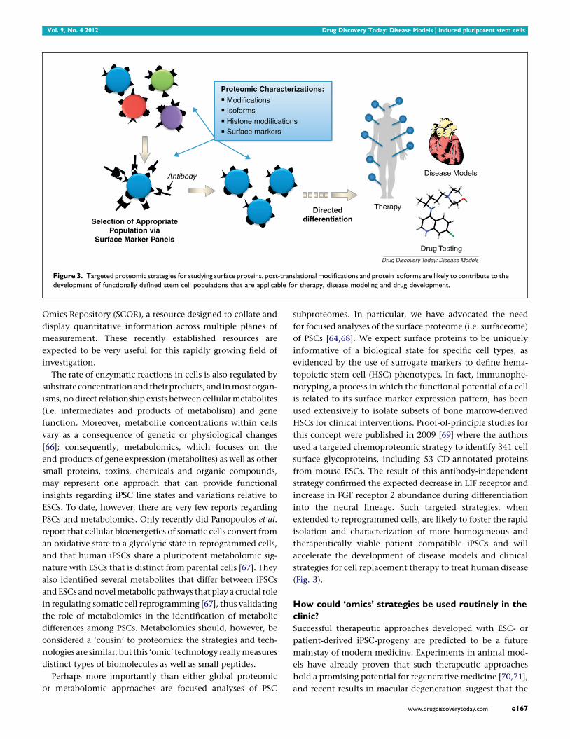
Figure 3. Targeted proteomic strategies for studying surface proteins, post-translational modifications and protein isoforms are likely to contribute to the
development of functionally defined stem cell populations that are applicable for therapy, disease modeling and drug development.
Omics Repository (SCOR), a resource designed to collate and
display quantitative information across multiple planes of
measurement. These recently established resources are
expected to be very useful for this rapidly growing field of
investigation.
The rate of enzymatic reactions in cells is also regulated by
substrate concentration and their products, and in most organ-
isms, no direct relationship exists between cellular metabolites
(i.e. intermediates and products of metabolism) and gene
function. Moreover, metabolite concentrations within cells
vary as a consequence of genetic or physiological changes
[66]; consequently, metabolomics, which focuses on the
end-products of gene expression (metabolites) as well as other
small proteins, toxins, chemicals and organic compounds,
may represent one approach that can provide functional
insights regarding iPSC line states and variations relative to
ESCs. To date, however, there are very few reports regarding
PSCs and metabolomics. Only recently did Panopoulos et al.
report that cellular bioenergetics of somatic cells convert from
an oxidative state to a glycolytic state in reprogrammed cells,
and that human iPSCs share a pluripotent metabolomic sig-
nature with ESCs that is distinct from parental cells [67]. They
also identified several metabolites that differ between iPSCs
and ESCs and novel metabolic pathways that play a crucial role
in regulating somatic cell reprogramming [67], thus validating
the role of metabolomics in the identification of metabolic
differences among PSCs. Metabolomics should, however, be
considered a ‘cousin’ to proteomics: the strategies and tech-
nologies are similar, but this ‘omic’ technology really measures
distinct types of biomolecules as well as small peptides.
Perhaps more importantly than either global proteomic
or metabolomic approaches are focused analyses of PSC
subproteomes. In particular, we have advocated the need
for focused analyses of the surface proteome (i.e. surfaceome)
of PSCs [64,68]. We expect surface proteins to be uniquely
informative of a biological state for specific cell types, as
evidenced by the use of surrogate markers to define hema-
topoietic stem cell (HSC) phenotypes. In fact, immunophe-
notyping, a process in which the functional potential of a cell
is related to its surface marker expression pattern, has been
used extensively to isolate subsets of bone marrow-derived
HSCs for clinical interventions. Proof-of-principle studies for
this concept were published in 2009 [69] where the authors
used a targeted chemoproteomic strategy to identify 341 cell
surface glycoproteins, including 53 CD-annotated proteins
from mouse ESCs. The result of this antibody-independent
strategy confirmed the expected decrease in LIF receptor and
increase in FGF receptor 2 abundance during differentiation
into the neural lineage. Such targeted strategies, when
extended to reprogrammed cells, are likely to foster the rapid
isolation and characterization of more homogeneous and
therapeutically viable patient compatible iPSCs and will
accelerate the development of disease models and clinical
strategies for cell replacement therapy to treat human disease
(Fig. 3).
How could ‘omics’ strategies be used routinely in the
clinic?
Successful therapeutic approaches developed with ESC- or
patient-derived iPSC-progeny are predicted to be a future
mainstay of modern medicine. Experiments in animal mod-
els have already proven that such therapeutic approaches
hold a promising potential for regenerative medicine [70,71],
and recent results in macular degeneration suggest that the
www.drugdiscoverytoday.com e167

Drug Discovery Today: Disease Models | Induced pluripotent stem cells Vol. 9, No. 4 2012
day is rapidly approaching for therapeutic applications in
human [72].
Before these cells and their derivatives can be routinely
employed clinically, careful molecular, immunological and
functional assays must be performed. Conventional assays
like G-banding are not sufficient because only large genetic
abnormalities can be detected. By contrast, a combination of
‘omics’ techniques represents a promising approach to assess
these cells – particularly in preclinical stages. However, to
date there are no definitive criteria on how to best define
pluripotent cells and their specific cell derivatives, as well as
assess the functional consequences of potential alterations in
PSC genomes, epigenomes, transcriptomes, proteomes and
metabolomes. Moreover, genetic defects are generally spora-
dic, thus complicating standard clinically accessible analyses.
Even if a certain cell line has been evaluated with a combina-
tion of ‘omics’ technologies and aberrations relative to a
potential gold standard cell line have been pinpointed, it is
not clear which changes or what degree of genetic changes are
still acceptable for clinical use. Therefore, as an important
step towards the clinical use of ESCs and iPSCs, well-defined
standards must be formulated to best identify cells suited for
transplantation in patients and to minimize patient’s risks in
Table 1. Selected bioinformatics websites relevant to ‘Omics’ r
URL K
General http://seqanswers.com/wiki/
Software/list
S
Pluripotency test http://pluritest.org B
Genomic/transcriptomic http://genecodes.com S
f
http://www.astridbio.com G
http://www.avadis-ngs.com A
a
http://www.biobase-international.com G
s
http://www.clcbio.com C
S
http://www.dnastar.com L
http://www.genomatix.de G
t
http://www.integromics.com S
http://www.omicsoft.com A
(
http://www.partek.com P
http://www.phenosystems.com G
http://www.realtimegenomics.com R
http://www.softgenetics.com N
http://www.spiralgenetics.com S
Proteomic http://scor.chem.wisc.edu/ S
http://www.ebi.ac.uk/pride/ G
https://proteomecommons.org/tranche/ G
http://www.peptideatlas.org/ P
http://gpmdb.thegpm.org/ G
NGS – Next Generation Sequencing.
e168 www.drugdiscoverytoday.com
terms of tumorigenicity and immunogenicity. Thus, gener-
ally accepted guidelines regarding generation, expansion,
manipulation, purification and evaluation of stem cells
and stem cell derivatives must be developed (reviewed in
[73]) before ‘omics’ technologies can be routinely applied
preclinically. But with that said, one ‘omics’ technology has
the potential of facilitating the daily use of PSCs and their
derivatives for therapeutics. This is based on the identifica-
tion of cell surface proteins as surrogate markers of a cell’s
phenotype and/or function analogous to that already
described for HSCs. The generation of non-genetic, immu-
nophenotyping methods (Fig. 3) for the isolation of defined
cell states should permit the efficient isolation of desired cell
types for clinical applications.
Conclusions
Differences between ESC and iPSC genomes, epigenomes,
transcriptomes, proteomes and metabolomes are well estab-
lished. While we have emphasized the differences, good
quality iPSCs are almost identical to ESCs, but currently,
there are no fully accepted criteria to make this determina-
tion in human cells. Omics approaches, especially when
coupled with bioinformatics tools (Table 1) and functional
esearch
ey features
ummary of useful software for data analysis
ioinformatic assay for pluripotency based on microarray data
equencher software for DNA sequence assembly and analysis tools
or DNA datasets
enoMiner – NGS data analysis
vadis NGS – desktop software platform for NGS (RNA-Seq, DNA-Seq
nd ChIP-Seq analysis)
enome Trax – identification of human genome variations of functional
ignificance
LC Genomics Workbench for analyzing and visualizing Next Generation
equencing data
asergene for next-gen sequence assembly and analysis.
enomatix Mining Station – mapping of NGS reads onto genomes,
ranscriptomes and splice junction libraries
eqSolve – for analysis of Next Generation Sequencing data
rray Studio – statistics and visualization for high dimensional data
NGS, microarray, SNP, CNV)
artek Genomics Suite – for analysis of microarray – and NGS data
ensearchNGS – software solution for Next Generation Sequencing
TG Investigator – software for NGS sequence analysis
extGENe – for analysis of Next Generation Sequencing data
piral Studio – for analysis of Next Generation Sequencing dataset
tem Cell Omics Repository
eneral proteomic data repository that contains stem cell data
eneral proteomic data repository that contains stem cell data
eptide data repository, especially useful for developing quantitative MS assays
eneral proteomic data repository that contains stem cell data

Vol. 9, No. 4 2012 Drug Discovery Today: Disease Models | Induced pluripotent stem cells
assays are likely to play a crucial role in establishing, char-
acterizing and eventually defining which populations of
iPSCs are appropriate for translational research.
Acknowledgements
The authors are supported by 4R00HL094708-03 (RLG), the
Innovation Center at the Medical College of Wisconsin
(RLG), the Intramural Research Program of the NIH, National
Institute on Aging (KRB) and NIH Induced Pluripotent Stem
Cell Center (NiPSCC) Pilot Study Award (KRB).
References1 Wobus, A.M. and Boheler, K.R. (2005) Embryonic stem cells: prospects for
developmental biology and cell therapy. Physiol. Rev. 85, 635–678
2 Takahashi, K. and Yamanaka, S. (2006) Induction of pluripotent stem cells
from mouse embryonic and adult fibroblast cultures by defined factors.
Cell 126, 663–676 (Epub 2006 Aug 2010)
3 Okita, K. et al. (2007) Generation of germline-competent induced
pluripotent stem cells. Nature 448, 313–317 (Epub 2007 Jun 2006)
4 Boheler, K.R. (2010) Pluripotency of human embryonic and induced
pluripotent stem cells for cardiac and vascular regeneration. Thromb.
Haemost. 104, 23–29
5 Lin, S.L. et al. (2011) Regulation of somatic cell reprogramming through
inducible mir-302 expression. Nucleic Acids Res. 39, 1054–1065
6 Anokye-Danso, F. et al. (2011) Highly efficient miRNA-mediated
reprogramming of mouse and human somatic cells to pluripotency. Cell
Stem Cell 8, 376–388
7 Zhao, T.B. et al. (2011) Immunogenicity of induced pluripotent stem cells.
Nature 474, 212–251
8 Robert, C. (2010) Microarray analysis of gene expression during early
development: a cautionary overview. Reproduction 140, 787–801
9 Perkins, D. et al. (2011) Advances of genomic science and systems biology
in renal transplantation: a review. Semin. Immunopathol. 33, 211–218
10 Wilmut, I. et al. (1997) Viable offspring derived from fetal and adult
mammalian cells. Nature 385, 810–813 (see comments; published erratum
appears in Nature 1997 Mar 13; 386(6621):200)
11 Mayshar, Y. et al. (2010) Identification and classification of chromosomal
aberrations in human induced pluripotent stem cells. Cell Stem Cell 7,
521–531
12 Gore, A. et al. (2011) Somatic coding mutations in human induced
pluripotent stem cells. Nature 471, 63–76
13 Hussein, S.M. et al. (2011) Copy number variation and selection during
reprogramming to pluripotency. Nature 471, 58–67
14 Laurent, L.C. et al. (2011) Dynamic changes in the copy number of
pluripotency and cell proliferation genes in human ESCs and iPSCs during
reprogramming and time in culture. Cell Stem Cell 8, 106–118
15 Howden, S.E. et al. (2011) Genetic correction and analysis of induced
pluripotent stem cells from a patient with gyrate atrophy. Proc. Natl. Acad.
Sci. U. S. A. 108, 6537–6542
16 Weber, M. et al. (2007) Distribution, silencing potential and evolutionary
impact of promoter DNA methylation in the human genome. Nat. Genet.
39, 457–466
17 Meissner, A. et al. (2008) Genome-scale DNA methylation maps of
pluripotent and differentiated cells. Nature 454, 766–791
18 Mikkelsen, T.S. et al. (2007) Genome-wide maps of chromatin state in
pluripotent and lineage-committed cells. Nature 448 553-U552
19 Polo, J.M. et al. (2010) Cell type of origin influences the molecular and
functional properties of mouse induced pluripotent stem cells. Nat.
Biotechnol. 28 848-U130
20 Ohi, Y. et al. (2011) Incomplete DNA methylation underlies a
transcriptional memory of somatic cells in human iPS cells. Nat. Cell Biol.
13 541-U328
21 Kim, K. et al. (2010) Epigenetic memory in induced pluripotent stem cells.
Nature 467 285-U260
22 Chin, M.H. et al. (2009) Induced pluripotent stem cells and embryonic
stem cells are distinguished by gene expression signatures. Cell Stem Cell 5,
111–123
23 Nishino, K. et al. (2011) DNA methylation dynamics in human induced
pluripotent stem cells over time. PLoS Genet. 7, e1002085
24 Deng, J. et al. (2009) Targeted bisulfite sequencing reveals changes in DNA
methylation associated with nuclear reprogramming. Nat. Biotechnol. 27,
353–360
25 Bock, C. et al. (2010) Quantitative comparison of genome-wide DNA
methylation mapping technologies. Nat. Biotechnol. 28, 1106–1196
26 Butcher, L.M. and Beck, S. (2010) AutoMeDIP-seq: a high-throughput,
whole genome, DNA methylation assay. Methods 52, 223–231
27 Li, N. et al. (2010) Whole genome DNA methylation analysis based on high
throughput sequencing technology. Methods 52, 203–212
28 Huangfu, D.W. et al. (2008) Induction of pluripotent stem cells from
primary human fibroblasts with only Oct4 and Sox2. Nat. Biotechnol. 26,
1269–1275
29 Huangfu, D.W. et al. (2008) Induction of pluripotent stem cells by defined
factors is greatly improved by small-molecule compounds. Nat. Biotechnol.
26, 795–797
30 Zhao, X.D. et al. (2007) Whole-genome mapping of histone H3 Lys4 and
27 trimethylations reveals distinct genomic compartments in human
embryonic stem cells. Cell Stem Cell 1, 286–298
31 Pan, G.J. et al. (2007) Whole-genome analysis of histone H3 lysine 4 and
lysine 27 methylation in human embryonic stem cells. Cell Stem Cell 1,
299–312
32 Ringrose, L. and Paro, R. (2004) Epigenetic regulation of cellular memory
by the polycomb and trithorax group proteins. Annu. Rev. Genet. 38, 413–
443
33 Ringrose, L. et al. (2004) Distinct contributions of histone H3 lysine 9 and
27 methylation to locus-specific stability of polycomb complexes. Mol.
Cell 16, 641–653
34 Bernstein, B.E. et al. (2006) A bivalent chromatin structure marks key
developmental genes in embryonic stem cells. Cell 125, 315–326
35 Marion, R.M. et al. (2009) Telomeres acquire embryonic stem cell
characteristics in induced pluripotent stem cells. Cell Stem Cell 4, 141–154
36 Mali, P. et al. (2010) Butyrate greatly enhances derivation of human
induced pluripotent stem cells by promoting epigenetic remodeling and
the expression of pluripotency-associated genes. Stem Cells 28, 713–720
37 Jiang, H. et al. (2011) Role for Dpy-30 in ES cell-fate specification by
regulation of H3K4 methylation within bivalent domains. Cell 144, 513–
525
38 Wang, Y. et al. (2010) A transcriptional roadmap to the induction of
pluripotency in somatic cells. Stem Cell Rev. Rep. 6, 282–296
39 Lowry, W.E. et al. (2008) Generation of human induced pluripotent stem
cells from dermal fibroblasts. Proc. Natl. Acad. Sci. U. S. A. 105, 2883–2888
40 Maherali, N. et al. (2008) A high-efficiency system for the generation and
study of human induced pluripotent stem cells. Cell Stem Cell 3, 340–345
41 Soldner, F. et al. (2009) Parkinson’s disease patient-derived induced
pluripotent stem cells free of viral reprogramming factors. Cell 136, 964–
977
42 Yu, J.Y. et al. (2009) Human induced pluripotent stem cells free of vector
and transgene sequences. Science 324, 797–801
43 Bock, C. et al. (2011) Reference maps of human ES and iPS cell variation
enable high-throughput characterization of pluripotent cell lines. Cell
144, 439–452
44 Muller, F.J. et al. (2011) A bioinformatic assay for pluripotency in human
cells. Nat. Methods 8, 315–354
45 Lemischka, I.R. and Pritsker, M. (2006) Alternative splicing increases
complexity of stem cell transcriptome. Cell Cycle 5, 347–351
46 Pritsker, M. et al. (2005) Diversification of stem cell molecular repertoire by
alternative splicing. Proc. Natl. Acad. Sci. U. S. A. 102, 14290–14295
47 Salomonis, N. et al. (2010) Alternative splicing regulates mouse embryonic
stem cell pluripotency and differentiation. Proc. Natl. Acad. Sci. U. S. A. 107,
10514–10519
48 Yeo, G.W. et al. (2007) Alternative splicing events identified in human
embryonic stem cells and neural progenitors. PLoS Comput. Biol. 3,
1951–1967
www.drugdiscoverytoday.com e169

Drug Discovery Today: Disease Models | Induced pluripotent stem cells Vol. 9, No. 4 2012
49 Wang, E.T. et al. (2008) Alternative isoform regulation in human tissue
transcriptomes. Nature 456, 470–476
50 Stamm, S. et al. (2005) Function of alternative splicing. Gene 344, 1–20
51 Lewis, B.P. et al. (2003) Evidence for the widespread coupling of alternative
splicing and nonsense-mediated mRNA decay in humans. Proc. Natl. Acad.
Sci. U. S. A. 100, 189–192
52 Atlasi, Y. et al. (2008) OCT4 spliced variants are differentially expressed in
human pluripotent and nonpluripotent cells. Stem Cells 26, 3068–3074
53 Cauffman, G. et al. (2006) POU5F1 isoforms show different expression
patterns in human embryonic stem cells and preimplantation embryos.
Stem Cells 24, 2685–2691
54 Lee, J. et al. (2006) The human OCT-4 isoforms differ in their ability to
confer self-renewal. J. Biol. Chem. 281, 33554–33565
55 Rao, S. et al. (2010) Differential roles of Sall4 isoforms in embryonic stem
cell pluripotency. Mol. Cell. Biol. 30, 5364–5380
56 Gabut, M. et al. (2011) An alternative splicing switch regulates embryonic
stem cell pluripotency and reprogramming. Cell 147, 132–146
57 Yu, J. et al. (2007) Induced pluripotent stem cell lines derived from human
somatic cells. Science 318, 1917–1920 (Epub 2007 Nov 1920)
58 Viswanathan, S.R. et al. (2008) Selective blockade of MicroRNA processing
by Lin28. Science 320, 97–100
59 Bushati, N. and Cohen, S.M. (2007) microRNA functions. Annu. Rev. Cell
Dev. Biol. 21, 21
60 Judson, R.L. et al. (2009) Embryonic stem cell-specific microRNAs promote
induced pluripotency. Nat. Biotechnol. 27, 459–461
61 Kozomara, A. and Griffiths-Jones, S. (2011) miRBase: integrating microRNA
annotation and deep-sequencing data. Nucleic Acids Res. 39, D152–D157
62 Houbaviy, H.B. et al. (2003) Embryonic stem cell-specific MicroRNAs. Dev.
Cell 5, 351–358
e170 www.drugdiscoverytoday.com
63 Suh, M.R. et al. (2004) Human embryonic stem cells express a unique set of
microRNAs. Dev. Biol. 270, 488–498
64 Gundry, R.L. et al. (2011) Pluripotent stem cell heterogeneity and the
evolving role of proteomic technologies in stem cell biology. Proteomics
11, 3947–3961
65 Phanstiel, D.H. et al. (2011) Proteomic and phosphoproteomic
comparison of human ES and iPS cells. Nat. Methods 8, 821–884
66 Raamsdonk, L.M. et al. (2001) A functional genomics startegy that uses
metabolome data to reveal the phenoytpe of silent mutations. Nat.
Biotechnol. 19, 45–50
67 Panopoulos, A.D. et al. (2012) The metabolome of induced pluripotent
stem cells reveals metabolic changes occurring in somatic cell
reprogramming. Cell Res. 2012, 168–177
68 Gundry, R.L. et al. (2008) A novel role for proteomics in the discovery of
cell-surface markers on stem cells: scratching the surface. Proteomics Clin.
Appl. 2, 892–903
69 Wollscheid, B. et al. (2009) Mass-spectrometric identification and relative
quantification of N-linked cell surface glycoproteins. Nat. Biotechnol. 27,
378–386
70 Hanna, J. et al. (2007) Treatment of sickle cell anemia mouse model with
iPS cells generated from autologous skin. Science 318, 1920–1923
71 Wernig, M. et al. (2008) Neurons derived from reprogrammed fibroblasts
functionally integrate into the fetal brain and improve symptoms of rats
with Parkinson’s disease. Proc. Natl. Acad. Sci. U. S. A. 105, 5856–5861
72 Schwartz, S.D. et al. (2012) Embryonic stem cell trials for macular
degeneration: a preliminary report. Lancet 2012 Jan 24 (Epub ahead of
print)
73 Goldring, C.E. et al. (2011) Assessing the safety of stem cell therapeutics.
Cell Stem Cell 8, 618–628