Human cathelicidin LL-37 enhance the antibiofilm effect of EGCG on ...
8
RESEARCH ARTICLE Open Access Human cathelicidin LL-37 enhance the antibiofilm effect of EGCG on Streptococcus mutans Yi-jie Guo 1,3*† , Bo Zhang 1,2† , Xue-song Feng 1,3 , Hui-xun Ren 1,3 and Ji-ru Xu 1,3* Abstract Background: Streptococcus mutans forms biofilms as a resistance mechanism against antimicrobial agents in the human oral cavity. We recently showed that human cathelicidin LL-37 exhibits inhibitory effects on biofilm formation of S. mutans through interaction with lipoteichoic acid (LTA), but without antibacterial or biofilm dispersal abilities. (-)-Epigallocatechin gallate (EGCG) is the most abundant constituent of tea catechins that has the greatest anti-infective potential to inhibit the growth of various microorganisms and biofilm formation. Therefore, in this study, we evaluated whether LL-37 interacts with EGCG to enhance the antibiofilm effect of EGCG on S. mutans biofilm formation. Methods: Clinical S. mutans strains (n = 10) isolated from children’s saliva were tested in a biofilm formation assay. The antibiofilm effect of EGCG with and without LL-37 was analyzed by the minimum biofilm eradication concentration assay and confirmed using field emission-scanning electron microscopy. In addition, the interaction among EGCG, LL-37, and LTA of S. mutans was determined using quartz crystal microbalance analysis. Results: EGCG killed 100 % of planktonic S. mutans within 5 h, inhibited biofilm formation within 24 h, and reduced bacteria cells in preformed biofilms within 3 h at a concentration of 0.2 mg/mL. However, EGCG did not appear to interact with LTA. LL-37 effectively enhanced the bactericidal activity of EGCG against biofilm formation and preformed biofilms as determined by quantitative crystal violet staining and field emission-scanning electron microscopy. In addition, quartz crystal microbalance analysis revealed that LL-37 interacted with EGCG and promoted binding between EGCG and LTA of S. mutans. Conclusions: We show that LL-37 enhances the antibiofilm effect of EGCG on S. mutans. This finding provides new knowledge for dental treatment by using LL-37 as a potential antibiofilm compound. Keywords: LL-37, EGCG, Streptococcus mutans, Biofilm Background Pathogenic bacteria can form biofilms that cause much infectious disease in the human oral cavity. Among these diseases, dental caries is one of the most ubiquitous biofilm-dependent oral diseases worldwide, which frequently occurs in both children and adults [1]. Oral streptococci, including Streptococcus mutans and Streptococcus sobrinus, are generally considered the primary etiologic agents of dental caries [2, 3]. One of the most well-documented virulence characteristics of S. mutans is its ability to establish cariogenic biofilms on tooth surfaces as an assembled extracellular matrix [4, 5]. Extracellular polymeric substances, especially water- insoluble glucans, are the main constituents of the matrix and can mediate S. mutans adherence [6, 7]. Other con- stituents such as extracellular DNA [8] and lipoteichoic acids (LTA) have also been found in high concentrations in the matrix of cariogenic biofilms [9, 10]. Tea is the most frequently consumed beverage in the world after water [11]. The antimicrobial activity of tea was first demonstrated almost 100 years ago by * Correspondence: [email protected]; [email protected] † Equal contributors 1 Department of Pathogenic Microbiology and Immunology, School of Basic Medical Sciences, Xi’an Jiaotong University, Yanta West Road No.76, Xi’an 710061, ShaanXi, China Full list of author information is available at the end of the article © 2016 The Author(s). Open Access This article is distributed under the terms of the Creative Commons Attribution 4.0 International License (http://creativecommons.org/licenses/by/4.0/), which permits unrestricted use, distribution, and reproduction in any medium, provided you give appropriate credit to the original author(s) and the source, provide a link to the Creative Commons license, and indicate if changes were made. The Creative Commons Public Domain Dedication waiver (http://creativecommons.org/publicdomain/zero/1.0/) applies to the data made available in this article, unless otherwise stated. Guo et al. BMC Oral Health (2016) 16:101 DOI 10.1186/s12903-016-0292-y RETRACTED ARTICLE
Transcript of Human cathelicidin LL-37 enhance the antibiofilm effect of EGCG on ...
RESEARCH ARTICLE Open Access
Human cathelicidin LL-37 enhance theantibiofilm effect of EGCG on StreptococcusmutansYi-jie Guo1,3*†, Bo Zhang1,2†, Xue-song Feng1,3, Hui-xun Ren1,3 and Ji-ru Xu1,3*
Abstract
Background: Streptococcus mutans forms biofilms as a resistance mechanism against antimicrobial agents in thehuman oral cavity. We recently showed that human cathelicidin LL-37 exhibits inhibitory effects on biofilm formationof S. mutans through interaction with lipoteichoic acid (LTA), but without antibacterial or biofilm dispersal abilities.(−)-Epigallocatechin gallate (EGCG) is the most abundant constituent of tea catechins that has the greatestanti-infective potential to inhibit the growth of various microorganisms and biofilm formation. Therefore, in thisstudy, we evaluated whether LL-37 interacts with EGCG to enhance the antibiofilm effect of EGCG on S. mutansbiofilm formation.
Methods: Clinical S. mutans strains (n = 10) isolated from children’s saliva were tested in a biofilm formationassay. The antibiofilm effect of EGCG with and without LL-37 was analyzed by the minimum biofilm eradicationconcentration assay and confirmed using field emission-scanning electron microscopy. In addition, the interactionamong EGCG, LL-37, and LTA of S. mutans was determined using quartz crystal microbalance analysis.
Results: EGCG killed 100 % of planktonic S. mutans within 5 h, inhibited biofilm formation within 24 h, and reducedbacteria cells in preformed biofilms within 3 h at a concentration of 0.2 mg/mL. However, EGCG did not appearto interact with LTA. LL-37 effectively enhanced the bactericidal activity of EGCG against biofilm formation andpreformed biofilms as determined by quantitative crystal violet staining and field emission-scanning electronmicroscopy. In addition, quartz crystal microbalance analysis revealed that LL-37 interacted with EGCG andpromoted binding between EGCG and LTA of S. mutans.
Conclusions: We show that LL-37 enhances the antibiofilm effect of EGCG on S. mutans. This finding providesnew knowledge for dental treatment by using LL-37 as a potential antibiofilm compound.
Keywords: LL-37, EGCG, Streptococcus mutans, Biofilm
BackgroundPathogenic bacteria can form biofilms that cause muchinfectious disease in the human oral cavity. Among thesediseases, dental caries is one of the most ubiquitousbiofilm-dependent oral diseases worldwide, whichfrequently occurs in both children and adults [1]. Oralstreptococci, including Streptococcus mutans andStreptococcus sobrinus, are generally considered the
primary etiologic agents of dental caries [2, 3]. One of themost well-documented virulence characteristics of S.mutans is its ability to establish cariogenic biofilms ontooth surfaces as an assembled extracellular matrix [4, 5].Extracellular polymeric substances, especially water-insoluble glucans, are the main constituents of the matrixand can mediate S. mutans adherence [6, 7]. Other con-stituents such as extracellular DNA [8] and lipoteichoicacids (LTA) have also been found in high concentrationsin the matrix of cariogenic biofilms [9, 10].Tea is the most frequently consumed beverage in the
world after water [11]. The antimicrobial activity of teawas first demonstrated almost 100 years ago by
* Correspondence: [email protected]; [email protected]†Equal contributors1Department of Pathogenic Microbiology and Immunology, School of BasicMedical Sciences, Xi’an Jiaotong University, Yanta West Road No.76, Xi’an710061, ShaanXi, ChinaFull list of author information is available at the end of the article
© 2016 The Author(s). Open Access This article is distributed under the terms of the Creative Commons Attribution 4.0International License (http://creativecommons.org/licenses/by/4.0/), which permits unrestricted use, distribution, andreproduction in any medium, provided you give appropriate credit to the original author(s) and the source, provide a link tothe Creative Commons license, and indicate if changes were made. The Creative Commons Public Domain Dedication waiver(http://creativecommons.org/publicdomain/zero/1.0/) applies to the data made available in this article, unless otherwise stated.
Guo et al. BMC Oral Health (2016) 16:101 DOI 10.1186/s12903-016-0292-y
RETRACTED ARTIC
LE
McNaught [12]. Green tea (Camellia sinensis) is a non-fermented tea that has more beneficial effects than blacktea or oolong tea [13]. It has been observed that catechincomponents of green tea are responsible for its antibac-terial activity, and (−)-epicatechin gallate, (−)-epigallo-catechin (EGC), and (−)-epigallocatechin gallate (EGCG)constitute the most important antibacterial agents of teacatechins [14, 15].EGCG is the most abundant constituent of tea catechins
that has the greatest anti-infective ability by inhibiting thegrowth of various microorganisms [16], biofilm formation[17], and quorum sensing [18, 19]. Xu et al. reported thatEGCG inhibits the growth of S. mutans, decreases gluco-syltransferases activity, and suppresses gtf genes, whichare associated with bacterial biofilm formation [20, 21].However, the effects of EGCG on biofilm and cariogenicvirulence factors of oral streptococci other than glucosyl-transferases have not been well documented, especiallyregarding its potential interaction with LTA.The antibacterial peptide LL-37 is the only member of
the cathelicidin family found in humans. LL-37 is an 18-kDa peptide [22] with 37 amino acid residues that startwith two leucines [23]. LL-37 is expressed in many tis-sues and body fluids, such as saliva, gingiva, sweat, am-niotic fluids, and seminal plasma [24]. It has beenreported that the intravital concentration of LL-37 isabout 86 μg/mL in seminal plasma of healthy donors[25], and ranges from less than 1.2 to more than 80 μg/mL in unstimulated and nonpurulent nasal secretions[26], respectively. Cariogenic species, such as S. sobrinus,Lactobacillus paracasei, and Actinomyces viscosus havebeen reported to be resistant to LL-37 through growthinhibition and bactericidal tests [27], especially whenbiofilm formation occurred. We recently showed thatLL-37 has the ability to bind to LTA of S. mutans, inhi-biting biofilm formation [28]. Therefore, in this study,we evaluated whether LL-37 could be used as a bindingagent to enhance the interaction between EGCG andLTA of S. mutans to increase the antibiofilm effect ofEGCG.
MethodsBacterial strains and culture conditionsTen clinical S. mutans strains isolated from children’ssaliva were kindly provided by Dr. E. Isogai [29]. Bac-teria were grown in brain heart infusion (BHI) broth(Merck KGaA, Darmstadt, Germany) for 24 h at 37 °C.The present study was approved by the Ethics Commit-tee of Xi’an Jiaotong University Faculty of Medicine(XJTU2014-014).
Catechin and antimicrobial peptideEGCG (NH020403) was purchased from Nagara ScienceCo. Ltd. (Gifu, Japan). EGCG was dissolved in absolute
ethyl alcohol at a concentration of 10 mg/mL. Then,EGCG/ethanol was used a concentration of 2 % to yielda maximum concentration of 0.2 mg/mL. LL-37 (se-quence: LLGDF FRKSK EKIGK EFKRI VQRIK DFLRNLVPRT ES) powder was synthesized as described previ-ously using the solid-phase method [30]. Briefly, thepeptide was purified (>99.9 %) by reverse-phase high-performance liquid chromatography (Model LC-8A;Shimadzu Corporation, Kyoto, Japan) on a YMC-A 302column (YMC Co. Ltd., Kyoto, Japan). The final productwas confirmed by electrospray ionization mass spec-trometry and conserved by suspension in Hank’sbalanced salt solution (HBSS, pH 7.4; Gibco, GrandIsland, NY, USA).
Growth inhibition testPrecultured bacteria were measured at an optical density(OD) of 660 nm using an ultraviolet/visible spectropho-tometer (Ubest-35; JASCO Corporation, Tokyo, Japan).Samples were adjusted to OD660 = 0.5 using BHI broth.Then, bacteria were diluted to a final concentration of103–104 Colony-forming unit (CFU)/mL with BHI broth,and 1 mL of adjusted culture and 1 mL of EGCG solu-tion were mixed together [28, 31]. The EGCG solutionwas prepared by two-fold serial dilution in BHI broth.Each mixture solution was incubated at 37 °C. The bac-tericidal activity of EGCG was estimated by measuringthe minimum inhibitory concentration (MIC) and mini-mum bactericidal concentration (MBC) at 3, 5, and 24 hof incubation according to Clinical and Laboratory Stan-dards Institute 2014 (CLSI) guidelines. After each incu-bation, 100 μL of EGCG–cell suspension was removedand inoculated on BHI agar plates. The plates were incu-bated for 48 h at 37 °C, followed by colony counting(CFU/mL). Control samples were prepared by mixing1 mL of bacterial suspension with 0.9 mL of BHI brothand 0.1 mL of HBSS. The MIC was defined as the lowestconcentration of EGCG that inhibited visible bacterialgrowth after overnight incubation. The MBC was de-fined as the lowest concentration of EGCG that killed99.9 % of the initial inoculum.
Biofilm formation assaysBiofilm formation was measured under two static con-ditions using a quantitative crystal violet assay on apolystyrene 96-well, minimum biofilm eradication con-centration plate (MBEC; Biosurface Technologies,Bozeman, MT, USA) as described previously [32]. TheMBEC biofilm assay used 96 polystyrene pegs that fitthe wells of a conventional plate. Briefly, overnight cul-tures of S. mutans were diluted to OD660 = 0.5 (1 ×10⁷ CFU/mL) in fresh BHI. Two hundred microliters ofbacterial suspensions were transferred to a 96-well mi-crotiter plate with 20 μL of varying concentrations of
Guo et al. BMC Oral Health (2016) 16:101 Page 2 of 8
RETRACTED ARTIC
LE
EGCG in the presence or absence of LL-37 (final con-centration of 80 μL/mL). The microtiter plates werethen incubated at 37 °C with shaking for 12, 24, and36 h. After incubation, the pegs were washed severaltimes with phosphate buffered solution (PBS) and thenfixed with 200 μL of 100 % ethanol prior to staining for2 min with 200 μL of 0.41 % (wt/vol) crystal violet in12 % ethanol (Bio Chemical Sciences, Swedesboro, NJ,USA). The pegs were washed several times with PBS toremove excess stain. Quantitative assessment of biofilmformation was performed by the immersion of pegs in asterile microtiter plate containing 200 μL of 100 %ethanol and incubation at room temperature for10 min. The absorbance at 550 nm was then deter-mined. Three independent experiments were performedfor each abovementioned assay.
Biofilm susceptibility assaysTo measure the antimicrobial susceptibility of S. mutansgrowing in biofilms, the MBEC High-throughput (HTP)Assay (Innovotech Inc., Edmonton, Alberta, Canada)was performed [33]. Briefly, overnight cultures of S.mutans were diluted to OD660 = 0.5 (1 × 10⁷ CFU/mL) infresh BHI. Then, 200 μL of bacterial suspensions wastransferred to the wells of a MBEC microtiter plate andthe MBEC lid was placed on top of the wells. Biofilmswere grown on the MBEC pegs at 37 °C under shakingconditions for 24 h. The lid was removed and trans-ferred to a new plate with wells filled with varyingconcentrations of EGCG in the presence or absence ofLL-37 (final concentration of 80 μL/mL), and incubatedfor 3 and 5 h at 37 °C. After each incubation, the lid wasgently washed twice with 200 μL of PBS to remove non-adherent cells. Adherent biofilms were fixed with 200 μLof 100 % ethanol prior to staining for 2 min with 200 μLof 0.41 % (wt/vol) crystal violet in 12 % ethanol. Thepegs were washed several times with PBS to remove ex-cess stain. Quantitative assessment of biofilm formationwas performed by the immersion of pegs in a sterilepolystyrene microtiter plate which contained 200 μL of100 % ethanol. The plate was incubated at roomtemperature for 10 min, then the absorbance at 550 nmwas determined using a microplate spectrophotometer.Three independent experiments were performed foreach assay.
Field emission-scanning electron microscopy (FE-SEM)To visualize the effects of EGCG on planktonic bacteria[28, 31], 10 μL of EGCG solution (0.2 mg/mL) wasplated on a glass slide followed by 10 μL of bacteriamixed with the solution. This cell suspension was thenincubated at 37 °C for 24 h and dried. Saturated 70 %ethanol was then applied for 5 min, followed by satu-rated 100 % ethanol for 5 min, and then the sample was
dried. The sample was then coated with palladium alloyand examined using FE-SEM (SU8000; Hitachi High-Technologies Corporation, Tokyo, Japan).To visualize the effect of EGCG on biofilms, the
MBEC HTP Assay (Innovotech Inc.) was used [28]. Pegswith adherent biofilms were removed from the MBECbiofilm assay and rinsed in 0.9 % saline for 1 min to re-move non-adherent bacteria. The samples were thenfixed with 2.5 % glutaraldehyde (Kanto Chemical Co., Inc.,Tokyo, Japan) in 0.1 M cacodylic acid (Wako Pure Chem-ical Industries, Ltd., Miyagi, Japan) at 4 °C for 16 h. Thepegs were washed in 0.1 M cacodylic acid and thenwashed once in distilled water for approximately 10 min.Saturated 70 % ethanol was applied for 15–20 min andthen the sample was air dried for a minimum of 24 h.Specimens were finally mounted and examined usingFE-SEM.
EGCG, LL-37 and LTA interaction analysis using quartzcrystal microbalance (QCM)QCM was used to analyze the potential enhancement ef-fect of LL-37 on binding between EGCG and bacterialLTA. EGCG was used as a ligand and LTA from S.mutans (L4152; Sigma-Aldrich, St. Louis, MO, USA)was used as an analyst. QCM measurements were per-formed using AFFINIX QN μ (INITIUM Inc., Kanagawa,Japan). This instrument has a 500-μL cell equipped witha 27-MHz QCM plate at the bottom of the cell, inaddition to a stirring bar and temperature control sys-tem. Gold-coated quartz crystal sensor surfaces werewashed twice by mounting 3:1 mixture of concentratedsulfuric acid and hydrogen peroxide solution for 5 min.An AFFINIX immobilization kit was then used follow-ing the manufacturer’s instruction. Briefly, the surfacesof the sensors were coated with carboxylic acid-terminated thiol and carboxylic acids were activated asN-hydroxysuccinimidyl esters as previously described[34]. Activated carboxyl groups reacted with EGCG(0.2 mg/mL). After the binding of EGCG to the sensor,unreacted esters were deactivated using 1 mol ethanol-amine solution. The amount of LTA solution (10 μg/mL)and/or LL-37 (80 μg/mL) injected each time was 5 μLeach. HBSS was used as a measurement buffer. The stir-ring speed was set at 1000 rpm, and experiments wereconducted at 25 °C. Frequency changes in response to theaddition of LTA solution were monitored in real time. Asa control, 10 μg/mL of LTA was injected without initiallybinding EGCG on the sensor.
Statistical analysisData are presented as the means and standard devia-tions. Significant differences were determined by thet-test using Microsoft Excel 2007 (Microsoft Corporation).
Guo et al. BMC Oral Health (2016) 16:101 Page 3 of 8
RETRACTED ARTIC
LE
Values were considered to be statistically significantat p < 0.05.
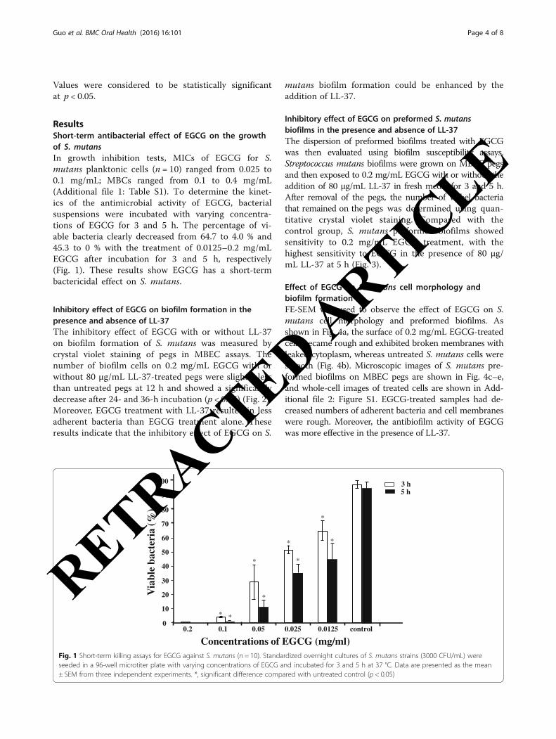
ResultsShort-term antibacterial effect of EGCG on the growthof S. mutansIn growth inhibition tests, MICs of EGCG for S.mutans planktonic cells (n = 10) ranged from 0.025 to0.1 mg/mL; MBCs ranged from 0.1 to 0.4 mg/mL(Additional file 1: Table S1). To determine the kinet-ics of the antimicrobial activity of EGCG, bacterialsuspensions were incubated with varying concentra-tions of EGCG for 3 and 5 h. The percentage of vi-able bacteria clearly decreased from 64.7 to 4.0 % and45.3 to 0 % with the treatment of 0.0125–0.2 mg/mLEGCG after incubation for 3 and 5 h, respectively(Fig. 1). These results show EGCG has a short-termbactericidal effect on S. mutans.
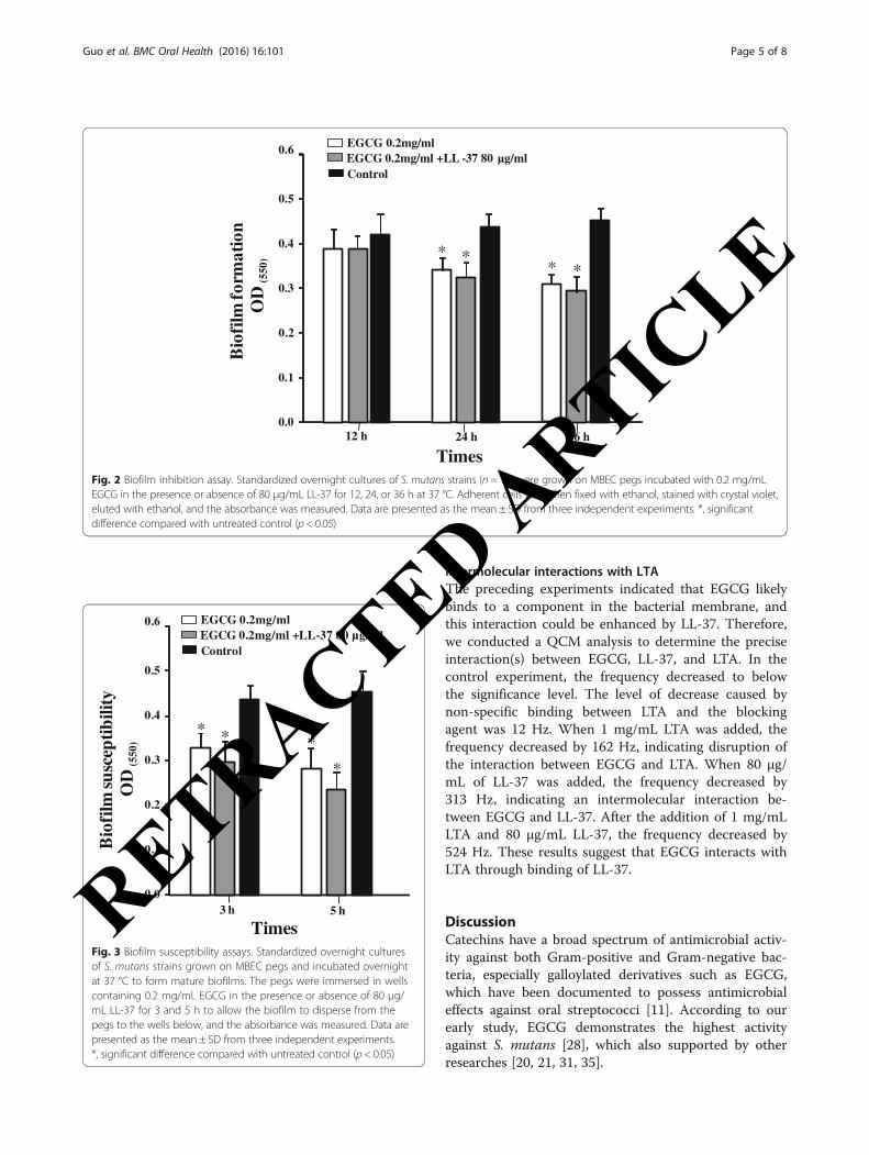
Inhibitory effect of EGCG on biofilm formation in thepresence and absence of LL-37The inhibitory effect of EGCG with or without LL-37on biofilm formation of S. mutans was measured bycrystal violet staining of pegs in MBEC assays. Thenumber of biofilm cells on 0.2 mg/mL EGCG with orwithout 80 μg/mL LL-37-treated pegs were slightly lessthan untreated pegs at 12 h and showed a significantlydecrease after 24- and 36-h incubation (p < 0.05) (Fig. 2).Moreover, EGCG treatment with LL-37 resulted in lessadherent bacteria than EGCG treatment alone. Theseresults indicate that the inhibitory effect of EGCG on S.
mutans biofilm formation could be enhanced by theaddition of LL-37.
Inhibitory effect of EGCG on preformed S. mutansbiofilms in the presence and absence of LL-37The dispersion of preformed biofilms treated with EGCGwas then evaluated using biofilm susceptibility assays.Streptococcus mutans biofilms were grown on MBEC pegsand then exposed to 0.2 mg/mL EGCG with or without theaddition of 80 μg/mL LL-37 in fresh media for 3 and 5 h.After removal of the pegs, the number of viabel bacteriathat remained on the pegs was determined using quan-titative crystal violet staining. Compared with thecontrol group, S. mutans preformed biofilms showedsensitivity to 0.2 mg/mL EGCG treatment, with thehighest sensitivity to EGCG in the presence of 80 μg/mL LL-37 at 5 h (Fig. 3).
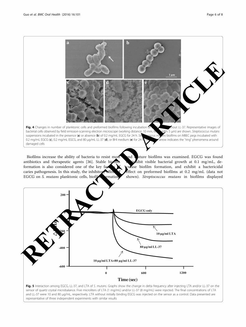
Effect of EGCG on S. mutans cell morphology andbiofilm formationFE-SEM was used to observe the effect of EGCG on S.mutans cell morphology and preformed biofilms. Asshown in Fig. 4a, the surface of 0.2 mg/mL EGCG-treatedcells became rough and exhibited broken membranes withleaked cytoplasm, whereas untreated S. mutans cells weresmooth (Fig. 4b). Microscopic images of S. mutans pre-formed biofilms on MBEC pegs are shown in Fig. 4c–e,and whole-cell images of treated cells are shown in Add-itional file 2: Figure S1. EGCG-treated samples had de-creased numbers of adherent bacteria and cell membraneswere rough. Moreover, the antibiofilm activity of EGCGwas more effective in the presence of LL-37.
0
10
20
30
40
50
60
70
80
90
100
5 h3 h
0.2 0.1 0.05 0.025 0.0125
)%(
airetcabelbai
V
Concentrations of EGCG (mg/ml)
*
control
*
*
* *
* *
*
Fig. 1 Short-term killing assays for EGCG against S. mutans (n = 10). Standardized overnight cultures of S. mutans strains (3000 CFU/mL) wereseeded in a 96-well microtiter plate with varying concentrations of EGCG and incubated for 3 and 5 h at 37 °C. Data are presented as the mean± SEM from three independent experiments. *, significant difference compared with untreated control (p < 0.05)
Guo et al. BMC Oral Health (2016) 16:101 Page 4 of 8
RETRACTED ARTIC
LE
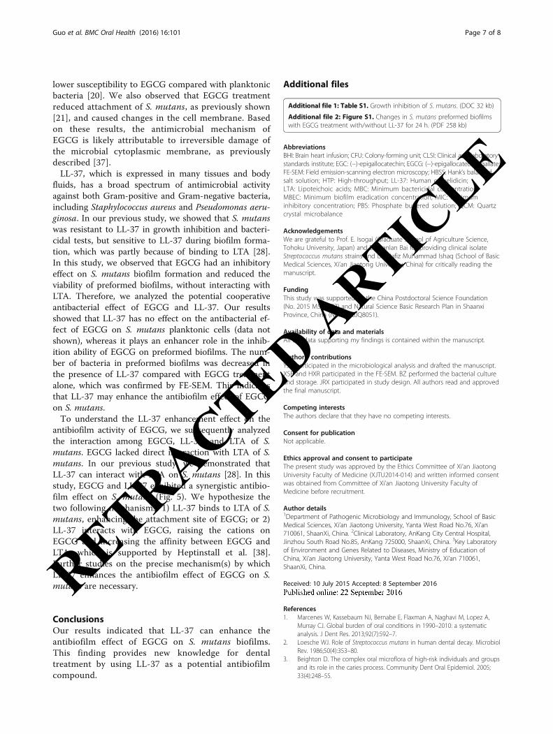
Intermolecular interactions with LTAThe preceding experiments indicated that EGCG likelybinds to a component in the bacterial membrane, andthis interaction could be enhanced by LL-37. Therefore,we conducted a QCM analysis to determine the preciseinteraction(s) between EGCG, LL-37, and LTA. In thecontrol experiment, the frequency decreased to belowthe significance level. The level of decrease caused bynon-specific binding between LTA and the blockingagent was 12 Hz. When 1 mg/mL LTA was added, thefrequency decreased by 162 Hz, indicating disruption ofthe interaction between EGCG and LTA. When 80 μg/mL of LL-37 was added, the frequency decreased by313 Hz, indicating an intermolecular interaction be-tween EGCG and LL-37. After the addition of 1 mg/mLLTA and 80 μg/mL LL-37, the frequency decreased by524 Hz. These results suggest that EGCG interacts withLTA through binding of LL-37.
DiscussionCatechins have a broad spectrum of antimicrobial activ-ity against both Gram-positive and Gram-negative bac-teria, especially galloylated derivatives such as EGCG,which have been documented to possess antimicrobialeffects against oral streptococci [11]. According to ourearly study, EGCG demonstrates the highest activityagainst S. mutans [28], which also supported by otherresearches [20, 21, 31, 35].
0.0
0.1
0.2
0.3
0.4
0.5
0.6
mlifoiB
ytilibit
pecsus OD
(550
)
3 h 5 h
Times
EGCG 0.2mg/ml +LL-37 80 µg/ml EGCG 0.2mg/ml
Control
*
*
* *
Fig. 3 Biofilm susceptibility assays. Standardized overnight culturesof S. mutans strains grown on MBEC pegs and incubated overnightat 37 °C to form mature biofilms. The pegs were immersed in wellscontaining 0.2 mg/mL EGCG in the presence or absence of 80 μg/mL LL-37 for 3 and 5 h to allow the biofilm to disperse from thepegs to the wells below, and the absorbance was measured. Data arepresented as the mean ± SD from three independent experiments.*, significant difference compared with untreated control (p < 0.05)
0.0
0.1
0.2
0.3
0.4
0.5
12 h 24 h 36 h
0.6
Times
mlifoiB
noitamrof
OD
(550
)
EGCG 0.2mg/ml +LL -37 80 µg/ml EGCG 0.2mg/ml
Control
* ** *
Fig. 2 Biofilm inhibition assay. Standardized overnight cultures of S. mutans strains (n = 10) were grown on MBEC pegs incubated with 0.2 mg/mLEGCG in the presence or absence of 80 μg/mL LL-37 for 12, 24, or 36 h at 37 °C. Adherent cells were then fixed with ethanol, stained with crystal violet,eluted with ethanol, and the absorbance was measured. Data are presented as the mean ± SD from three independent experiments. *, significantdifference compared with untreated control (p < 0.05)
Guo et al. BMC Oral Health (2016) 16:101 Page 5 of 8
RETRACTED ARTIC
LE
Biofilms increase the ability of bacteria to resist mostantibiotics and therapeutic agents [36]. Stable biofilmformation is also considered one of the key factors ofcaries pathogenesis. In this study, the inhibitory effect ofEGCG on S. mutans planktonic cells, biofilm formation,
and mature biofilms was examined. EGCG was foundto inhibit visible bacterial growth at 0.1 mg/mL, de-crease biofilm formation, and exhibit a bactericidaleffect on preformed biofilms at 0.2 mg/mL (data notshown). Streptococcus mutans in biofilms displayed
-400
-200
0
200
-600
0 600 1200
Time (sec)
10 µg/ml LTA+80 µg/ml LL-37
80 µg/ml LL-37
10 µg/ml LTA
EGCG only
zH/
F
Fig. 5 Interaction among EGCG, LL-37, and LTA of S. mutans. Graphs show the change in delta frequency after injecting LTA and/or LL-37 on thesensor of quartz crystal microbalance. Five microliters of LTA (1 mg/mL) and/or LL-37 (8 mg/mL) were injected. The final concentrations of LTAand LL-37 were 10 and 80 μg/mL, respectively. LTA without initially binding EGCG was injected on the sensor as a control. Data presented arerepresentative of three independent experiments with similar results
a b
1 µm
c d e
Fig. 4 Changes in number of planktonic cells and preformed biofilms following incubation of EGCG with/without LL-37. Representative images ofbacterial cells observed by field emission-scanning electron microscope (working distance: 10 mm, field width: 5 μm) are shown. Streptococcus mutanssuspensions incubated in the presence (a) or absence (b) of 0.2 mg/mL EGCG for 24 h. Streptococcus mutans biofilms on MBEC pegs incubated with0.2 mg/mL EGCG (c), 0.2 mg/mL EGCG, and 80 μg/mL LL-37 (d), or BHI medium (e) for 24 h. The white arrow indicates the “ring” phenomena arounddamaged cells
Guo et al. BMC Oral Health (2016) 16:101 Page 6 of 8
RETRACTED ARTIC
LE
lower susceptibility to EGCG compared with planktonicbacteria [20]. We also observed that EGCG treatmentreduced attachment of S. mutans, as previously shown[21], and caused changes in the cell membrane. Basedon these results, the antimicrobial mechanism ofEGCG is likely attributable to irreversible damage ofthe microbial cytoplasmic membrane, as previouslydescribed [37].LL-37, which is expressed in many tissues and body
fluids, has a broad spectrum of antimicrobial activityagainst both Gram-positive and Gram-negative bacteria,including Staphylococcus aureus and Pseudomonas aeru-ginosa. In our previous study, we showed that S. mutanswas resistant to LL-37 in growth inhibition and bacteri-cidal tests, but sensitive to LL-37 during biofilm forma-tion, which was partly because of binding to LTA [28].In this study, we observed that EGCG had an inhibitoryeffect on S. mutans biofilm formation and reduced theviability of preformed biofilms, without interacting withLTA. Therefore, we analyzed the potential cooperativeantibacterial effect of EGCG and LL-37. Our resultsshowed that LL-37 has no effect on the antibacterial ef-fect of EGCG on S. mutans planktonic cells (data notshown), whereas it plays an enhancer role in the inhib-ition ability of EGCG on preformed biofilms. The num-ber of bacteria in preformed biofilms was decreased inthe presence of LL-37 compared with EGCG treatmentalone, which was confirmed by FE-SEM. This indicatesthat LL-37 may enhance the antibiofilm effect of EGCGon S. mutans.To understand the LL-37 enhancement effect on the
antibiofilm activity of EGCG, we subsequently analyzedthe interaction among EGCG, LL-37, and LTA of S.mutans. EGCG lacked direct interaction with LTA of S.mutans. In our previous study, we demonstrated thatLL-37 can interact with LTA on S. mutans [28]. In thisstudy, EGCG and LL-37 exhibited a synergistic antibio-film effect on S. mutans (Fig. 5). We hypothesize thetwo following mechanisms: 1) LL-37 binds to LTA of S.mutans, enhancing the attachment site of EGCG; or 2)LL-37 interacts with EGCG, raising the cations onEGCG and increasing the affinity between EGCG andLTA, which is supported by Heptinstall et al. [38].Further studies on the precise mechanism(s) by whichLL-37 enhances the antibiofilm effect of EGCG on S.mutans are necessary.
ConclusionsOur results indicated that LL-37 can enhance theantibiofilm effect of EGCG on S. mutans biofilms.This finding provides new knowledge for dentaltreatment by using LL-37 as a potential antibiofilmcompound.
Additional files
Additional file 1: Table S1. Growth inhibition of S. mutans. (DOC 32 kb)
Additional file 2: Figure S1. Changes in S. mutans preformed biofilmswith EGCG treatment with/without LL-37 for 24 h. (PDF 258 kb)
AbbreviationsBHI: Brain heart infusion; CFU: Colony-forming unit; CLSI: Clinical and laboratorystandards institute; EGC: (−)-epigallocatechin; EGCG: (−)-epigallocatechin gallate;FE-SEM: Field emission-scanning electron microscopy; HBSS: Hank’s balancedsalt solution; HTP: High-throughput; LL-37: Human cathelidicin;LTA: Lipoteichoic acids; MBC: Minimum bactericidal concentration;MBEC: Minimum biofilm eradication concentration; MIC: Minimuminhibitory concentration; PBS: Phosphate buffered solution; QCM: Quartzcrystal microbalance
AcknowledgementsWe are grateful to Prof. E. Isogai (Graduate School of Agriculture Science,Tohoku University, Japan) and Dr. Lanlan Bai for providing clinical isolateStreptococcus mutans strains and Dr. Hafiz Muhammad Ishaq (School of BasicMedical Sciences, Xi’an Jiaotong University, China) for critically reading themanuscript.
FundingThis study was supported by the China Postdoctoral Science Foundation(No. 2015 M582672) and Natural Science Basic Research Plan in ShaanxiProvince, China (No. 2016JQ8051).
Availability of data and materialsAll the data supporting my findings is contained within the manuscript.
Authors’ contributionsYJG participated in the microbiological analysis and drafted the manuscript.XSF and HXR participated in the FE-SEM. BZ performed the bacterial cultureand storage. JRX participated in study design. All authors read and approvedthe final manuscript.
Competing interestsThe authors declare that they have no competing interests.
Consent for publicationNot applicable.
Ethics approval and consent to participateThe present study was approved by the Ethics Committee of Xi’an JiaotongUniversity Faculty of Medicine (XJTU2014-014) and written informed consentwas obtained from Committee of Xi’an Jiaotong University Faculty ofMedicine before recruitment.
Author details1Department of Pathogenic Microbiology and Immunology, School of BasicMedical Sciences, Xi’an Jiaotong University, Yanta West Road No.76, Xi’an710061, ShaanXi, China. 2Clinical Laboratory, AnKang City Central Hospital,Jinzhou South Road No.85, AnKang 725000, ShaanXi, China. 3Key Laboratoryof Environment and Genes Related to Diseases, Ministry of Education ofChina, Xi’an Jiaotong University, Yanta West Road No.76, Xi’an 710061,ShaanXi, China.
Received: 10 July 2015 Accepted: 8 September 2016
References1. Marcenes W, Kassebaum NJ, Bernabe E, Flaxman A, Naghavi M, Lopez A,
Murray CJ. Global burden of oral conditions in 1990–2010: a systematicanalysis. J Dent Res. 2013;92(7):592–7.
2. Loesche WJ. Role of Streptococcus mutans in human dental decay. MicrobiolRev. 1986;50(4):353–80.
3. Beighton D. The complex oral microflora of high-risk individuals and groupsand its role in the caries process. Community Dent Oral Epidemiol. 2005;33(4):248–55.
Guo et al. BMC Oral Health (2016) 16:101 Page 7 of 8
RETRACTED ARTIC
LE
4. Hamada S, Slade HD. Biology, immunology, and cariogenicity ofStreptococcus mutans. Microbiol Rev. 1980;44(2):331–84.
5. Bowen WH, Koo H. Biology of Streptococcus mutans-derivedglucosyltransferases: role in extracellular matrix formation of cariogenicbiofilms. Caries Res. 2011;45(1):69–86.
6. Mattos-Graner RO, Smith DJ, King WF, Mayer MP. Water-insoluble glucansynthesis by mutans streptococcal strains correlates with caries incidence in12- to 30-month-old children. J Dent Res. 2000;79(6):1371–7.
7. Vacca SAM, Scott-Anne KM, Whelehan MT, Berkowitz RJ, Feng C, BowenWH. Salivary glucosyltransferase B as a possible marker for caries activity.Caries Res. 2007;41(6):445–50.
8. Perry JA, Cvitkovitch DG, Levesque CM. Cell death in Streptococcus mutansbiofilms: a link between CSP and extracellular DNA. FEMS Microbiol Lett.2009;299(2):261–6.
9. Rolla G, Oppermann RV, Bowen WH, Ciardi JE, Knox KW. High amounts oflipoteichoic acid in sucrose-induced plaque in vivo. Caries Res. 1980;14(4):235–8.
10. Denapaite D, Bruckner R, Hakenbeck R, Vollmer W. Biosynthesis of teichoicacids in Streptococcus pneumoniae and closely related species: lessons fromgenomes. Microb Drug Resist. 2012;18(3):344–58.
11. Sannella AR, Messori L, Casini A, Francesco VF, Bilia AR, Majori G, Severini C.Antimalarial properties of green tea. Biochem Biophys Res Commun. 2007;353(1):177–81.
12. McNaught JG. On the action of cold or lukewarm tea on Bacillus typhosus.J R Army Med Corps. 1906;7:372–3.
13. Jazani NH, Sh S, Ali AA. Antibacterial effects of water soluble green teaextracts on multi-antibiotic resistant isolates of Pseudomonas aeruginosa.Pak J Biol Sci. 2007;10(9):1544–6.
14. Yam TS, Hamilton-Miller JM, Shah S. The effect of a component of tea(Camellia sinensis) on methicillin resistance, PBP2’ synthesis, and beta-lactamase production in Staphylococcus aureus. J Antimicrob Chemother.1998;42(2):211–6.
15. Bidlack WR. Green Tea: Health Benefits and Applications. J Am Coll Nutr.2001;20(6):656–8.
16. Kohda C, Yanagawa Y, Shimamura T. Epigallocatechin gallate inhibitsintracellular survival of Listeria monocytogenes in macrophages. BiochemBiophys Res Commun. 2008;365(2):310–5.
17. Blanco AR, Sudano-Roccaro A, Spoto GC, Nostro A, Rusciano D.Epigallocatechin gallate inhibits biofilm formation by ocular staphylococcalisolates. Antimicrob Agents Chemother. 2005;49(10):4339–43.
18. Huber B, Eberl L, Feucht W, Polster J. Influence of polyphenols on bacterialbiofilm formation and quorum-sensing. Z Naturforsch C. 2003;58(11–12):879–84.
19. Lee KM, Kim WS, Lim J, Nam S, Youn M, Nam SW, Kim Y, Kim SH, Park W,Park S. Antipathogenic properties of green tea polyphenol epigallocatechingallate at concentrations below the MIC against enterohemorrhagicEscherichia coli O157:H7. J Food Prot. 2009;72(2):325–31.
20. Xu X, Zhou XD, Wu CD. The tea catechin epigallocatechin gallatesuppresses cariogenic virulence factors of Streptococcus mutans. AntimicrobAgents Chemother. 2011;55(3):1229–36.
21. Xu X, Zhou XD, Wu CD. Tea catechin epigallocatechin gallate inhibitsStreptococcus mutans biofilm formation by suppressing gtf genes. Arch OralBiol. 2012;57(6):678–83.
22. Agerberth B, Gunne H, Odeberg J, Kogner P, Boman HG, Gudmundsson GH.FALL-39, a putative human peptide antibiotic, is cysteine-free and expressedin bone marrow and testis. Proc Natl Acad Sci. 1995;92(1):195–9.
23. Ramos LD R, Gama M. LL37, a human antimicrobial peptide withimmunomodulatory properties. Sci Against Microb Pathogens. 2011;9:915–25.
24. Johansson J, Gudmundsson GH, Rottenberg ME, Berndt KD, Agerberth B.Conformation-dependent antibacterial activity of the naturally occurringhuman peptide LL-37. J Biol Chem. 1998;273(6):3718–24.
25. Malm J, Sorensen O, Persson T, Frohm-Nilsson M, Johansson B, Bjartell A,Lilja H, Stahle-Backdahl M, Borregaard N, Egesten A. The human cationicantimicrobial protein (hCAP-18) is expressed in the epithelium of humanepididymis, is present in seminal plasma at high concentrations, and isattached to spermatozoa. Infect Immun. 2000;68(7):4297–302.
26. Lysenko ES, Gould J, Bals R, Wilson JM, Weiser JN. Bacterialphosphorylcholine decreases susceptibility to the antimicrobial peptide LL-37/hCAP18 expressed in the upper respiratory tract. Infect Immun. 2000;68(3):1664–71.
27. Altman H, Steinberg D, Porat Y, Mor A, Fridman D, Friedman M, Bachrach G.In vitro assessment of antimicrobial peptides as potential agents againstseveral oral bacteria. J Antimicrob Chemother. 2006;58(1):198–201.
28. Bai LL, Takagi S, Guo YJ, Kuroda K, Ando T, Yoneyama H, Ito K, Isogai E.Inhibition of Streptococcus mutans Biofilm by LL-37. IntJ Med Sci Biotech.2013;I(I):56–64.
29. Hirose H, Hirose K, Isogai E, Miura H, Ueda I. Close association betweenStreptococcus sobrinus in the saliva of young children and smooth-surfacecaries increment. Caries Res. 1993;27(4):292–7.
30. Isogai E, Isogai H, Matuo K, Hirose K, Kowashi Y, Okumuara K, Hirata M.Sensitivity of genera Porphyromonas and Prevotella to the bactericidalaction of C-terminal domain of human CAP18 and its analogues. OralMicrobiol Immunol. 2003;18(5):329–32.
31. Takagi S, Hayashi S, Takahashi K, Isogai H, Bai L, Yoneyama H, Ando T, Ito K,Isogai E. Antimicrobial activity of a bovine myeloid antimicrobial peptide(BMAP-28) against methicillin-susceptible and methicillin-resistantStaphylococcus aureus. Anim Sci J. 2012;83(6):482–6.
32. Wei GX, Campagna AN, Bobek LA. Effect of MUC7 peptides on the growthof bacteria and on Streptococcus mutans biofilm. J Antimicrob Chemother.2006;57(6):1100–9.
33. Coraca-Huber DC, Fille M, Hausdorfer J, Pfaller K, Nogler M. Evaluation ofMBEC-HTP biofilm model for studies of implant associated infections.J Orthop Res. 2012;30(7):1176–80.
34. Lahiri J, Isaacs L, Tien J, Whitesides GM. A strategy for the generation ofsurfaces presenting ligands for studies of binding based on an active esteras a common reactive intermediate: a surface plasmon resonance study.Anal Chem. 1999;71(4):777–90.
35. Sasaki H, Matsumoto M, Tanaka T, Maeda M, Nakai M, Hamada S, Ooshima T.Antibacterial activity of polyphenol components in oolong tea extract againstStreptococcus mutans. Caries Res. 2004;38(1):2–8.
36. Costerton JW, Stewart PS, Greenberg EP. Bacterial biofilms: a common causeof persistent infections. Science. 1999;284(5418):1318–22.
37. Taylor PW, Hamilton-Miller JM, Stapleton PD. Antimicrobial properties ofgreen tea catechins. Food Sci Technol Bull. 2005;2:71–81.
38. Heptinstall S, Archibald AR, Baddiley J. Teichoic acids and membranefunction in bacteria. Nature. 1970;225(5232):519–21.
• We accept pre-submission inquiries
• Our selector tool helps you to find the most relevant journal
• We provide round the clock customer support
• Convenient online submission
• Thorough peer review
• Inclusion in PubMed and all major indexing services
• Maximum visibility for your research
Submit your manuscript atwww.biomedcentral.com/submit
Submit your next manuscript to BioMed Central and we will help you at every step:
Guo et al. BMC Oral Health (2016) 16:101 Page 8 of 8
RETRACTED ARTIC
LE