Anatomy & Physiology of the Excretory System Anatomy & Physiology 13-14.

Human Anatomy & Physiology
Neuroscience
Okayama University Year 2008
alpha-lipoic acid suppresses
6-hydroxydoparnine-induced ROS
generation and apoptosis through the
stimulation of glutathione synthesis but
not by the expression of heme
oxygenase-1Hirofumi Fujita, Department of Cytology and Histology, OkayamaUniversity Graduate School of Medicine, Dentistry and Pharmaceu-tical SciencesMasahiko Shiosaka, Department of Cytology and Histology, OkayamaUniversity Graduate School of Medicine, Dentistry and Pharmaceu-tical SciencesTetsuya Ogino, Department of Pathological Research, Okayama Uni-versity Graduate School of Medicine, Dentistry and PharmaceuticalSciencesYuya Okimura, Department of Cytology and Histology, OkayamaUniversity Graduate School of Medicine, Dentistry and Pharmaceu-tical SciencesToshihiko Utsumi, Department of Biological Chemistry, Faculty ofAgriculture, Yamaguchi UniversityEisuke F. Sato, Department of Biochemistry and Molecular Pathol-ogy Osaka City University Medical SchoolReiko Akagi, Faculty of Health and Welfare Science, Okayama Pre-fectural UniversityMasayasu Inoue, Department of Biochemistry and Molecular Pathol-ogy Osaka City University Medical School

Kozo Utsumi, Department of Cytology and Histology, Okayama Uni-versity Graduate School of Medicine, Dentistry and PharmaceuticalSciencesJunzo Sasaki, Department of Cytology and Histology, Okayama Uni-versity Graduate School of Medicine, Dentistry and PharmaceuticalSciences
This paper is posted at eScholarship@OUDIR : Okayama University Digital InformationRepository.
http://escholarship.lib.okayama-u.ac.jp/neuroscience/3

1
aaaa----Lipoic acid suppresses 6-hydroxydopamine-induced ROS generation and
apoptosis through the stimulation of glutathione synthesis but not by the
expression of heme oxygenase-1
Hirofumi Fujitaa*, Masahiko Shiosakaa, Tetsuya Oginob, Yuya Okimuraa, c, Toshihiko
Utsumid, Eisuke F. Satoe, Reiko Akagif, Masayasu Inouee, Kozo Utsumia and Junzo
Sasakia
aDepartment of Cytology & Histology, Okayama University Graduate School, Medicine,
Dentistry and Pharmaceutical Sciences, Okayama, Okayama 700-8558, Japan,
bDepartment of Pathological Research, Okayama University Graduate School, Medicine,
Dentistry and Pharmaceutical Sciences, Okayama, Okayama 700-8558, Japan,
cDoonan Institute of Medical Science, Akiyama Memorial Hospital, Hakodate 041-8502, Japan,
dDepartment of Biological Chemistry, Faculty of Agriculture, Yamaguchi University, Yamaguchi
753-8515, Japan,
eDepartment of Biochemistry & Molecular Pathology Osaka City University Medical School 1-
4-3 Asahimachi, Abeno, Osaka 545-8585, Japan
fFaculty of Health and Welfare Science, Okayama Prefectural University, 111 Kuboki, Soja-shi
719-1197, Japan
The number of text pages: 37
The number of figures and tables: 9 and 0
*Corresponding author: Department of Cytology and Histology, Okayama University
Graduate School of Medicine, Dentistry and Pharmaceutical Sciences, 2-5-1 Shikatacho,
* 3. Manuscript-title pg, abst, fig lgnd, refs, tbls

2
Okayama 700-8558, Japan. TEL: +81-86-235-7081, Fax: +81-86-235-7079
E-mail:[email protected] (H. Fujita)
Abbreviations;
Ac-DEVD-MCA, acetyl-Asp-Glu-Val-Asp-MCA; Ac-IETD-MCA, acetyl-Ile-Glu-Thr-
Asp-MCA; Ac-LEHD-MCA, acetyl-Leu-Glu-His-Asp-MCA; AMC, 7-amino-4-methyl-
coumarin; BSO, buthionine sulphoximine; CMFDA, 5-chloromethylfluorescein
diacetate; DHLA, dihydro lipoic acid; FBS, Fetal Bovine Serum; JC-1, 5,5’6,6’-
tetrachloro-1,1’,3,3’- tetraethylbenzimidazol carbocyanine iodide; g-GCS, gamma-
glutamylcystein synthetase ; GSH, gluthathione; HE, hydroethidine; HO-1, heme
oxygenase-1; LA, a-lipoic acid; Nrf2, Nuclear factor E2-related factor 2; 6-OHDA, 6-
hydroxydopamine; PC12, pheochromocytoma 12; ROS, reactive oxygen species; Sn-
MP, Sn-mesoporphyrin; SOD, superoxide dismutase;

3
Abstract
We previously reported that the generation of reactive oxygen species (ROS) is the
initial event in cell death induced by 6-hydroxydopamine (6-OHDA), an experimental
model of Parkinsonism. Since recent studies suggested the important role of
antioxidant activity of a-lipoic acid (LA) in the suppression of apoptosis of various
types, we studied the effect on 6-OHDA-induced apoptosis of PC12 cells.
Biochemical analysis revealed that LA suppressed the 6-OHDA-induced ROS
generation, increase of caspase-like activity and chromatin condensation. The
suppression of 6-OHDA-induced apoptosis by LA required preincubation of PC12 cells
with LA for 12-24h. LA increased the intracellular levels of heme oxygenase-1 (HO-
1) and glutathione (GSH) and stimulated the expression of GSH synthesis-related genes
such as cystine/glutamate antiporter and g-glutamylcysteine synthetase (g-GCS).
However, Sn-mesoporphyrin-IX, an inhibitor of HO-1, did not attenuate the LA-induced
suppression of apoptosis. In contrast, buthionine sulfoximine, an inhibitor of g-GCS,
attenuated the LA-induced suppression of ROS generation and chromatin condensation.
In addition, a transcription factor Nrf2, which regulates the expression of antioxidant
enzymes such as g-GCS, translocated to the nucleus by LA. These results suggested
that LA suppressed the 6-OHDA induced-apoptosis by the increase in cellular
glutathione through stimulation of the GSH synthesis system but not by the expression
of HO-1.
Section/Category: Neurophysiology; Neuropharmacology; other forms of Intercellular
Communication

4
Keywords: apoptosis; glutathione; g-glutamylcysteine synthetase; heme oxygenase-1; 6-
hydroxydopamine; a-lipoic acid; Nrf2

5
1. Introduction
Evidences obtained over the past two decades have shown that reactive oxygen
species (ROS) are involved in several diseases (Christophe and Nicolas, 2006).
Excessive production of ROS may lead to oxidative stress, loss of cell function, and
ultimately apoptosis or necrosis (Engel and Evens, 2006). ROS are also known to be
mediators of intracellular signaling cascades (Sauer and Wartenberg, 2005).
Mitochondria, which have a critical role in cell death or survival, are the primary
intracellular source of ROS through the electron transport system and ROS induces
huge numbers of oxidation-reduction reactions (Lemasters et al., 1998.; Jezek and
Hlavata, 2005).
Glutathione (GSH, L-g-glutamyl-L-cysteinyl-glycine) is one of the low molecular
weight thiol-containing molecules (Reliene and Schiestl, 2006). GSH protects cells
against ROS-induced oxidative stress and regulates intracellular redox status. GSH is
the most abundant intracellular antioxidant, having an important role in protection
against ROS toxicity (Anderson, 1998.; Wu, 2004).
g-glutamylcysteine synthetase (g-GCS) is the rate-limiting enzyme for GSH synthesis
(Griffith, 1982). g-GCS is a heterodimer composed of a heavy subunit (73 kDa) and a
light subunit (29 kDa). The heavy subunit exhibits catalytic activity and is subject to
non-allosteric inhibition by GSH, while the light subunit (g-GCSL) regulates the
catalytic activity of the heavy subunit (Seelig et al., 1984). L-buthionine-sulfoximine
(BSO) is an irreversible inhibitor of g-GCS and thereby depletes intracellular GSH.
Thus, BSO is often used to investigate the role of GSH under oxidative stress conditions

6
in cells and animals (Griffith 1982; Powell et al., 2001). Cystine/ glutamate antiporter
also regulates GSH synthesis and apoptosis via cystine uptake into cytosol (Wang,
2006). This antiporter is a heterodimeric complex between the ubiquitous CD98 heavy
chain, also referred to as 4F2hc, and the xCT light chain responsible for the
determination of substrate specificity.
Heme oxygenase-1 (HO-1) has emerged recently as a crucial mediator of
antioxidant and tissue-protective actions. Increased HO-1 expression leads to
degradation of heme and accumulation of iron, bilirubin, and CO, followed by reduced
sensitivity of tissues to oxidant damage (Cheng et al., 2006). Of these metabolites,
bilirubin acts as a direct antioxidant (Stocker et al., 2006) whereas CO may exert tissue-
protective actions primarily through its vasodilator and antiplatelet effects (Maines,
1997).
It has been widely accepted that g-GCS, HO-1 and cystine/ glutamate antiporter
can be transcriptionally regulated by NF-E2-related factor 2 (Nrf2), a central
transcription factor that interacts with the antioxidant response elements to activate the
antioxidant- and detoxication-related gene transcription in response to oxidative stress
(Gail et al. 2004; Jeong et al. 2006; Sasaki et al., 2002).
a-lipoic acid (LA) and its reduced form, dihydrolipoic acid (DHLA), are
components of the multi enzyme complex that catalyzes the decarboxylation of a -
ketoacids (Smith et al., 2004; Perham et al. 1992; Harris et al., 1975) and are involved
in the regulation of carbohydrate and lipid metabolisms (Harris et al., 1975). Aside
from their co-enzymatic roles, both LA and DHLA exhibit antioxidant activity (Smith et
al. 2004; Bast and Haenen, 2003; Packer, 1998;Smith et al., 2004; Mioni et al.,

7
2002;Hagen et al, 2002). Furthermore, LA protects biomembranes from the damage
induced by various kinds of oxidative stresses (Persson et al. 2001). In addition, LA
protects against programmed cell death in some cells (Persson et al., 2001; Vloboueva
et al., 2005) and induces GSH increase and g-GCS gene expression (Suh et al., 2004a;
Suh et al., 2004b; Bharat et al., 2002). However, the anti-apoptotic molecular
mechanism of LA has remained unclear.
Previously we described that 6-hydroxydopamine (6-OHDA)-induced apoptosis of
PC12 cells was initiated by ROS generation followed by increase of caspase-like
activities and chromatin condensation (Fujita et al., 2006). In this study, we examined
the effect of LA on the 6-OHDA-induced apoptosis in PC12 cells. We found that LA
suppressed the 6-OHDA-induced apoptosis of PC12 cells by up-regulation of the GSH
synthesis system including nuclear translocation of Nrf2, up-regulation of g-GCSL and
4F2hc mRNA, and intracellular GSH elevation, but not by LA-induced HO-1.
2. Results
2.1. Suppression of 6-OHDA-induced ROS generation in PC12 cells by LA.
We previously showed that intracellular ROS production initiates apoptotic signal
transduction in 6-OHDA-induced PC12 cell apoptosis (Fujita et al., 2006).
Intracellular ROS generation was detected by using the fluorescent probe
hyderoethidine (HE) which ROS oxidize to ethidium. As shown in Fig. 1A and B,
intracellular ROS generation was increased by the treatment with 6-OHDA. The
intracellular ROS generation was significantly suppressed by preincubation with 300

8
mM LA for 24 h (Fig. 1A and B). Notably, longer preincubation with LA resulted in
more potent inhibition of 6-OHDA-induced ROS production (Fig. 1B and C).
Fig. 1
2.2. Suppression of 6-OHDA-induced apoptosis in PC12 cells by LA.
Since LA suppressed 6-OHDA-induced ROS production in PC12 cells, we next
investigated the effect of LA on the downstream apoptotic events of the ROS production
including increase of caspase-like activity and chromatin condensation (Fujita et al.,
2006). As shown in Fig. 2A, B and C, 6-OHDA increased caspase-3, 8 and 9-like
activities in PC12 cells, and LA significantly suppressed these increases in a dose-
dependent manner. LA suppressed the 6-OHDA-induced chromatin condensation in a
concentration-dependent manner (Fig. 2D). Similar to the suppression of intracellular
ROS generation (Fig. 1B and C), the suppressive action of LA against 6-OHDA-
induced chromatin condensation also depended on the pre-incubation time (Fig. 2E).
Preincubation with 300 mM LA for 0 - 3 h did not attenuate the chromatin condensation
significantly, and 24 h preincubation resulted in a maximal inhibition.
Fig. 2
2.5. Effect of LA on the content of apoptosis-related proteins and antioxidant enzymes in
PC12 cells

9
Since the sufficient suppressive action of LA required 24 h pretreatment (Fig. 1 and
Fig. 2) despite LA being rapidly ingested into a cell (Constantinescu et al., 1995), it was
considered that the cytoprotective action of LA was indirect. Recently, many reports
have shown that LA promotes induction of antioxidant enzymes (Powell et al., 2001;
Voloboueva et al., 2005; Suh et al., 2004). The ratio between anti-apoptotic Bcl-2
family proteins (Bcl-2, Bcl-xL, etc.) and pro-apoptotic Bcl-2 family proteins (Bax, etc.)
regulates apoptotic cell death, and LA increases this ratio in rat endothelial cells (Marsh
et al., 2005). To study the involvement of these proteins expression in the antioxidant
and anti-apoptotic actions of LA, we investigated the effects of LA on the content of
classic antioxidant enzymes (Cu/Zn-SOD and Mn-SOD) and major Bcl-2 family
proteins (Bcl-xL and Bax) in PC12 cells. As shown in Fig 3A and B, LA treatment for
24 h had no significant effect on the expression of apoptosis-related protein Bcl-xL and
Bax. Furthermore, LA also had no significant effect on the content of antioxidant
enzymes Cu/Zn-SOD and Mn-SOD (Fig. 3C). These results suggested that the
expression changes of Bcl-xL, Bax and SODs were not involved in the LA-induced
suppression of apoptosis.
Fig. 3
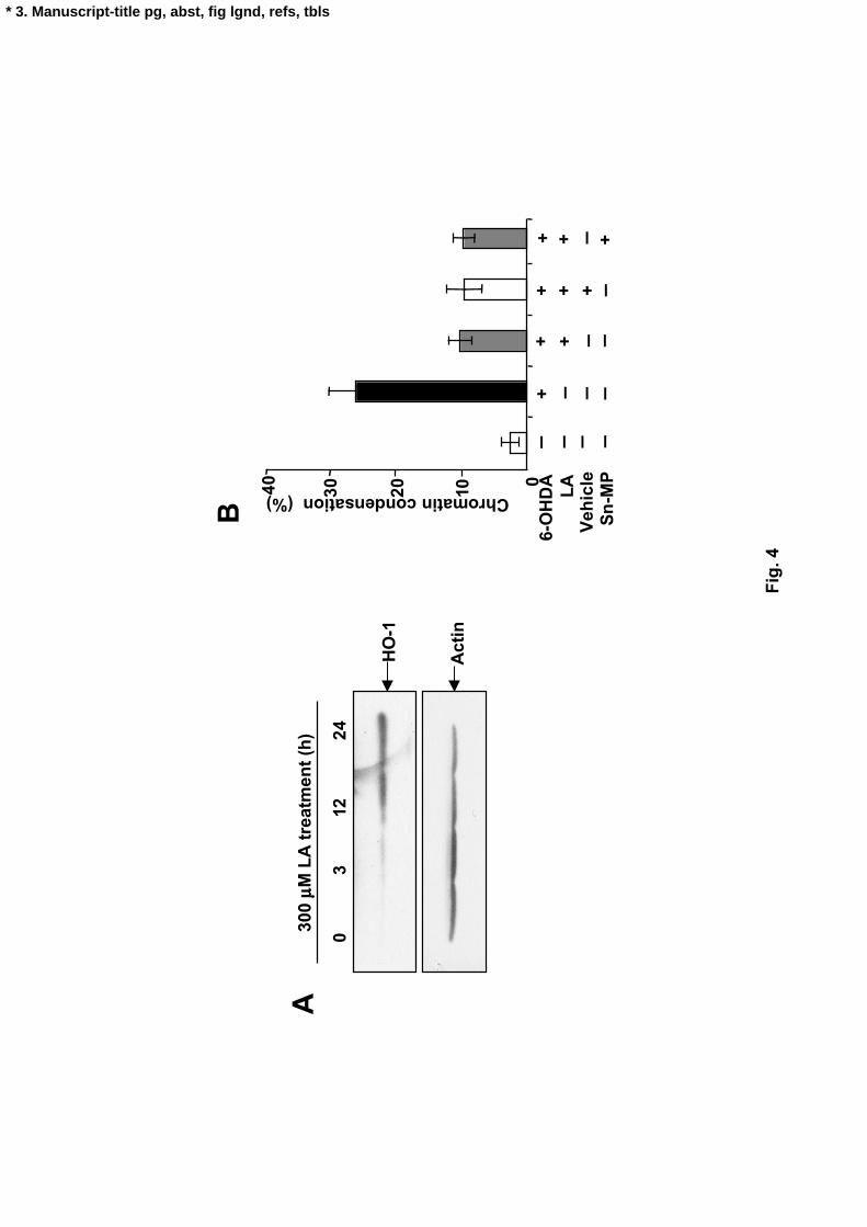
2.6. Heme oxygenase-1 was not involved in the LA-induced suppression of 6-OHDA-
induced apoptosis.
LA also induced the expression of stress response antioxidant enzyme HO-1 which
attenuated the action of intracellular ROS in other cells (Ogborne e al., 2005; Ryter and
Choi, 2005). Furthermore, it has been reported that NGF suppressed the 6-OHDA-

10
induced oxidative stress by increasing expression of HO-1 (Salinas et al., 2006). Thus,
the effect of LA on the HO-1 content of PC12 cells was examined. As shown in Figure
6A, LA significantly increased the content of HO-1 in PC12 cells in a-time dependent
manner. To obtain further insight into the involvement of HO-1 in LA-induced
suppression of apoptosis, we investigated the effect of HO-1 inhibitor Sn-
mesoporphyrin IX (Sn-MP) on LA-suppressed chromatin condensation. Contrary to
the previous reports, 100 mM Sn-MP pretreatment for 1h before LA treatment failed to
attenuate LA-suppression of the chromatin condensation (Fig. 4B).
Fig. 4
2.7. LA increased the intracellular content of glutathione and the gene expression of g-
glutamylcysteine synthetase light subunit.
The GSH content in various cells is very high in order to maintain the redox state
(Dringen, 2000; Takata et al., 2005), and LA promotes GSH synthesis (Suh et al.,
2004a; Suh et al., 2004b, Bharat et al., 2002). Thus the effect of LA on the content of
GSH in PC12 cells was examined. LA increased the content of GSH in PC12 cells in a
time-dependent manner and this increase reached a maximum at 12 h (Fig. 5A). In
this case, we confirmed that most of the GSH was reduced form and only 1/100 was
GSSG (data not shown). Since intracellular GSH is regulated by g-GCS (Seelig et al.,
1984) and by cystine/glutamate antiporter (Wang et al., 2006; Han et al. 1997) the effect
of LA on the gene expression of these proteins was examined. Semi-quantitative RT-
PCR analysis showed that LA increased the expression of g-GCSL mRNA (Fig.5B) and

11
4F2hc mRNA (Fig.5C). LA had no significant effects on the expression of xCT
mRNA (Fig.5D).
Fig. 5
2.8. GSH depletion by BSO attenuated the LA-induced suppression of apoptosis in
PC12 cells.
To obtain further insight into the involvement of GSH in LA-induced suppression
of apoptosis, we investigated the effect of g-GCS inhibitor BSO on the LA-suppressed
apoptosis. First, we confirmed whether BSO depleted LA-increased GSH in PC12
cells using the enzymatic method and the GSH detectable fluorescent probe 5-
chloromethylfluorescein diacetate (CMFDA) (Fig 6A and B). After incubation of
PC12 cells for 24 h with BSO in the presence or absence of 300 mM LA, the LA-
increased GSH content was significantly decreased by BSO (Fig 6B) in a dose-
dependent manner (Fig 6A).
Since BSO markedly depleted the intracellular GSH content, we next investigated
the effect of BSO on various LA-suppressed apoptotic events. As shown in Figure 7,
BSO attenuated the LA-induced suppression of caspase-3, 8, and 9-like activities (Fig
7A, B and C), ROS generation (Fig. 7D), and chromatin condensation (Fig. 7E). It
should be noted that the induction of chromatin condensation (Fig. 7E) correlated
negatively with the cellular GSH level (Fig. 6A). These results indicated that LA-
induced suppression of apoptosis involved the generation of GSH.

12
Figs. 6 and 7
2.9. Effect of LA on the level of nuclear Nrf2 in PC12 cells
Since the transcription factor Nrf2 has been implicated as the central protein that
interacts with the antioxidant response element, also referred to as the electrophile
response element, to induce g-GCS (Cheng et al., 2006) and cystine/glutamate antiporter
(Sasaki et al., 2002), we investigated the effect of LA on the level of nuclear Nrf2 in
PC12 cells. As shown in Figure 8A and B, the nuclear Nrf2 level was increased by the
treatment with LA in a time-dependent manner.
Fig. 8
3. Discussion
Although LA suppressed the apoptosis of various cells through antioxidant ability,
its molecular mechanism is not fully elucidated. In the previous paper, we
demonstrated that ROS generation was an initial step in 6-OHDA-induced apoptosis of
PC12 cells (Fujita et al., 2006). In the present study, we showed the molecular
mechanism of suppression of 6-OHDA-induced apoptosis by LA in PC12 cells. Our
results suggested that LA-stimulated GSH synthesis was a major factor in the
suppression of 6-OHDA-induced ROS generation and apoptosis of PC12 cells and that
LA-stimulated HO-1 expression was not a major factor.
Short time preincubation or no preincubation was needed for some antioxidants
such as Tiron, N-acetylcysteine and GSH to protect PC12 cells from toxicity of 6-

13
OHDA (Fujita et al., 2006; Blum et al., 2001). Although LA is also widely accepted as
an antioxidant, short time preincubation or no preincubation (0 – 3 h) with LA did not
significantly suppress the 6-OHDA-induced apoptosis (Fig. 2E), while long time
preincubation (12 - 24 h) with LA significantly suppressed the apoptosis (Fig. 2E).
These findings indicate that long time preincubation of PC12 is required to mediate the
protective effect of LA and suggest that the antioxidant action is indirect.
Accumulated evidences indicate that LA might suppress cell death through GSH
synthesis (Packer, 1998; Voloboueva et al., 2005). It has been suggested that LA
inhibits TNF-alpha-induced ROS generation, GSH reduction and apoptosis in human
bone marrow stromal cells (Byun et al., 2005) and potently prevents high glucose-
induced oxidative stress and cell death (Vincent et al., 2005). In addition, LA
promoted glutathione synthesis via g-GCS expression and activation (Powell et al.,
2001; Suh et al., 2004a; Suh et al., 2004b; Catherwood et al., 2002) and via cystine
uptake by cystine/glutamine antiporter (Suh et al., 2004b; Han et al., 1997). In the
present experiment, we observed that LA suppressed 6-OHDA-induced ROS generation
(Fig. 1) and chromatin condensation (Figs. 2E and F) and induced gene expression of g-
GCSL and 4F2hc (Figs. 5B and C), and that the LA-suppressed ROS generation and
chromatin condensation were attenuated by BSO (Figs. 7D and E). These findings
suggested that the LA-suppressed ROS generation and apoptosis depended on GSH
synthesis. In this context, the effect of LA on the suppression of ROS generation
depended on the pretreatment time with LA (Fig. 1C) and BSO completely attenuated
the suppression of ROS generation (Fig. 7D). These results suggested that LA worked
through the induction of GSH-elevating proteins, although LA’s direct antioxidant

14
activity could not be excluded.
We also observed that intracellular GSH reached a maximum at 12 hours after LA
treatment, but the peak times of ROS and apoptosis suppression were at 24 hours after
LA pretreatment (Figs. 1C, 2F and 5A). It is likely that GSH regeneration capacity is
more important than GSH level itself. When 6-OHDA produces oxidants in the cell,
GSH is likely to be oxidized and no longer works as an antioxidant. Under these
conditions, a rapid GSH regeneration, either by GSSG reduction or new GSH synthesis,
is important for the continuous cell protection. As the mRNA level for g-GCS is
substantially higher than basal level at 12h, the enzyme protein synthesis appears to be
enhanced for the following time and the enzyme activity might be increased at 24h.
A previous study showed that NGF suppresses 6-OHDA-induced oxidative stress
and apoptosis in PC12 cells by increasing the expression of HO-1 in a
phosphatidylinositol 3-kinase-dependent manner (Salinas et al., 2003). In another
report, LA increased the HO-1 protein level and the resistance of vascular smooth
muscle cells to hydrogen-peroxide-induced cell death, and this protection of LA
depended on HO activity (Cheng et al., 2006). In the present experiment, however, the
LA-suppressed apoptosis did not depend on HO-1 activity despite increasing expression
of HO-1 by LA (Fig. 4). The reason for this discrepancy is not clear at this time, but it
may be due to the different experimental conditions, such as the cell culture conditions,
or to a difference in the kind of ROS which is involved in cell death. Further studies
are needed to answer this question.
It is believed that GSH synthesis is transcriptionally regulated by activating
protein-1 (AP-1) and Nrf2. AP-1 regulates mRNA expression of rate limiting proteins

15
in GSH synthesis including xCT and g-GCS heavy subunit (Rahman et al., 1998; Sato et
al., 2000). It is reported that LA activated AP-1 and its upstream pathway ERK
signaling pathway in vascular smooth muscle cells (Cheng et al., 2006). These findings
suggested that AP-1 had an important role in the increase of GSH synthesis by LA. In
this study, we showed that Nrf2 was significantly translocated into nuclei by LA
treatment (Fig. 8). In addition, Nrf2 also regulates gene expression of g-GCSL and
cystine/glutamate antiportor in some cells (Gail et al., 2004; Moinova and Mulacahy,
1998; Sasaki et al., 2002). These results suggested that Nrf2 might cooperate with AP-
1 in promoting gene expression of GSH synthesis-related protein by LA.
Recently, the adaptive response induced by pro-oxidants including ROS has
received increased attention. It has been reported that low levels of ROS and end
product of lipid peroxidation can induce adaptive responses including GSH synthesis
and Nrf2 nuclear translocation, and enhance tolerance against subsequent oxidative
stress in culture cells (Chen et al., 2006; Seo et al., 2004). LA may also work as a pro-
oxidant under some conditions (Moini et al., 2002; Aoyama et al., 2006). In our
experiment, 1 mM LA alone had cytotoxicity in PC12 cells (data not shown) and 300
mM LA was hardly attainable physiologically. Thus, 300 mM LA might induce low
levels of oxidative stress and enhance tolerance against subsequent oxidative stress.
We also showed that LA suppressed 6-OHDA-induced oxidative stress through
increasing GSH content and increased Nrf2 nuclear translocation in PC12 cells (Figs. 1,
5A and 8). In addition, it has been reported that LA significantly increased ROS and
suppressed subsequent hydrogen-peroxide-induced oxidant stress in vascular smooth
muscle cells (Cheng et al., 2006). These results did not exclude the possibility that

16
induction of an adaptive response by LA might suppress 6-OHDA-induced oxidative
stress in PC12 cells.
Since 6-OHDA generated the intracellular ROS in PC12 cells (Fig 1), the contents
of GSH and GSSG in 6-OHDA-treated cells were examined. Interestingly, 50 mM 6-
OHDA-treatment for 12 h significantly increased the intracellular GSH content and did
not increase GSSG content (data not shown). No change of GSSG content by 6-
OHDA might be due to rapid reduction by glutathione reductase (Ochiai et al., 2004).
In this study, LA suppressed apoptosis due to the enhancement of GSH synthesis in
PC12 cells. These findings suggested that the 6-OHDA-increased GSH might be the
response to avoid 6-OHDA-induced oxidative stress in non-apoptotic PC12 cells.
Thus intracellular GSH in non-apoptotic adherent cells and apoptotic non-adherent cells
was examined. In this case, intracellular GSH content in adherent cells was
significantly higher than in non-adherent cells (data not shown). In this context,
oxidative stress induced by lipid peroxidation products and oxysterols at sublethal
concentrations significantly increased the cellular GSH (Chen et al., 2006). These
results suggested that non-apoptotic cells treated by 6-OHDA might respond to the 6-
OHDA-induced oxidative stress via induction of GSH. It is likely that LA might
promote this cell response in the suppression of 6-OHDA-induced apoptosis by LA.
Although caspase-8 mediates the death receptor signal pathway in death ligand-
induced apoptosis, 6-OHDA induced an increase of caspase-8 like activity in PC12 cells
(Fig. 2B). In this context, the oxidative stress-induced p38 phosphorylation in the 6-
OHDA-induced apoptosis pathway was linked to the activation of caspase-8 in neuronal
culture cells (Choi et al., 2004). In addition, we have reported that 6-OHDA induced

17
p38 phosphorylation, increase of caspase-8-like activity and tBid in PC12 cells (Fujita
et al. 2006). Thus, presumably, caspase-8 is upstream of tBid and downstream of p38
phosphorylation (Fig. 9).
Taken together, we propose the following causal sequence of 6-OHDA-induced
apoptosis of PC12 cells and its suppression by LA: intracellular generation of ROS by
6-OHDA is an initial event in 6-OHDA-induced apoptosis, and the ROS stimulates p38
phosphorylation, activating caspase-8, the cleavage of Bid, cytochrome c release,
activating caspase-9, and activating caspase-3, thereby inducing chromatin condensation.
LA stimulates the translocation of Nrf2 into nuclei, and thereby promotes the gene
expression of GSH synthesis-related proteins such as g-GCSL and 4F2hc. The
increased g-GCS then increases intracellular GSH content, thereby suppressing the 6-
OHDA-induced ROS and subsequent chromatin condensation independently from HO-
1 expression (Fig. 9).
4. Materials and Methods
4.1 Chemicals
HE and CMFDA were obtained from Molecular Probes (OR, USA). 6-
hydroxydopamine, fetal bovine serum (FBS) and buthionine sulfoximine (BSO) were
obtained from Sigma Chemical Co. (St. Louis, MO). Sn-MP was obtained from
Frontier Scientific Inc. (Logan, UT). Sn-MP was dissolved in a solvent consisting of
40% propanediol, 10% ethanol, 153mM arginine and H2O. Fluorogenic tetrapeptide
substrates, such as Ac-DEVD-MCA for caspase-3-like protease, Ac-IETD-MCA for

18
caspase-8-like protease and Ac-LEHD-MCA for caspase-9-like protease were obtained
from the Peptide Institute (Osaka, Japan). Polyclonal antibodies of HO-1 and Mn-
SOD were from Stressgen Biotechnology Corp (BC, Canada). Anti-Cu/Zn-SOD
antiserum was prepared in our laboratory. Polyclonal antibodies of Bax, Bcl-xL, lamin
B and Nrf2 were obtained from Santa Cruz Biotechnology (Santa Cruz, CA). LA was
donated by Cargill Co. Ltd. (USA). BIOXYTECH GSH / GSSG-412 Assay Kit was
obtained from OXIS international, Inc. (Portland, OR) All other chemicals were of
analytical grade and obtained from Nacalai Tesque (Kyoto, Japan).
4.2. Cell culture
PC12 cell line was maintained in DMEM supplemented with 10% FBS, 100 U/ml
penicillin and 100 mg/ml streptomycin on collagen type I-coated dishes as described in a
previous paper (Furuno et al., 2001). Cells were grown in a humidified incubator at
37 °C under 5 % CO2 / 95 % air and used for experiments during the exponential phase
of growth.
4.3. Assay for ROS generation
Intracellular ROS generation was measured using ROS-sensitive fluorescent
precursor, HE (Fujita et al., 2006; Yamada et al., 2003). Cells (2 x 10⁵ cells/ml) were
plated onto collagen coated-24 well plates in 0.5 ml culture medium and incubated for
24 h before LA treatment. Cells were pretreated with or without 300 mM LA for the
indicated times and incubated with 75 mM 6-OHDA for 30 min at 37 °C. Cells were
washed with PBS and stained with 10 mM hydroethidine for 15 min at 37 °C in the dark.

19
Then the cells were analyzed with light and fluorescence microscopy and a FACScan
flow cytometer to determine the ROS generation.
4.4. Assay for caspase activity
Activities of caspases were determined as described previously (Fujita et al.,
2005; Yabuki et al., 2000) in 20 mM HEPES buffer (pH 7.5) containing 0.1 M NaCl and
5 mM dithiothreitol at 37°C using 10 mM of Ac-DEVD-MCA, Ac-IETD-MCA or Ac-
LEHD-MCA as substrates for caspase-3, 8 and 9-like protease, respectively.
Fluorescence of released AMC was measured using a fluorescence plate reader with
excitation and emission wavelengths of 355 and 460 nm, respectively.
4.5. Assay for chromatin condensation
Cells (2 x 10⁵ cells/ml) were plated onto collagen coated-24 well plates in 0.5 ml
culture medium and incubated for 24 h before LA treatment. Cells were pretreated
with or without LA for the indicated times. After incubation with 6-OHDA for 12 h,
cells were stained with Hoechst33342 and the chromatin-condensed cells were
determined under fluorescence microscopy. Total cells (500-1000 cells) and
chromatin-condensed cells were counted in the same field, and the percentage of
chromatin condensation was calculated (Fujita et al., 2005)
4.6. Western blot analysis
Cells (2 x 10⁵ cells/ml) were plated onto collagen coated-100 mm dishes in 15 ml
culture medium and incubated for 24 h before LA treatment. Cells were treated with

20
or without LA for the indicated times. Cell lysates were prepared as described
elsewhere (Fujita et al., 2005). Cells were dissolved in ice-cold lysis buffer [20 mM
Tris-HCl (pH 7.5), 150 mM NaCl 1% Triton X-100, 1 mM EDTA, 1 mM EGTA]
supplemented with protease inhibitors (0.1 mM PMSF, 100 mg/ml leupeptin, 5 mg/ml
pepstatin A). Lysates were cleared by centrifugation.
Nuclear proteins were fractionated from PC12 cells (Wielandt et al., 2006).
Briefly, cells were collected by centrifugation at 800 x g for 5min at 4°C and then were
resuspended in 400 ml of ice-cold buffer A (10 mM HEPES (pH 7.8), 10 mM KC1, 1.5
mM MgCl2, 0.2 mM PMSF, and 0.5 mM DTT). After 15 min incubation on ice, NP-
40 was added to a final concentration of 0.6%, and cells were vortexed and centrifuged
for 1 min at 16,000 x g. The nuclear pellet was extracted with 50 ml of ice-cold buffer
B [l0 mM HEPES (pH 7.8), 420 mM NaCl, 1.5 mM MgCl2, 0.2 mM EDTA, 0.5 mM
PMSF, 0.5 mM DTT, and 25% glycerol] for 30 min at 4°C on a rocking platform, and
debris were removed by centrifugation at 16,000 x g for 20 min at 4 °C.
Lysates and nuclear proteins were added with the same volume of SDS sample
buffer (125 mM Tris-HCl, pH 6.8, 4% SDS, 10% 2-mercaptoethanol, 20% glycerol, and
0.002% bromophenol blue) and boiled at 100°C for 5 min. The samples (10-50 mg
protein) were then subjected to SDS-polyacrylamide gel electrophoresis. Proteins in
the gel were transferred onto a PVDF membrane, and then incubated with the primary
antibody (1:5000 dilution for HO-1 and actin, 1:1000 dilution for others) and finally
with horseradish peroxide-linked second antibody (1:2000 or 1:25000 dilution) and
analyzed using ECL plus kit (Amersham). Protein concentration was determined by

21
the method of Bradford (Bradford, 1976) using bovine serum albumin as a standard.
4.7. Assay for intracellular glutathione content
Measurements of GSH were performed using the BIOXYTECH GSH / GSSG-412
Assay kit. Cells (2 x 10⁵ cells/ml) were plated onto collagen-coated 60 mm dishes in 5
ml culture medium and incubated for 24 h before LA treatment. Cells were treated
with or without LA and BSO for the indicated time. Cells were suspended in ice-cold
5% trichloroacetic acid (w/v) and lyzed by vortex mixer. The supernatant was
centrifuged for 10 min at 20,000 x g and 4°C. After removing insoluble materials by
centrifugation, samples were assayed spectrophotometrically following the
manufacturer’s instructions. The protein contents of the samples were determined by the
method of Bradford (Bradford et al., 1976).
The thiol-reactive fluorescent dye CMFDA was used for GSH determination.
CMFDA forms a GSH adduct in a reaction catalyzed by glutathione-S-transferase. After
conjugation with GSH, CMFDA is hydrolyzed to the fluorescent 5- chloromethyl-
fluorescein by cellular esterase (Poot et al., 1991). Cells were treated with or without
LA and BSO for the indicated times, incubated for an additional 30 min in a CFMDA
(30 mM)-containing culture medium at 37 °C, and then analyzed with a FACScan flow
cytometer to determine the GSH content.
4.8. Semi-quantitative RT-PCR analysis for mRNA expression of GSH synthesis-related
proteins
To determine the relative expression level of g-GCS transcripts, RT-PCR was

22
performed as follows. Total RNA was isolated from PC12 cells using TRIzol reagent
(Invirogen) following the manufacturer’s instructions. Oligo dT-primed cDNA was
prepared from 3-5 mg of total RNA using Superscript III (Invitrogen). One-fiftieth of
the cDNA obtained was used for each PCR. Primer sequences and product sizes were
as follows: 5’-TAA TAC GAC TCA CTA TAG TGC GGC GCC TCA GTG ACG CTT
TTT G-3’ (containing T7 promoter sequence) and 5’-GAT TTA GGT GAC ACT ATA
GAA CCA AGT TAA TCT TGC CTC C-3’ (containing SP6 promoter sequence) for g-
GCSL (456 bp); 5’-CCTGGCATTTGGACGCTACAT-3’ and 5’-
TCAGAATTGCTGTGAGCTTGC-3’ for xCT (182 bp); 5’-
CTCCCAGGAAGATTTTAAAGACCTTCT-3’ and 5’-
TTCATTTTGGTGGCTACAATGTCAG-3’ for 4F2hc (141 bp); 5’-ATT TGG CAC
CAC ACT TT TAC A-3’ and 5’-TCA CGC ACG ATT TCC CTC TCA G-3’ for actin
(379 bp). The cDNA were amplified by 28 cycles (g-GCSL or actin) or 35 cycles (xCT
or 4F2hc) with the following conditions: denaturing at 94 oC for 0.5 min, annealing at
52oC (g-GCSL or actin) or 60 oC (xCT or 4F2hc) for 1 min, extension at 72oC for 1 min
and final elongation at 72 oC for 3 min. The amplified products were separated on
1.5% agarose gels and visualized by ethidium bromide staining. The intensity of the
bands was quantified using an image analyzer (NIH Image Software). The identity of
PCR products was verified by sequencing.
4.9. Statistical analysis
Results are expressed as means +/- SD. The significance of differences between

23
experimental conditions was determined using the two-tailed Student t-test. A
probability of p < 0.05 was considered significant.

24
References
Anderson, M. E., 1998. Glutathione: an overview of biosynthesis and modulation.
Chem. Biol. Interact. 111-112, 1-14.
Aoyama, S., Okimura, Y., Fujita, H., Sato, E.F., Abe, K., Inoue, M., Utsumi, K., Sasaki,
J., 2006. Stimulation of membrane permeability transition by alpha-lipoic acid
and its biochemical characteristics. Physiol. Chem. Phys. & Med. NMR. 38, 1-20.
Bast, A., Haenen, G.R., 2003. Lipoic acid: a multifunctional antioxidant. Biofactors. 17,
207-213.
Bharat, S., Cochran, B.C., Hsu, M., Liu, J., Ames, B.N., Andersen, J.K., 2002. Pre-
treatment with R-lipoic acid alleviates the effects of GSH depletion in PC12 cells:
implications for Parkinson's disease therapy. Neurotoxicology. 23,479-486.
Blum, D., Torch, S., Lambeng, N., Nissou, M., Benabid, A.L., Sadoul, R., Verna, J.M.
2001. Molecular pathways involved in the neurotoxicity of 6-OHDA, dopamine
and MPTP: contribution to the apoptotic theory in Parkinson's disease. Prog
Neurobiol. 65, 135-172.
Bradford, M.M., 1976. A rapid and sensitive method for the quantitation of microgram
quantities of protein utilizing the principle of protein-dye binding. Anal. Biochem.
72, 248-254.
Byun, C.H., Koh, J.M., Kim, D.K., Park, S.I., Lee, K.U., Kim, G.S., 2005. Alpha-lipoic
acid inhibits TNF-alpha-induced apoptosis in human bone marrow stromal cells. J.
Bone Miner. Res. 20,1125-1135.
Catherwood, M.A., Powell, L.A., Anderson, P. M., 2002. Glucose-induced oxidative
stress in mesangial cells. Kidney Int. 61, 599-608.
Cheng, P.Y., Lee, Y.M., Shih, N.L., Chen, Y.C., Yen, M.H., 2006. Heme oxygenase-1

25
contributes to the cytoprotection of alpha-lipoic acid via activation of p44/42
mitogen-activated protein kinase in vascular smooth muscle cells. Free Radic. Biol.
Med. 40, 1313-1322.
Chen, Z.H., Yoshida, Y., Saito, Y., Sekine, A., Noguchi, N., Niki, E., 2006. Induction
of adaptive response and enhancement of PC12 cell tolerance by 7-
hydroxycholesterol and 15-deoxy-delta(12,14)-prostaglandin J2 through up-
regulation of cellular glutathione via different mechanisms. J. Biol. Chem.
281,14440-14445.
Christophe, M., Nicolas, S., 2006. Mitochondria: a target for neuroprotective
interventions in cerebral ischemia-reperfusion. Curr. Pharm. Des. 12,739-757.
Choi, WS., Eom, D.S., Han, B.S., Kim, W.K., Han, B.H., Choi, E.J., Oh, T.H.,
Markelonis, G.J., Cho, J.W., Oh, Y.J., 2004. Phosphorylation of p38 MAPK induced
by oxidative stress is linked to activation of both caspase-8- and -9-mediated
apoptotic pathways in dopaminergic neurons. J Biol Chem. 279, 20451-20460.
Coceani, F., 2000. Carbon monoxide in vasoregulation: the promise and the challenge.
Circ. Res. 86, 1184-1186.
Constantinescu, A., Pick, U., Handelman, G.J., Haramaki, N., Han, D., Podda, M.,
Tritschler, H.J., Packer, L., 1995. Reduction and transport of lipoic acid by
human erythrocytes. Biochem. Pharmacol. 50, 253-261.
Dringen, R., 2000. Metabolism and functions of glutathione in brain. Prog. Neurobiol.
62, 649-671.
Engel, R. H., Evens, A. M. 2006. Oxidative stress and apoptosis: a new treatment
paradigm in cancer. Front. Biosci. 11, 300-312.
Fujita, H., Utsumi, T., Muranaka, S., Ogino, T., Yano, H., Akiyama, J., Yasuda, T.,

26
Utsumi, K., 2005. Involvement of Ras/extracellular signal-regulated kinase, but
not Akt pathway in risedronate-induced apoptosis of U937 cells and its suppression
by cytochalasin B. Biochem. Pharmacol. 69, 1773-1784.
Fujita, H., Ogino, T., Kobuchi, H., Fujiwara, T., Yano, H., Akiyama, J., Sasaki, J.,
Utsumi, K., 2006. Cell-permeable cAMP analog suppresses 6-hydroxydopamine-
induced apoptosis in PC12 cells through the activation of Akt pathway. Brain Res.
1113, 10-23.
Furuno, T., Kanno, T., Arita, K., Asami, M., Utsumi, T., Doi, Y., Inoue, M., Utsumi, K.,
2001. Roles of long chain fatty acids and carnitine in mitochondrial membrane
permeability transition. Biochem. Pharmacol. 62, 1037-1046.
Gail, K., McWalter, L.G., Higgins, L.I., McLellan, C.J., Henderson, L. S., Paul J. T.,
Itoh, K., Yamamoto, M., John D.H., 2004. Transcription Factor Nrf2 Is Essential
for Induction of NAD(P)H:Quinone Oxidoreductase 1, Glutathione S-Transferases,
and Glutamate Cysteine Ligase by Broccoli Seeds and Isothiocyanates. J. Nutr. 134,
3499-3506.
Griffith, O.W. 1982. Mechanism of action, metabolism, and toxicity of buthionine
sulfoximine and its higher homologs, potent inhibitors of glutathione synthesis. J.
Biol. Chem. 257, 13704-13712.
Hagen, T.M., Moreau, R., Suh, J.H., Visioli, F., 2002. Mitochondrial decay in the
aging rat heart: evidence for improvement by dietary supplementation with acetyl-L-
carnitine and/or lipoic acid. Ann. N. Y. Acad. Sci.959, 491-507.
Han, D., Handelman, G., Marcocci, L., Sen, C.K., Roy, S., Kobuchi, H., Tritschler, H.J.,
Flohe, L., Packer, L., 1997. Lipoic acid increases de novo synthesis of cellular
glutathione by improving cystine utilization. Biofactors. 6, 321-338.

27
Hanrott, K., Gudmunsen, L., O'Neill, M.J., Wonnacott, S., 2006. 6-hydroxydopamine-
induced apoptosis is mediated via extracellular auto-oxidation and caspase 3-
dependent activation of protein kinase Cdelta. J. Biol. Chem. 281,5373-5382.
Harris, J.B., Alonso, D., Park, O.H., Cornfield, D., Chacin, J., 1975. Lipoate effect on
carbohydrate and lipid metabolism and gastric H+ secretion. Am. J. Physiol. 228,
964-971.
Jeong, W.S., Jun, M., Kong A, N., 2006. Nrf2: a potential molecular target for cancer
chemoprevention by natural compounds. Antioxid. Redox Signal. 8, 99-106.
Jezek, P., Hlavata, L., 2005. Mitochondria in homeostasis of reactive oxygen species
in cell, tissues, and organism. Int. J. Biochem. Cell Biol. 37, 2478-2503.
Lemasters, J.J, Nieminen, A.L, Qian, T., Trost, L.C., Elmore, S.P., Nishimura, Y., Crowe,
R.A., Cascio, W.E., Bradham, C.A., Brenner, D.A., Herman, B., 1998. The
mitochondrial permeability transition in cell death: a common mechanism in
necrosis, apoptosis and autophagy. Biochim. Biophys. Acta. 1366, 177-196.
Maines, M.D., 1997. The heme oxygenase system: a regulator of second messenger
gases. Annu. Rev. Pharmacol. Toxicol. 37, 517-554.
Marsh, S,A., Laursen, P.B., Pat, B.K., Gobe, G.C., Coombes, J.S., 2005. Bcl-2 in
endothelial cells is increased by vitamin E and alpha-lipoic acid supplementation
but not exercise training. J. Mol. Cell Cardiol. 38,445-451.
Mizuno, M., Packer, L., 1994. Effects of alpha-lipoic acid and dihydrolipoic acid on
expression of proto-oncogene c-fos. Biochem Biophys Res Commun. 200, 1136-
1142.
Moini, H., Packer, L., Saris, N.E.,2002. Antioxidant and prooxidant activities of
alpha-lipoic acid and dihydrolipoic acid. Toxicol. Appl. Pharmacol. 182, 84-90.

28
Moinova, H.R., Mulcahy, R.T. 1998. An Electrophile Responsive Element (EpRE)
Regulates beta -Naphthoflavone Induction of the Human gamma -
Glutamylcysteine Synthetase Regulatory Subunit Gene; CONSTITUTIVE
EXPRESSION IS MEDIATED BY AN ADJACENT AP-1 SITE. J. Biol. Chem.
273, 14683-14689.
Ochiai, T., Soeda, S., Ohno, S., Tanaka, H., Shoyama, Y., Shimeno, H., 2004. Crocin
prevents the death of PC-12 cells through sphingomyelinase-ceramide signaling by
increasing glutathione synthesis. Neurochem. Int. 44, 321-330.
Ogborne, R.M., Rushworth, S.A., O'Connell M.A., 2005. Alpha-lipoic acid-induced
heme oxygenase-1 expression is mediated by nuclear factor erythroid 2-related
factor 2 and p38 mitogen-activated protein kinase in human monocytic cells.
Arterioscler. Thromb. Vasc. Biol. 25,2100-2105.
Packer, L., 1998. Alpha-Lipoic acid: a metabolic antioxidant which regulates NF-
kappa B signal transduction and protects against oxidative injury. Drug. Metab. Rev.
30, 245-275.
Perham, R.N., Borges, A., Dardel, F., Graham, L.D., Hawkins, C.F., Laue, E.D.,
Packman, L.C., 1992. The lipoyl domain and its role in thiamin diphosphate-
dependent oxidative decarboxylation. J. Nutr. Sci. Vitaminol (Tokyo). Spec No, 457-
460.
Persson, H.L., Svensson, A.I, Brunk, U.T., 2001. Alpha-lipoic acid and alpha-
lipoamide prevent oxidant-induced lysosomal rupture and apoptosis. Redox Rep. 6,
327-334.
Poot, M., Kavanagh, T.J., Kang, H.C., Haugland, R.P., Rabinovitch, P.S., 1991. Flow
cytometric analysis of cell cycle-dependent changes in cell thiol level by combining

29
a new laser dye with Hoechst 33342. Cytometry. 12, 184-187.
Powell, L.A., Nally, S.M., McMaster, D., Catherwood, M.A., Trimble, E.R., 2001.
Restoration of glutathione levels in vascular smooth muscle cells exposed to high
glucose conditions. Free Radic. Biol. Med. 31, 1149-1155.
Rahman, I., Smith, C.A., Antonicelli, F., MacNee, W., 1998. Characterisation of
gamma-glutamylcysteine synthetase-heavy subunit promoter: a critical role for AP-1.
FEBS Lett. 427, 129-33.
Reliene, R., Schiestl, R.H., 2006. Glutathione depletion by buthionine sulfoximine
induces DNA deletions in mice. Carcinogenesis. 27, 240-244.
Ryter, S.W., Choi, A.M., 2005. Heme oxygenase-1: redox regulation of a stress
protein in lung and cell culture models. Antioxid. Redox Signal. 7, 80-91.
Salinas, M., Diaz, R., Abraham, N.G., Ruiz, de Galarreta, C.M., Cuadrado, A., 2003.
Nerve growth factor protects against 6-hydroxydopamine-induced oxidative stress
by increasing expression of heme oxygenase-1 in a phosphatidylinositol 3-kinase-
dependent manner. J. Biol. Chem. 278, 13898-13904.
Sasaki, H., Sato, H., Kuriyama-Matsumura, K., Sato, K., Maebara, K., Wang, H., Tamba,
M., Itoh, K., Yamamoto, M., Bannai, S., 2002. Electrophile response element-
mediated induction of the cystine/glutamate exchange transporter gene expression.
J. Biol. Chem. 277,44765-44771.
Sato, H., Tamba, M., Kuriyama-Matsumura, K., Okuno, S., Bannai, S., 2000.
Molecular cloning and expression of human xCT, the light chain of amino acid
transport system xc-. Antioxid. Redox Signal. 2,665-671
Sauer, H., Wartenberg, M., 2005. Reactive oxygen species as signaling molecules in
cardiovascular differentiation of embryonic stem cells and tumor-induced

30
angiogenesis. Antioxid. Redox Signal. 7, 1423-1434.
Seelig, G. F., Simondsen, R. P., Meister, A., 1984. Reversible dissociation of g-GCS
into two subunits. J. Biol. Chem. 259, 9345-9347.
Seo, Y.J., Lee, J.W., Lee, E.H., Lee, H.K., Kim, H.W., Kim, Y.H., 2004. Role of
glutathione in the adaptive tolerance to H2O2. Free Radic. Biol. Med. 37,1272-1281.
Shih, A.Y., Johnson, D.A., Wong, G., Kraft, A.D., Jiang, L., Erb, H., Johnson, J.A.,
Murphy, T.H., 2003. Coordinate regulation of glutathione biosynthesis and release
by Nrf2-expressing glia potently protects neurons from oxidative stress. J Neurosci.
23,3394-3406.
Smith, A.R., Shenvi, S.V., Widlansky, M., Suh, J.H., Hagen, T.M., 2004. Lipoic acid
as a potential therapy for chronic diseases associated with oxidative stress. Curr.
Med. Chem. 11, 1135-1146.
Stocker, R., Yamamoto, Y., McDonagh, A.F., Glazer, A.N., Ames, B.N., 1987. Bilirubin
is an antioxidant of possible physiological importance. Science. 235, 1043-1046.
Suh, J.H., Shenvi, S.V., Dixon, B.M., Liu, H., Jaiswal, A.K., Liu, R.M., Hagen, T.M.,
2004. Decline in transcriptional activity of Nrf2 causes age-related loss of
glutathione synthesis, which is reversible with lipoic acid. Proc. Natl. Acad. Sci.
USA. 101, 3381-3386.
Suh, J.H., Wang, H., Liu, R.M., Liu, J., Hagen, T.M., 2004. (R)-alpha-lipoic acid
reverses the age-related loss in GSH redox status in post-mitotic tissues: evidence
for increased cysteine requirement for GSH synthesis. Arch. Biochem. Biophys.
423,126-135.
Takata, M.K., Yamaguchi, F., Nakanose, K., Watanabe, Y., Hatano, N, Tsukamoto, I.,

31
Nagata, M., Izumori, K., Tokuda, M., 2005. Neuroprotective effect of D-psicose on
6-hydroxydopamine-induced apoptosis in rat pheochromocytoma (PC12) cells. J.
Biosci. Bioeng. 100, 511-516.
Vincent, A.M., McLean, L.L., Backus, C., Feldman, E.L., 2005. Short-term
hyperglycemia produces oxidative damage and apoptosis in neurons.FASEB. J.
19,638-640.
Voloboueva, L.A., Liu, J., Suh, J.H., Ames, B.N., Miller, S.S., 2005. (R)-alpha-lipoic
acid protects retinal pigment epithelial cells from oxidative damage. Invest.
Ophthalmol. Vis. Sci. 46, 4302-4310.
Wang, L., Hinoi, E., Takemori, A., Nakamichi, N., Yoneda, Y., 2006. Glutamate
Inhibits Chondral Mineralization through Apoptotic Cell Death Mediated by
Retrograde Operation of the Cystine/Glutamate Antiporter. J. Biol. Chem., 281,
24553-24565.
Wielandt, A.M., Vollrath, V., Farias, M., Chianale, J., 2006. Bucillamine induces
glutathione biosynthesis via activation of the transcription factor Nrf2. Biochem.
Pharmacol. 72,455-462.
Wu, G., Fang, Y.Z., Yang, S., Lupton, J.R., Turner, N.D., 2004. Glutathione
metabolism and its implications for health. J. Nutr. 134, 489-492.
Yabuki, M., Tsutsui, K., Horton, A.A., Yoshioka, T., Utsumi, K. 2000. Caspase
activation and cytochrome c release during HL-60 cell apoptosis induced by a nitric
oxide donor. Free Radic. Res. 32, 507-514.
Yamada, J., Yoshimura, S., Yamakawa, H., Sawada, M., Nakagawa, M., Hara, S., Kaku,
Y., Iwama, T., Naganawa, T., Banno, Y., Nakashima, S., Sakai, N., 2003. Cell
permeable ROS scavengers, Tiron and Tempol, rescue PC12 cell death caused by

32
pyrogallol or hypoxia/reoxygenation. Neurosci Res. 45, 1-8.

33
Figure legends
Fig. 1. Generation of ROS in PC12 cells by 6-OHDA and its suppression by LA in a
pre-incubation time dependent manner.
Cells were pretreated with or without 300 mM LA for 1-24 h and then incubated with 75
mM 6-OHDA for 30 min. Cells were stained with 10 mM hydroethidine. PC12 cells
generated ROS by the treatment with 6-OHDA. (A) Suppression of ROS generation
by LA treatment for 24 h was analyzed by bright field and fluorescence microscopy.
Red fluorescence showed the hydroethidine reacted with ROS. (B) The FACScan
analyzed patterns of pre-incubation time-dependency by LA. (C) Percentage change
of ethidium fluorescence positive cells by the preincubation with LA. Ethidium
fluorescence positive cells were defined as cells in the gate of panel B. The asterisk
indicates that LA significantly suppressed 6-OHDA-induced ROS generation (p<0.05).
These results are representative of at least three independent experiments.
Fig. 2. Suppression of 6-OHDA-induced apoptotic events by LA.
Cells were preincubated with the indicated concentration of LA for the indicated time
and then incubated with 50 mM 6-OHDA for 12 h. (A-C) Effects of LA on the 6-
OHDA-induced caspase-3-like (A), caspase-8-like (B) and caspase-9-like (C) protease
activity. The activities of various caspases were measured by using synthetic peptide
substrates (Ac-DEVD-MCA for caspase-3-like protease, Ac-IETD-MCA for caspase-8-

34
like protease and Ac-LEHD-MCA for caspase-9-like protease) after treatment with 6-
OHDA. (D) Concentration-dependent suppression of 6-OHDA-induced chromatin
condensation by LA pretreatment. (E) Preincubation time-dependent suppression of 6-
OHDA-induced chromatin condensation by LA. Chromatin condensation was
detected with Hoechst33342 staining and fluorescence microscopy, and the percentage
of chromatin condensation was calculated. The asterisk indicates that LA significantly
suppressed 6-OHDA-induced events (p < 0.05). Data are the means +/- SD derived
from three independent experiments.
Fig. 3. Effect of LA on the content of Bcl2 family protein and SOD in PC12 cells.
PC12 cells were incubated with 300 mM LA for various times and Bcl-xL, Bax, Cu/Zn-
SOD and Mn-SOD were detected by Western blotting each with specific antibodies.
(A) Time-dependent change in the content of Bcl-xL after incubation with LA. (B)
Effect of LA on the content of Bax. (C) Effect of LA on the content of Cu/Zn-SOD and
Mn-SOD. Similar results were obtained in three separate experiments.
Fig. 4. Effect of LA on the content of heme oxygenase-1 and effect of Sn-
mesoporphyrin IX on the suppression of 6-OHDA-induced apoptosis by LA.
(A) Effect of LA on the content of Heme oxygenase-1 (HO-1). Experimental
conditions were the same as described for Figure 3. Similar results were obtained in
three separate experiments. (B) Effect of Sn-mesoporphyrin IX (Sn-MP) on the

35
suppression of 6-OHDA-induced chromatin condensation by LA. Cells were pretreated
with or without 300 mM LA in the presence or absence of 100 mM Sn-MP for 24 h and
then incubated with 50 mM 6-OHDA for 12 h. Data are the means +/- SD derived from
three independent experiments.
Fig. 5. Effects of LA on the glutathione synthesis system in PC12 cells
Experimental conditions were the same as described for Figure 3. PC12 cells were
incubated for the indicated time with 300 mM LA. (A) Time dependent change in the
content of GSH after incubation with LA. The content of GSH was determined by the
method described in the text. (B-D) Effect of LA on the mRNA expression of g-
glutamylcysteine synthetase L subunit (g-GCSL) (B), 4F2hc (C) and xCT (D) in PC12
cells. Expression of the enzyme gene was measured by RT-PCR method as described in
the text. The asterisk indicates that LA significantly increased the indicated molecules
(p < 0.05). Data are the means +/- SD derived from three independent experiments.
Fig. 6. Effect of g-glutamylcysteine synthetase inhibitor on the GSH content in PC12
cells treated with LA.
Cells were incubated for 24 h with 0.3-30 mM buthionine sulfoximine (BSO) in the
presence or absence of 300 mM LA. (A) Concentration dependent suppression of LA-

36
induced GSH by BSO was determined with the BIOXYTECH GSH / GSSG-412 Assay
kit. The asterisk indicates that BSO significantly suppressed LA-induced GSH (p <
0.05). Data are the means +/- SD derived from three independent experiments. (B)
Suppression of LA-induced GSH by BSO was determined with thiol-reactive
fluorescent dye CMFDA. GSH, the intracellular major non-protein thiol in cell, was
detected by CMFDA and quantified using a flow cytometer. Similar results were
obtained in three separate experiments.
Fig. 7. Effect of g-glutamylcysteine synthetase inhibitor on the suppression of various 6-
OHDA-induced apoptotic events by LA.
PC12 cells were incubated for 24 h with 0.3-30 mM buthionine sulfoximine (BSO) in
the presence or absence of 300 mM LA, and then treated with 6-OHDA. (A-C) Effect of
BSO on the suppression of 6-OHDA-induced caspase-3 like (A), caspase-8 like (B) and
caspase-9 like (C) protease activity by LA. (D) Effect of BSO on the suppression of 6-
OHDA-induced ROS generation by LA. (E) Effect of BSO on the suppression of 6-
OHDA-induced chromatin condensation by LA. Experimental conditions of (A-C), (D)
and (E) were the same as described for Figs. 3, 1 and 4, respectively. The asterisk
indicated that BSO significantly attenuated the LA-induced suppression (p<0.05).
Data are the means +/- SD from three independent experiments.
Fig. 8. Effect of LA on the level of nuclear Nrf2 in PC12 cells.
Experimental conditions were the same as described in Fig. 5. Cells were incubated
with 300 mM LA for 0-12 h and the nuclear fraction extracted. Nrf2 were detected by

37
Western blotting with Nrf2 specific antibodies. (A) Time-dependent increase in Nrf2
nuclear translocation. (B) The ratio of Nrf2/lamin B band densities is shown in panel A.
The asterisk indicates that LA significantly increased nuclear Nrf2 (p < 0.05). Data are
the means +/- SD from three independent experiments.
Fig. 9. Schematic diagram summarizing the cross talk of 6-OHDA-activated apoptosis
pathway and LA-activated glutathione synthesis pathway.
6-OHDA probably induces PC12 cell apoptosis by the following mechanisms:
intracellular ROS production by 6-OHDA is an initial event, and then the ROS increases
the activating caspase cascade and chromatin condensation. LA rapidly increases the
nuclear Nrf2 levels and then enhances gene expression of antioxidant related enzymes
such as HO-1, g-GCSL and 4F2hc, thereby activating the glutathione synthesis system.
LA-induced GSH suppresses ROS induced by 6-OHDA. HO-1 and g-GCSL were heme
oxygenase-1 and g-gluthamylcystein synthetase L subunit, respectively.

B 4080 0
100
101
102
103
104
Eth
idiu
m f
luo
resc
ence
(F
L-2
H)
No
ne
6-O
HD
A, 0
.5h
+ L
A, 1
2 h
pre
-in
cub
atio
n
+ L
A, 2
4 h
pre
-in
cub
atio
n
Counts 4080 0
40
80 0
4080 0
Fig
. 1
C Ethidium fluorescence positive cells (%)
+L
ANon
e24
121
3
6-O
HDA0
1020304050
pre
-tre
atm
ent
tim
e (h
)
AB
rig
ht
Hyd
roet
hid
ine
6-O
HD
A
+ L
A
No
ne
6-O
HD
A
+ L
A
No
ne
*
* 3. Manuscript-title pg, abst, fig lgnd, refs, tbls

Fig
. 2
D
+L
A (mmmm M
)Non
e30
030
100
6-O
HDA
+L
A (mmmm M
)Non
e30
030
100
6-O
HDA0
1020
30
40
Caspase-3 like activity(nmol/h/mg protein)
0
0.5
1.0
1.5
2.0
+L
A (mmmm M
)Non
e30
030
100
6-O
HDA0
0.4
0.8
1.2
1.6
Caspase-8 like activity(nmol/h/mg protein)
Caspase-9 like activity(nmol/h/mg protein)
*
**
BC
A
Chromatin condensation (%)
Ep
retr
eatm
ent
for
24 h
wit
h L
A
None
300
10
0
6-O
HDA
+L
A (mmmm M
)30
010
20
30
40
50
*
0
102030
40
Chromatin condensation (%)
+ L
Ap
re-t
reat
men
t ti
me
(h)
None
241
21
3
6-O
HDA
pre
trea
tmen
t w
ith
300
mmmmM
LA
**
0
pre
trea
tmen
t fo
r 24
h w
ith
LA
pre
trea
tmen
t fo
r 24
h w
ith
LA
pre
trea
tmen
t fo
r 24
h w
ith
LA
* 3. Manuscript-title pg, abst, fig lgnd, refs, tbls

300 mmmm M
LA
tre
atm
ent
(h)
03
122
4A
Act
in
Bcl
-xL
Bax
300 mmmm M
LA
tre
atm
ent
(h)
03
122
4
Act
in
B
Fig
. 3
Cu
/Zn
-SO
D
300 mmmm M
LA
tre
atm
ent
(h)
03
122
4
Mn
-SO
D
C
Act
in
* 3. Manuscript-title pg, abst, fig lgnd, refs, tbls
B
LA
6-O
HD
A
Sn
-MP
Veh
icle
++
++
++
++
+
0
1020
3040
Chromatin condensation (%)
HO
-1
300 mmmm M
LA
tre
atm
ent
(h)
03
122
4
A
Act
in
Fig
. 4
* 3. Manuscript-title pg, abst, fig lgnd, refs, tbls

Fig
.5
RT
-PC
R (
28 c
ycle
)B
C
Act
in
gggg ----G
CS
L
LA
(h
)0
13
12
Relative gggg-GCSL/Actin
012345
RT
-PC
R (
35 c
ycle
)
Act
in
4F2h
c
LA
(h
)0
13
12
Relative 4F2hc/Actin
DR
T-P
CR
(35
cyc
le)
Act
in
xCT
LA
(h
)0
13
12
0
0.5
1.0
1.5
2.0
2.5
3.0
Relative xCT/Actin0
0.5
1.0
1.5
2.0
*
*
A
0
4080
120
16
0
06
121
82
4
GSH (nmol/mg protein)
300 mmmm M
LA
**
* 3. Manuscript-title pg, abst, fig lgnd, refs, tbls

Fig
.6
A
050100
15
0
20
0
GSH (nmol/mg protein)
LA
BS
O(mmmm
M)
303
30
++
+
0.3+
Counts200 010
010
110
210
3
CM
FD
A (
FL
-1)
300 mmmm M
LA
No
ne
+ 3
0 mmmm M
BS
O
Incu
bat
ion
wit
h L
A a
nd
BS
O f
or
24 h
B
*
*
* 3. Manuscript-title pg, abst, fig lgnd, refs, tbls

Fig
. 7E
6-O
HD
AL
AB
SO
(mmmmM
)+
++
++
303
++ 30
Chromatin condensation (%)
02040
60
++ 0.3
01020
30
40
50
D
6-O
HD
AL
AB
SO
(mmmmM
)+
30++ 30
++
01020
30
40
50
Caspase 3-like activity(nmol/hour/mgprotein)
BA
C
LA
6-O
HD
A
BS
O+
+ +
++
++
+ +
++
++
+ +
++
+L
A6-
OH
DA
BS
OL
A6-
OH
DA
BS
O
0123
Caspase 8-like activity (nmol/hour/mgprotein)
Caspase 9-like activity (nmol/hour/mgprotein)
Ethidium fluorescence positive cells (%)
0
0.4
0.8
1.2
1.6
**
*
**
*
++
+
* 3. Manuscript-title pg, abst, fig lgnd, refs, tbls

Fig
. 8
A0
LA
(h
)
Nu
clea
rfr
acti
on
112
Nrf
2
3
Lam
in B
01
312
LA
(h
)
Relative Nrf2/Lamin B*
B
*
0
0.51
1.52
2.53
3.5
* 3. Manuscript-title pg, abst, fig lgnd, refs, tbls

Mit
och
on
dri
a
Cas
pas
e-8
6-O
HD
A
RO
S
p38
ph
osp
ho
ryla
tio
n
Cel
l mem
bran
e
tBid
Cyt
ochr
ome
c r
elea
se
Cas
pas
e-9
Cas
pas
e-3
Ch
rom
atin
co
nd
ensa
tio
nGS
H
BSO
gggg -G
CS
(L)
LA
HO
-1
Sn-MP
Nrf
2 n
ucl
ear
tran
slo
cati
on
GS
Hsy
nth
esis
syst
em
Cys
tin
e/
glu
tam
ate
anti
po
rter
(4F
2hc)
Fig
. 9
* 3. Manuscript-title pg, abst, fig lgnd, refs, tbls