Human Anatomy and Physiology: Unit 1 Unit 1: An Introduction to Human Anatomy and Physiology Chapter...
78
Human Anatomy and Physiology: Unit 1 • Unit 1: An Introduction to Human Anatomy and Physiology • Chapter 1 © 2013 Pearson Education, Inc.
-
Upload
buck-richard -
Category
Documents
-
view
244 -
download
2
Transcript of Human Anatomy and Physiology: Unit 1 Unit 1: An Introduction to Human Anatomy and Physiology Chapter...

Human Anatomy and Physiology: Unit 1
• Unit 1: An Introduction to Human Anatomy and Physiology
• Chapter 1
© 2013 Pearson Education, Inc.

© 2013 Pearson Education, Inc.
Chapter 1 Learning Targets
• 1-1
• Describe the basic functions of living organisms.
• 1-2
• Explain the relationship between anatomy and physiology, and describe various specialties of each discipline.
• 1-3
• Identify the major levels of organization in living organisms.
• 1-4
• Identify the 11 organ systems of the human body and contrast their major functions.
© 2013 Pearson Education, Inc.
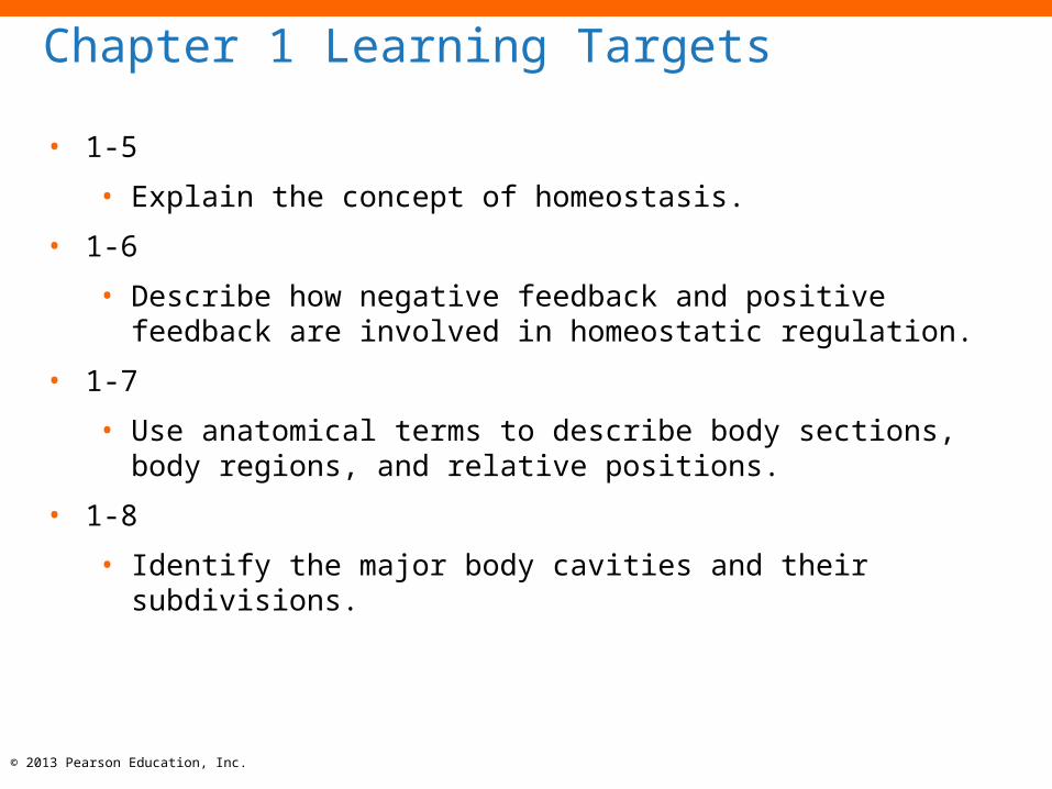
Chapter 1 Learning Targets
• 1-5
• Explain the concept of homeostasis.
• 1-6
• Describe how negative feedback and positive feedback are involved in homeostatic regulation.
• 1-7
• Use anatomical terms to describe body sections, body regions, and relative positions.
• 1-8
• Identify the major body cavities and their subdivisions.

Do you remember…
• The Characteristics of All Living Things???
• http://www.youtube.com/watch?v=uM_CgOgJGG0
© 2013 Pearson Education, Inc.
© 2013 Pearson Education, Inc.
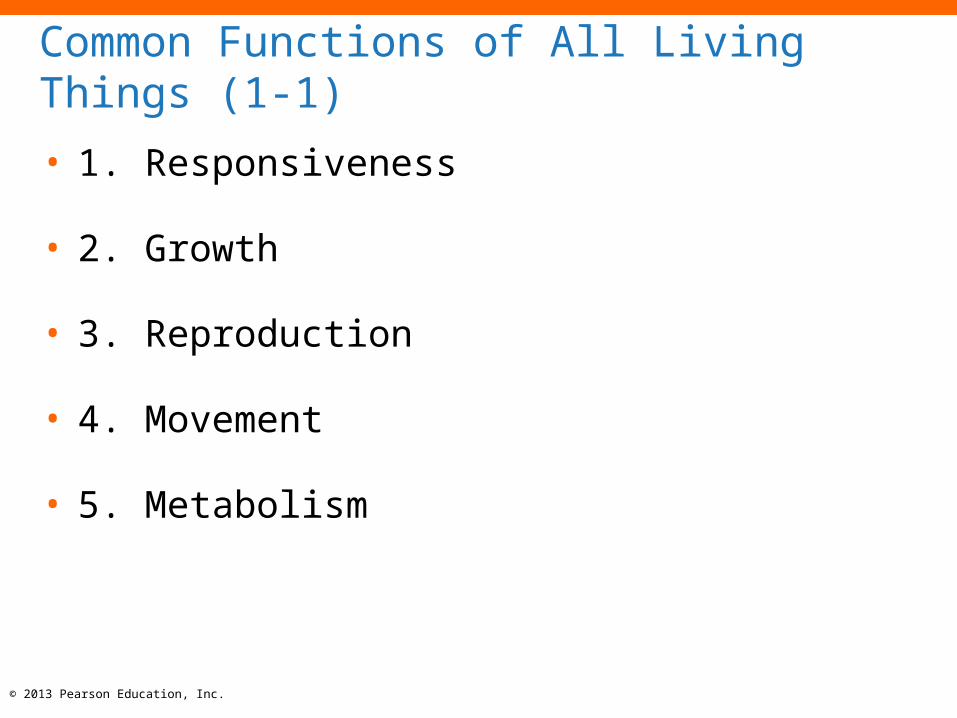
Common Functions of All Living Things (1-1)
• 1. Responsiveness
• 2. Growth
• 3. Reproduction
• 4. Movement
• 5. Metabolism
© 2013 Pearson Education, Inc.
Responsiveness and Growth (1-1)
• 1. Responsiveness
• Also called irritability
• An organism changes in response to its environment
• Longer term change is called adaptation
• 2. Growth
• An increase in size, number of cells, complexity of cells, or all
three
• The process of developing a variety of cells is called
differentiation

© 2013 Pearson Education, Inc.
Reproduction and Movement (1-1)
• 3. Reproduction
• Creation of new generations of similar organisms
• 4.Movement
• Ability to transport things within the internal environment of
the organism
• Ability to transport the organism through the external
environment
© 2013 Pearson Education, Inc.
Metabolism (1-1)
• 5. Metabolism
• The sum total of all the chemical reactions in the organism
• Uses resources absorbed from the environment
• Uses respiration for cellular production of energy
• Organism excretes any waste products left over from the
chemical reactions
© 2013 Pearson Education, Inc.
Checkpoint (1-1)
1. How are vital functions such as responsiveness,
growth, reproduction, and movement dependent
on metabolism?
What is Anatomy and Physiology?
• Intro to Anatomy and Physiology
• http://www.youtube.com/watch?v=f_v-3mW5uq4
© 2013 Pearson Education, Inc.

© 2013 Pearson Education, Inc.
Anatomy (1-2)
• The word anatomy means "a cutting open"
• The structure of things or how things are built
• Specifics of:
• Where things are
• What they are made of
• Physical relationships between parts

Anatomy (1-2)
• Two categories:• 1.Gross Anatomy
• 2. Microscopic Anatomy
© 2013 Pearson Education, Inc.

© 2013 Pearson Education, Inc.
1. Gross Anatomy (1-2)
• Also called macroscopic anatomy
• Studies visible structures
• Includes:
• Surface anatomy
• Study of general form and superficial markings
• Regional anatomy
• Study of all the superficial and internal features of a specific
region of the body
• Systemic anatomy
• Study of the structure of major organ systems

© 2013 Pearson Education, Inc.
2. Microscopic Anatomy (1-2)
• Studies structures that cannot be seen without
magnification
• Includes:
• Cytology
• Study of internal structure of individual cells
• Histology
• Study of tissues, groups of specialized cells and cell products
that work together to perform specific functions

© 2013 Pearson Education, Inc.
Physiology (1-2)
• The function of the anatomical structures
• Specifics of:
• How structures, organs, and systems work separately and
together

© 2013 Pearson Education, Inc.
Physiology (1-2)
• Human physiology specialties include:
• Cell physiology
• Study of the functions of living cells
• Special physiology
• Study of the physiology of specific organs
• Systemic physiology
• Study of all aspects of the function of specific organ systems
• Pathological physiology or pathology
• Study of the effects of diseases on organ or system functions

© 2013 Pearson Education, Inc.
Checkpoint (1-2)
2. Describe how anatomy and physiology are
closely related.
3. Would a histologist more likely be considered a
specialist in microscopic anatomy or gross
anatomy? Why?

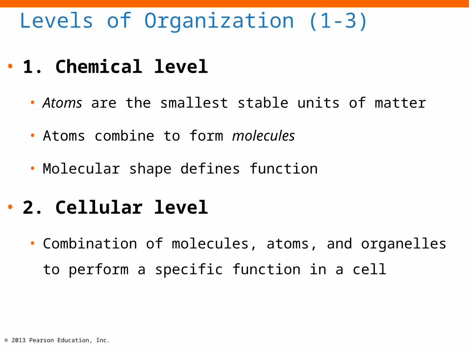
Levels of Organization (1-3)
• 1. Chemical level
• 2. Cellular level
• 3. Tissue level
• 4. Organ level
• 5. Organ system level
• 6. Organism level
© 2013 Pearson Education, Inc.
© 2013 Pearson Education, Inc.
Levels of Organization (1-3)
• 1. Chemical level
• Atoms are the smallest stable units of matter
• Atoms combine to form molecules
• Molecular shape defines function
• 2. Cellular level
• Combination of molecules, atoms, and organelles to perform a
specific function in a cell

© 2013 Pearson Education, Inc.
Levels of Organization (1-3)
• 3. Tissue level
• A collection of cells working together to perform a specific
function
• 4. Organ level
• Two or more tissues working together to perform specific
functions

© 2013 Pearson Education, Inc.
Levels of Organization (1-3)
• 5. Organ system level
• Two or more organs working together to perform specific
functions
• 6. Organism level
• Multiple organ systems working together to maintain health
© 2013 Pearson Education, Inc.
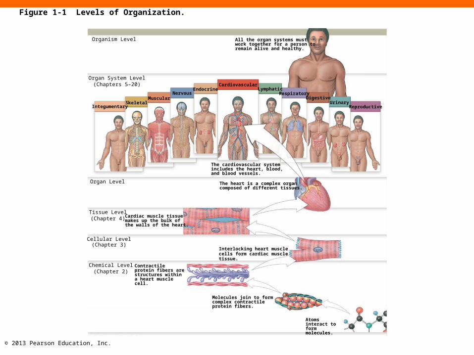
Organism Level All the organ systems mustwork together for a person toremain alive and healthy.
Organ System Level(Chapters 5–20)
IntegumentarySkeletal
MuscularNervous
EndocrineCardiovascular
LymphaticRespiratory
DigestiveUrinary
Reproductive
The cardiovascular systemincludes the heart, blood,and blood vessels.
The heart is a complex organcomposed of different tissues.
Organ Level
Tissue Level(Chapter 4) Cardiac muscle tissue
makes up the bulk ofthe walls of the heart.
Cellular Level(Chapter 3)
Interlocking heart muscle cells form cardiac muscle tissue.
Chemical Level(Chapter 2)
Contractile protein fibers are structures within a heart muscle cell.
Molecules join to form complex contractile protein fibers.
Atoms interact to form molecules.
Figure 1-1 Levels of Organization.

© 2013 Pearson Education, Inc.
Checkpoint (1-3)
4. Identify the major levels of organization of the
human body from the simplest to the most
complex.

© 2013 Pearson Education, Inc.
The 11 Organ Systems of the Human Body (1-4)
1. Integumentary
2. Skeletal
3. Muscular
4. Nervous
5. Endocrine
6. Cardiovascular
© 2013 Pearson Education, Inc.
The 11 Organ Systems of the Human Body (1-4)
7. Lymphatic
8. Respiratory
9. Digestive
10.Urinary
11.Reproductive

© 2013 Pearson Education, Inc.
The Integumentary System
Protects against environmental hazards; helps control body temperature
Hair
Skin
Nails
Figure 1-2 The Organ Systems of the Human Body. (1 of 12)
© 2013 Pearson Education, Inc.
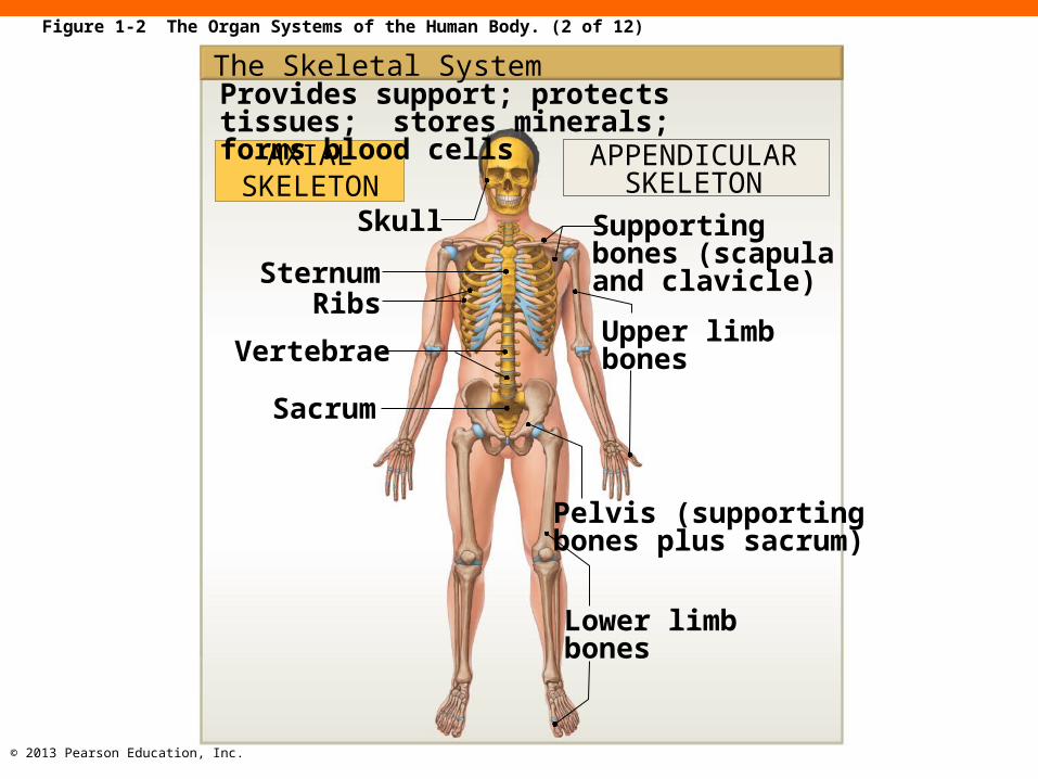
Figure 1-2 The Organ Systems of the Human Body. (2 of 12)
The Skeletal SystemProvides support; protects tissues; stores minerals; forms blood cells
AXIALSKELETON
APPENDICULARSKELETON
Skull
SternumRibs
Vertebrae
Sacrum
Supporting bones (scapula and clavicle)
Upper limbbones
Pelvis (supportingbones plus sacrum)
Lower limbbones

© 2013 Pearson Education, Inc.
Figure 1-2 The Organ Systems of the Human Body. (3 of 12)
The Muscular SystemAllows for locomotion; provides support; produces heat
Axialmuscles
Appendicularmuscles
Tendons

© 2013 Pearson Education, Inc.
Figure 1-2 The Organ Systems of the Human Body. (4 of 12)
The Nervous System
Directs immediate responses to stimuli, usually by coordinating the activities of other organ systems
CENTRALNERVOUS SYSTEM
BrainSpinal cord
PERIPHERAL NERVOUS SYSTEM
Peripheralnerves
© 2013 Pearson Education, Inc.
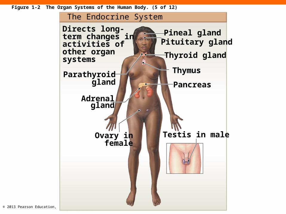
Figure 1-2 The Organ Systems of the Human Body. (5 of 12)
The Endocrine System
Directs long-term changes in activities of other organ systems
Parathyroidgland
Adrenalgland
Ovary infemale
Pineal glandPituitary gland
Thyroid gland
Thymus
Pancreas
Testis in male

© 2013 Pearson Education, Inc.
Figure 1-2 The Organ Systems of the Human Body. (6 of 12)
The Cardiovascular SystemTransports cells and dissolved materials, including nutrients, wastes, and gases
ArteryVein
Heart
Capillaries

© 2013 Pearson Education, Inc.
Figure 1-2 The Organ Systems of the Human Body. (7 of 12)
The Lymphatic SystemDefends against infection and disease; returns tissue fluid to the bloodstream
Thymus Lymphnodes
Spleen
Lymphaticvessel

© 2013 Pearson Education, Inc.
Figure 1-2 The Organ Systems of the Human Body. (8 of 12)
The Respiratory SystemDelivers air to sites where gas exchange can occur between the air and circulating blood; produces sound
Pharynx
Lung
Diaphragm
Nasal cavitySinus
Larynx
Trachea
Bronchi
© 2013 Pearson Education, Inc.
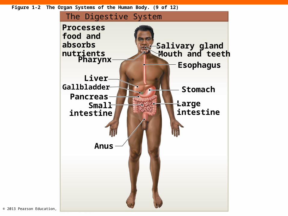
The Digestive SystemProcesses food and absorbs nutrients
Pharynx
LiverGallbladder
PancreasSmall
intestine
Anus
Salivary glandMouth and teeth
Esophagus
Stomach
Large intestine
Figure 1-2 The Organ Systems of the Human Body. (9 of 12)
© 2013 Pearson Education, Inc.
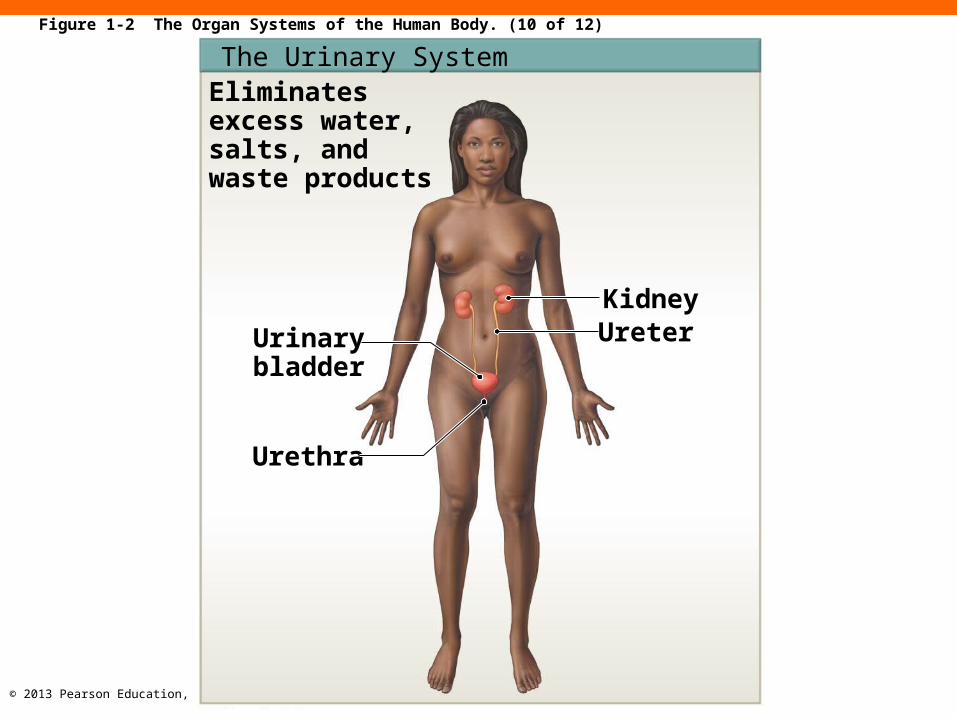
The Urinary SystemEliminates excess water, salts, and waste products
Urinarybladder
Urethra
KidneyUreter
Figure 1-2 The Organ Systems of the Human Body. (10 of 12)

© 2013 Pearson Education, Inc.
Figure 1-2 The Organ Systems of the Human Body. (11 of 12)
The Male Reproductive SystemProduces sex cells and hormones
Prostategland
Seminalgland
Ductusdeferens
Urethra
EpididymisTestisPenis
Scrotum

© 2013 Pearson Education, Inc.
The Female Reproductive SystemProduces sex cells and hormones; supports embryonic and fetal develop-ment fromfertilization to birth
Mammarygland
UterinetubeOvaryUterusVaginaExternalgenitalia
Figure 1-2 The Organ Systems of the Human Body. (12 of 12)

© 2013 Pearson Education, Inc.
Checkpoint (1-4)
5. Identify the organ systems of the body and list
their major functions.
6. Which organ system includes the pituitary gland
and directs long-term changes in the activities of
other systems?
© 2013 Pearson Education, Inc.
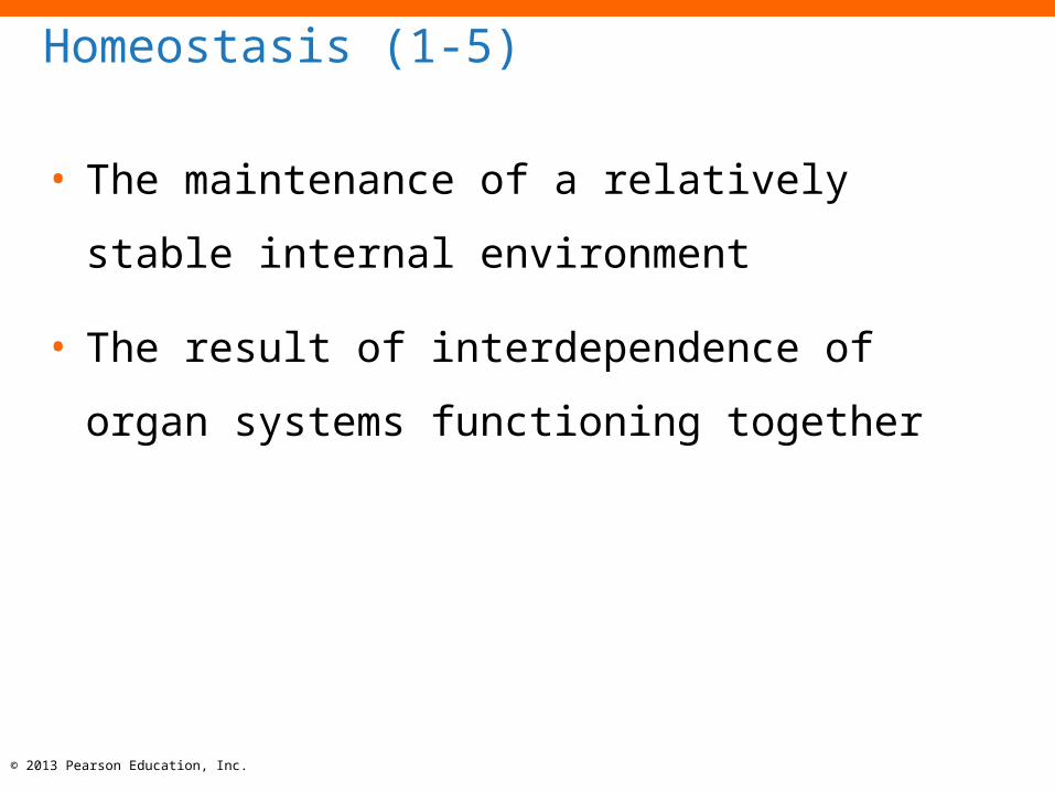
Homeostasis (1-5)
• The maintenance of a relatively stable internal
environment
• The result of interdependence of organ systems
functioning together
© 2013 Pearson Education, Inc.
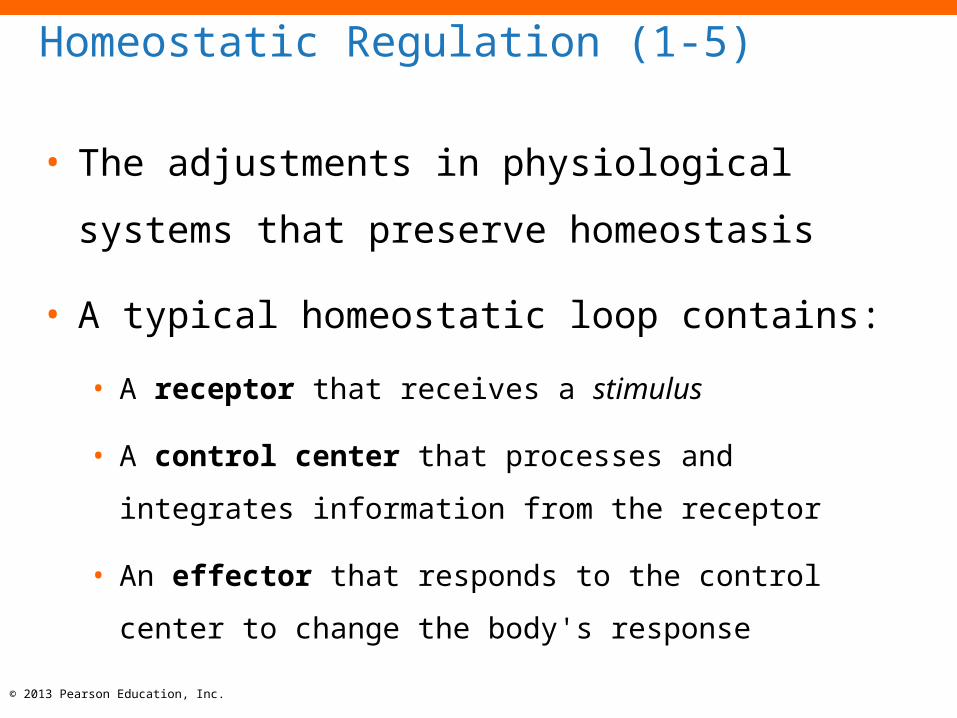
Homeostatic Regulation (1-5)
• The adjustments in physiological systems that
preserve homeostasis
• A typical homeostatic loop contains:
• A receptor that receives a stimulus
• A control center that processes and integrates information
from the receptor
• An effector that responds to the control center to change the
body's response

© 2013 Pearson Education, Inc.
Normalconditiondisturbed
RECEPTOR
ThermometerInformation
affects
STIMULUS:Room temperature
rises
HOMEOSTASIS
Normal roomtemperature
RESPONSE:Room temperature
drops
Normalconditionrestored
EFFECTOR
Air conditionerturns on
Sendscommands
to
CONTROL CENTER(Thermostat)
208
308
408
Figure 1-3 The Control of Room Temperature.

© 2013 Pearson Education, Inc.
Checkpoint (1-5)
7. Define homeostasis.
8. Why is homeostatic regulation important to an
organism?
© 2013 Pearson Education, Inc.
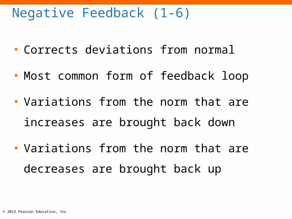
Negative Feedback (1-6)
• Corrects deviations from normal
• Most common form of feedback loop
• Variations from the norm that are increases are
brought back down
• Variations from the norm that are decreases are
brought back up
© 2013 Pearson Education, Inc.
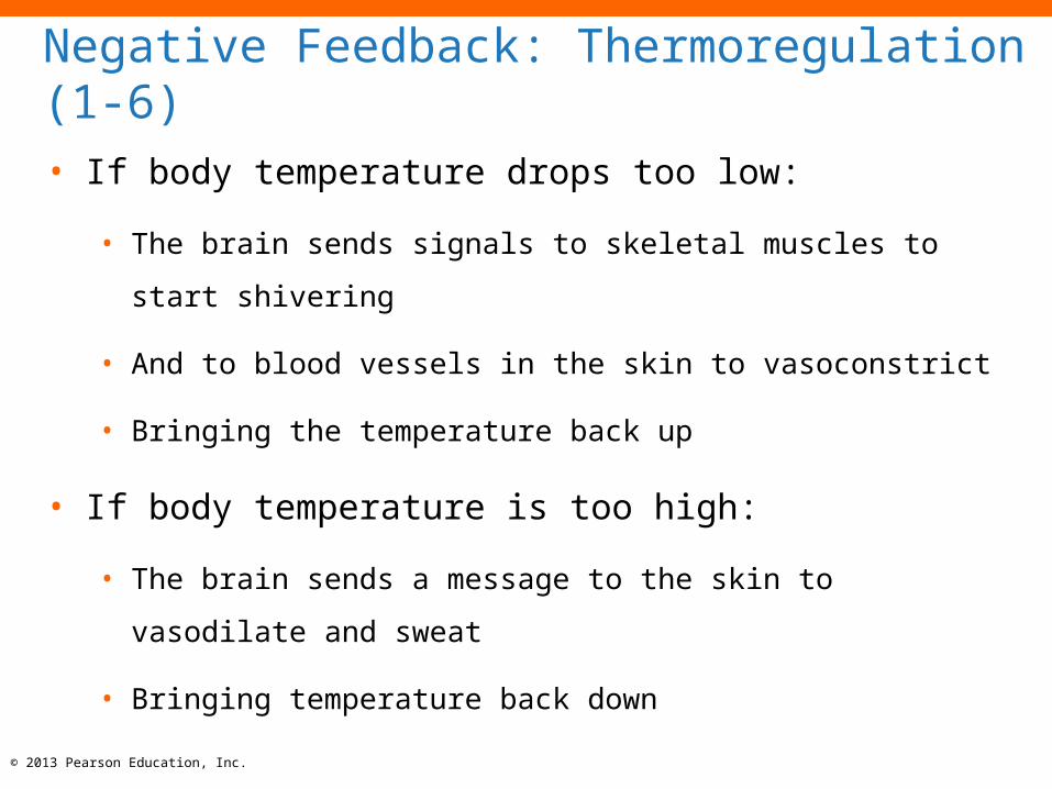
Negative Feedback: Thermoregulation (1-6)
• If body temperature drops too low:
• The brain sends signals to skeletal muscles to start shivering
• And to blood vessels in the skin to vasoconstrict
• Bringing the temperature back up
• If body temperature is too high:
• The brain sends a message to the skin to vasodilate and sweat
• Bringing temperature back down

© 2013 Pearson Education, Inc.
Positive Feedback (1-6)
• Reinforces or exaggerates deviations from normal
• Variations from the norm that are increases are
further increased
• Fairly rare occurrence and must have an "off
switch"
© 2013 Pearson Education, Inc.
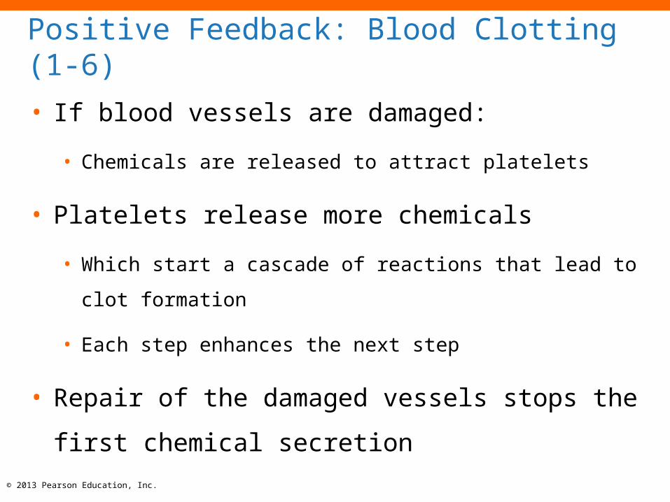
Positive Feedback: Blood Clotting (1-6)
• If blood vessels are damaged:
• Chemicals are released to attract platelets
• Platelets release more chemicals
• Which start a cascade of reactions that lead to clot formation
• Each step enhances the next step
• Repair of the damaged vessels stops the first
chemical secretion

© 2013 Pearson Education, Inc.
RECEPTORS
Body'stemperature
sensorsSTIMULUS
Body temperaturerises above 37.2°C(99°F)
RESPONSE
Increased blood flowto skinIncreased sweatingStimulus removedHomeostasis restored
Negativefeedback
EFFECTORS
Blood vessels and sweat glands
in skin
Sendscommands to
Thermoregulatorycenter in brain
If body temperature climbs above 37.2°C (99°F), heat loss is increased through enhanced blood flow to the skin and increased sweating.
Informationaffects
CONTROLCENTER
Controlmechanismwhen bodytemperature
rises
Sendscommands to
EFFECTORS
Blood vessels and sweat glands
in skinSkeletal muscles
Negativefeedback
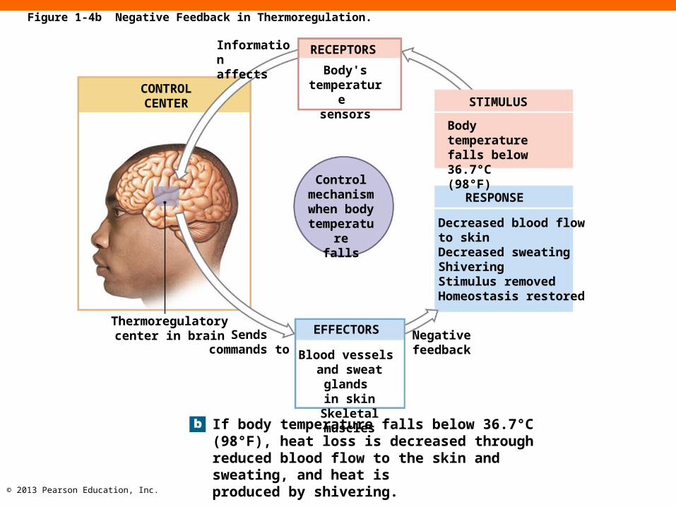
If body temperature falls below 36.7°C (98°F), heat loss is decreased through reduced blood flow to the skin and sweating, and heat isproduced by shivering.
Controlmechanismwhen bodytemperature
fallsDecreased blood flowto skinDecreased sweatingShiveringStimulus removedHomeostasis restored
RESPONSE
STIMULUS
Body temperaturefalls below 36.7°C(98°F)
Informationaffects
RECEPTORS
Body'stemperature
sensors
Figure 1-4 Negative Feedback in Thermoregulation.

© 2013 Pearson Education, Inc.
Figure 1-4a Negative Feedback in Thermoregulation.
RECEPTORS
Body'stemperature
sensorsSTIMULUS
Body temperaturerises above 37.2°C(99°F)
RESPONSE
Increased blood flowto skinIncreased sweatingStimulus removedHomeostasis restored
Negativefeedback
EFFECTORS
Blood vessels and sweat glands
in skin
Sendscommands to
If body temperature climbs above 37.2°C (99°F), heat loss is increased through enhanced blood flow to the skin and increased sweating.
Informationaffects
Controlmechanismwhen bodytemperature
rises
Thermoregulatorycenter in brain
CONTROLCENTER
© 2013 Pearson Education, Inc.
Figure 1-4b Negative Feedback in Thermoregulation.
Thermoregulatorycenter in brain
CONTROLCENTER
Sendscommands to
EFFECTORS
Blood vessels and sweat glands
in skinSkeletal muscles
Negativefeedback
If body temperature falls below 36.7°C (98°F), heat loss is decreased through reduced blood flow to the skin and sweating, and heat isproduced by shivering.
Controlmechanismwhen bodytemperature
falls
RESPONSE
STIMULUS
Informationaffects
RECEPTORS
Body'stemperature
sensors
Decreased blood flowto skinDecreased sweatingShiveringStimulus removedHomeostasis restored
Body temperaturefalls below 36.7°C(98°F)
© 2013 Pearson Education, Inc.
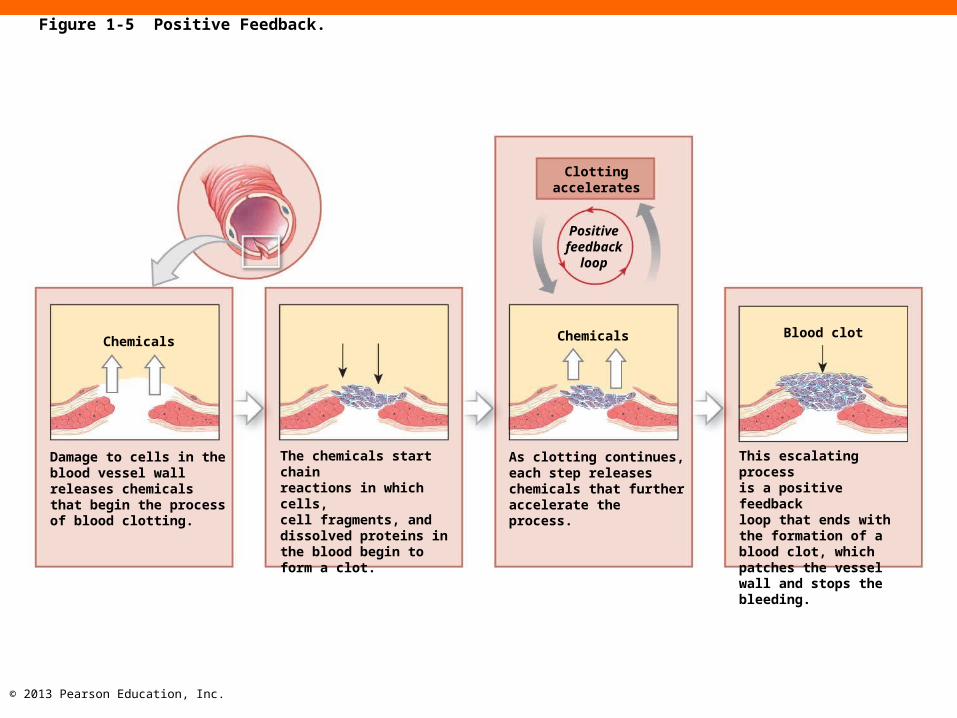
Chemicals
Damage to cells in the blood vessel wall releases chemicals that begin the process of blood clotting.
The chemicals start chainreactions in which cells,cell fragments, and dissolved proteins in the blood begin to form a clot.
As clotting continues, each step releases chemicals that further accelerate the process.
This escalating processis a positive feedbackloop that ends with the formation of a blood clot, which patches the vessel wall and stops thebleeding.
Chemicals Blood clot
Clottingaccelerates
Positive feedback
loop
Figure 1-5 Positive Feedback.

© 2013 Pearson Education, Inc.
Chemicals
Damage to cells in the blood vessel wall releases chemicals that begin the process of blood clotting.
Figure 1-5 Positive Feedback. (1 of 4)

© 2013 Pearson Education, Inc.
The chemicals start chainreactions in which cells,cell fragments, and dissolved proteins in the blood begin to form a clot.
Figure 1-5 Positive Feedback. (2 of 4)
© 2013 Pearson Education, Inc.
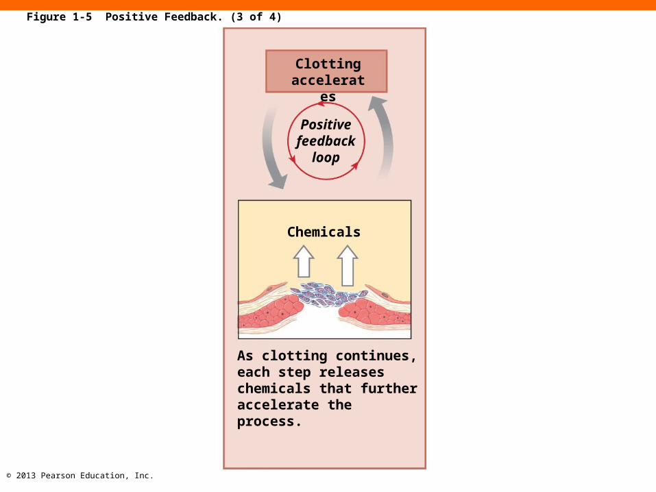
As clotting continues, each step releases chemicals that further accelerate the process.
Chemicals
Clottingaccelerates
Positive feedback
loop
Figure 1-5 Positive Feedback. (3 of 4)

© 2013 Pearson Education, Inc.
This escalating processis a positive feedbackloop that ends with the formation of a blood clot, which patches the vessel wall and stops thebleeding.
Blood clot
Figure 1-5 Positive Feedback. (4 of 4)
© 2013 Pearson Education, Inc.
Checkpoint (1-6)
9. Explain the function of negative feedback
systems.
10.Why is positive feedback helpful in blood clotting
but unsuitable for the regulation of body
temperature?
11.What happens to the body when homeostasis
breaks down?

© 2013 Pearson Education, Inc.
Anatomical Terminology (1-7)
• Medical terminology
• Describes body regions, anatomical positions and
directions, and body sections
• 1. Surface anatomy
• 2. Sectional anatomy
1. Surface Anatomy
• 1a.Anatomical Landmarks
• Anatomical position: hands at sides, palms forward and feet together
• Supine: lying down, face up
• Prone: lying down, face down
• 1b.Anatomical Regions
• Body regions
• Abdominopelvic quadrants
• Abdominopelvic regions
• 1c.Anatomical Directions
• Reference terms based on subject
© 2013 Pearson Education, Inc.
© 2013 Pearson Education, Inc.
1a. Anatomical Landmarks (1-7)
• 1a. Anatomical position
• Hands at the sides with the palms facing forward and feet
together
• Supine (face up)
• Prone (face down)

© 2013 Pearson Education, Inc.
Forehead (frontal)
Craniumor skull
(cranial)Face
(facial)
Cephalonor head
(cephalic)
Oris or mouth (oral)Chin
(mental)Axilla or armpit
(axillary)Brachium
or arm(brachial)
Antecubitis orfront of elbow
(antecubital)Antebrachium
or forearm(antebrachial)
Carpus orwrist (carpal)
Palm(palmar)
Pollexor thumb Digits (phalanges)
or fingers (digitalor phalangeal)
Patellaor kneecap
(patellar)Leg (crural)
Tarsus orankle
(tarsal)Digits (phalanges)
or toes (digital orphalangeal)
Hallux or great toe
Nasus or nose (nasal)
Oculus or eye (orbital or ocular)
Auris or ear (otic)
Cheek (buccal)
Cervicis or neck (cervical)
Thoracis orthorax, chest(thoracic)
Mammaor breast(mammary)
Abdomen(abdominal)
Umbilicusor navel(umbilical)
Pelvis(pelvic)
Manusor hand(manual)
Groin(inguinal)
Pubis(pubic)
Femur orthigh (femoral)
Pes or foot(pedal)
Trunk
Anterior view in the anatomical position
Figure 1-6a Anatomical Landmarks.

© 2013 Pearson Education, Inc.
Figure 1-6b Anatomical Landmarks.
Cephalonor head (cephalic)
Cervicisor neck (cervical)
Shoulder(acromial)
Dorsum orback
(dorsal)
Olecranonor back
of elbow(olecranal)
Lumbus or loin(lumbar)
Gluteusor buttock
(gluteal)Popliteus orback of knee
(popliteal)
Calf (sural)
Calcaneus orheel of foot(calcaneal)
Planta orsole of foot (plantar)
Lowerlimb
Upperlimb
Posterior view in the anatomical position
© 2013 Pearson Education, Inc.
1b. Anatomical Regions (1-7)
• Major regions of the body
• Two methods to map the surface of the abdomen
and pelvis
1. Abdominopelvic quadrants
2. Abdominopelvic regions

© 2013 Pearson Education, Inc.
Right UpperQuadrant
(RUQ)
Right LowerQuadrant
(RLQ)
Left UpperQuadrant(LUQ)
Left LowerQuadrant(LLQ)
Abdominopelvic quadrants. The four abdominopelvic quadrants are formed by two perpendicular lines that intersect at the navel (umbilicus). The terms for these quadrants, or their abbreviations, are most often used in clinical discussions.
Figure 1-7a Abdominopelvic Quadrants and Regions.

© 2013 Pearson Education, Inc.
Figure 1-7b Abdominopelvic Quadrants and Regions.
Righthypochondriac
region
Right lumbarregionRight
inguinalregion
Lefthypochondriacregion
Left lumbarregionLeft inguinalregion
Epigastricregion
Umbilicalregion
Hypogastric(pubic)region
Abdominopelvic regions. The nine abdominopelvicregions provide more precise regional descriptions.
© 2013 Pearson Education, Inc.
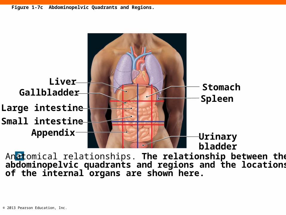
LiverGallbladder
Large intestine
Small intestineAppendix
StomachSpleen
Urinarybladder
Anatomical relationships. The relationship between the abdominopelvic quadrants and regions and the locations of the internal organs are shown here.
Figure 1-7c Abdominopelvic Quadrants and Regions.

© 2013 Pearson Education, Inc.
1c. Anatomical Directions (1-7)
• Provide an orientation of structures relative to
anatomical position
• Left and right always refer to the left and right
sides of the subject, not the observer
© 2013 Pearson Education, Inc.
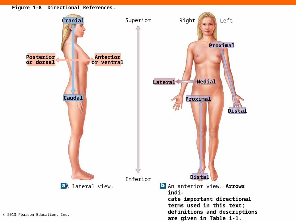
Posterioror dorsal
Cranial
Anterioror ventral
Caudal
A lateral view.
Superior Right Left
Lateral
Proximal
Medial
Proximal
Distal
DistalInferiorAn anterior view. Arrows indi-cate important directional terms used in this text; definitions and descriptions are given in Table 1-1.
Figure 1-8 Directional References.

© 2013 Pearson Education, Inc.
Table 1-1 Directional Terms

© 2013 Pearson Education, Inc.
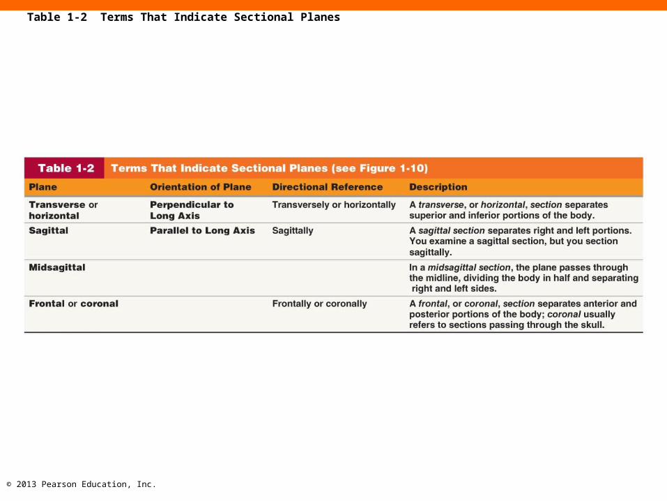
2. Sectional Anatomy (1-7)
• A "slice" through the body in three primary sectional
planes
1. Transverse plane
• Divides body into superior and inferior portions
2. Frontal plane
• Divides body into anterior and posterior portions
3. Sagittal plane
• Divides body into left and right portions

© 2013 Pearson Education, Inc.
Frontal plane
Transverse plane
Sagittal plane
Figure 1-10 Planes of Section.
© 2013 Pearson Education, Inc.
Table 1-2 Terms That Indicate Sectional Planes

© 2013 Pearson Education, Inc.
Checkpoint (1-7)
12. What is the purpose of anatomical terms?
13. Describe an anterior view and a posterior view in
the anatomical position.
14. What type of section would separate the two
eyes?
© 2013 Pearson Education, Inc.
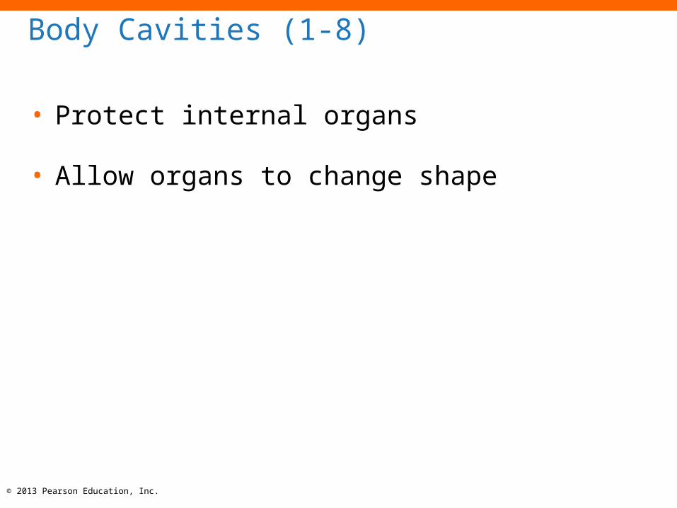
Body Cavities (1-8)
• Protect internal organs
• Allow organs to change shape

© 2013 Pearson Education, Inc.
Ventral Body Cavity (1-8)
• Contains viscera:
• Organs of the respiratory, cardiovascular, digestive, urinary, and
reproductive systems
• Cavities are lined (parietal layer) and organs are enclosed
(visceral layer) by serous membranes
• Two major divisions of Ventral Body Cavity
1. Thoracic cavity
2. Abdominopelvic cavity

© 2013 Pearson Education, Inc.
1. Thoracic Cavity (1-8)
• Three internal chambers
• One pericardial cavity and two pleural cavities
• Each cavity is defined by a serous membrane

© 2013 Pearson Education, Inc.
Pericardial Cavity of the Thoracic Cavity (1-8)
• Contains the heart, and is found in the
mediastinum
• Defined by serous membrane, pericardium
• Visceral pericardium is the layer covering the
heart
• Parietal pericardium is the outer layer
• In between two layers is serous fluid to reduce
friction

© 2013 Pearson Education, Inc.
Pleural Cavities of the Thoracic Cavity (1-8)
• Each lung is found within its own pleural cavity
• Serous membrane is the pleura
• Visceral pleura is the layer covering the lung
• Parietal pleura defines the edge of the
mediastinum and lines the inner surface of the
chest wall
© 2013 Pearson Education, Inc.
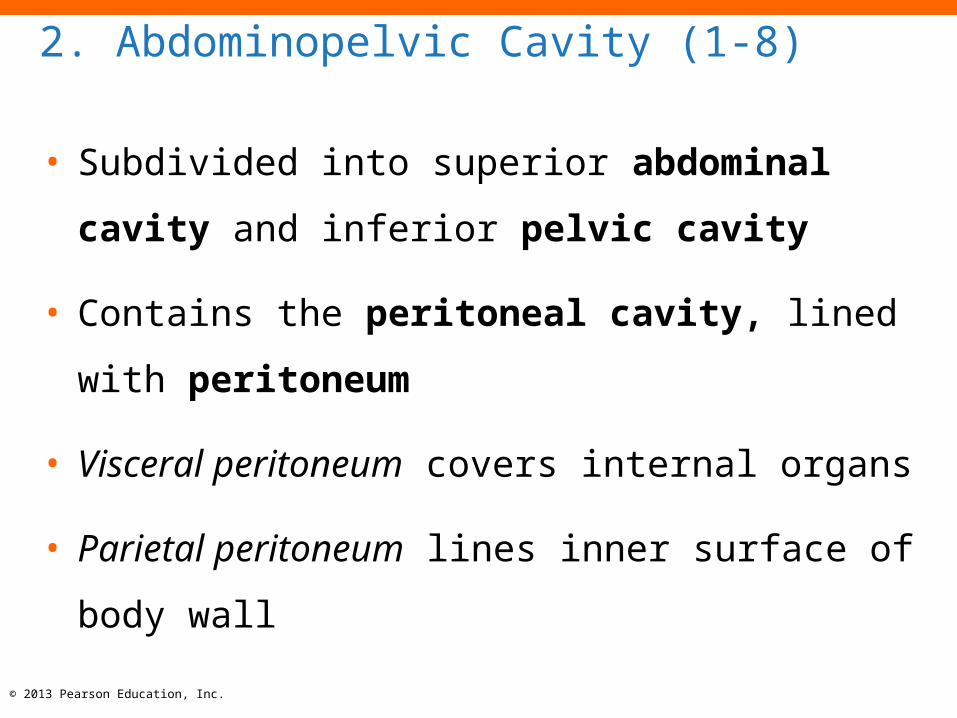
2. Abdominopelvic Cavity (1-8)
• Subdivided into superior abdominal cavity and
inferior pelvic cavity
• Contains the peritoneal cavity, lined with
peritoneum
• Visceral peritoneum covers internal organs
• Parietal peritoneum lines inner surface of body
wall
© 2013 Pearson Education, Inc.
Checkpoint (1-8)
15.Describe two essential functions of body
cavities.
16. Identify the subdivisions of the ventral body
cavity.
17. If a surgeon makes an incision just inferior to the
diaphragm, what body cavity will be opened?