Human A-I geneexpressionincreases highdensity ... · Proc. Natl. Acad. Sci. USA Vol. 91, pp....
5
Proc. Natl. Acad. Sci. USA Vol. 91, pp. 9607-9611, September 1994 Genetics Human apolipoprotein A-I gene expression increases high density lipoprotein and suppresses atherosclerosis in the apolipoprotein E-deficient mouse ANDREW S. PLUMP, CHRISTOPHER J. SCOrr, AND JAN L. BRESLOW* The Rockefeller University, 1230 York Avenue, New York, NY 10021 Communicated by Alexander G. Bearn, June 9, 1994 ABSTRACT Atherosclerosis is a complex disease with both genetic and environmental determinants. Apolipoprotein (Apo) E-deficient mice have been created that are hibly ssceptible to atherosclerosis. In order to assess the role of human apolipoprotein (hApo) A-I and hig density lipoprotein (HDL) in atherosclerosis susceptibility, traenlc mice over- expressing the hApo A-I gene were crossed with Apo E-defi- dent mice. Apo E -/-, hApo A-I mice with two-fold elevation in HDL cholesterol have markedly shed atherosclerosis with less fibroproliferative lesions by 8 months of age. A strong reciprocal relationship between HDL cholesterol levels and atherosclerosis was found with HDL levels accounting for 78% of the observed variance in mean lesion area. The effect of HDL on atherosclerosis resistance was independent of non-HDL cholesterol. Atherosclerosis is influenced by many genetic and environ- mental factors. In particular, altered lipoprotein levels caused by diet or mutant genes have been strongly associated with susceptibility to atherosclerotic coronary heart disease. Epidemiological studies have shown a strong inverse corre- lation between plasma high density lipoprotein (HDL) cho- lesterol levels and coronary heart disease (1). The principal structural protein of HDL is apolipoprotein (Apo) A-I, a 28-kDa protein found in human plasma at a mean concentra- tion of 125 mg/dl (2). HDL cholesterol levels are highly correlated with plasma Apo A-I levels. Individuals have been described with Apo A-I mutations precluding synthesis of normal Apo A-I (3-6). These individuals have low HDL cholesterol levels and severe premature coronary heart dis- ease. Transgenic mice expressing the human Apo (hApo) A-I gene have increased HDL cholesterol levels (7, 8). These two observations suggest that Apo A-I genetic variation can affect HDL cholesterol levels, which in turn may alter atheroscle- rosis susceptibility. Although epidemiological studies in hu- mans (1) and studies of diet-induced fatty streak lesions in laboratory animals (8, 9) suggest a role for HDL in mediating atherosclerosis susceptibility, direct proof that genetic vari- ability in HDL cholesterol levels can diminish atherosclerosis susceptibility would require specifically increasing the HDL cholesterol levels and demonstrating a decrease in fibropro- liferative atherosclerotic lesions. In addition to documenting the role of HDL in atherosclerosis susceptibility, this system should provide understanding of the mechanism by which HDL acts. Gene targeting has been used to create Apo E-deficient mice (10-12). These animals develop severe hypercholester- olemia on a low-fat, low-cholesterol, mouse chow diet. The predominant lipoprotein abnormality in the Apo E-deficient mice is the accumulation of (3 very low density lipoprotein (f3-VLDL), a cholesterol ester-enriched atherogenic lipopro- tein particle. On the mouse chow diet, Apo E-deficient mice develop extensive atherosclerosis with lesions progressing from endothelial-monocyte adhesions to lipid-laden fatty streaks to advanced fibroproliferative lesions by 20-30 weeks of age (13, 14). This progression of atherosclerotic lesions is seen in humans. The Apo E-deficient mouse model is differ- ent from the previous mouse model of atherosclerosis, the C57BL/6 diet-induced model (15). In the latter, an athero- genic, high-cholesterol (1%), cholic acid (0.5%)-containing diet is required to produce foam cells at the base of the aorta. Apo A-I overexpression in this model leads to diminished foam-cell formation (8). Though encouraging, the model is limited because the atherogenic diet is not physiological (16) and has significant hepatotoxicity. Furthermore, lesions re- sulting from this diet fail to progress through the phases of atherosclerosis to fibroproliferative plaques. Thus, the Apo E-deficient mouse model is a more suitable one for assessing the effect of HDL cholesterol levels on atherosclerosis. MATERIALS AND METHODS Mice. The creation of the transgenic hApo A-I mice line 179) (7) and the Apo E-knockout mice (11) used in this study has been described. To generate the study populations, a C57BL/6 x 12901a hybrid mouse containing the Apo E -/- mutation was crossed with a C57BL/6 x CBA hybrid mouse containing the hApo A-I transgene. Resulting F1 Apo E +/- mice containing the hApo A-I transgene were backcrossed to C57BL/6 x 12901A Apo E -/- hybrid mice. All experi- ments performed with mice were in accordance with the Rockefeller University Animal Care and Use Committee. Apolipoproteln and ipoprotein Analysis. Lipoprotein mea- surements were made on 10- to 15-week-old chow-fed mice after an 8-hr fast. The lipoprotein and apolipoprotein data in the text and in Table 1 are from pooled mice from the 4- and 8-month-old study groups depicted in Fig. 2. These data were pooled because serial chronological examination of individ- ual mice and comparison of groups of mice of different ages have revealed that mouse age does not affect plasma choles- terol levels. Cholesterol measurements were made by enzy- matic assay (Boehringer Mannheim, no. 816302). For HDL cholesterol measurement, individual mouse plasma was first separated at a density (d) = 1.006 g/ml in a Beckman Airfuge. Cholesterol measurement was then made on the d > 1.006 g/ml fraction after dextran sulfate precipitation. Human Apo A-I levels were measured by ELISA using a goat anti-human Apo A-I antibody. FPLC (Pharmacia) analysis of plasma lipoproteins was performed as described (11). Quantitative Athe Measurements. Mice were sac- rificed at 4 or 6 months of age and perfused at physiologic pressure with 0.9%6 NaCl by cardiac intraventricular canali- zation. The heart and proximal aorta were isolated and fixed Abbreviations: Apo, apolipoprotein; hApo, human apolipoprotein; HDL, high density lipoprotein; VLDL, very low density lipoprotein. *To whom reprint requests should be addressed. 9607 The publication costs of this article were defrayed in part by page charge payment. This article must therefore be hereby marked "advertisement" in accordance with 18 U.S.C. §1734 solely to indicate this fact. Downloaded by guest on May 28, 2020
Transcript of Human A-I geneexpressionincreases highdensity ... · Proc. Natl. Acad. Sci. USA Vol. 91, pp....

Proc. Natl. Acad. Sci. USAVol. 91, pp. 9607-9611, September 1994Genetics
Human apolipoprotein A-I gene expression increases high densitylipoprotein and suppresses atherosclerosis in the apolipoproteinE-deficient mouseANDREW S. PLUMP, CHRISTOPHER J. SCOrr, AND JAN L. BRESLOW*The Rockefeller University, 1230 York Avenue, New York, NY 10021
Communicated by Alexander G. Bearn, June 9, 1994
ABSTRACT Atherosclerosis is a complex disease with bothgenetic and environmental determinants. Apolipoprotein(Apo) E-deficient mice have been created that are hiblyssceptible to atherosclerosis. In order to assess the role ofhuman apolipoprotein (hApo) A-I and hig density lipoprotein(HDL) in atherosclerosis susceptibility, traenlc mice over-expressing the hApo A-I gene were crossed with Apo E-defi-dent mice. Apo E -/-, hApo A-I mice with two-fold elevationin HDL cholesterol have markedly shed atherosclerosiswith less fibroproliferative lesions by 8 months ofage. A strongreciprocal relationship between HDL cholesterol levels andatherosclerosis was found with HDL levels accounting for 78%of the observed variance in mean lesion area. The effect ofHDLon atherosclerosis resistance was independent of non-HDLcholesterol.
Atherosclerosis is influenced by many genetic and environ-mental factors. In particular, altered lipoprotein levelscaused by diet or mutant genes have been strongly associatedwith susceptibility to atherosclerotic coronary heart disease.Epidemiological studies have shown a strong inverse corre-lation between plasma high density lipoprotein (HDL) cho-lesterol levels and coronary heart disease (1). The principalstructural protein of HDL is apolipoprotein (Apo) A-I, a28-kDa protein found in human plasma at a mean concentra-tion of 125 mg/dl (2). HDL cholesterol levels are highlycorrelated with plasma Apo A-I levels. Individuals have beendescribed with Apo A-I mutations precluding synthesis ofnormal Apo A-I (3-6). These individuals have low HDLcholesterol levels and severe premature coronary heart dis-ease. Transgenic mice expressing the human Apo (hApo) A-Igene have increased HDL cholesterol levels (7, 8). These twoobservations suggest thatApo A-I genetic variation can affectHDL cholesterol levels, which in turn may alter atheroscle-rosis susceptibility. Although epidemiological studies in hu-mans (1) and studies of diet-induced fatty streak lesions inlaboratory animals (8, 9) suggest a role for HDL in mediatingatherosclerosis susceptibility, direct proof that genetic vari-ability in HDL cholesterol levels can diminish atherosclerosissusceptibility would require specifically increasing the HDLcholesterol levels and demonstrating a decrease in fibropro-liferative atherosclerotic lesions. In addition to documentingthe role ofHDL in atherosclerosis susceptibility, this systemshould provide understanding of the mechanism by whichHDL acts.Gene targeting has been used to create Apo E-deficient
mice (10-12). These animals develop severe hypercholester-olemia on a low-fat, low-cholesterol, mouse chow diet. Thepredominant lipoprotein abnormality in the Apo E-deficientmice is the accumulation of (3 very low density lipoprotein(f3-VLDL), a cholesterol ester-enriched atherogenic lipopro-
tein particle. On the mouse chow diet, Apo E-deficient micedevelop extensive atherosclerosis with lesions progressingfrom endothelial-monocyte adhesions to lipid-laden fattystreaks to advanced fibroproliferative lesions by 20-30weeksof age (13, 14). This progression of atherosclerotic lesions isseen in humans. The Apo E-deficient mouse model is differ-ent from the previous mouse model of atherosclerosis, theC57BL/6 diet-induced model (15). In the latter, an athero-genic, high-cholesterol (1%), cholic acid (0.5%)-containingdiet is required to produce foam cells at the base ofthe aorta.Apo A-I overexpression in this model leads to diminishedfoam-cell formation (8). Though encouraging, the model islimited because the atherogenic diet is not physiological (16)and has significant hepatotoxicity. Furthermore, lesions re-sulting from this diet fail to progress through the phases ofatherosclerosis to fibroproliferative plaques. Thus, the ApoE-deficient mouse model is a more suitable one for assessingthe effect of HDL cholesterol levels on atherosclerosis.
MATERIALS AND METHODSMice. The creation of the transgenic hApo A-I mice line
179) (7) and the Apo E-knockout mice (11) used in this studyhas been described. To generate the study populations, aC57BL/6 x 12901a hybrid mouse containing the Apo E -/-mutation was crossed with a C57BL/6 x CBA hybrid mousecontaining the hApo A-I transgene. Resulting F1 Apo E +/-mice containing the hApo A-I transgene were backcrossed toC57BL/6 x 12901A Apo E -/- hybrid mice. All experi-ments performed with mice were in accordance with theRockefeller University Animal Care and Use Committee.
Apolipoproteln and ipoprotein Analysis. Lipoprotein mea-surements were made on 10- to 15-week-old chow-fed miceafter an 8-hr fast. The lipoprotein and apolipoprotein data inthe text and in Table 1 are from pooled mice from the 4- and8-month-old study groups depicted in Fig. 2. These data werepooled because serial chronological examination of individ-ual mice and comparison of groups of mice of different ageshave revealed that mouse age does not affect plasma choles-terol levels. Cholesterol measurements were made by enzy-matic assay (Boehringer Mannheim, no. 816302). For HDLcholesterol measurement, individual mouse plasma was firstseparated at a density (d) = 1.006 g/ml in a Beckman Airfuge.Cholesterol measurement was then made on the d > 1.006g/ml fraction after dextran sulfate precipitation. Human ApoA-I levels were measured by ELISA using a goat anti-humanApo A-I antibody. FPLC (Pharmacia) analysis of plasmalipoproteins was performed as described (11).
Quantitative Athe Measurements. Mice were sac-rificed at 4 or 6 months of age and perfused at physiologicpressure with 0.9%6 NaCl by cardiac intraventricular canali-zation. The heart and proximal aorta were isolated and fixed
Abbreviations: Apo, apolipoprotein; hApo, human apolipoprotein;HDL, high density lipoprotein; VLDL, very low density lipoprotein.*To whom reprint requests should be addressed.
9607
The publication costs of this article were defrayed in part by page chargepayment. This article must therefore be hereby marked "advertisement"in accordance with 18 U.S.C. §1734 solely to indicate this fact.
Dow
nloa
ded
by g
uest
on
May
28,
202
0

Proc. Natl. Acad. Sci. USA 91 (1994)
for 5 days or more in phosphate-buffered formaldehyde.After fixation, hearts were embedded in 25% gelatin andcryostat sectioned at 10-lm thickness. Processing and stain-ing of tissues were carried out according to Paigen et al. (17).Lesion area was quantified by the method of Rubin et al. (8)with one addition. Because the lesions in the Apo E-deficientmice are not limited to fatty streaks but progress to fibro-proliferative lesions, we quantified not only the fatty portionof the lesion but the entire lesion area from the lumen to thepoint of normal vessel wall.
Qualitative Atherosclerosis Analysis. After intraventricularperfusion with 0.9% NaCl the heart and proximal aorta wereremoved and quick frozen in liquid N2. Frozen sections weregenerated at 6-,Am thickness and placed on lysine-coatedslides. With standard histologic techniques slides were thenstained with one of three stains: oil red 0 and hematoxylinwith a light green counterstain, hematoxylin and eosin, orMasson's trichrome.
Statistical Analysis. Plasma hApo A-I, non-HDL choles-terol, andHDL cholesterol levels are reported as mean ± SD.Comparisons between groups were made by using the Stu-dent t test with significance at P < 0.05. Simple and multiplelinear regressions were used to determine the correlation ofHDL and non-HDL cholesterol to mean lesion area.
RESULTSTo assess the role of HDL in determining susceptibility toatherosclerosis, a hApo A-I transgene was bred onto the ApoE-deficient mouse background. The line of hApo A-I trans-genic mice used to generate the study population, line 179from Walsh et al. (7), has a wide range of plasma hApo A-Ilevels. Because of this, the study populations were dividedinto three groups, Apo E -/-; Apo E -/-, low-expressing(low) hApo A-I (plasma hApo A-I < 200 mg/dl); and Apo E-/-, high-expressing (high) hApo A-I (hApo A-I > 200mg/dl) mice. Control mice and low and high hApo A-I micewere generated within the same litter. hApo A-I levels were153 ± 26 mg/dl in a representative group of low hApo A-Imice, and 275 ± 50 mg/dl in a group of high hApo A-I mice(Table 1). Sizing FPLC (Fig. 1) showed a significant differ-ence in the plasma lipoprotein pattern between the Apo E-/- mice and the Apo E -/-, high hApo A-I mice. HDLcholesterol levels were significantly elevated in the lattergroup, whereas non-HDL cholesterol levels were similar. Nodifferences were detected between the Apo E -/-, lowhApo A-I mice and the Apo E -/- mice. Control Apo E -/-mice had HDL cholesterol levels of50 ± 17 mg/dl, while ApoE -/-, high hApo A-I mice had HDL cholesterol levels of105 + 32 mg/dl (P < 0.0001) (Table 1). Total cholesterol andnon-HDL cholesterol were not significantly different be-tween the two groups (Fig. 1, Table 1). In the Apo E -/-,low hApo A-I mice as compared to the Apo E -/- mice therewas no significant difference between HDL or non-HDLcholesterol.
0
,300IA
200 -
i 100 L
00
15 20 25 30 35Fraction (ml)
FIG. 1. Sizing FPLC ofplasmafrom ApoE --and ApoE --,high hApo A-I transgenic mice. As previously described (11), 100 p1
of plasma pooled from several mice was loaded onto two Superose6 columns linked in series. Samples were eluted at a constant flowrate of 0.3 ml/min with 1 mM EDTA/0.15 M NaCl. Fractions of 0.5ml were collected and cholesterol concentration was measuredenzymatically. Cholesterol levels are shown as pig per fraction per100 y4 of plasma. Solid line, Apo E -/-; dashed line, Apo E -/-,high hApo A-I.
To assess the effect of elevated plasma hApo A-I levels onthe development of atherosclerosis, groups of chow-fed ApoE -/-; Apo E -/-, low hApo A-I; and Apo E -/-, highhApo A-I mice were analyzed for lesion formation at 4months of age. On a chow diet Apo E -/- mice are knownto develop extensive fatty streak lesions by 4 months of age(13). Animals were analyzed quantitatively for proximal aortaatherosclerosis. Atherosclerosis was quantified by assessingmean lesion area over an =350-pum segment of the proximalaorta (17). Unlike previous studies assessing lesion area inthe mouse, the current studies made use of the observationthat lesions in the Apo E-deficient mice are not limited tofatty streaks, but progress to fibroproliferative lesions. Assuch, results include quantification of the entire lesion fromthe lumen to the point of normal vessel wall.High expression of hApo A-I led to a significant decrease
in the development of atherosclerotic lesions. At 4 months ofage, Apo E -/- mice had a mean lesion area of 22,964 ±
23,030 pm2 (n = 34) compared with a mean lesion area of470± 825 pm2 in Apo E -/-, high hApo A-I mice (n = 12, P <0.0001) (Fig. 2A). Six out of 12 of the Apo E -/-, high ApoA-I mice had no detectable lesions at this time point. At 4months Apo E -/- mice had well-defined fatty streaklesions (Fig. 3A), and the Apo E -/-, high hApo A-I micethat did have detectable pathology had nascent fatty streaklesions (Fig. 3B). The Apo E -/-, low hApo A-I mice did nothave significantly different mean lesion size fromApo E -/-mice (18,099 ± 10,153 uMm2, n = 8). These lesions werepredominantly fatty streak lesions resembling those seen inthe Apo E -/- mice.To further assess the role of hApo A-I in atherosclerosis
formation and progression, a second group of animals wasanalyzed for lesion formation at 8 months of age. High hApoA-I expression led to a decrease in lesion area, whereas low
Table 1. Lipoprotein profile of Apo E -/- and Apo E -/-, hApo A-I miceCholesterol, mg/dl
Mice Total Non-HDL HDL hApo A-I, mg/dIApo E -/-(n = 34) 663 ± 245 613 ± 245 50 ± 17Apo E -/-,low hApo A-I (n =8) 533 ± 122 469 ± 110 65 ± 27 153 ± 26 (n = 11)Apo E -/-,high hApo A-I (n=14) 577 ± 137 472 ±151 105 ± 32* 275 ± 50 (n = 9)Apo E +/+ (n = 12) 97 ± 8* 33 3* 64 ± 5**
Lipoprotein measurements were made on 10- to 15-week-old chow-fed mice after an 8-hr fast. Cholesterol measurementswere made by enzymatic assay (Boehringer Mannheim, no. 816302). For HDL cholesterol measurement, individual mouseplasma was first separated at d = 1.006 g/ml. Cholesterol measurement was then made on the d > 1.006 g/ml fraction afterdextran sulfate precipitation. hApo A-I levels were measured by ELISA using a goat anti-hApo A-I antibody. For reference,plasma cholesterol measurements of Apo E +/+ (C57BL/6 x 129)F1 mice are shown.*, P < 0.0001; **, P < 0.01 as compared with Apo E -/- mice.
%08 Genetics: Plump et A
Dow
nloa
ded
by g
uest
on
May
28,
202
0

Proc. Natl. Acad. Sci. USA 91 (1994) 9609
A i10000 -
_ 70000-N
EEt 60000-
50000-
° 40000-
e 30000-
z 20000-
10000
0.
B 650000-
- 300000-N
E
ote 200000-
0
'U3
2 100
0-
a
8
0
300
DooDo00
00
MI ri
A
AA
AAAA
E -/-E /' hApo A-I
Low
E -/-hApo A-I
High
0
A
A
0
0
0
0
E -/-
A
A
000
0
E -/-hApo A-ILow
E -/-hApo A-I
High
FIG. 2. Mean lesion area in Apo E -/-; Apo E -/-, low hApoA-I; and Apo E -/-, high hApo A-I mice. (A) Chow-fed mice weresacrificed at 4 months of age. The proximal aorta was sectioned at10-jam intervals and stained with oil red 0, hematoxylin, and a lightgreen counterstain, and mean lesion area was assessed by calculatinglesion area every 80 pm over an -350-pm proximal aortic segment(17). Apo E -/- mean lesion area (± SD) = 22,964 ± 23,030 pm2(n = 34); Apo E -/-, low hApo A-I mean lesion area = 18,099 +
10,153 pm2 (n = 8); Apo E -/-, high hApo A-I mean lesion area =470 ± 825 pm2 (n = 12, P < 0.0001 compared with Apo E -/- mice).There were no significant weight differences in the animals from eachof the groups. (B) Chow-fed mice sacrificed at 8 months of age weresimilarly assessed for mean lesion area. Apo E -/- mean lesion area= 243,200 ± 202,698 pm2 (n = 6); Apo E -/-, low hApo A-I micemean lesion area = 221,536 ± 83,211 pm2 (n = 7); Apo E -/-, highhApo A-I mice mean lesion area = 45,222 ± 35,631 pam2 (n = 7, P<0.05 compared with Apo E -/- mice). There were no significantweight differences in the animals from each ofthe groups. Lesion sizeat both time points was quantified by assessing lipid and nonlipidstaining material within the atherosclerotic plaque. Male and femalemice were included in the study; there was no difference in meanlesion area between the two.
hApo A-I expression had no effect on mean lesion area (Fig.2B). Apo E -/- mice at 8 months of age developed meanatherosclerotic lesion area of 243,200 ± 202,698 jum2 (n = 6).Apo E -/-, low hApo A-I mice had a mean lesion area of221,536 ± 83,211 ,um2 (n = 7). Apo E -/-, high hApo A-Imice had a mean lesion area of 45,222 ± 35,631 ,um2 (n = 7,P < 0.05).
Previous studies have demonstrated that by 20-40 weeksof age Apo E -/- mice have developed advanced fibropro-
liferative lesions (13, 14). As seen by a representative histo-logical section in Fig. 3C, 8-month-old Apo E -/- mice inthe present study also developed advanced lesions. Com-pared with the 4-month-old Apo E -/- mice the lesions inthe 8-month-old mice were comparatively lipid depleted.Other histological stains have demonstrated that these samelesions are rich in cells and extracellular collagen and elastin.On the other hand, as seen by a representative section in Fig.3D, 8-month-old Apo E -/-, high hApo A-I mice had lessadvanced lesions. The lesion shown in Fig. 3D is a fattystreak lesion with lipid accumulation similar to that seen inthe 4-month-old Apo E -/- mice. Additional histologicalstains of lesions from older Apo E -/-, high hApo A-I micesuggested significantly decreased cell content and extracel-lular matrix as well.To determine the effect of lipoprotein patterns on athero-
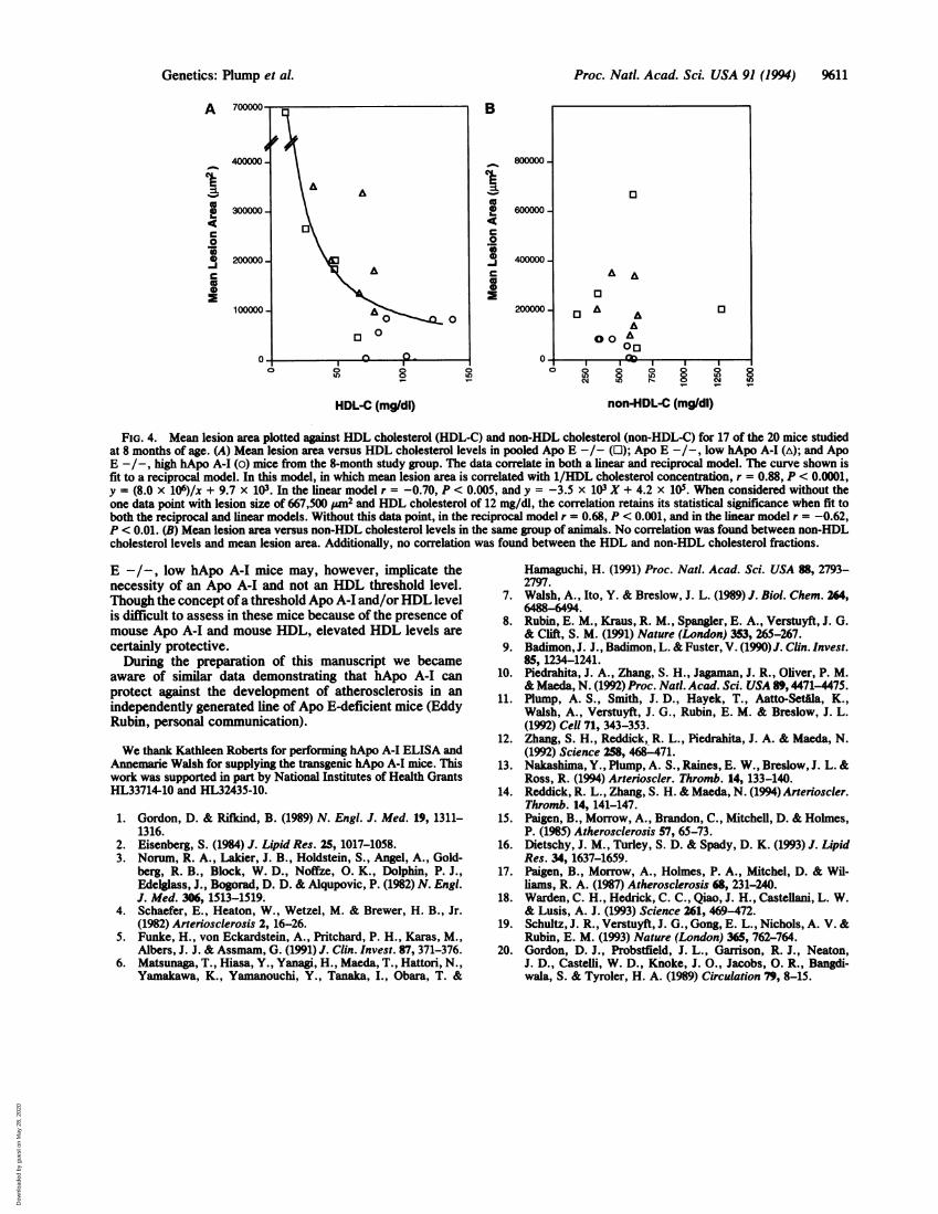
sclerosis susceptibility, we correlated mean lesion area withboth HDL and non-HDL cholesterol measured in 17 ofthe 20eight-month-old mice. A strong inverse relationship wasfound between HDL cholesterol levels and atherosclerosis(Fig. 4A). When fit to a linear model, HDL predicted 49% ofthe variance in mean lesion area (r = -0.70, P < 0.005).However, the relationship of HDL cholesterol and athero-sclerosis appears hyperbolic, and when mean lesion area wascorrelated with the inverse of the HDL cholesterol concen-tration, HDL levels predicted 78% of the observed variancein mean lesion area (r = 0.88, P < 0.0001). The data werereexamined after removal of the one data point from themouse with the lowestHDL cholesterol and the highest meanlesion area. As described in the legend to Fig. 4 the corre-lation remained statistically significant, with a reciprocalmodel better describing the data. No correlation was foundbetween non-HDL cholesterol levels and atherosclerosis inthese animals (Fig. 4B). In multiple linear-regression analy-sis, non-HDL cholesterol levels failed to enhance the pre-dictive value of HDL cholesterol levels.
DISCUSSIONThe mechanism by which Apo A-I and HDL cholesterol actto inhibit the development of atherosclerosis is unknown.There are three existing hypotheses to explain the role ofHDL in protecting against the development of atherosclero-sis: reverse cholesterol transport, direct protective effects ofHDL on the vessel wall or on lipoprotein oxidation, and aninverse relationship between HDL and atherogenic ApoB-containing lipoproteins. In the present study the hApo A-Itransgene specifically raised HDL cholesterol levels withoutsignificantly affecting non-HDL cholesterol levels. Thus, theeffect of HDL cholesterol may be a direct one and not areflection of altered levels of Apo B-containing lipoproteins.This strongly suggests that HDL may act via direct protectiveeffects on the vessel wall or by enhancing reverse cholesteroltransport from the vessel wall to the liver. Subsequent studiesexamining cholesterol flux and various vessel wall parame-ters, such as oxidative state and expression ofendothelial celladhesion molecules, should provide evidence to support oneor both of these hypotheses.
Several factors may influence future studies examining theprotective mechanism of elevated HDL cholesterol. Theeffect of HDL may be modified by its composition, as theexpression ofthe other majorHDL apolipoprotein, Apo A-II,in transgenic mice appears to nullify the protective effect ofApo A-I and may in fact be proatherogenic (18, 19); however,this has yet to be tested in the Apo E-deficient mouse model.Furthermore, in addressing mechanism of protection, an-other point to consider is the mechanism of atherogenesis.There are several means by which a vessel can becomeatherosclerotic, including plasma abnormalities such as lipo-protein alterations, hemodynamic derangements such as hy-
Genetics: Plump et A
Dow
nloa
ded
by g
uest
on
May
28,
202
0
Proc. Natl. Acad. Sci. USA 91 (1994)
-.CdenteS*S;
.. . .. : £ : .0 .:-¶.
Vf .. o
Ii
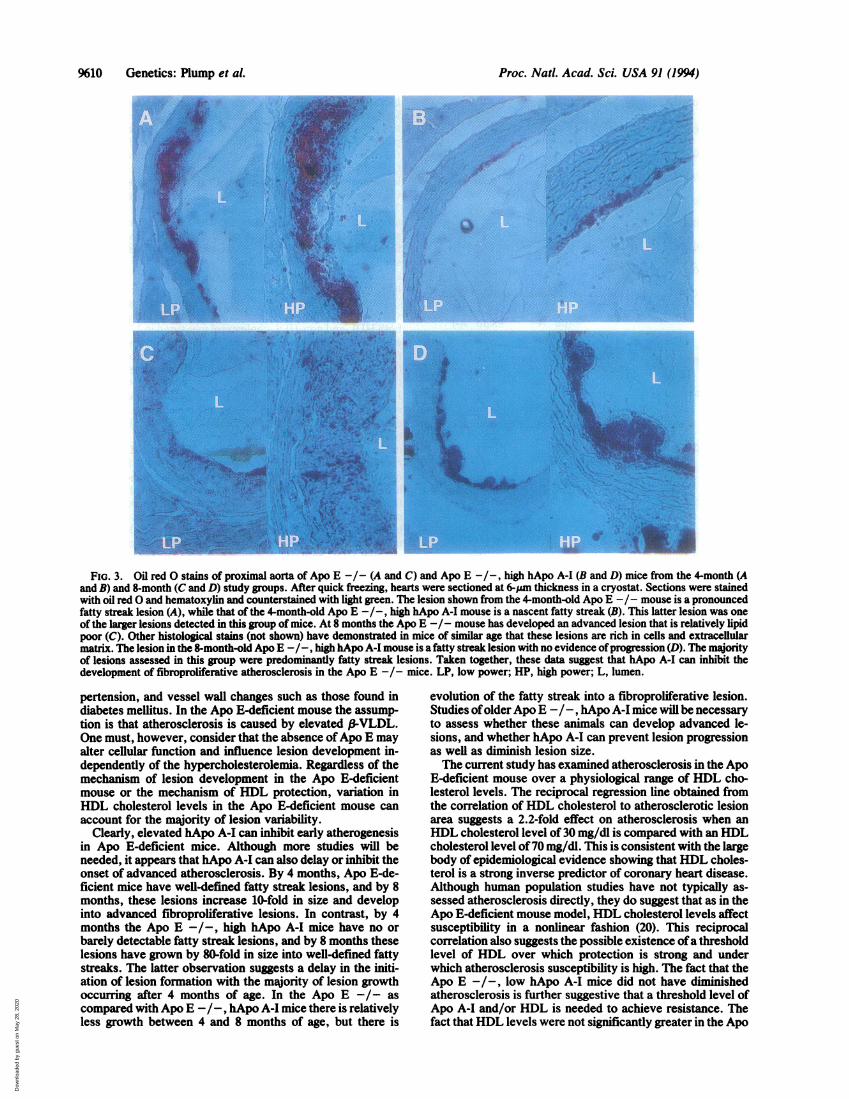
FIG. 3. Oil redO stains of proximal aorta of Apo E -/- (A and C) and Apo E -/-, high hApo A-I (B and D) mice from the4-month (Aand B) and 8-month (C and D) study groups. After quick freezing, hearts were sectioned at 6-Mm thickness in a cryostat. Sections were stainedwith oil redO and hematoxylin and counterstained with light green. The lesion shown from the 4-month-old Apo E -/- mouse is a pronouncedfatty streak lesion (A), while that of the 4-month-old Apo E -/-, high hApo A-I mouse is a nascent fatty streak (B). This latter lesion was oneof the larger lesions detected in this group of mice. At 8 months the Apo E -/- mouse has developed an advanced lesion that is relatively lipidpoor (C). Other histological stains (not shown) have demonstrated in mice of similar age that these lesions are rich in cells and extracellularmatrix. The lesion in the 8-month-old Apo E -/-, high hApo A-I mouse is afatty streak lesion with no evidence ofprogression (D). The majorityof lesions assessed in this group were predominantly fatty streak lesions. Taken together, these data suggest that hApo A-I can inhibit thedevelopment of fibroproliferative atherosclerosis in the Apo E -I- mice. LP, low power; HP, high power; L, lumen.
pertension, and vessel wall changes such as those found indiabetes mellitus. In the Apo E-deficient mouse the assump-tion is that atherosclerosis is caused by elevated f-VLDL.One must, however, consider that the absence ofApo E mayalter cellular function and influence lesion development in-dependently of the hypercholesterolemia. Regardless of themechanism of lesion development in the Apo E-deficientmouse or the mechanism of HDL protection, variation inHDL cholesterol levels in the Apo E-deficient mouse canaccount for the majority of lesion variability.
Clearly, elevated hApo A-I can inhibit early atherogenesisin Apo E-deficient mice. Although more studies will beneeded, it appears that hApo A-I can also delay or inhibit theonset of advanced atherosclerosis. By 4 months, Apo E-de-ficient mice have well-defined fatty streak lesions, and by 8months, these lesions increase 10-fold in size and developinto advanced fibroproliferative lesions. In contrast, by 4months the Apo E -/-, high hApo A-I mice have no orbarely detectable fatty streak lesions, and by 8 months theselesions have grown by 80-fold in size into well-defined fattystreaks. The latter observation suggests a delay in the initi-ation of lesion formation with the majority of lesion growthoccurring after 4 months of age. In the Apo E -/- ascompared with Apo E -/-, hApo A-I mice there is relativelyless growth between 4 and 8 months of age, but there is
evolution of the fatty streak into a fibroproliferative lesion.Studies ofolder Apo E -/-, hApo A-I mice will be necessaryto assess whether these animals can develop advanced le-sions, and whether hApo A-I can prevent lesion progressionas well as diminish lesion size.The current study has examined atherosclerosis in the Apo
E-deficient mouse over a physiological range of HDL cho-lesterol levels. The reciprocal regression line obtained fromthe correlation ofHDL cholesterol to atherosclerotic lesionarea suggests a 2.2-fold effect on atherosclerosis when anHDL cholesterol level of 30 mg/dl is compared with an HDLcholesterol level of70 mg/dl. This is consistent with the largebody of epidemiological evidence showing that HDL choles-terol is a strong inverse predictor of coronary heart disease.Although human population studies have not typically as-sessed atherosclerosis directly, they do suggest that as in theApo E-deficient mouse model, HDL cholesterol levels affectsusceptibility in a nonlinear fashion (20). This reciprocalcorrelation also suggests the possible existence ofa thresholdlevel of HDL over which protection is strong and underwhich atherosclerosis susceptibility is high. The fact that theApo E -/-, low hApo A-I mice did not have diminishedatherosclerosis is further suggestive that a threshold level ofApo A-I and/or HDL is needed to achieve resistance. Thefact thatHDL levels were not significantly greater in the Apo
%10 Genetics: Plump et al.
Dow
nloa
ded
by g
uest
on
May
28,
202
0
Proc. Nat. Acad. Sci. USA 91 (1994) 9611
A
I
0
C0
C00
B
I
0-4c
0'r
600000-
400000-
200000.
nI , I - I . .
~~~~~~~00LO 8 ol LO° g 8 ° 8
HDL-C (mg/dl) non-HDL-C (mg/dl)
FIG. 4. Mean lesion area plotted against HDL cholesterol (HDL-C) and non-HDL cholesterol (non-HDL-C) for 17 of the 20 mice studiedat 8 months of age. (A) Mean lesion area versus HDL cholesterol levels in pooled Apo E -/- (0); Apo E -/-, low hApo A-I (A); and ApoE -/-, high hApo A-I (o) mice from the 8-month study group. The data correlate in both a linear and reciprocal model. The curve shown isfit to a reciprocal model. In this model, in which mean lesion area is correlated with 1/HDL cholesterol concentration, r = 0.88, P < 0.0001,y = (8.0 x 10⁶)/x + 9.7 x 10³. In the linear model r = -0.70, P < 0.005, and y = -3.5 x 10³ X + 4.2 x 10⁵. When considered without theone data point with lesion size of 667,500 Wn2 and HDL cholesterol of 12 mg/dl, the correlation retains its statistical significance when fit toboth the reciprocal and linear models. Without this data point, in the reciprocal model r = 0.68, P < 0.001, and in the linear model r = -0.62,P < 0.01. (B) Mean lesion area versus non-HDL cholesterol levels in the same group of animals. No correlation was found between non-HDLcholesterol levels and mean lesion area. Additionally, no correlation was found between the HDL and non-HDL cholesterol fractions.
E -/-, low hApo A-I mice may, however, implicate thenecessity of an Apo A-I and not an HDL threshold level.Though the concept ofa threshold Apo A-I and/orHDL levelis difficult to assess in these mice because of the presence ofmouse Apo A-I and mouse HDL, elevated HDL levels are
certainly protective.During the preparation of this manuscript we became
aware of similar data demonstrating that hApo A-I canprotect against the development of atherosclerosis in anindependently generated line of Apo E-deficient mice (EddyRubin, personal communication).
We thank Kathleen Roberts for performing hApo A-I ELISA andAnnemarie Walsh for supplying the transgenic hApo A-I mice. Thiswork was supported in part by National Institutes of Health GrantsHL33714-10 and HL32435-10.
1. Gordon, D. & Rifiind, B. (1989) N. Engl. J. Med. 19, 1311-1316.
2. Eisenberg, S. (1984) J. Lipid Res. 25, 1017-1058.3. Norum, R. A., Lakier, J. B., Holdstein, S., Angel, A., Gold-
berg, R. B., Block, W. D., Noffze, 0. K., Dolphin, P. J.,Edelglass, J., Bogorad, D. D. & Alqupovic, P. (1982) N. Engl.J. Med. 306, 1513-1519.
4. Schaefer, E., Heaton, W., Wetzel, M. & Brewer, H. B., Jr.(1982) Arteriosclerosis 2, 16-26.
5. Funke, H., von Eckardstein, A., Pritchard, P. H., Karas, M.,Albers, J. J. & Assmam, G. (1991) J. Clin. Invest. 87, 371-376.
6. Matsunaga, T., Hiasa, Y., Yanagi, H., Maeda, T., Hattori, N.,Yamakawa, K., Yamanouchi, Y., Tanaka, I., Obara, T. &
Hamaguchi, H. (1991) Proc. Natl. Acad. Sci. USA 88, 2793-2797.
7. Walsh, A., Ito, Y. & Breslow, J. L. (1989) J. Biol. Chem. 264,6488-6494.
8. Rubin, E. M., Kraus, R. M., Spangler, E. A., Verstuyft, J. G.& Clift, S. M. (1991) Nature (London) 353, 265-267.
9. Badimon, J. J., Badimon, L. & Fuster, V. (1990)J. Clin. Invest.85, 1234-1241.
10. Piedrahita, J. A., Zhang, S. H., Jagaman, J. R., Oliver, P. M.& Maeda, N. (1992) Proc. Natl. Acad. Sci. USA 89, 4471-4475.
11. Plump, A. S., Smith, J. D., Hayek, T., Aatto-Setfila, K.,Walsh, A., Verstuyft, J. G., Rubin, E. M. & Breslow, J. L.(1992) Cell 71, 343-353.
12. Zhang, S. H., Reddick, R. L., Piedrahita, J. A. & Maeda, N.(1992) Science 258, 468-471.
13. Nakashima, Y., Plump, A. S., Raines, E. W., Breslow, J. L. &Ross, R. (1994) Arterioscler. Thromb. 14, 133-140.
14. Reddick, R. L., Zhang, S. H. & Maeda, N. (1994) Arterioscler.Thromb. 14, 141-147.
15. Paigen, B., Morrow, A., Brandon, C., Mitchell, D. & Holmes,P. (1985) Atherosclerosis 57, 65-73.
16. Dietschy, J. M., Turley, S. D. & Spady, D. K. (1993) J. LipidRes. 34, 1637-1659.
17. Paigen, B., Morrow, A., Holmes, P. A., Mitchel, D. & Wil-liams, R. A. (1987) Atherosclerosis 68, 231-240.
18. Warden, C. H., Hedrick, C. C., Qiao, J. H., Castellani, L. W.& Lusis, A. J. (1993) Science 261, 469-472.
19. Schultz, J. R., Verstuyft, J. G., Gong, E. L., Nichols, A. V. &Rubin, E. M. (1993) Nature (London) 365, 762-764.
20. Gordon, D. J., Probstfield, J. L., Garrison, R. J., Neaton,J. D., Castelli, W. D., Knoke, J. O., Jacobs, 0. R., Bangdi-wala, S. & Tyroler, H. A. (1989) Circulation 79, 8-15.
13
A A
0
03A A 03AA00
Genetics: Plump et al.
Dow
nloa
ded
by g
uest
on
May
28,
202
0