
How To Read an Eye Report - Welcome to Southeast...
30
TECHNICAL ASSISTANCE and CONTINUING EDUCATION SOUTHEAST TACE A Project of the Burton Blatt Institute at Syracuse University in collaboration with the DBTAC: Southeast ADA HOW TO READ AN EYE REPORT November 1, 2012 ROUGH EDITED COPY REMOTE CART PROVIDED BY: ALTERNATIVE COMMUNICATION SERVICES, LLC PO BOX 278 LOMBARD, IL 60148 * * * * * This is being provided in a rough draft format. Remote Communication Access Realtime Translation (CART) is provided in order to facilitate communication accessibility and may not be a totally verbatim record of the proceedings. * * * * * I just want to say good afternoon and welcome to the 2012 Southeast TACE webinar "How to Read an Eye Report". My name is Celestia Ohrazda and I'm the Information Technology consultant for the Southeast TACE. The Southeast TACE center's mission is to improve the quality and effectiveness of Vocational Rehabilitation Services and enhance employment out comes for individuals with disabilities in the 8 southeastern states. TACE is in coordination with southeast ADA Center and both TACE and southeast ADA Center are managed by Burton at Syracuse University we have a lot of webinars this year if you're signed up that's great if not please visit the Web site at TACEsoutheast.org. Before we get started I wanted to share some information and answer the Frequently Asked Questions we have received about webinars. Today's webinar is being conducted using Blackboard Collaborate. Blackboard Collaborate is a webconferencing system. It is fully accessible to everybody regardless of the assistive technology that they may use. This system makes it possible for us to conduct workshops over the Internet from just about any computer with an Internet connection and web browser. There are many computer issues that are beyond our control. But there are a few things you must do to enhance your experience. First of all, it's really important that you check your system prior to the session. As we are unable to troubleshoot technical issues right before the webinar is scheduled to begin.
Transcript of How To Read an Eye Report - Welcome to Southeast...

TECHNICAL ASSISTANCE and CONTINUING EDUCATION SOUTHEAST TACE
A Project of the Burton Blatt Institute at Syracuse University in collaboration with the DBTAC: Southeast ADA
HOW TO READ AN EYE REPORT
November 1, 2012
ROUGH EDITED COPY
REMOTE CART PROVIDED BY: ALTERNATIVE COMMUNICATION SERVICES, LLC
PO BOX 278 LOMBARD, IL 60148
* * * * *
This is being provided in a rough draft format. Remote Communication Access Realtime Translation (CART) is provided in order to facilitate communication accessibility and
may not be a totally verbatim record of the proceedings.
* * * * *
I just want to say good afternoon and welcome to the 2012 Southeast TACE webinar "How to Read an Eye Report". My name is Celestia Ohrazda and I'm the Information Technology consultant for the Southeast TACE. The Southeast TACE center's mission is to improve the quality and effectiveness of Vocational Rehabilitation Services and enhance employment out comes for individuals with disabilities in the 8 southeastern states. TACE is in coordination with southeast ADA Center and both TACE and southeast ADA Center are managed by Burton at Syracuse University we have a lot of webinars this year if you're signed up that's great if not please visit the Web site at TACEsoutheast.org. Before we get started I wanted to share some information and answer the Frequently Asked Questions we have received about webinars. Today's webinar is being conducted using Blackboard Collaborate. Blackboard Collaborate is a webconferencing system. It is fully accessible to everybody regardless of the assistive technology that they may use. This system makes it possible for us to conduct workshops over the Internet from just about any computer with an Internet connection and web browser. There are many computer issues that are beyond our control. But there are a few things you must do to enhance your experience.
First of all, it's really important that you check your system prior to the session. As we are unable to troubleshoot technical issues right before the webinar is scheduled to begin.
110112 Webinar Transcript– How To Read an Eye report
Phone: (866) 518-7750 [voice/tty] • Fax: (404) 541-9002 Web: TACEsoutheast.org | Email : [email protected]
A Project of the Burton Blatt Institute (BBI) at Syracuse University | bbi.syr.edu Funded by the U.S. Department of Education, Rehabilitation Services Administration (RSA), Grant # H264A080021
2
This session is being captioned. To turn on captioning please select the CC icon in the upper toolbar to open a separate window with captioning. Today's session is also being recorded and archived for future use. You will be sent a link to the archive after the session of the please share this freely with colleagues who may have missed the opportunity to participate in today's session. At this time we are advising you to close all other applications you may have running on your computer because they may interfere with your experience today. If you are connected to a network do not forget to periodically tap the space bar once in a while just to let the webinar system know that it you're still there.
And last, participants may submit questions at this point during the webinar by typing them into the chat area. Questions will be addressed at appropriate times throughout the session and an open forum at the end of today's presentation.
We are privileged to be joined by the feature presenter BJ LeJeune. BJ is the training coordinator for the national research and training center on blindness and low vision at Mississippi State University. BJ has served as the Project Director and primary instructor of the vision special ist graduate certificate program since 2007 she provides the bulk of in service training for the center including developing workshops and national conferences she is also working closely to coordinate training activities with other -- several other TACE centers. She holds a center in best practices in distance education by the center on teaching and learning at Mississippi State University and she's a certified vision rehabilitation therapist currently finishing up her doctorate program.
And at this time I'm going to turn the session over to BJ. BJ, if you're there, you're on.
Hi, BJ, are you there?
BJ LeJEUNE: Yes I'm here but I forgot to turn on my mic.
Oh we got to turn on the mic in order for us to hear you. Wonderful.
(Chuckles).
BJ LeJEUNE: Thank you very much. It's very good to be with you this morning -- or this afternoon for some of you and I appreciate you taking the time out to come and participate. We have quite a few slides we're going to be going through. And I did also send Celestia a couple of examples of eye reports. I know many of you are probably working in states that you have a form that needs to be filled out if somebody goes for an eye report you receive those reports back. And because everybody's forms are a

110112 Webinar Transcript– How To Read an Eye report
Phone: (866) 518-7750 [voice/tty] • Fax: (404) 541-9002 Web: TACEsoutheast.org | Email : [email protected]
A Project of the Burton Blatt Institute (BBI) at Syracuse University | bbi.syr.edu Funded by the U.S. Department of Education, Rehabilitation Services Administration (RSA), Grant # H264A080021
3
little bit different I'm going to be using more of a letter format but a lot of it is getting to know the terms and the way things are used in an eye report. So we'll be going through a lot of that information.
So let's just jump right in here. I jumped in and I didn't get where I thought I was going. There we go. Sorry; I'm new to this particular webinar format so we'll see how it goes. First thing because there is a lot of terminology and sometimes you'll come across things that we're not mentioning here there are two resources that I want to tell you about one is the -- the Dictionary of Eye Terminology it's a really good place to look up definitions of words and find out things you're not sure about. The other is a textbook we use in our vision special ist course by Riordan & Whitcher and it's called Vaughn & Asbury's General Ophthalmology book. And it's a little bit technical but a little less so than some other medical books but it will give you an over view of particular things that may come up in a person's eye report that you're in and out -- not familiar with. We're going to be talking about some of the more general things but obviously there are things that we don't cover. So the information about how to -- about those two books is on Slide 2.
Slide 3 entitled: Two types of eye exams we're going to be talking about two different kinds eye -- eye exams we'll we're -- make sure we're on the same page the first is what I call a regular eye exam you go to the doctor get your eyes checked see if there's a problem the second is a low vision exam where you have some kind of problem where cannot be corrected by glasses or contact lenses and as a result of that your vision isn't as good as it should be so you're going to find if there's some other kind of thing that can enhance and improve your vision.
Slide 4, the goal of a regular eye exam. So if you go for a regular eye exam the first thing the doctor is going to want -- you're going to want to know is your general eye health you want a diagnosis of any particular eye problems that you may have. You want to develop a treatment plan to address any of those problems or refraction errors, refraction would be when your focus is a little bit off either near or far and you want to maximize your vision as much as possible using traditional glasses or contact lenses or maybe referring somebody for lasik surgery or perhaps there's going to have to be some surgery or some other kinds of treatment.
Slide 5, what constitutes a regular eye exam and I'm going to use what many physicians use here which is the SOAP format and we use it in other areas of service delivery also human service delivery but taking each one of those letters and we're looking at the subject, objective, assessment and -- subjective objective assessment

110112 Webinar Transcript– How To Read an Eye report
Phone: (866) 518-7750 [voice/tty] • Fax: (404) 541-9002 Web: TACEsoutheast.org | Email : [email protected]
A Project of the Burton Blatt Institute (BBI) at Syracuse University | bbi.syr.edu Funded by the U.S. Department of Education, Rehabilitation Services Administration (RSA), Grant # H264A080021
4
and plan. So subjective here is where -- usually when you sit down in the doctor's office you'll have a little discussion about the patient history. What's going on. Where is the problem, which eye? When did it start? Was there something that caused it? Did you get hit in the eye with something? Did you have a hemorrhage? Something started bleeding? What kind of things. And then also they want to know how severe it is. You're coming in saying I have these floaters in my vision and they might ask you how often do you have those? Does it help you if you just close your eyes for a few minutes what kinds of things are you doing that may or may not help you?
Slide 7 continuing on then they will ask you a little bit more about a medical history. Your general health, the medications you take. This is pretty important because some medications can have an impact on your vision. And I'll give you an example of that. I worked -- I have a friend an older woman and she didn't have any vision impairments but started having these hemorrhages in her eyes and it turned out she was taking a lot of over - the - counter medications for memory. So things like some vitamin supplements she was taking Centrum silver a variety of thing -- things like that it was causing a lot of blood to move to her head and that was causing hemorrhages in her eyes sometimes it's important to find out -- sometimes they will tell you what the prescriptions are but they may not mention the other things so it's really important that all of that be covered. They'll ask about previous eye care and if especially if it's somebody who is older and they are going to get a Medicare reimbursement she'll -- they'll have to ask medical orientation questions Sims -- sometimes called O times 3 if they are oriented to person, place and time do you know where you are, why you're here, who is the President what day is it those kinds of things. Slide 8 objective testing this is where we get into more technical types of things. The first thing you'll see sometimes on an eye report are the letters VA for visual acuity. Not Virginia, not Veterans affairs but visual acuity and there are several terms here related to in a if there's a smallers that means without correction without correction means without your glasses on. C with correction. So that's with glasses or contact lenses. If you have a DVA that's your distance visual acuity. NVA your near distance acuity there may be terms like OD, OS or OU the OD is oculus dexter your right eye, OS, oculus sinister, which is your left eye and OU oculus -- I'm not sure I'm pronouncing this correctly -- uterque -- between both eyes You'll see that you'll have a visual acuity for your right eye one for your left eye and one for both eyes together sometimes see -- something called BVA not Bureau of Veterans affairs but this is best corrected visual acuity. Now there are several different charts that are used. And initially you'll be looking at distance vision and looking at something called the Snellen chart or Snellen like chart.

110112 Webinar Transcript– How To Read an Eye report
Phone: (866) 518-7750 [voice/tty] • Fax: (404) 541-9002 Web: TACEsoutheast.org | Email : [email protected]
A Project of the Burton Blatt Institute (BBI) at Syracuse University | bbi.syr.edu Funded by the U.S. Department of Education, Rehabilitation Services Administration (RSA), Grant # H264A080021
5
Snellen chart is made -- it's the one with the big E on the top that we usually think of when you think of an eye chart and it's made originally designed to be looked at at 20 feet but most offices are not 20 feet as long so they may have measured out from the patient's chair to the wall ten feet perhaps or they will have an adjusted size chart but now they have a POC a projected chart usually white letters with black background or the other way around and they have adjusted for the size of the chair -- the room. When you get the Snellen chart traditionally it would be 20 over 20 which is normal vision 20 over 40, which is the top number is -- excuse me here I just kind of blurred out for a second let me go back again 20 over 200 we often thought of that what a person with normal vision sees at 20 feet. A person with a visual impairment would see what the normal person sees at 200 person with abnormal vision sees at 20 feet that's not really what that means but basically it's the size of the letter you're looking the bigger the bottom no, ma'am -- number the worse your vision. 20/20 would be what we consider normal vision, 20/40 would be not so good. 20/70 usually you can't drive a car, 20 over 100 you may have difficulty reading print. 20 over 200 is considered legally blind with part of the definition legally blind and that's considered to be very poor vision.
Okay. Moving on here. Here is a picture of a Snellen chart again there's a big E at the top that's 20 over 200 so if a person can read a Snellen chart if they can read the top letter if they can see it sometimes now they do change especially on the project - o - chart they change the top letter because everybody knows it's an E but then you'll notice the second line down in this particular example what I have it's an F and a P. Two letters that look somewhat similar that's actually 20 over 100. So if it's a very big space over 20 over 100 and 20 over 200. And the issue that's often brought up is what if somebody is 20 over 170.well, if they can't read that second line, then they are considered legally blind so sometimes you hear that people -- more people are blinded by definition than any other cause. But the way the law is written for legal blindness, it's on a Snellen chart or Snellen like chart one of the things that may come up with -- if you have somebody with poor vision they may go for a low vision and they come back with a different visual acuity than what they had on the regular eye exam because different measuring techniques are used and so then you may not be sure if that person is legally blind or not but the legal blindness measurement which is 20 over 200 that part of the legal blind definition is based on visual acuity on the Snellen like chart.
Some of the other things that will be done there's something -- one of the things we want to look at are the pupils make sure they are reacting properly and you may see a

110112 Webinar Transcript– How To Read an Eye report
Phone: (866) 518-7750 [voice/tty] • Fax: (404) 541-9002 Web: TACEsoutheast.org | Email : [email protected]
A Project of the Burton Blatt Institute (BBI) at Syracuse University | bbi.syr.edu Funded by the U.S. Department of Education, Rehabilitation Services Administration (RSA), Grant # H264A080021
6
term on our report called PERRLA, PERRLA. And that means pupils equally round and reactive to light and accommodation.
And essentially often what you'll see is the doctor will have a little pin light and they will shine it in your eye or move it away or over to the other eye what's happening here they are wanting to see if it responds positively when the light is shining in your eyes and I'm not sure how many of you have really been through an anatomy of the eye but just real quickly. The iris which is the color part of the eye, if there's bright light it's going to close up the pupil which is the center hole that images -- light images come into the eye through if it's dark it's going to open up to make more space for more light to come in .
And what they are doing here is they are checking to see if that is working correctly. So when they shine the bright light in your eye, your pupil should get smaller. The constriction of the kind of aperture of the iris of the eye and when they move it away, it should open up a little bit. And that will be called either a positive or negative effect and sometimes they will say Marcus-Gunn pupils, gunn, Marcus-Gunn and that's primarily just the way they are measuring that. How much change is there. How much is it not doing what it's supposed to do.
Another term you'll see a lot is EOM. Extraocular muscles. Or eye movements. So one of the things that the doctor will say is look at my finger and they will move their finger across in front of you and they are looking for a smooth and full movement. So you're able to keep your head pointed straight a head and move your eyes all the way from the left to the right up and down. And that goes smoothly you don't have any jerkiness in it and that would be called ans & F, smooth and full. Then they also indicate that there's some kind of a restriction. So maybe you have difficulty looking up. So it may say EOM restricted upper right quadrant. So you may see some terminology like that.
Continuing with the objective another thing that's often looked at is how your eyes are aligned. Now what you want is for your eyes to move in tandem to the right to the left. If they turn in a little bit that's called a tropia. If they constantly turn I'm sorry if they constantly turn that's a tropia if they are intermittent it's phoria he is so is in exo is out if it's exophoric that means your eyes will turn out constantly if it's exo phoria that means they will move out but intermittent. Looking at visual fields this is what we think of legal blindness as a restricted field of vision you're going to see how much of the environment you can see normal a -- normally a person would see about 180 degrees and sometimes you'll see on an eye report that will say confrontations that means when the person is standing right in front of you and here I have a slide that has a picture of

110112 Webinar Transcript– How To Read an Eye report
Phone: (866) 518-7750 [voice/tty] • Fax: (404) 541-9002 Web: TACEsoutheast.org | Email : [email protected]
A Project of the Burton Blatt Institute (BBI) at Syracuse University | bbi.syr.edu Funded by the U.S. Department of Education, Rehabilitation Services Administration (RSA), Grant # H264A080021
7
a gentleman standing he's holding up a thumb and two fingers. And that's often what will happen they will hold up a couple of fingers in each quadrant when I say quadrant looking straight a head you have the straight up and down axis and straight horizontal axis and dividing into those four basic quadrants they will be looking to measure how much fingers you see in each quadrant they may ask you to do that with one eye or both eyes.
And that can be used kind of just a general to map out if there's some kind of visual loss in the peripheral areas.
Moving on to Slide 12.something called perimetry. I'm sorry I'm kind of not used to be -- to being where I'm not inter acting with people so I'm getting a little blank from time to time this is where you're measuring on the visual fields again and there's several different ways you can measure these there's auto mated and some of you may be familiar with the Humphrey or the Dicon or the Octopus. These give you basically a static perimetry what you're looking at here for example the Humphrey a large bowl kind of effect somebody looking to related to a machine they are looking in and lights are coming -- coming -- coming on when the light come on that gives you an indication that the person can see in the area and they have a button indicating they are seeing a light if there's an area where lights aren't not being shown and they are not seeing lights then they will know they are having a problem if you are looking at a central point and they are indicating if they see lights coming on there's also something called Goldmann, -- Goldmann, goldmann. This is more of a looking light you're looking to see how lights are moving out and into your field done in a manual way if you get an auto mated report back looking at the visual fields you're going to have places that will either have several different ways it can be marked. It could be little xs or little boxes or it could just be kind of a graph and it will show kind of darkened areas and the more dark the area, the poorer the vision is. So if you have a very dark area that means there would be no vision in that area.
The Goldmann sometimes you'll see an eye report where they will be concentric circles and one for each other -- each eye and there will be a place for a manual writing in of where there are difficult areas to see and sometimes they will be blocked out entirely the sometimes you'll see an -- a blocking out where there's a natural blind spot in everybody's vision and then also you'll mark -- some are done in color and some in black and white depends how that person reports it again you're evaluating the field of vision.

110112 Webinar Transcript– How To Read an Eye report
Phone: (866) 518-7750 [voice/tty] • Fax: (404) 541-9002 Web: TACEsoutheast.org | Email : [email protected]
A Project of the Burton Blatt Institute (BBI) at Syracuse University | bbi.syr.edu Funded by the U.S. Department of Education, Rehabilitation Services Administration (RSA), Grant # H264A080021
8
Okay. So here -- I have a picture of the Humphrey you're looking in it's kind of a bowl effect you're looking at one point and notice on this particular machine there's a screen on the side so the person who is testing can see what's happening. How that person is responding. And she has in her hand a little button that she's going to push every time she sees the light come on. Moving on to Slide 14 is how you will see the results on the left you have the hem free where in the picture at the top you have the four quadrants the top two quadrants are entirely blocked out -- blacked out and then a little blocking out in the lower right hand quadrant that tells you that area the person is not able to see and then you have some other below just other ways you might see it in stead of being little spaces between the blocks but still in another way and then on the right a the Goldmann where you have the actual drawn in of the areas of the problem the area would be within the enclosed area that's drawn this one is not filled in but it's just got the lines.
Okay. Near -- another thing that's kind of done in maybe a less technical way looking at sometimes field vision is an Amsler grid but not so much used for measuring fields as it is to look at changes in how the macula is taking in information.
And you can see them in different numbers of grids, 10 by 10 is usually the one and there's a -- there's a dot in the middle a lot of times people put them on their refrigerator it's a way to, -- mark if there's progress in the development of mark collar degeneration it's something that affects the central vision it's something that also may affect some field vision, too so it's primarily measuring central but you may see for example on the right here I have a picture of the grid and there's a big gray spot slightly off to the right and it's more dense in the middle and less dense in the outside that kind of gives you an idea of where this opacity is. But then also the lines are kind of wavy so that will give you an idea of kind of how they are seeing that.
And this is something that the patient will report to you. They say you know -- they would describe it to you. As opposed to being something that you would measure on a machine or something. Oops. I'm sorry; I hit the wrong button. And I might have to ask for a little help here. I do apologize for this. There's two little buttons to push and one of them takes you to the very end. And that's what I just did.
You're there okay great.
(Chuckles).
BJ LeJEUNE: Sorry. Okay. Sorry about that.

110112 Webinar Transcript– How To Read an Eye report
Phone: (866) 518-7750 [voice/tty] • Fax: (404) 541-9002 Web: TACEsoutheast.org | Email : [email protected]
A Project of the Burton Blatt Institute (BBI) at Syracuse University | bbi.syr.edu Funded by the U.S. Department of Education, Rehabilitation Services Administration (RSA), Grant # H264A080021
9
Okay then continuing on this is kind of just some of the general testing of the health of the eye and what the vision is like. Next thing you might look at is called refraction. Here there's two ways that it's determined. First one is called manifest. You might see that term on an eye report. And that just means traditional. There's no dilation of the eyes or anything like that. You're just looking your normal vision how you see with your glasses without your glasses kind of thing.
Cycloplegic is dilated. They put dots in your eyes they are letting light in but interfering with your ability to focus typically they will do most of the refraction testing before they dilate your eyes when they dilate they look at the inside of the health of the retina they may also as far as refraction ask you to read a Jaegar near vision chart. And I believe I can go forward here a little bit. Well, I'm sorry, I'm going to go back a little bit so I don't get too lost. We'll move up here a little bit I think I have it in low vision materials. A copy what the chart looks like. But it's just a little sentence or paragraph and as you read them you hold them about 12 inches from your face and if you have more difficulty you move them up closer or further away and that's the way they can measure what your near visual acuity is.
They will also be measuring your intra ocular pressures which is called an IOP. Looking at -- sometimes it's referred to as tonometry looking at what the pressure is inside your eye testing for glaucoma and normal pressure although this may -- this changes a lot from our understanding but normal pressure is between 14 and 20.
And this is measured basically the pressure against the eye. It used to be that a puff of air was put before the eye but now they will put a little something that will number your eye and they have a little pin or sometimes they will use a measure -- machine and press against the eye. A slit lamp is sometimes before -- part of that. Sorry; I've got some of these slides further on down the road I guess. A slit lamp is something that is used to measure the health of the outside of the eye here I have an -- a picture of an eye on Slide 17 and there's kind of like a little very fine crescent shape here where the little slit in the land that's allowing the light to go across the front of the eye as it moves across the front of the eye they can see what the shape of the cornea is. And one of the things that happens is that cornea is slightly misshapen or if instead of being round like a ping pong ball the eye is slightly elongated a little bit this will show up on this particular test to see. If it is elongated or if there are irregularities there it's referred to as a -- an a stigmatism with different focal points coming into the eye they can check to see if -- they will be looking through something that's magnified so they can check and see if there's any abnormalities in the -- along the eyelid or in the conjunctiva which is

110112 Webinar Transcript– How To Read an Eye report
Phone: (866) 518-7750 [voice/tty] • Fax: (404) 541-9002 Web: TACEsoutheast.org | Email : [email protected]
A Project of the Burton Blatt Institute (BBI) at Syracuse University | bbi.syr.edu Funded by the U.S. Department of Education, Rehabilitation Services Administration (RSA), Grant # H264A080021
10
the membrane that covers from the edge of the cornea back around to kind of secure the eye area around the cornea and the cornea being the clear part in the center of the eye.
And they look in the interior at the front chamber of the eye which is the iris and check and see what they might see in terms of the health of the eye.
Here is another photo of the slit lamp. And you can see the curvature of the eye. And in this case, because there's a bright light being shined the pupil has gotten very small and the iris is larger.
Okay. Now the next thing that might be done under the objective testing is a dilated fundus exam and a 237-- fundus exam is when they want to look straight into the eye I'm going to back up a second here to the picture of the slit lamp as you see when you have a slight -- light and shine it into the eye the pie -- pupil gets very small that makes it difficult to look in the back of the eye and look at how the retina is the majority of the people we work with that have a visual problem it's going to be Row -- related to the retina retina pigmentosa or retinopathy they want to look at the back of the iris they put drops in the eyes to kind of dilate the eyes to move the iris back so they can see in that's -- they can take pictures at the back of the eye that's called a dilated fundus exam to look at the internal workings of the eyes they will be checking at the retina they will look at the optic nerve which I have a photo here of a fundus view and in the center there's kind of a yellow -- the retina itself is very pinkish in the center there's kind of a little yellow round area and there's blood vessels coming out of that area that are coming -- covering various parts of the retina and that yellow part is the optic nerve. And one of the things that we are seeing now is a change in how we look at glaucoma. We used to look at glaucoma and just see how high the pressure was we would say if it's over 20 it's a real problem over 25 even more so we need to give you drops but that's not what they are looking at now. Now they are looking at the ratio between a little indentation in the middle of the optic nerve if you look at the slide there's a little brighter yellow in the middle that would be what's known as the cup then the outside margin of the disc of the object -- optic nerve and the larger the cup is and the smaller the distance is between the edge of the cup and the outside edge of the retinal nerve, the more impact there is of glaucoma.
So that's where all of that is happening. So now they are much more concerned about that. So you may see on an exam something that says CD which is the cup to disc ratio. And that would couple -- come up under the DFE the dilated fundus exam and will also be looking at vitreous which is the fluid in the back of the eye they will want to

110112 Webinar Transcript– How To Read an Eye report
Phone: (866) 518-7750 [voice/tty] • Fax: (404) 541-9002 Web: TACEsoutheast.org | Email : [email protected]
A Project of the Burton Blatt Institute (BBI) at Syracuse University | bbi.syr.edu Funded by the U.S. Department of Education, Rehabilitation Services Administration (RSA), Grant # H264A080021
11
see if there's any floaters in there or scar tissue anything that's kind of mucking it up so to speak and they will be looking at the blood vessels making sure they are healthy not enlarged or gorged or sometimes you get neo vascularization with something like diabetic retinopathy so little tiny blood vessels will begin to develop and then they will hemorrhage so they will be looking at some of that in particular with people who have deities. Okay -- diabetes moving on to Slide 20 this is another picture this one is the optic nerve is off to the left side. And in the middle what you're saying -- seeing is the macula and that's the darker pink area in the very center of that it's even a little darker pink and that would be the fovea and the blood vessels that come all around in the retina or you are actually seeing them in the layer under the retina but what you see developing there they don't go across to the macula so the macula if there are blood vessels across that that's something you would want to report here on Slide 21 these are different fundus views of a diseased retina just so you can understand what it looks like when it's not doing so good so being the person that's more used to kind of didactic things where I would see if you can identify what these are so since we're not in that kind of an atmosphere the one in the upper left is a -- it's very gray kind of shades around it. If you look at the optic nerve in the center, it's very bright, very white all the way almost out to the edges of it. And what we have here is glaucoma and this is an enlarged optic disc so if you look you have to have a trained eye to do this between the outside edge of the outside edge of the optic nerve where the really bright white part is that's a very narrow margin all around so that would indicate a pretty advanced case of glaucoma. The second one in the upper right hand corner is atrilated macular degeneration in this particular slide you have the retina looks a little bit pale and you have a large area of blood that has hemorrhaged. And it's hemorrhaged around the macula. This is called an exudative hemorrhage so it's from wet macular degeneration and you have a big area here now in some macular degeneration especially the dry form you might have yellow scrambled egg kind of things but this particular one doesn't have so much of the drusen although there are yellow patterns but there's this big area of blood that has kind of seeped out across the retina.
Now, I'm not really sure how much all of you know about the anatomy of the eye but just very briefly the retinal layer itself has several layers behind that is the choroid behind that is the white sclera the outside of the eye that we see when we look at the eye. And the choroid is where most of these blood vessels are traveling. And there's an area that kind of connects between the retina and the choroid called the retinal pigment epithelium and that's where a lot of those blood vessels are moving to get nutrition to the retina. The retina needs so much nutrition. It's basically brain tissue in

110112 Webinar Transcript– How To Read an Eye report
Phone: (866) 518-7750 [voice/tty] • Fax: (404) 541-9002 Web: TACEsoutheast.org | Email : [email protected]
A Project of the Burton Blatt Institute (BBI) at Syracuse University | bbi.syr.edu Funded by the U.S. Department of Education, Rehabilitation Services Administration (RSA), Grant # H264A080021
12
the inside of the back of your eye. And so anything that's disrupting the blood supply or somehow impacting that is going to have an impact on your vision.
In the lower left hand corner is diabetic retinopathy. And here this is peripheral. Diabetic retinopathy where you have as you can see it's kind of yellowed in some areas because you're not getting good blood flow it's white in some areas because the blood flow is almost stopped you have a lot of little tiny blood vessels that have developed and there's some hemorrhaging around some of those and there's a lot of mottling so to speak in the color of the retina. And then on the right I've got kind of a mid stage retinitis pigmentosa photo and what you have here is a picture of the retina but again there's a lot of little discoloration kind of black almost looks like it has the measles around the outside edge where it's got these little black splotches some very white splotches the retinal -- splotches the pigment if it's not evenly distributed you have clumps of color and so forth all of that is part of what's going -- being looked at when you're doing a fundus examination.
Ah here is my Jaegar chart sorry so what you have here is a chart that has letters or -- they are small at the top bigger at the bottom and they are measuring the size of those letters so what size can a person read and that's usually an M number maybe M 1 M 2 M 3 the larger the number the larger the print.
Okay. Now intraocular pressure. Here again I mentioned I said on the other slide 14 to 20 that's what used to be considered normal. 20,21 is still the normal range but here we have a picture of a device that's putting -- it's going to press against the eye to see how much pressure it is to kind of cause the eye to in dent a little bit and that's done as the eye is numb so you don't feel that but it gives you an idea of what that would look like.
This I've already said some of this in this next slide, slide 24, the numbers, it is expressed when you look at intraocular pressure you're looking in terms of millimeters of mercury just like the barometer. And the -- one of the things that we see now is somebody can have high intraocular pressure but not have any change in the cup to disc ratio of the optic nerve in that case even their inter -- if their intraocular pressure is 28 it may not be considered glaucoma on the other hand you can have somebody who is intra ocular pressure is maybe 9 or 8 and they are considered to have glaucoma because it is causing a problem in that cup to disc ratio. So a little dill -- little different way than how we have thought about things in the past.

110112 Webinar Transcript– How To Read an Eye report
Phone: (866) 518-7750 [voice/tty] • Fax: (404) 541-9002 Web: TACEsoutheast.org | Email : [email protected]
A Project of the Burton Blatt Institute (BBI) at Syracuse University | bbi.syr.edu Funded by the U.S. Department of Education, Rehabilitation Services Administration (RSA), Grant # H264A080021
13
So this, a variety of measurements there are other things they can do also in terms of the objective measurements they may be looking at contrast or if somebody has -- is suspected of having some kind of a color deficiency they may do some additional testing electro retinal gram those kinds of things but for the basic eye exam generally we've gone over most of the things you've seen covered in the report. And then we come to an area of our SOAP remember we have done the subjective and objective and now we're doing the assessment and here in the assessment they are basically coming up with a conclusion because -- based on what they have seen the information they have collected what is their impression of what's happening in the eye. So the examples would be they may have -- these are some abbreviations you may see. AMT which is atrilated macular degeneration sometimes ARMD sometimes both of those sometimes just MD they might say GLC which would be glaucoma. Maybe BDR which is background diabetic retinopathy.
PDR, proliferative diabetic retinopathy and they may say NS which is nuclear sclerosis which means they have cataracts they may say myopia which is near-sighted. Hyperopia being far-sighted. Presbyopia that's a term that happens for those of us over 40 where we need bifocals don't have the ability to kind of change our focus quite as well as we used to.
Other things you may see maybe retinitis pigmentosa retinopathy of pre maturity if you're working with children. LCA Leber congenital -- amaurosis they have the lenses removed so they have no lenses in their eyes. Just more of the terminology you might see.
So then the last part of the SOAP is the plan. How is the patient going to be treated? And here in Slide 27 we look at some possibilities. There may be medical management for example if the person has glaucoma maybe they are going to be taking drops in their eyes. They might be having surgery. And that could be either some type of laser surgery or a conventional surgery. Maybe that they just want to look at it for a little more have them come back in a regular three, six, nine month basis they may do some things called optical management where they will give them either glasses or contacts give them a prescription for what kind of refraction they might need one of the things we hope they will do is if somebody has a severe vision impairment they will make a referral then to rehabilitation services or low vision services unfortunately we know this doesn't happen as much as we would like to see.
I just want to see if there's any questions at this point. Just to take a moment here. That's primarily what the information we're going to be covering -- we are going to go

110112 Webinar Transcript– How To Read an Eye report
Phone: (866) 518-7750 [voice/tty] • Fax: (404) 541-9002 Web: TACEsoutheast.org | Email : [email protected]
A Project of the Burton Blatt Institute (BBI) at Syracuse University | bbi.syr.edu Funded by the U.S. Department of Education, Rehabilitation Services Administration (RSA), Grant # H264A080021
14
on what the reports will look like but this is just kind of what's involved in a regular eye exam. Any questions?
Okay. Then I'm going to shoot on here. In a low vision evaluation. And in low vision basically what we're going to do is, first of all, confirm the findings of an eye report there may be quite a bit time between an eye report and a low vision exam you may have somebody referred to your agency and you arrange for a low vision exam for them by the if I am -- time you get your eligibility processed and everything a couple of months may have passed hopefully it won't be too much time but what a low vision exam wants to do is maximize somebody's functional vision sometimes you'll see the term functional vision assessment a functional vision assessment is a little different than a low vision exam because the functional vision assessment takes place in a natural environment not in an office so a functional vision assessment would take place in the person's workplace or their home or maybe school. And it may be something that's done by what we would call a rehabilitation professional. So it could be a low vision therapist, it could be rehab counselor, a vision teacher. It could be a teacher of the visually impaired or an orientation mobility instructor they may see a certain situation a person is having difficulty they do a formalized functional vision you'll assessment or an informal one just saying they notice people have difficulty seeing branches above them orientation mobility instructor might make that observation.
There are some assessment tools that can be used to do this in a more prescriptive kind of way. But one of the things about a low vision -- a functional low vision assessment it's very helpful to do that prior to a low vision exam. Because it's one thing to see how well a person is going to see in the doctors office but that may or may not relate to how they will see in their home. And we often can see people get magnification devices they go home they go in the drawer we do know there's an interesting study done on the abandonment of low vision devices and they found that magnification devices that were handheld were not electronic were much more likely to be abandoned and left in a drawer somewhere so apparently if there's a cost related or perhaps they just help them see better we don't know but there's more of a commitment to use those electronic devices that's all part of the low vision evaluation.
So low vision it's the same kinds of procedures or processes. First of all we're going to take a history but the questions are going to be what is the person or patient having trouble doing and they are going to be very specific they may say I have trouble watching TV so the next question is how far from the TV do you sit or how big is injure -- your screens -- screen nowadays somebody could have a 60 inch screen what type of

110112 Webinar Transcript– How To Read an Eye report
Phone: (866) 518-7750 [voice/tty] • Fax: (404) 541-9002 Web: TACEsoutheast.org | Email : [email protected]
A Project of the Burton Blatt Institute (BBI) at Syracuse University | bbi.syr.edu Funded by the U.S. Department of Education, Rehabilitation Services Administration (RSA), Grant # H264A080021
15
reading light do you use how much reading do you -- do you actually need to do is there a glare do you have enough light what kind of hobbies do you do what kind of computer do you use what kind of job do you have if you're a student are you taking courses that are in a large lab or a small classroom big lecture hall a lot of just very specific information about how the person is going to be using their vision and if they have glasses how long have you had the glasses if you have contacts how long have you had that's -- those do you use magnifiers now a lot of people go down to the local drugstore or Wal-Mart or target and they pick up a magnifier you know if you have something that's really good at a low vision evaluation for them to bring with them any devices they are using.
And the big thing here is you want to find out what is it the person wants to do. You know what is it they want to do with their vision? Are they wanting to -- you can spend a lot of time trying to help somebody learn how to read better and to get larger print and get everything set up for them to read and then you find out that what they really want to do is be able to read street signs when they are going for walks so you need to make sure you understand what it is that the person wants to do.
There are some similarities some of the same areas might be checked. But the emphasis is more on functionality. Here you're already going to have a diagnosis because they have gone for a regular exam very rarely is somebody going to go to a low vision exam without having a regular exam usually they want some referral information from the previous doctor. And most important is functionality you want them -- to also see what eye medications they are taking particularly if they are taking any eye drops. All of that would come with them.
Then here on Slide 32 I'm supposed to remember to say that every time I'm probably hitting about 50 % now. But you want to include what kind of living arrangements if a person lives by themself that might be very different than somebody who lives with a family.
Are there transportation issues? Are they still driving? A lot of us who are rehab workers we've had lots of experiences where you go to meet somebody who is legally blind and they drive up to the house. Had that happen not too long ago and the person's wife who didn't have a visual impairment was the passenger in the car you do have people still driving it's good to find that out almost all of the eye conditions have a negative impact of smoking and alcohol and drug abuse certainly could impact your vision, too. So you want to find out those kinds of things.
110112 Webinar Transcript– How To Read an Eye report
Phone: (866) 518-7750 [voice/tty] • Fax: (404) 541-9002 Web: TACEsoutheast.org | Email : [email protected]
A Project of the Burton Blatt Institute (BBI) at Syracuse University | bbi.syr.edu Funded by the U.S. Department of Education, Rehabilitation Services Administration (RSA), Grant # H264A080021
16
Visual acuity again you want to look at what that would be. Distance and near. And the distance is going to be tested on different types of charts a Feinbloom which has numbers on it EDTRS, lea there's a variety of different ones and what you're wanting to see is a gradation between what would be the lines on the Snellen chart you want to check that under low and high contrast .
So the Feinbloom chart is one many of you may have seen. It's probably 10 by 16 or something like that. And it's a flip -- kind of like a spiral notebook on one side and it flips up and the biggest letter I think I can't remember off the top of my head but I want to see -- to say it's like 20 over 700 or something like that and then the numbers get smaller and more pages and gives you much more variety. You can test at -- for 170,140, 130 a lot more flexibility to get more precise idea of what the person is going to be seeing. What they can see.
And then the near vision may also be tested most likely not on a Jaegar chart like it was on the regular exam but something like the Bailey Lovie the Hoeft or the Mnread the Minnesota Read here again it's held maybe 12,16 inches you're going to look at the size of the print that somebody is able to read.
One of the things, too, you're going to be looking again at trying to map out any scotomas or blind spots in the person's vision their field vision again you may use the Amsler grid most likely you're not going to have the same kind of precise measurement that you had in a regular eye exam for checking the field vision and the low vision exam.
Most of what's of a low vision exam is to improve the central vision, kind of maximize what you have in the central vision. And one of the things you'll be looking at in terms of trying to check out for blind spots, too is the shape of those blind spots you want to know for example if somebody has a blind spot that's shaped a little bit like a C, if that's -- if that C is where they are trying to read and then coming away from their blind spot to read, that's not going to be nearly as difficult for them if the blind spot is on the right side and they are reading into their blind spot. They will have to learn then to turn their head more so you want to know exactly what the shape and size and placement of those blind spots would be and if there's going to be any kind of distortion.
They might do some of the -- some testing but again it's not as much with the over all fields as it is looking at central scotomas and what might -- could sometimes for example somebody could have a floater which would be a moving thing they might have difficulty seeing around that floater if it's very thick sometimes people that have

110112 Webinar Transcript– How To Read an Eye report
Phone: (866) 518-7750 [voice/tty] • Fax: (404) 541-9002 Web: TACEsoutheast.org | Email : [email protected]
A Project of the Burton Blatt Institute (BBI) at Syracuse University | bbi.syr.edu Funded by the U.S. Department of Education, Rehabilitation Services Administration (RSA), Grant # H264A080021
17
retinopathy of pre maturity may have some fibrous tissue that's in the vitreous fluid and that may move around in different places and so the blind spots may actually move a little bit. And it's interesting to me, too, because the brain is a wonderful thing.
And the brain has a tendency to fill in the blanks for us.
So when we're looking through both eyes, oftentimes -- well all of us have some kind of a small blind spot but we may not be able to detect where it is because our brain has filled in the blank spots.
And if somebody has a field restriction vision oftentimes we picture it of something looking through a paper towel tube or something like that. But -- so there's this area of voidness around it. But that's not who the person sees. They have their entire field of vision filled up with that small image. So oftentimes people don't know what they don't see.
And the brain may play some tricks on them and fill in some of the blanks so sometimes people will go for a low vision exam or even a regular exam and think they see much better than they do. If any of you have seen the video Going Blind there's a couple of instances in there where the -- in one the gentleman is walking down the sidewalk and suddenly this person just appears in front of them and they don't know where they came from but what happened is the person had been walking down the sidewalk in their blind spot when he got close enough he filled in beyond where the blind spot was so the brain had just filled in all of that blind spot with sidewalk. So he didn't realize that there was anything in the sidewalk.
So that's one of the things with low vision exam can help you to do is learn how to utilize -- where those blind spots are and how to make up for that.
Again they are going to be looking at how well the eyes move together. One of the things about a low vision exam is that you almost never -- and I always say almost because there's always an exception out there some place but you don't dilate the eye for a low vision exam buzz -- because you're going to maximize the vision you already had the other exam where you're looking at the over all eye health. But here you're not going to want to dilate the eyes because you're looking at different things you already have the diagnosis you're looking for function.
I do want to mention I have a Web site on here biopticdrivingUSA.com we have about 35 states that have bioptic places and low vision places are places where people would go to get a bioptic lens this is a gentleman wearing a bioptic lens it's a carrier lens

110112 Webinar Transcript– How To Read an Eye report
Phone: (866) 518-7750 [voice/tty] • Fax: (404) 541-9002 Web: TACEsoutheast.org | Email : [email protected]
A Project of the Burton Blatt Institute (BBI) at Syracuse University | bbi.syr.edu Funded by the U.S. Department of Education, Rehabilitation Services Administration (RSA), Grant # H264A080021
18
going on top of the glasses if you notice it's kind of across his eyebrows -- eyebrows almost sometimes people don't understand people don't put on the lenses and look through the lenses when they are driving actually they are using them just to spot certain things so there may be something in the road they are not sure what it is they drop their head look through that carrier lens and then they can identify it or maybe they are looking for a street and they want to be able to read the street sign and they just use that just momentarily because if you look at this lens here the way it's constructed, when the person is looking through it, they are actually legally blind because of the restricted field -- field of vision and you need your peripheral vision as much as possible to drive.
So I wanted to mention this because sometimes people go to low vision clinics in order to get bioptic lenses they are rather expensive lenses they involve training in the clinic how to use them and involve training on the road and then the person has to wear them is the other part. And there are sometimes some restrictions.
If you're interesting -- interested you can go to this bioptic driving USA and I believe they have a list of all the states that have bioptic and -- to what the requirements are can vary from 20/70 vision in Mississippi it's 20/200. Here is another one again they are going to check the fields they want to make sure you can look quickly at something that you have the ability to track that you can interpret what it is you see so they are looking at injure visual acuity see if you have -- if you need additional light. If you have dominance in one eye over the other you want to make sure the stronger eye is the one you would be looking through the bioptic with. They want to check your contrast sensitivity. If you have motivation and enthusiasm about doing it.
It is -- it's not the most cosmetically beautiful piece of equipment but can make a huge difference in people being able to be employed. There's a very high percentage of employment among people who are legally blind or who have severe visual impairments and get bioptics most of them do report to work if they are of working age.
So when -- when people go for a low vision evaluation often the thing they are most interested -- ed from -- interested in is reading. And I think many -- many years ago I got into a real strong debate I was a rehab teacher and I had a low vision kid I -- kit that I took out to peoples homes and was working with an older blind program we had a Jaegar chart they would read and we had a little chart to tell us what magnifier to try if they can only read certain kinds of print we used that and we thought it was great and I got into a disagreement with somebody saying you need to go to a low vision clinic it's really important you don't just throw magnifiers at them and one of the things

110112 Webinar Transcript– How To Read an Eye report
Phone: (866) 518-7750 [voice/tty] • Fax: (404) 541-9002 Web: TACEsoutheast.org | Email : [email protected]
A Project of the Burton Blatt Institute (BBI) at Syracuse University | bbi.syr.edu Funded by the U.S. Department of Education, Rehabilitation Services Administration (RSA), Grant # H264A080021
19
I guess that changed my view because one of the things I've seen over the years is that low vision, a good low vision clinic and there's -- there's a lot of variations but a good low vision clinic will really make a difference in what you're able to see and function and the quality of the magnifiers does make a difference some states are now having low vision clinics being done in conjunction with field staffers you may have a rehab teacher or vision therapist or low vision therapist or OEM instructor who is working with the clinic to try to get kind of that functional piece going.
Another thing with the low vision exam which is very helpful is to bring along examples of what you want to see I think it's important to be thoughtful in advance because sometimes people go and they expect to get their vision back just like it was before and that's not going to happen. They may be using certain magnifiers but there is some distortion with magnifiers there's some limitations of how much you can see he with the magnifier bigger is not always better. We do know now that a lot of times people when they read if they can see the first and last letter of the word they can read faster than if they see a larger clearer -- part of the word so sometimes the clearest isn't the best. So it is trickier than we used to think it was. So I just wand -- want to stress the importance of going to a clinic, if possible.
But I have a couple of examples here of nutrition facts for somebody that has diabetic retinopathy it's important for them to maintain good control of their diabetes they need to be able to read these nutrition facts and I have a -- I think it's campbell's soup can here where you have black print on red label doesn't give you much contrast that can be very difficult for some people to read to bring in the examples of things they want to see or they want to read if they want to be able to bring in the Bible they need to brick in the -- bring in the Bible they want to read or the phonebook bring it in or newspaper bring it in if they need to bring their medicines bring that in that can be very helpful.
They do give you a diagnosis in low vision clinic that's primarily for billing purposes and they may or may not get aids at their first visit sometimes it may be two or three visits the typical low vision exam a good low vision exam is going to take at least an hour sometimes much longer than that depending on. There may be referral for people to come to their home. Low vision clinics are much more likely to refer for O & M for VRT services or RT services VR services or things like -- things like that if they are not already been sponsored by those agencies there are some reasons why low vision aids are abandoned in ability to use aid correctly wrong aid for situation may be poor lighting maybe the aid is too strong or not strong enough. Something to check into.

110112 Webinar Transcript– How To Read an Eye report
Phone: (866) 518-7750 [voice/tty] • Fax: (404) 541-9002 Web: TACEsoutheast.org | Email : [email protected]
A Project of the Burton Blatt Institute (BBI) at Syracuse University | bbi.syr.edu Funded by the U.S. Department of Education, Rehabilitation Services Administration (RSA), Grant # H264A080021
20
So -- so reading reports that's why you came to the seminar, right? So Slide 43 here we're going to be looking at first of all who is the report from. The primary care provider the primary eye care provider sometimes referred to as the ECP. Eye care provider it can be an OD or MDOD would be optometrist MD ophthalmologist low vision special ist perhaps that could be an OD or MD it to be OT or CLVT or CVRT or it could be I don't know I guess those are the primary ones. Occasionally you might have somebody who is a TBI a low vision special ist or O & M a low vision special ist. Sometimes these will happen within the agencies so you may be working with a state agency that has low vision clinic and may have their own procedures they follow in making reports.
The thing that becomes the most confusing I think if it comes from another agency or another doctors office from outside where you're just not real sure you're not using the same forms and things.
It could come -- come from a neurologist or neuro optometrist or ophthalmologist we have a lot of people with traumatic brain injury also have visual problems -- problems -- about 40 % coming back from Middle East have visual impairments so it might be a neuro optometrist or ophthalmologist giving you reports instead of it all being about the eyes some of it is about the brain so the eyes may be working fine there's no difficulty with the eyes at all but the brain is not interpreting of -- what it's seeing because of the injury to the brain or pathways all of these people will have slightly different lingo and terminology so just be alert to that.
You're going to want to be looking if the report comes from a medical doctor an eye care professional you want to be looking at objective findings, the diagnosis if there's some kind of treatment plan. When and how often they plan to see the patient most often geared towards other medical professionals in terms of the way they write the report.
You may just get a Xerox copy of their chart notes. More people now are -- not Xerox necessarily they may just print out because -- because more of them are doing laptops they report it on a laptop or computer and print it out and you will not get any opinion usually from regular eye exam on low vision aids.
So here -- I've got several slides an example of a letter you might get if you were to get that report back in letter form. So it says dear so-and-so I had the pleasure of meeting Mrs. Whatever a very pleasant let's say 72 - year - old woman female for a comprehensive eye exam on such-and-such date. The chief complaint was blurred
110112 Webinar Transcript– How To Read an Eye report
Phone: (866) 518-7750 [voice/tty] • Fax: (404) 541-9002 Web: TACEsoutheast.org | Email : [email protected]
A Project of the Burton Blatt Institute (BBI) at Syracuse University | bbi.syr.edu Funded by the U.S. Department of Education, Rehabilitation Services Administration (RSA), Grant # H264A080021
21
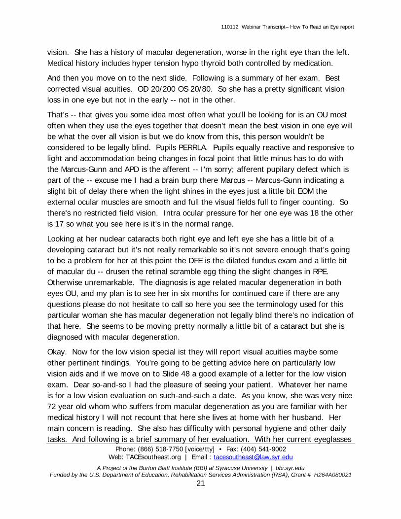
vision. She has a history of macular degeneration, worse in the right eye than the left. Medical history includes hyper tension hypo thyroid both controlled by medication.
And then you move on to the next slide. Following is a summary of her exam. Best corrected visual acuities. OD 20/200 OS 20/80. So she has a pretty significant vision loss in one eye but not in the early -- not in the other.
That's -- that gives you some idea most often what you'll be looking for is an OU most often when they use the eyes together that doesn't mean the best vision in one eye will be what the over all vision is but we do know from this, this person wouldn't be considered to be legally blind. Pupils PERRLA. Pupils equally reactive and responsive to light and accommodation being changes in focal point that little minus has to do with the Marcus-Gunn and APD is the afferent -- I'm sorry; afferent pupilary defect which is part of the -- excuse me I had a brain burp there Marcus -- Marcus-Gunn indicating a slight bit of delay there when the light shines in the eyes just a little bit EOM the external ocular muscles are smooth and full the visual fields full to finger counting. So there's no restricted field vision. Intra ocular pressure for her one eye was 18 the other is 17 so what you see here is it's in the normal range.
Looking at her nuclear cataracts both right eye and left eye she has a little bit of a developing cataract but it's not really remarkable so it's not severe enough that's going to be a problem for her at this point the DFE is the dilated fundus exam and a little bit of macular du -- drusen the retinal scramble egg thing the slight changes in RPE. Otherwise unremarkable. The diagnosis is age related macular degeneration in both eyes OU, and my plan is to see her in six months for continued care if there are any questions please do not hesitate to call so here you see the terminology used for this particular woman she has macular degeneration not legally blind there's no indication of that here. She seems to be moving pretty normally a little bit of a cataract but she is diagnosed with macular degeneration.
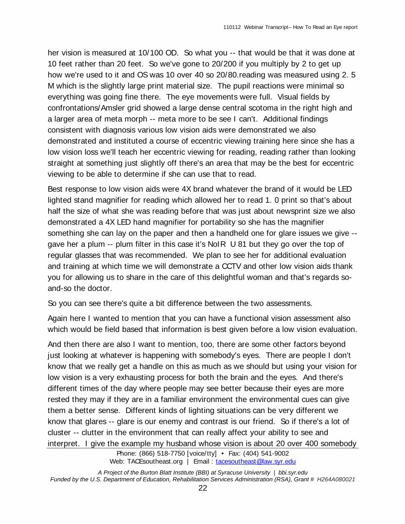
Okay. Now for the low vision special ist they will report visual acuities maybe some other pertinent findings. You're going to be getting advice here on particularly low vision aids and if we move on to Slide 48 a good example of a letter for the low vision exam. Dear so-and-so I had the pleasure of seeing your patient. Whatever her name is for a low vision evaluation on such-and-such a date. As you know, she was very nice 72 year old whom who suffers from macular degeneration as you are familiar with her medical history I will not recount that here she lives at home with her husband. Her main concern is reading. She also has difficulty with personal hygiene and other daily tasks. And following is a brief summary of her evaluation. With her current eyeglasses
110112 Webinar Transcript– How To Read an Eye report
Phone: (866) 518-7750 [voice/tty] • Fax: (404) 541-9002 Web: TACEsoutheast.org | Email : [email protected]
A Project of the Burton Blatt Institute (BBI) at Syracuse University | bbi.syr.edu Funded by the U.S. Department of Education, Rehabilitation Services Administration (RSA), Grant # H264A080021
22
her vision is measured at 10/100 OD. So what you -- that would be that it was done at 10 feet rather than 20 feet. So we've gone to 20/200 if you multiply by 2 to get up how we're used to it and OS was 10 over 40 so 20/80.reading was measured using 2. 5 M which is the slightly large print material size. The pupil reactions were minimal so everything was going fine there. The eye movements were full. Visual fields by confrontations/Amsler grid showed a large dense central scotoma in the right high and a larger area of meta morph -- meta more to be see I can't. Additional findings consistent with diagnosis various low vision aids were demonstrated we also demonstrated and instituted a course of eccentric viewing training here since she has a low vision loss we'll teach her eccentric viewing for reading, reading rather than looking straight at something just slightly off there's an area that may be the best for eccentric viewing to be able to determine if she can use that to read.
Best response to low vision aids were 4X brand whatever the brand of it would be LED lighted stand magnifier for reading which allowed her to read 1. 0 print so that's about half the size of what she was reading before that was just about newsprint size we also demonstrated a 4X LED hand magnifier for portability so she has the magnifier something she can lay on the paper and then a handheld one for glare issues we give -- gave her a plum -- plum filter in this case it's NoIR U 81 but they go over the top of regular glasses that was recommended. We plan to see her for additional evaluation and training at which time we will demonstrate a CCTV and other low vision aids thank you for allowing us to share in the care of this delightful woman and that's regards so-and-so the doctor.
So you can see there's quite a bit difference between the two assessments.
Again here I wanted to mention that you can have a functional vision assessment also which would be field based that information is best given before a low vision evaluation.
And then there are also I want to mention, too, there are some other factors beyond just looking at whatever is happening with somebody's eyes. There are people I don't know that we really get a handle on this as much as we should but using your vision for low vision is a very exhausting process for both the brain and the eyes. And there's different times of the day where people may see better because their eyes are more rested they may if they are in a familiar environment the environmental cues can give them a better sense. Different kinds of lighting situations can be very different we know that glares -- glare is our enemy and contrast is our friend. So if there's a lot of cluster -- clutter in the environment that can really affect your ability to see and interpret. I give the example my husband whose vision is about 20 over 400 somebody

110112 Webinar Transcript– How To Read an Eye report
Phone: (866) 518-7750 [voice/tty] • Fax: (404) 541-9002 Web: TACEsoutheast.org | Email : [email protected]
A Project of the Burton Blatt Institute (BBI) at Syracuse University | bbi.syr.edu Funded by the U.S. Department of Education, Rehabilitation Services Administration (RSA), Grant # H264A080021
23
near us painted a house bright pink and so we were driving by and I said wow look at the color of that house and he looked over it's a big two story house it's fairly near the curb and he didn't see it. It somehow just faded away in his vision but then the next day we were standing on a corner downtown and the schwann's truck they have a distinctive shape going by and he said what is that truck doing in town I'm thinking he saw this small truck but didn't see the big house sometimes those things are hard for us to get a grasp on the way people use their vision just depends on so many factors beyond just their visual acuity.
So visual acuity what the numbers mean and probably I should have covered this at first but just kind of tying it up here a little bit near the end, the first number is the test distance. Second number is letter sides -- letter size and the larger the second number the worse is the vision. Many low vision doctors work in smaller areas so you can multiply in order to get the numbers if they are using it at 10 feet and they say 10/40 that equates to 20/80 and so on.
And. Legal blindness this is something that was determined by the Social Security Administration back in the ' 30s what they wanted to know what they felt was the level of visual impairment that would make it very difficult for somebody to work. And so the definition of legal blindness is 20/200 or worse in the better eye with best correction or a field restriction of 20 degrees or less.
So many times you'll hear people say I'm legally blind without my glasses or I'm legally blind in one eye. That's not true. They may have 20/200 in one eye but they are not legally blind if they put their glasses on and they see 20/400 sometimes we have quite a bit confusion over legal blindness often we ask our consumers are you legally blind and they will say yes but they may or may not be actually legally blind again this is on a Snellen chart.
Just want to take a little bit of time here to hear about eccentric viewing. I have a photo graph here of a retina it's a dilated fundus exam approach you get a picture of the back of the eye this person has macular degeneration and there's an X on the slide just slightly off the side of the macula and this is something that's referred to as the preferred retinal locus this is the place that the person sees the best off the side of their macula.
And then we have this Big Mac collar scar which is a yellow area in the middle.
So if you're working on eccentric viewing this is just the two second over view of the eccentric viewing, you have light coming in through the pupil. It hits the back of the
110112 Webinar Transcript– How To Read an Eye report
Phone: (866) 518-7750 [voice/tty] • Fax: (404) 541-9002 Web: TACEsoutheast.org | Email : [email protected]
A Project of the Burton Blatt Institute (BBI) at Syracuse University | bbi.syr.edu Funded by the U.S. Department of Education, Rehabilitation Services Administration (RSA), Grant # H264A080021
24
eye of the macular -- macula but -- but that area is obscured you have problems with scarring and things and that's not going to be functional so what you want to do is move the 357-- paper slightly off to the side if you're trying to read print on paper so it's sitting off -- your focus point is going to be on the retina where you have that preferred visual location.
Or locus. And learning to use that is not an easy process but you may see that more and more recommended on some of the low vision exams that you get. Refraction wearing glasses. I have a young lady here that has some big glasses on. I think they are the new -- now we see glasses are very sporty some of those very sporty glasses are not as functional as the older ones these are very big and probably are fine but some that have very wide arms on them may cause some difficulty.
Filters here. We have people who are photo phobic that doesn't mean they are afraid of light but they may have difficulty out in bright light. So sometimes the plums are amber colored glasses go over regular glasses can be helpful I have a picture of a young man riding a bike and he has some yellow filters on and that will increase the contrast.
An older gentleman working in his garden. Oops.
Okay. Magnification sometimes you talk diopters is a measure for magnification. And we often hear of something as 4 X which is magnification so just so you're aware, 12 diopters equals 3 X so you take the diopters you divide by 4 and that's what the X is. I don't know if that comes up on any of your reports where you have confusion like that but just to be alert.
So the results of the low vision exam and a regular exam should be that the person is giving -- given the tools that they need to function appropriately.
And to maximize their vision as much as possible I have two photographs here the one on the left is a woman physician who is -- has the chart out and she's explaining to a gentleman what's going on with his eyes so he can kind of get an idea of what's happening. Eye education is certainly a very important factor on the right somebody is using a handheld electronic magnifier to look at some fishing information.
So any questions or comments? This is the time for questions. I'm going to have to ask Celestia to activate that person that has a question.
Hello there, BJ. If participants have any questions, type them in the chat area. At this time. All microphones and phone lines are muted.

110112 Webinar Transcript– How To Read an Eye report
Phone: (866) 518-7750 [voice/tty] • Fax: (404) 541-9002 Web: TACEsoutheast.org | Email : [email protected]
A Project of the Burton Blatt Institute (BBI) at Syracuse University | bbi.syr.edu Funded by the U.S. Department of Education, Rehabilitation Services Administration (RSA), Grant # H264A080021
25
BJ LeJEUNE: Okay. There's one question down here I don't know if the person is wanting to comment or want he -- wanting me to comment on a diagnosis of 20/400 in both eyes that's what my husband has so that's one I'm fairly comfortable with one of the things about that kind of a diagnosis it's worse than legal blindness so the person is legally blind worse than the minimal definition of legal blindness and if it's in both eyes that person probably can read print with a magnifier but they will need something that has quite a bit of power to it in order to read they might benefit from CCTV. And I'm not sure what else you might want to know you might -- might ask more questions.
Any other questions?
BJ, mary Ellen has a question.
BJ LeJEUNE: Yeah, I'll just read that in case people have difficulty with that. What methods did you use as a rehab teacher to help someone with low vision to be more receptive to the assessment process I have a client who is hesitant to participate. That's a very common thing that it people are not sure -- part of it I think is understanding what low vision is. A lot of times people particularly if they have something that's involved a lot of treatment are very hesitant to go for another exam. And they are not sure what to expect. And they also don't want to be disappointed. I think part of it is having a very realistic discussion with them about what are the components of a good low vision exam. For example, you know you would -- you would not have your eyes dilated there's no invasiveness in a low vision exam if there's something that can happen -- help you sometimes and this is one issues too is low vision clinics are not always conveniently located and somebody has to go quite a ways to get to a low vision clinic and it seems like too much of a hassle and they don't expect a good out come.
So that's part of the issue is helping them see the value of it.
You might be able to give them some example of some of the kinds of equipment that are available in low vision exam that they might see. You can report certainly using confidentiality but some of the successes that people have been able to use low vision devices to do things they hadn't been able to do mover -- before.
So some of those kinds of things.
When I was a rehab teacher one of the things that I found that was really helpful was to integrate some of those low vision devices into some of the instructional processes if you're teaching cooking using the various devices and I would have to tell you as a

110112 Webinar Transcript– How To Read an Eye report
Phone: (866) 518-7750 [voice/tty] • Fax: (404) 541-9002 Web: TACEsoutheast.org | Email : [email protected]
A Project of the Burton Blatt Institute (BBI) at Syracuse University | bbi.syr.edu Funded by the U.S. Department of Education, Rehabilitation Services Administration (RSA), Grant # H264A080021
26
practicing rehab teacher or visual rehabilitation therapist it's been a few years so a lot of the technology is a lot better now than it was back when I was doing that kind of instruction but to help them -- I think the thing that motivates people is finding a solution to a problem that they are having with their vision. So sometimes just you know what are you having difficulty doing having trouble reading your mail well you know let's go and see if you can find something that will help you do that. I'm going to have to back up on the questions here a little bit.
This presentation is archived to view later. And I think Celestia may have sent in the information.
Yes, BJ, the session will be archived. And a recording of it both the audio and also with the slides will be available on the TACE Web site and everyone who is registered for this webinar will also receive a link.
BJ LeJEUNE: Okay. Great. Thanks. I don't have a Feinbloom chart in front of me and Cheryl has asked with the Feinbloom chart the numbers at the bottom for example 49 M and 160 feet is that referencing how far away the chart is from the person? 49 is usually -- that 49 M, that has to do with the size of the letter. And the 160 is what the person's visual acuity will be if they can read that letter. It's not -- it interprets -- now the Feinbloom usually when you see the measurement of that it's going to see -- to say 160 at 6 feet. So it doesn't translate exactly the same.
The -- so you might -- the person might be holding the chart of -- 6 feet away or might be holding it 12 feet away so they can remember -- read -- remember the -- one of the difficulties that we have sometimes with the measuring has to do with the distance in there. And so what you're doing here, the way I always use the Feinbloom I'm trying to talk around this and I'm not doing a good job is the size of the letter and how far away you have to hold it. And that's the way I've always used the Feinbloom. You say 140 and this is at 6 feet or 10 feet. And I'm sorry I don't have one right in front of me.
Somebody with a head injury training for someone with a head injury okay visual mechanism but difficulty with functional seeing, who does this sort of thing -- sort of training and I guess the question is who is going to be working with the person who has a neuro logical visual impairment and a neuropathologist, a neurologist. There are some low vision clinics I can tell you right now they are very limited in terms of the number of low vision clinics that are equipped to handle somebody that has a brain injury I referenced my husband a lot because he has -- also has a brain injury. And we have -- he had left side neglect. He had difficulty -- his reading has changed quite a bit

110112 Webinar Transcript– How To Read an Eye report
Phone: (866) 518-7750 [voice/tty] • Fax: (404) 541-9002 Web: TACEsoutheast.org | Email : [email protected]
A Project of the Burton Blatt Institute (BBI) at Syracuse University | bbi.syr.edu Funded by the U.S. Department of Education, Rehabilitation Services Administration (RSA), Grant # H264A080021
27
not because of his ability to see the letter but his ability to process what he reads and none of that has shown up on any of the low vision clinics we have gone to and -- but if you're in an area where you have a neuropathologist or neurologist those are people who might be able to help you. You might find that an OT may be able to help in some instances with some of the functional -- they need to be working with a neurologist. On the eye exam report when I look at the visual fields round bulls eye images with the numbers vertical and horizontal and diagonal what do those numbers represent? I'm not sure if -- you're talking about a Goldmann report and there are numbers on there. I believe that what you're referring to -- and I'm not positive that I'm going to be referring to this correctly but the numbers represent where in the area of the bulges eyes I like that way of using it -- bulls eyes -- the scotoma is appearing. And it may also indicate some extent to the -- how dense that scotoma is.
And some of them do it with color pens some do a handwritten -- I kind of have to see what's there in order to be able to answer that correctly. Lila asked about approximate if the cup to disc ratio is more important to the diagnosis of glaucoma then what use of drops specifically? Well what you've got here, you've got high pressure is what's causing the disc to enlarge because of pressure against it. So you still want to reduce the pressure. The thing that's -- we see now is that sometimes they want people to get their pressure down to 8 or 9 or they may find that people can tolerate higher pressures like 20,24 without any problems, any damage to the eye.
So you still -- if you're having some damage, if your disc -- cup to disc ratio is in -- is causing a problem what you want to do is take drops to lower the pressure because the pressure is what's causing that to happen. I think that's a good question.
What visual field is better to use with somebody that has RP? I guess you're asking what tool is good to measure somebody with RP either of the ones that I mentioned, either the auto mated or the manual. I didn't mention the specific difference between the static and the kinetic. You have some that are stilled where you have a flash and others that are moving.
I think either one. Sometimes you see the retinal -- the electro retinal gram is looking at some of the areas of people with RP.
I don't know -- I've heard -- I haven't heard that one is better than the other for somebody with RP. Karen asked as a low vision and rehabilitation professional if we learn that a client with a vision of less than 20/70 is still driving are we legally liable to

110112 Webinar Transcript– How To Read an Eye report
Phone: (866) 518-7750 [voice/tty] • Fax: (404) 541-9002 Web: TACEsoutheast.org | Email : [email protected]
A Project of the Burton Blatt Institute (BBI) at Syracuse University | bbi.syr.edu Funded by the U.S. Department of Education, Rehabilitation Services Administration (RSA), Grant # H264A080021
28
report the individual to the DMV or other authorities or does that fall to the eye doctor they've seen. Just curious.
Well, I think anybody can report somebody. You know one of the things I think we need he to keep in mind, 20/70 is the legal limit and in some states doctors are required by law to report that person. I know for example in Massachusetts if somebody's vision comes below a certain level and I think it's legal blindness 20/200 they are required by law to report but I do not know in Tennessee I don't think that they are required -- they are required by law to report it and you probably are not required by law to report it, too -- either so that would be a determination of your agency. If your agency I think I would check with them and see what their policy is. And the reason I see that is -- I say that is because if you do report somebody that's driving that's going to damage your relationship with that client so that's a consideration. At the same time that person might danger somebody else's health so I think that would be something that I would want to have my agency make a decision on.
Where can I order the video Going Blind? Actually if you're in the south -- Ivy I don't know where you're located but if you're in the southeast region it was my understanding that each of the state VR agencies was going to order that video. That was in a discussions -- discussion with some of the state directors I don't know if they actually have done it I think you can just Google Going Blind. The Web site and I don't have it off the top of my head but it's something like goingblindmovie.com but I'm not positive that's it but just put Going Blind movie in Google and that will bring it up.
Cheryl is asking if -- is there a pot on the eye reports that will give you a degree of visual field loss. I struggle getting this information from doctors they say restricted or constricted but not the degree. Unless they write on there what the degree is, you're not going to get it. What you might do is ask -- I don't know what your position is. If you are the one requesting the report, you can ask them for the actual degree if they say there's a restricted or constricted field degree you can ask them what that is now they will have to do some additional testing on either the Goldmann or hum frees or -- humphreys or Dicon or something like that so if you ask them for that they should be able to get that back for you.
If you're in an agency where you're requesting it and paying for the exam then for sure you can get it but that I -- they may not test it but it won't appear in the eye report unless they have done it and I think you'll find they haven't done it.

110112 Webinar Transcript– How To Read an Eye report
Phone: (866) 518-7750 [voice/tty] • Fax: (404) 541-9002 Web: TACEsoutheast.org | Email : [email protected]
A Project of the Burton Blatt Institute (BBI) at Syracuse University | bbi.syr.edu Funded by the U.S. Department of Education, Rehabilitation Services Administration (RSA), Grant # H264A080021
29
Oh, donna, oh I'm so glad Donna posted it as the goingblindmovie.com I'm always glad when I can remember something. Karen asked about the reporting the eye report she said a friend of hers had a -- their neurologist had the license revoked because of seizure disorder there are certain medical conditions that by law in whatever state you are in require somebody to -- the doctor to report. But that's done at the state level.
Can you tell the degree of vision loss from the Humphrey eye exam -- Humphrey eye diagram yes you can however one of the things that's a little tricky is you don't have -- you know we always talk about looking down a paper towel tube but it's not a perfectly round area. It's somewhat you know -- it could have different bits and pieces in different places in the vision. It can be very non- symmetrical so it does take a little bit of calculation to be able to do that.
So if you're asking somebody to do that, they should be able to interpret it. It's my understanding that they can interpret it from those reports but they would need to report that to you. That would be part of their job. Just to report the degree of vision loss. And to interpret those reports.
Okay, BJ, this is Celestia and we have hit the hour of 2: 30 probably a lot of people have to get back to work and things I just want to remind everybody to please take a moment and complete the evaluation for this investigation -- session. The link is in the chat room. This session is also approved for a CRCC credits just log into your my TACE account and take the posttest. And then you can apply for your CRCC credits.
If you have any questions that were not answered today by BJ, our contact information is on the convenient here. You can send any questions you might have to [email protected] and we'll make sure to get those to BJ Chip, if you're on there, do you have anything else to say and I would like to thank BJ for this wonderful webinar.
I am on here. I'm trying to be stealth but I got caught. So yeah, I, too, would like to thank BJ and the audience. It seemed like there were a lot of great questions and that's usually the result of a great presentation. And a lot of people on the webinar. So it looks like a great success. So thank all of you so much. And Celestia, thank you. And Val and our Technical Team once again for a great job.
BJ LeJEUNE: And this is BJ.
So that's it.
110112 Webinar Transcript– How To Read an Eye report
Phone: (866) 518-7750 [voice/tty] • Fax: (404) 541-9002 Web: TACEsoutheast.org | Email : [email protected]
A Project of the Burton Blatt Institute (BBI) at Syracuse University | bbi.syr.edu Funded by the U.S. Department of Education, Rehabilitation Services Administration (RSA), Grant # H264A080021
30
BJ LeJEUNE: This is BJ I would like to thank, everybody, too. It's a little challenging sometimes on these webinars when you don't see the responses of people and you can't ask those questions but I just really appreciate you hanging in there for an hour and a half it was great.


![Dr.Aghaie-management of dry eye [Read-Only]erc.mums.ac.ir/images/erc/eyecenter/Dr.aghaei.lacri.pdf · Dry Eye : treatment Dry eye /ocular surface disorder is : progressive , life-long](https://static.fdocuments.in/doc/165x107/5c5f186309d3f2515c8d2852/draghaie-management-of-dry-eye-read-onlyercmumsacirimageserceyecenterdr.jpg)


