How to evaluate tumor response? - kasl.org Trans-arterial... · OS equivalent to EASL criteria in...
39
How to evaluate tumor response? Yonsei University College of Medicine Kim, Beom Kyung
Transcript of How to evaluate tumor response? - kasl.org Trans-arterial... · OS equivalent to EASL criteria in...

How to evaluate tumor response?
Yonsei University College of Medicine
Kim, Beom Kyung

End points in research for solid cancers
• Overall survival (OS) The most ideal one, but requires long follow-up
duration
• Progression (or recurrence)-free survival/ • Shrinkage of tumor burden Representative surrogates for OS Decrease in tumor size, or complete and
sustained remission after treatment influence OS in the majority of cancers including HCC.

• Introduced in 1979 (ref: Miller et al. Cancer 1981)
• “Measurable lesions” / “non-measurable lesion”
• Bi-dimensional measurement (=long axis x short axis)
• Difference between baseline and after treatment
CR (complete respone): disappearance of all lesions
PR (partial response): ≥50% decrease
SD (stable disease): neither PR nor PD
PD (progressive disease) : ≥25% increase / appearance of new lesions
Tumor response criteria -World Health Organization (WHO) criteria

Tumor response criteria - RECIST (Response Evaluation Criteria in Solid Tumors)
• Introduced in 2000 (ref: Therasse et al. J Natl Cancer Inst 2000)
• Target lesion (measurable) / non-target lesion (measurable or non-measurable)
• Uni-dimensional measurement (longest diameter)
• Difference between baseline and after treatment CR: disappearance of all lesions
PR: ≥30% decrease
SD: neither PR nor PD
PD: ≥20% increase / appearance of new lesions

Tumor response criteria - RECIST (Response Evaluation Criteria in Solid Tumors)
• Compared to WHO criteria, RECIST is the
simple method with good predictive ability of survival outcome and more detailed recommendation.
• So, now, RECIST is regarded as a standard method in oncology field

Clinical application of size-based criteria for hepatocellular carcinoma
• WHO criteria and RECIST were primarily designed to measure tumor shrinkage through cytotoxic chemotherapy.
• What about hepatocellular carcinoma(HCC)?
Type of treatment Objective response Survival benefit
Conventional treatments in HCC Objective response Survival benefit
Local ablative therapies (RF ablation and/or PEI) 70 – 80% (CR) Yes
Chemoembolization 35 – 40% (PR) Yes
Internal radiation (I131, Y90) 20 – 30% (PR) Unknown
Intraarterial chemotherapy 15 – 20% (PR) Unknown
Systemic chemotherapy ~ 10% (PR) No
Molecular targeted therapies in oncological practice †
Small-molecule kinase inhibitors
EGFR: erlotinib (NSCLC) (41) 9% (PR) Yes
Raf/VEGFR: sorafenib (HCC) (18) 2.7% (PR) Yes
mTOR: temsirolimus (RCC) (42) 8% (PR) Yes
Monoclonal antibodies
Anti-VEGF: bevacizumab (metastatic CRC) (43) 10% (PR) Yes
Hudes etl al. N Engl J Med 2007; Shepherd et al. N Engl J Med 2005; Hurwitz et al. N Engl J Med 2004; Llovet etl al. J Clin Oncol 2007
Llovet et al. J Natl Cancer Inst 2008

Problems of size-based criteria for HCC • Trans-arterial chemoembolization (TACE) or
radiofrequency ablation (RFA) are the mainstay of non-surgical locoregional treatment
Tumor necrosis, irrespective of tumor shrinkage, is significantly associated with the better OS, the most solid endpoint.
• Sorafenib
Using sorafenib or other investigational molecular target agents, cytostatic agents, shrinkage of tumor would not be expected (about 2% observed in SHARP trial).
Llovet et al. N Engl J Med 2008

Problems of size-based criteria for HCC
• Therefore, they almost all underestimate the clinical benefit of tumor necrosis but without tumor shrinkage.
• And, most cases with “complete response” are missed using WHO or RECIST guidelines.
• Now, a concept of “viable tumor” should be adopted.

Enhancement criteria for HCC -European Association for the Study of the Liver (EASL) criteria
• Introduced in 2001 (ref; Bruix et al. J Hepatol 2001)
• “Viable lesion” defined as arterialized enhanced portion is assessed, instead of whole mass.
• Lipiodol deposit area or area without arterial enhancement after treatment is regarded as necrotic portion.
• Otherwise, same methods with WHO criteria, bi-dimensional measurement and 4 categorizations (CR, PR, SD, and PD), are applied.

Enhancement criteria for HCC -European Association for the Study of the Liver (EASL) criteria
CR: Disappearance of any intratumoral arterial enhancement in all
measurable arterially-enhancing liver lesions
PR: At least a 50% decrease in the sum of the product of bi-dimensional
diameters of viable (enhancement in the arterial phase) target lesions,
taking as reference the baseline sum of the diameters of target lesions.
SD: Any cases that do not qualify for either partial response or
progressive disease.
PD: An increase of at least 25% in the sum of the diameters of viable
(enhancing) target lesions, taking as reference the smallest sum of
the diameters of viable (enhancing) target lesions recorded since
treatment started.
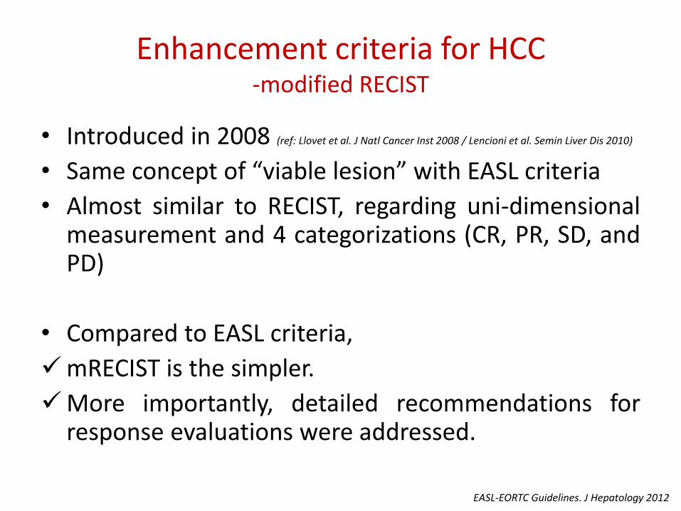
Enhancement criteria for HCC -modified RECIST
• Introduced in 2008 (ref: Llovet et al. J Natl Cancer Inst 2008 / Lencioni et al. Semin Liver Dis 2010)
• Same concept of “viable lesion” with EASL criteria
• Almost similar to RECIST, regarding uni-dimensional measurement and 4 categorizations (CR, PR, SD, and PD)
• Compared to EASL criteria,
mRECIST is the simpler.
More importantly, detailed recommendations for response evaluations were addressed.
EASL-EORTC Guidelines. J Hepatology 2012

WHO 1979. Available at: http://whqlibdoc.who.int/offset/WHO_OFFSET_48.pdf / Therasse P, et al. J Natl Cancer Inst 2000;92:205-16 / Bruix J, et al. J Hepatol 2001;35:421-30 / Llovet JM, et al. J Natl Cancer Inst 2008;100:698-791 /Lencioni R, et al. Semin Liver Dis 2010;30:52-60 / Eisenhauer EA, et al. Eur J Cancer 2009;45:228-47 / EASL-EORTC Guidelines. J Hepatology 2012;56:908-43
1979 2000 2001 2009 2010 2008
WHO criteria
RECIST
EASL criteria Conclusions from 2000 EASL
conference
RECIST v1.1 Revised RECIST
mRECIST for HCC
Proposed modifications to RECIST v1.1 for assessing response in HCC
mRECIST EASL-EORTC HCC
guideline
2012
Brief history of evolution of response evaluation criteria

mRECIST
• Image acquisition
contrast-enhanced spiral CT (preferably with use of multislice scanners contrast) or enhanced dynamic MRI
At least dual-phase (arterial & portal phase) imaging
Contiguous slice

Target lesions for mRECIST
• Only well-delineated, arterially enhancing lesions can be selected as target lesions.
Suitable for repeated measurement The longest diameter ≥ 1cm LN is categorized as non-target lesion Number of target lesions: RECIST 1.0 ~ 1.1 may apply
• Compared to target lesions for RECIST The longest diameter ≥ 1cm LN > 15 mm in the short diameter Number of target lesions: RECIST 1.0 (5 per organ, 10
Lesions) to RECIST 1.1 (2 per organ, 5 Lesions)

Target response of mRECIST
CR: disappearance of any intratumoral arterial enhancement in all
target lesions
PR: At least a 30% decrease in the sum of diameters of viable
(enhancement in the arterial phase) target lesions, taking as reference
the baseline sum of the diameters of target lesions
SD: Any cases that do not qualify for either partial response or
progressive disease
PD: An increase of at least 20% in the sum of the diameters of viable
(enhancing) target lesions, taking as reference the smallest sum of the
diameters of viable (enhancing) target lesions recorded since treatment
started
Lencioni et al. Semin Liver Dis 2010

Special consideration for target response
• The measurement of viable tumor is not necessarily located in the same scan plane with baseline evaluation.
• The longest diameter of viable tumor should be measured performed when contrast between viable tissue and non-enhancing tissue is the highest.
• The measurement of the viable tumor should not include any major intervening areas of necrosis.

Non-target lesions for mRECIST
• Non-measurable lesions
• Other measurable lesions other than target lesions
• Infiltrative HCC
• Previously treated with locoregional or systemic treatments (if it is poorly demarcated or exhibits atypical enhancement as a result of the previous treatment)

Special consideration for non-target response
• Malignant portal vein thrombosis is non-target lesion.
• Porta hepatis lymph node can be considered as malignant if its short axis is at least 20 mm.
Reactive lymph nodes at the porta hepatis is a common finding in patients with cirrhosis.
• Cytopathologic confirmation is necessary for neoplastic nature of any effusion (pleural effusion or ascites).

Non-target response of mRECIST
CR Disapperance of all nontarget lesions
IR (incomplete response)/SD Persistence of one or more target lesions
PD Appearance of new lesions
Unequivocal progression of existing nontarget lesions
Lencioni et al. Semin Liver Dis 2010
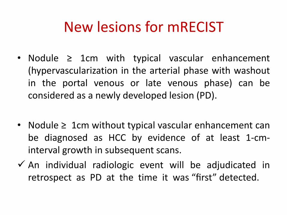
New lesions for mRECIST
• Nodule ≥ 1cm with typical vascular enhancement (hypervascularization in the arterial phase with washout in the portal venous or late venous phase) can be considered as a newly developed lesion (PD).
• Nodule ≥ 1cm without typical vascular enhancement can be diagnosed as HCC by evidence of at least 1-cm-interval growth in subsequent scans.
An individual radiologic event will be adjudicated in retrospect as PD at the time it was “first” detected.

Overall response for mRECIST determined by evaluation of target, non-target, and new lesions
Target lesions Non-target lesions New lesions Overall response
CR CR Absent CR
CR IR/SD Absent PR
PR Non PD Absent PR
SD Non PD Absent SD
PD Any Present or Absent PD
Any PD Present or Absent PD
Any Any Present PD Abbreviations: CR, complete response; PR, partial response; IR, incomplete response; SD, stable disease; PD,
progressive disease.
Lencioni et al. Semin Liver Dis 2010

Adapted from Shim JH et al. Radiology 2012
Size criteria
Enhancement criteria
Overview of schematics

Clinical application of enhancement criteria for HCC

Discrepancies between size-based and enhancement critetria
• There exist substantial discrepancies (kappa value 0.088-0.122) in response between enhancement criteria (EASL and mRECIST guidelines) and size-based criteria (WHO and RECIST guidelines).
EASL criteria mRECIST
WHO CR PR SD PD WHO CR PR SD PD
CR 1 0 0 0 CR 1 0 0 0
PR 10 4 0 0 PR 10 3 1 0
SD 22 30 22 0 SD 22 25 27 0
PD 1 0 3 5 PD 1 0 3 5
RECIST CR PR SD PD RECIST CR PR SD PD
CR 1 0 0 0 CR 1 0 0 0
PR 11 1 0 0 PR 11 1 0 0
SD 21 32 24 0 SD 21 26 30 0
PD 1 1 1 5 PD 1 1 1 5
Jung et al. J Hepatol 2013

Enhancement criteria for HCC treated with TACE
RECIST WHO
EASL criteria mRECIST
• EASL and mRECIST guidelines have better discriminatory ability for survival than WHO and RECIST guidelines among those treated with TACE.
Jung et al. J Hepatol 2013

Enhancement criteria for HCC treated with sorafenib
• For HCC treated with sorafenib, mRECIST were compared with RECIST.
• Patients with OR according to mRECIST had a longer OS than non-responders (median OS 18 vs. 8 months, respectively; P=0.013)
• Among 42 patients with SD according to RECIST, OS differed according to mRECIST; median OS with OR (n = 11), SD (n = 29), and PD (n = 2) was 17, 10 and 4 months, respectively (P = 0.016).
• Thus, mRECIST may be generally reliable in HCCs treated with TACE or sorafenib.
RECIST 1.1 OR: 2% SD: 79% PD: 19%
mRECIST OR: 23% SD: 57% PD: 21%
Edeline J, et al. Cancer. 2012 Jan 1;118(1):147-56
vs.

Which is the better between EASL and mRECIST guideline?
mRECIST
EASL criteria
CR PR SD PD
CR 113 0 0 0
PR 4 87 15 0
SD 0 6 56 0
PD 0 0 2 9
Kappa value 0.863
0.0
0.2
0.4
0.6
0.8
1.0
Overall Survival (months)
Pro
bab
ility
of
surv
ival
0 12 24 36
EASL criteria
0 12 24 36
0.0
0.2
0.4
0.6
0.8
1.0
Overall Survival (months)
Pro
bab
ility
of
surv
ival
mRECIST
Non-Responder Responders
Both P<0.001
Kim et al. Eur J Cancer 2012
• mRECIST, a simpler method, provided prognostic values for predicting OS equivalent to EASL criteria in patients with HCC undergoing TACE as an initial treatment modality.

Timing of assessment • More than half required repeated TACE session to achieve CR.
• Early vs. best response by mRECIST during “on-demand” TACE sessions from Yonsei experience
Both initial and best response well predicted OS, respectively.
0.0 20.0 40.0 60.0 80.0
OS (months)
0.0
0.2
0.4
0.6
0.8
1.0
PD
PR
SD
CR
Cu
mu
lati
ve
pro
ba
bil
ity
0.00 20.0 40.0 60.0 80.0
0.0
0.2
0.4
0.6
0.8
1.0
PD
PR
SD
CR
OS (months)
Cu
mu
lati
ve
pro
ba
bil
ity
Kim et al. Manuscript submitted
Both p<0.001

Optimal number of target lesions with reference to responses assessing all target lesions
Kim et al. Clin Cancer Res 2013
Prognostic value for OS of EASL and mRECIST guidelines
Maximum No. of targets C-index for OS
EASL criteria mRECIST
Up to 1 0.739 0.750
Up to 2 0.744 0.759
Up to 3 0.744 0.755
Up to 4 0.749 0.750
Up to 5 0.749 0.750
All targets 0.749 0.750
• Prognostic values for OS were similar regardless of number of target lesions. • However, assessing two targets could be recommended considering high
concordances from cross-sectional analyses

Further consideration of mRECIST
• mRECIST is a reliable method for assessing tumor response in HCC.
• Adequate skills and expertise were required in terms of inter-observer
and intra-observer variability.
Education and training
• Standardized software/ hardware protocols were required for reproducibility and reliable comparisons.
Uniform image acquisition parameters
Quality control
Blinded assessments
• The use of changes in serum levels of biomarkers (i.e. AFP levels) along with radiological response in HCC is under investigation.
Lencioni R, Llovet JM. Semin Liver Dis. 2010;30:52‐60. / EASL–EORTC Clinical Practice Guidelines. J Hepatology 2012;56:908-43

Combination of radiological response and biomarkers response -AFP change & mRECIST -
OS according to mRECIST, Child-Pugh, AFP ratio OS according to AFP ratio
Kawaoka T, et al. Oncology 2012
AFP ratio≤1 at 8weeks
AFP ratio≥1 at 8weeks
* : score was calculated as sum of the response by mRECIST (PD:0, CR or PR of SD:1), Child-Pugh score (B:0, A:1) and AFP ratio at 8 weeks from the start of the treatment (>1:0, ≤1:1)
*
• The combination of mRECIST and AFP ratio is useful for the assessment of prognosis of patients with advanced HCC treated with sorafenib.

Combination of radiological response and biomarkers response
-CP score change, AST change & EASL criteria -
• An ART (Assessment for Retreatment with TACE) score of ≥2.5 prior the second TACE identifies patients with a dismal prognosis who may not profit from further TACE sessions.
Sieghart et al. Hepatology 2013

Alternative evaluation methods
• Choi criteria Introduced in 2007 (Ref: Choi et al. J Clin Oncol 2007)
To resolve the limitation of RECIST for patients undergoing imatinib for GastroIntestinal Stromal Tumor
Tumor density, using CT attenuation coefficient (Hounsfield unit [HU]), was applied to reflect areas of tumors with reduced vascularization.
Combination of size criteria (long diameter) and tumor density

Alternative evaluation methods
• Choi criteria
4 categorization - CR: Disappearance of all lesions
- PR: Decrease in size of ≥10% or decrease in tumor density (HU) ≥15% on CT
- SD: Does not meet criteria for CR, PR, or PD
- PD: Increase in tumor size of ≥10% and does not meet criteria of PR by tumor density (HU) on CT/ New lesions

Alternative evaluation methods
• Choi criteria Also tried for HCC with good discrimination of OS
Choi criteria EASL criteria mRECIST RECIST 1.1
OR 32 13 12 2
SD 15 31 34 41
PD 17 19 17 20
Ronot et al. Oncologist 2014

Future perspectives
• Metabolic response by differences between standardized uptake values from PET scans
PET+ RECIST PERCIST Wahl et al. J Nucl Med. 2009
• 3D-Volumetric criteria (automated methods)
• Functional imaging using diffusion weighted MR
• Development of “new” biomarkers and combinations with radiological response

Future perspectives
• For the newer treatment modalities (drug-eluting bead TACE and TARE) and other investigational drugs, conventional assessment tools should be validated accordingly.
• For infiltrative HCC or HCC with atypical enhancement patterns, more optimized methods other than categorizing into “non-target lesions” are required.

Take home messages
• Enhancement criteria are now standard tools for HCC.
• So far, mRECIST provides equivalent efficacy, more convenience with a simpler method and detailed recommendation, compared to EASL criteria.
• Advances in imaging technologies will allow the better assessment protocol.
• Newer treatment modalities will require modification of current assessment tools.

경청해 주셔서 감사합니다~